Nano-World by Basta Anastasia
25
Nanoparticles and biomedicine Department of Molecular Biochemistry and Pharmacology Anastasia Basta
-
Upload
anastasia-ch-bastas -
Category
Documents
-
view
53 -
download
2
Transcript of Nano-World by Basta Anastasia
Nanoparticles and biomedicine
Department of Molecular Biochemistry and Pharmacology
AnastasiaBasta

Nano-World
• Synthesis of nanoparticles• In Vitro analysis - Nanotoxicity
• In vivo analysis
Milano, 1st March to 31th May 2014
AnastasiaBasta
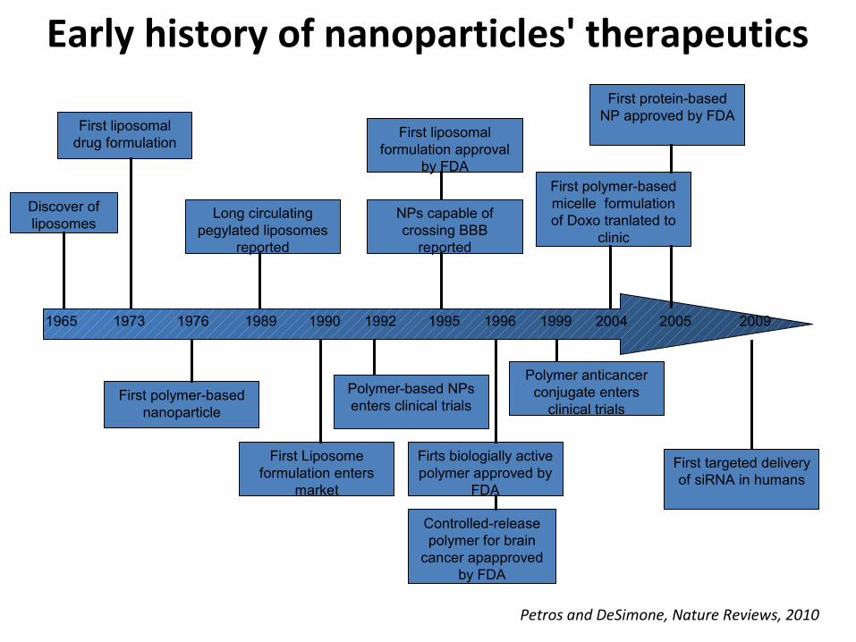
1965 1973 1976 1989 1990 1992 1995 1996 1999 2004 2005 2009
Discover of liposomes
First liposomal drug formulation
First polymer-based nanoparticle
Long circulating pegylated liposomes
reported
First Liposome formulation enters
market
Polymer-based NPs enters clinical trials
NPs capable of crossing BBB
reported
First liposomal formulation approval
by FDA
Firts biologially active polymer approved by
FDA
Controlled-release polymer for brain
cancer apapproved by FDA
Polymer anticancer conjugate enters
clinical trials
First polymer-based micelle formulation of Doxo tranlated to
clinic
First protein-based NP approved by FDA
First targeted delivery of siRNA in humans
Petros and DeSimone, Nature Reviews, 2010
Early history of nanoparticles' therapeutics

Synthesis of NPs
Structure ofLiposomes
Gaucher disease is a progressive lysosomal storage disorder (LSD) caused by deficiancy of glucocerebrosidase leading to the dysfunction in multiple organ systems. Associated clinical symptoms include hepatosplenomegaly, anaemia, thrombocytopenia and skeletal deterioration.
Glucocerebrosidase enzyme replacement therapy has become the standard of care for patients with symptomatic Gaucher disease due to its safety and efficacy profile. The problem here is that the enzyme administrated cannot reach the blood-brain barrier, but remains at the peripheral organs. However, the BBB is permeable to small and lipophilic molecules.
Liposomes are colloidal vesicles ranging from few nanometers to several micrometers in diameter with one or more lipid layers. They are prepared from natural or synthetic phospholipids and cholesterol. In addition, they are biodegradable, biocompatible, non-toxic and non-immunogenic. They can, also, entrap a wide variety of therapeutic drugs, such us hydrophilic drugs. Liposomes are able to cross the BBB when they are connected with specific peptides.
The objective of the present study is therefore to exploit nanotechnology to use functionalised liposomes in order to deliver glucocerebrosidase to the central nervous system. The final goal will be the evaluation of the enzyme replacement therapy as possible beneficial effect in a murine model of LSDs.
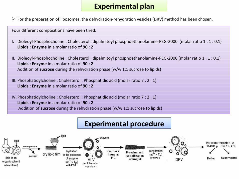
Experimental procedure
Experimental plan For the preparation of liposomes, the dehydration-rehydration vesicles (DRV) method has been chosen.
Four different compositions have been tried:
I. Dioleoyl-Phosphocholine : Cholesterol : dipalmitoyl phosphoethanolamine-PEG-2000 (molar ratio 1 : 1 : 0,1)Lipids : Enzyme in a molar ratio of 90 : 2
II. Dioleoyl-Phosphocholine : Cholesterol : dipalmitoyl phosphoethanolamine-PEG-2000 (molar ratio 1 : 1 : 0,1)Lipids : Enzyme in a molar ratio of 90 : 2 Addition of sucrose during the rehydration phase (w/w 1:1 sucrose to lipids)
III. Phosphatidylcholine : Cholesterol : Phosphatidic acid (molar ratio 7 : 2 : 1) Lipids : Enzyme in a molar ratio of 90 : 2
IV. Phosphatidylcholine : Cholesterol : Phosphatidic acid (molar ratio 7 : 2 : 1)Lipids : Enzyme in a molar ratio of 90 : 2 Addition of sucrose during the rehydration phase (w/w 1:1 sucrose to lipids)
(chloroform)
in evaporator
with PBS
with PBS(multilamellar
vesicle s)
184000 g
Supernatant

Through Bradford analysis we quantified the µg of enzyme entrapped in liposome and free in the supernatant. Inside liposomes we found about 38µg, while in the supernatant 810 µg. They correspond respectively to 3% and to 60 % of the initial amount of the added enzyme (1337μg) . These values remained stable also 24h after the synthesis.
Then we performed the measurement of enzymatic activity by using a fixed concentration of the substrate (3 mM) and a fixed incubation time (30 minutes). We correlated the fluorescence intensity obtained from the reaction with the concentration of the active enzyme in each sample. By matching these values we obtained the values of enzymatic activity expressed as units (µmol substrate transformed/min/mg protein).
Liposome contained 0.7 U, while in the supernatant there were 0.9 U of enzyme. Therefore, only 1,5% of the enzymatic activity is still measurable and remains stable also 24 hours after liposome preparation.The mixture containing free and entrapped enzyme corresponds to 1,6 U, i.e. to 50 µg of active enzyme.
µg of protein in 500 μl of sample In liposomes 38µg
In the supernatant 810µg
Total 848µg
μg of active protein in 500 μl of sample In liposome 21,8µg 0,7 U
In supernatant 28,4µg 0,9 U
total 50,2µg 1,6 U
Results - Discussion
Supernatant Supernatant
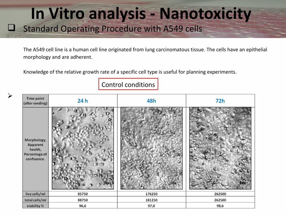
In Vitro analysis - Nanotoxicity Standard Operating Procedure with A549 cells
The A549 cell line is a human cell line originated from lung carcinomatous tissue. The cells have an epithelial morphology and are adherent.
Knowledge of the relative growth rate of a specific cell type is useful for planning experiments.
Seeding at 0.5x10⁵ cells/well in cell medium, 8 replicates per time period (24h, 48h, 72h)
10x 10x10x
Control conditions

hours
Cel
ls/m
l
0 24 48 720
50000
100000
150000
200000
250000
300000
350000
0,05% (w/v) Trypsin-EDTA has been added to cultures 24h, 48h, 72h after seeding to detach cells from the seeding plate.
An automatic cell counter has been used to measure the amount of living cells.
According to the results shown the cells are healthy, their morphology is appropriate and their doubling time is about 30 hours.

Standard Operating Procedure MTS cell viability assay
The MTS tetrazolium (Owen’s reagent) is reduced by mitochondria of viable cells into a colored formazan derivative that is soluble in tissue culture medium. Assays are performed by adding a small amount of the reagent directly to culture wells. After 1h of incubation the absorbance at 490nm was determined.
Cells were seeded at 1x10⁴ cells/well

1) Add MTS reagent 2) Incubate for 1h 3) Measure O.D at 490 nm
• NPs added in the cells culture: amine-modified polystyrene nanopartices, 50 nm diameter
carboxyl-modified polystyrene nanopartices, 40 nm diameter
• NP dilutions: 100 µg/ml, 50 µg/ml, 25 µg/ml, 10 µg/ml, 1 µg/ml
Steps of the assay
Nanotoxicity assay
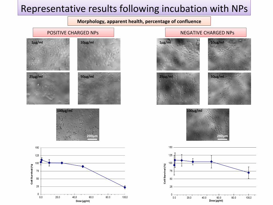
Representative results following incubation with NPs
1µg/ml 10µg/ml
25µg/ml 50µg/ml
100µg/ml
POSITIVE CHARGED NPs
1µg/ml 10µg/ml
25µg/ml 50µg/ml
100µg/ml
NEGATIVE CHARGED NPs
Morphology, apparent health, percentage of confluence
200μm 200μm

Mycoplasma testTest for mycoplasma should be performed as a quality check of the cell cultures.
Ctr
+ D
NA
M
ycoM
arke
r
Ctr
-
(H2O
)M
EM
26
-3
ME
M 2
8-3 M
EM
31-
3 ME
M 4
-4 M
EM
7-
4 ME
M 8
-4 M
EM
9-
4 ME
M 1
1-4
ME
M 1
4-4
1.5Kb
1.0Kb
0.5Kb
0.7 Kb
expected amplified product at 700bp
Negative mycoplasma results have been obtained by electrophoresis analysis

In Vivo analysis
• Administration (iv) of 20 mg/kg of NPs.• Two mice sacrificed 1h, 4h, 24h and 48h after administration• Two kinds of NPs used:
Core Shell NPs and PolyCaproLactone 3 (PCL3) NPs• Tumor and liver sections (20 µm thick) were acquired with fluorescent microscope• Nuclei are stained with HOECHST (blue signal) and NPs are visualized in red through
Rhodamine B (red) signal.• Only tumor samples of mice sacrified 24h and 48h after iv injection underwent to
histological analysis.
Biodistribution of Core Shell NPs and PolyCaproLactone 3 (PCL3) NPs loaded with Paclitaxel (PTX) in a mouse model of triple negative breast cancer
Core Shell NPs PCL3 NPs

Core shell NPs in Liver
NPs are concentrated in the veins where it is possible to see an association between circulating cells and red staining. No relevant migration was observed in parenchyma
A large amount of signal is still confined to the small vessels and almost disappeared in larger vessels. However, a significant migration to the parenchyma is clearly detectable.
4h - Liver1h - Liver
A strong migration in liver parenchyma can be observed. No presence of NPs are found in smallest vessels (brighter nuclei forming a circular structure). This widespread distribution suggests the presence of a specific association between nuclei and NPs.
24h - Liver 48h - Liver
A high amount of NPs are found in the liver parenchyma but not inside the big vessels.
40x
Epi Fluorescence Microscopy 20x
100μm 100μm
100μm 100μm
50μm

Core shell NPs in Tumors
Here, the general pattern of distribution in tumor sections is confirmed: NPs are visible after diffusing in the parenchyma both as internalized in cells and as big clusters not specifically associated with cellular internalization.
24h - Tumor 48h - Tumor
In this picture a very strong signal can be clearly observed: NPs spread in the parenchyma as clusters or as specified signal associated with cells, but vessels are out of signal.
Epi Fluorescence Microscopy 20x
100μm 100μm

PCL3 NPs in Liver
1h - Liver
It is possible to see blue dots inside the vein. This means that circulating cells are associated with NPs at this time. No relevant spread of NPs was here observed in parenchyma.
4h - Liver
In this picture, it is possible to see a strong signal associated with NPs at the external layer of this big vessel. It is also noteworthy that a signal associated to the migration of NPs -from the vessel to the parenchyma around- is observed.
24h - Liver
NPs can be found on both small vessels walls (all around endothelial layer) and in the parenchyma.
48h - Liver
NPs seem to be almost homogenously localized in the parenchyma. Vessels, either big or small ones, seem to be completely empty.
Epi Fluorescence Microscopy 20x
100μm
100μm100μm
100μm

PCL3 NPs in Tumors
A highly heterogeneous staining is visible. In many cases NPs seem to be uptaken by cells
24h - Tumor
NPs are heterogeneously diffused in the parenchyma. Many coronal vessels are contoured by a thin layer of NPs that extravasated.
48h - Tumor
Epi Fluorescence Microscopy 20x
100μm100μm
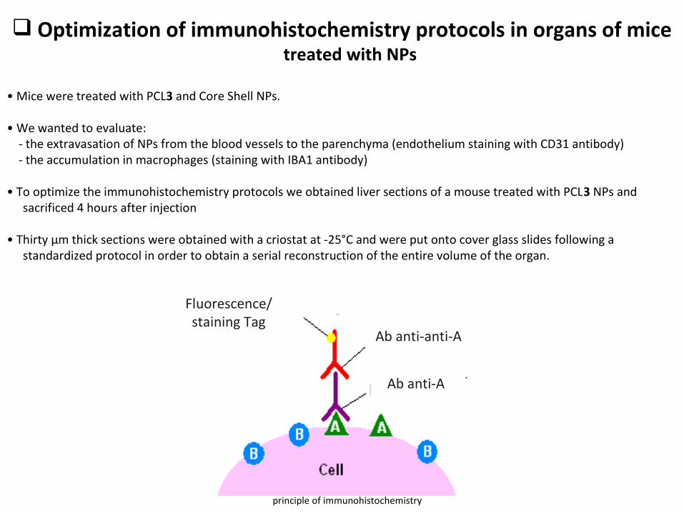
Optimization of immunohistochemistry protocols in organs of mice treated with NPs
• Mice were treated with PCL3 and Core Shell NPs.
• We wanted to evaluate: - the extravasation of NPs from the blood vessels to the parenchyma (endothelium staining with CD31 antibody) - the accumulation in macrophages (staining with IBA1 antibody)
• To optimize the immunohistochemistry protocols we obtained liver sections of a mouse treated with PCL3 NPs and sacrificed 4 hours after injection
• Thirty μm thick sections were obtained with a criostat at -25°C and were put onto cover glass slides following a standardized protocol in order to obtain a serial reconstruction of the entire volume of the organ.
Ab anti-A
Ab anti-anti-A
Fluorescence/staining Tag
principle of immunohistochemistry

Protocols used for the treatment of tissues with antibodies
CD31 Ab IBA I Ab
Confocalmicroscopy

RESULTS & DISCUSSION
CD31 NPs can be found both in the blood vessels and homogenously distributed in the
parenchyma of the organ . Partial colocalization of NPs and endothelium
are also able to be observed.
20μm
1:50
20x Epifluorescence images of HOECHST (blue), CD31 (green) and RhB (red)
Confocal Microscopy images of HOECHST (blue), CD31 (green) and Rhodamine B (red)
40x 60x
1:100 1:100
60x50μm 20μm
100μm 100μm
100μm 100μm
50μm

IBA I
A strong signal associated to NPs was confirmed both in vessels and in organ parenchyma.
100μm
100μm 100μm 20μm

By checking the general biodistribution of NPs it was able to find that both Core Shell NPs and PCL3 NPs show a similar behaviour :
In the first hour after administration, NPs can be observed almost exclusively inside the vessels of the organs.
However, 4 hours later of the administration, NPs are located both in vessels and in the parenchyma of the tissue.
Finally, at the time point of the 24 hours and over, NPs can be found spread in the parenchyma both in liver parenchima and in the tumor.
Conclusions

Ending…
Future steps of nanomedicine….
Synthesis
In vitro
In vivo

I have a dreamWe can envision a future where tiny nanomaterials (nanodroids,
ISAC ASIMOV) travel through the bloodstream searching for sites of infection or disease processes, and once any irregular cellular activities are found, they trigger the release of nanomaterial cargoes that repair damaged tissues or kills foreign invaders. (self-healing strategy)

Many thanks for this experience and
educational period to:Prof. Mario Salmona
Dr. Paolo BiginiDr. Leopoldo Sitia
Dr. Martina Violatto*Biochemistry Lab