Nano Structures Cellulose Acetate Ph Tha Late
10
Nanostructures of Cellulose Acetate Phthalate Obtained by Electrospinning from 2‑Methoxyethanol-Containing Solvent Systems: Morphological Aspects, Thermal Behavior, and Antimicrobial Activity Niculae Olaru,* ,† Liliana Olaru, † Nita Tudorachi, † Simona Dunca, ‡ and Manuela Pintilie † † “Petru Poni” Institute of Macromolecular Chemistry, Aleea Gr. Ghica-Voda 41 A, Iasi 700487, Romania ‡ “Al. I. Cuza” University, Bd. Carol I 11, Iasi 700506, Romania ABSTRACT: Cellulose acetate phthalate (CAP) is well-known and largely used as a pharmaceutical excipient for enteric coating of tablets and capsules. Recent literature has proved that this polymer exhibits antimicrobial and antiviral properties when it is used as a micronized powder. Nanostructures of CAP are also prepared, and their potential for antimicrobial and technological applications is recently reported. In the present work, we are focused on synthesis and characterization of CAP nanofibers and powders by electrospinning/electrospraying from solutions in solvent mixtures containing 2-methoxyethanol. The morphologies of the obtained nanostructures are investigated by scanning electron microscopy (SEM) and transmission electron microscopy (TEM) analyses and discussed in comparison with those previously reported for products obtained in 2-methoxyethanol alone. Thermal properties of CAP nanostructured as powders and fibers are studied in comparison with those of CAP films using thermogravimetric (TG), Fourier transform infrared (FTIR), and mass spectrometry. TG analysis reveals that CAP nanoproducts exhibit an apparent lower stability than CAP films. Further investigations of both residual polymer and the evolved degradation gas products evidence the presence of phthalic anhydride as a main degradation product in the initial stage of the process. In the case of nanostructures of CAP, due to their high porosity and very high specific surface areas, this degradation product is easier and continuously eliminated during the thermal treatment. This behavior explains their apparent lower thermal stability as compared with that of the films where the diffusion of phthalic anhydride proceeds slowly and therefore the weight loss of the sample is delayed. The paper also reports preliminary results of antimicrobial activities of the investigated products against Escherichia coli and Staphylococcus aureus. 1. INTRODUCTION Cellulosic polymers have gathered great interest in recent years in the field of nanoproducts useful for high-value applications. The biodegradability, biocompatibility, and versatility of these natural polymers are the reasons for those approaches regarding the possibilities to transform them in nanostructured materials with large applications especially in biomedicine, for tissue engineering scaffolds, wound dressing, controlled release, and technology purposes such as filtration, catalysis, sensors, affinity membranes, etc. 1-15 One of the most interesting cellulose derivative from this point of view is cellulose acetate phthalate (CAP), a mixed ester with acetic and o-phthalic acids. CAP has been known for several decades as a pharmaceutical excipient used for enteric coating of tablets and capsules. Recently, it has been shown that CAP formulated as a micronized powder exhibits activity against the human immunodeficiency virus type I (HIV-I), several herpes viruses (herpes simplex viruses I and II), and many nonviral sexually transmitted disease pathogens. 16-22 In a previous paper, 23 we have studied the possibility to obtain micro/nanostructured CAP with high specific surface areas via the electrospinning procedure. Several solvent systems (2-methoxyethanol, acetone-water mixture 85/15 (v/v), and 2-methoxyethanol-acetone-water mixture 50/42.5/7.5 (v/v/ v)) were investigated, and it has been found that by variation of the solvent system and the polymer concentration, powders and fibers of CAP with sizes in the submicrometer range and different morphologies can be easily obtained using a simple electrospinning laboratory setup. It has also been observed that the morphology of the electrospun CAP changes as a function of the solution characteristics like viscosity, surface tension, and conductivity. When using only 2-methoxyethanol as the electrospinning solvent, polymer concentrations higher than 25% (w/v) were necessary to obtain smooth filaments without beads, whereas at a polymer concentration as low as 12.5% (w/ v), only micro/nanoparticles that look like void hemispheres have been obtained. When a ternary system 2-methoxyetha- nol-acetone-water 50/42.5/7.5 (v/v/v) was used as the electrospinning solvent system for CAP, smooth filaments without beads and fiber diameters of about 600 nm could be produced at concentrations of 25% (w/v). The potential use of electrospun CAP fibers as a tool to prevent HIV transmission has been very recently discussed by Huang et al. 24 By taking into account the observation that the electro- spinnability of CAP from 2-methoxyethanol-containing sol- vents was better than that from acetone-water mixtures, 23 in Received: May 17, 2012 Revised: December 12, 2012 Accepted: December 12, 2012 Published: December 12, 2012 Article pubs.acs.org/IECR © 2012 American Chemical Society 696 dx.doi.org/10.1021/ie301299d | Ind. Eng. Chem. Res. 2013, 52, 696-705
-
Upload
adrianl136378 -
Category
Documents
-
view
45 -
download
0
description
art
Transcript of Nano Structures Cellulose Acetate Ph Tha Late

Nanostructures of Cellulose Acetate Phthalate Obtained byElectrospinning from 2‑Methoxyethanol-Containing SolventSystems: Morphological Aspects, Thermal Behavior, andAntimicrobial ActivityNiculae Olaru,*,† Liliana Olaru,† Nita Tudorachi,† Simona Dunca,‡ and Manuela Pintilie†
†“Petru Poni” Institute of Macromolecular Chemistry, Aleea Gr. Ghica-Voda 41 A, Iasi 700487, Romania‡“Al. I. Cuza” University, Bd. Carol I 11, Iasi 700506, Romania
ABSTRACT: Cellulose acetate phthalate (CAP) is well-known and largely used as a pharmaceutical excipient for enteric coatingof tablets and capsules. Recent literature has proved that this polymer exhibits antimicrobial and antiviral properties when it isused as a micronized powder. Nanostructures of CAP are also prepared, and their potential for antimicrobial and technologicalapplications is recently reported. In the present work, we are focused on synthesis and characterization of CAP nanofibers andpowders by electrospinning/electrospraying from solutions in solvent mixtures containing 2-methoxyethanol. The morphologiesof the obtained nanostructures are investigated by scanning electron microscopy (SEM) and transmission electron microscopy(TEM) analyses and discussed in comparison with those previously reported for products obtained in 2-methoxyethanol alone.Thermal properties of CAP nanostructured as powders and fibers are studied in comparison with those of CAP films usingthermogravimetric (TG), Fourier transform infrared (FTIR), and mass spectrometry. TG analysis reveals that CAPnanoproducts exhibit an apparent lower stability than CAP films. Further investigations of both residual polymer and the evolveddegradation gas products evidence the presence of phthalic anhydride as a main degradation product in the initial stage of theprocess. In the case of nanostructures of CAP, due to their high porosity and very high specific surface areas, this degradationproduct is easier and continuously eliminated during the thermal treatment. This behavior explains their apparent lower thermalstability as compared with that of the films where the diffusion of phthalic anhydride proceeds slowly and therefore the weightloss of the sample is delayed. The paper also reports preliminary results of antimicrobial activities of the investigated productsagainst Escherichia coli and Staphylococcus aureus.
1. INTRODUCTION
Cellulosic polymers have gathered great interest in recent yearsin the field of nanoproducts useful for high-value applications.The biodegradability, biocompatibility, and versatility of thesenatural polymers are the reasons for those approaches regardingthe possibilities to transform them in nanostructured materialswith large applications especially in biomedicine, for tissueengineering scaffolds, wound dressing, controlled release, andtechnology purposes such as filtration, catalysis, sensors, affinitymembranes, etc.1−15
One of the most interesting cellulose derivative from thispoint of view is cellulose acetate phthalate (CAP), a mixed esterwith acetic and o-phthalic acids. CAP has been known forseveral decades as a pharmaceutical excipient used for entericcoating of tablets and capsules. Recently, it has been shown thatCAP formulated as a micronized powder exhibits activityagainst the human immunodeficiency virus type I (HIV-I),several herpes viruses (herpes simplex viruses I and II), andmany nonviral sexually transmitted disease pathogens.16−22
In a previous paper,23 we have studied the possibility toobtain micro/nanostructured CAP with high specific surfaceareas via the electrospinning procedure. Several solvent systems(2-methoxyethanol, acetone−water mixture 85/15 (v/v), and2-methoxyethanol−acetone−water mixture 50/42.5/7.5 (v/v/v)) were investigated, and it has been found that by variation ofthe solvent system and the polymer concentration, powders
and fibers of CAP with sizes in the submicrometer range anddifferent morphologies can be easily obtained using a simpleelectrospinning laboratory setup. It has also been observed thatthe morphology of the electrospun CAP changes as a functionof the solution characteristics like viscosity, surface tension, andconductivity. When using only 2-methoxyethanol as theelectrospinning solvent, polymer concentrations higher than25% (w/v) were necessary to obtain smooth filaments withoutbeads, whereas at a polymer concentration as low as 12.5% (w/v), only micro/nanoparticles that look like void hemisphereshave been obtained. When a ternary system 2-methoxyetha-nol−acetone−water 50/42.5/7.5 (v/v/v) was used as theelectrospinning solvent system for CAP, smooth filamentswithout beads and fiber diameters of about 600 nm could beproduced at concentrations of 25% (w/v).The potential use of electrospun CAP fibers as a tool to
prevent HIV transmission has been very recently discussed byHuang et al.24
By taking into account the observation that the electro-spinnability of CAP from 2-methoxyethanol-containing sol-vents was better than that from acetone−water mixtures,23 in
Received: May 17, 2012Revised: December 12, 2012Accepted: December 12, 2012Published: December 12, 2012
Article
pubs.acs.org/IECR
© 2012 American Chemical Society 696 dx.doi.org/10.1021/ie301299d | Ind. Eng. Chem. Res. 2013, 52, 696−705

the present paper we further investigate the electrospinning ofCAP from 2-methoxyethanol and 2-methoxyethanol−watersystems with different compositions.A detailed characterization of CAP has been reported by
Roxin et al.25 including data about its thermal behavior whentreated in oxygen and inert atmospheres. Glass transitiontemperatures of CAP samples were also determined. Earlier26
we demonstrated that by treating CAP films at temperaturesbetween 120 and 220 °C for various times of exposure, one ofthe principal degradation products is the phthalic anhydride,which is partially evolved during the process.Having in mind the possibility to change drastically the
polymer properties by transforming it into nanostructuredmaterials and also taking into account the importance of thethermal behavior of these materials from an applicative point ofview, in the present paper we are concerned with a study onthermal properties of micro/nanopowders and fibers of CAP incomparison with those of the usual films obtained from thesame polymer. TG-DTG analysis, FTIR, ATR, and massspectrometry were used to investigate thermal stability ofnanostructured CAP.On the basis of literature reports, we expect that the obtained
nanostructures of CAP could enhance its potential asantimicrobial agent. Therefore, in this paper, preliminary testson the antimicrobial activity of the electrospun CAP productsare also reported in relation to their morphology.
2. MATERIALS AND METHODS
2.1. Materials. The chemicals were of reagent grade andwere used as received, without further purification.2.2. Synthesis and Characterization of CAP. Cellulose
acetate phthalate investigated in this work had a degree ofphthaloylation of 0.70 and a degree of acetylation of 1.73 andwas synthesized by phthaloylation of cellulose acetate (CA)with degree of acetylation of 1.77 and degree of polymerizationof 185. Both cellulose derivatives were prepared in ourlaboratory, as described in our previous papers.23,27
Characterization of CAP and CA with respect to theirdegrees of modification was performed by means of the knownmethods: refs 28 and 29, respectively.2.3. Electrospinning and Electrospraying Experi-
ments. The experiments for obtaining of both fibers andpowders of CAP were carried out using the same typicallaboratory setup, consisting of a high voltage source, a pumpand a syringe for polymer solution and a rotary collector disk.Solutions of CAP were prepared in 2-methoxyethanol and
mixtures of 2-methoxyethanol−water of three compositions:95/5 (v/v), 90/10 (v/v), and 85/15 (v/v). On the basis ofprevious results,23 the concentration of polymer solutions waschosen to be 10% and 30% (w/v) so as to obtain powders andfibers, respectively, with sizes in the submicrometer range.The same parameters were chosen for both electrospinning
and electrospraying experiments, as follows: voltage potential,15−20 kV; tip-to- collector distance, about 15 cm; flow rate ofthe solution through the syringe, 1.5 mL h−1. The processeswere carried out in air at room temperature.2.4. Characterization of CAP Micro/nanoproducts. The
morphology of CAP ultrafine fibers and powders wascharacterized by scanning electron microscopy (SEM; VEGAII SBH TESCAN) and transmission electron microscopy(TEM; TESLA BS 513A).
The particle size distribution curves for CAP powders wereobtained by means of a system Mastersizer 2000 version 5.31,Malvern Instruments.The electrical conductivity of polymer solutions was
measured using a CDM 210 (Radiometer, Copenhagen)Conductometer.Viscosity of CAP solutions was determined by means of a
Brookfield SYNCHRO-LECTRIC Viscometer, model LVF.2.5. Thermal Treatment. Thermal analysis was performed
by simultaneous TG, MS, and FTIR determinations using thefollowing equipment: TG-DSC model STA 449F1 Jupiter(Netzsch−Germania), QMS 403C Aeolos (Netzsch−Germa-nia), and FTIR spectrophotometer model Vertex-70 (Bruker)with an accessory for ATR (attenuated total reflectance) typeGolden Gate. The experiments were carried out in thefollowing conditions: heating rate 10 K min−1; gas purge He;reference material Al2O3; temperature range 30−600 °C.
2.6. Antimicrobial Tests. The in vitro antimicrobialactivity was assessed using the disk-diffusion method Kirby-Bauer30 on two strains of bacteria: Staphylococcus aureus ATCC25923 (S. aureus) and Escherichia coli ATCC 25922 (E. coli).CAP samples obtained from solutions in 2-methoxyethanol and2-methoxyethanol−water 85/15 (v/v) at 10% (w/v) polymerconcentration were used for these tests.The bacterial inoculum obtained from bacterial cultures
preincubated for 18 h at 37 °C was standardized according toMcFarland scale and a number of 107−108 CFU/mL wasobtained. The culture medium (LB-Lysogeny broth) wasseeded with the inoculum thus obtained and then microdisksof 1 cm diameter impregnated with CAP powders (0.01 g/microdisk) were placed on the surface of the medium. Threedeterminations were carried out for each sample, and theaverage values obtained for the diameters of the inhibitionzones are shown in Table 1.
Samples of CAP nanofibers were placed directly on thesurface of the culture medium. The plates were then incubatedat 37 °C for 24 h, and the antibacterial activity was evidencedby the occurrence of an inhibition zone around the sample area.
3. RESULTS AND DISCUSSION3.1. Morphological Aspects of Nanostructured CAPs.
3.1.1. Ultrafine CAP Powders. The morphology of CAP micro/nanoparticles obtained from 2-methoxyethanol−water systems,as revealed by SEM images for 10% (w/v) polymerconcentration in the electrospun solution (Figure 1), doesnot differ from that reported for similar products obtained from2-methoxyethanol solvent. The shape of void hemispheres forCAP micro/nanoparticles is preserved when water is present inthe electrospinning solvent.
Table 1. Antimicrobial Activity Expressed by the Diameter ofthe Inhibition Zone (mm) of CAP Nanopowders Obtainedfrom Solution in 2-Methoxyethanol for P1 and Solution in 2-Methoxyethanol−Water 85/15 (v/v) for P2 at 10% (w/v)Polymer Concentration
microorganism test
sample Staphylococcus aureus ATCC 25923 Escherichia coli ATCC 25922
P1 12 11P2 16 14
Industrial & Engineering Chemistry Research Article
dx.doi.org/10.1021/ie301299d | Ind. Eng. Chem. Res. 2013, 52, 696−705697
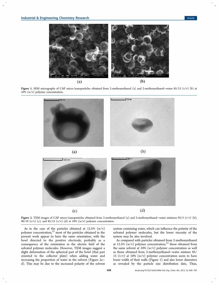
As in the case of the particles obtained at 12.5% (w/v)polymer concentration,23 most of the particles obtained in thepresent work appear to have the same orientation, with thebowl directed to the positive electrode, probably as aconsequence of the orientation in the electric field of thesolvated polymer molecules. However, TEM images suggest aslight deformation of the spherical part of the bowl (that partoriented to the collector plate) when adding water andincreasing the proportion of water in the solvent (Figure 2a−d). This may be due to the increased polarity of the solvent
system containing water, which can influence the polarity of thesolvated polymer molecules, but the lower viscosity of thesystem may be also involved.As compared with particles obtained from 2-methoxyethanol
at 12.5% (w/v) polymer concentration,23 those obtained fromthe same solvent at 10% (w/v) polymer concentration as wellas those obtained from 2-methoxyethanol−water mixture 85/15 (v/v) at 10% (w/v) polymer concentration seem to havelower width of their walls (Figure 1) and also lower diametersas revealed by the particle size distribution data. Thus,
Figure 1. SEM micrographs of CAP micro/nanoparticles obtained from 2-methoxyethanol (a) and 2-methoxyethanol−water 85/15 (v/v) (b) at10% (w/v) polymer concentration.
Figure 2. TEM images of CAP micro/nanoparticles obtained from 2-methoxyethanol (a) and 2-methoxyethanol−water mixtures 95/5 (v/v) (b),90/10 (v/v) (c), and 85/15 (v/v) (d) at 10% (w/v) polymer concentration.
Industrial & Engineering Chemistry Research Article
dx.doi.org/10.1021/ie301299d | Ind. Eng. Chem. Res. 2013, 52, 696−705698

distribution curves in Figure 3 clearly show that 10% polymerconcentration in the electrospun solutions for both solventsystems investigated in this paper gave 50% of the obtainedparticles with diameters lower than 5 μm as compared with 10μm previously reported for higher polymer concentrations.23 Atthe same time, the polymer solution in the solvent mixture 2-
methoxyethanol−water 85/15 (v/v) gave smaller particles withspecific surface area higher than of those produced from 2-methoxyethanol solution (Figure 3). The presence of water inthe electrospinning solution may be responsible for such effect,due to the higher electrical conductivity of the polymer solutioncontaining water (19.0 μS/cm as compared with 4.82 μS/cm
Figure 3. Particle size distribution of CAP micro/nanoparticles obtained from 2-methoxyethanol (a) and 2-methoxyethanol−water mixture 85/15(v/v) (b) at 10% (w/v) polymer concentration.
Figure 4. SEM micrographs of CAP nanofiber mats obtained from 2-methoxyethanol (a) and 2-methoxyethanol−water mixture 85/15 (v/v) (b) at30% (w/v) polymer concentration.
Industrial & Engineering Chemistry Research Article
dx.doi.org/10.1021/ie301299d | Ind. Eng. Chem. Res. 2013, 52, 696−705699

for the solution free of water), on one hand. On the other hand,the presence of water may contribute to the disruption of somemacromolecular associations linked together by strong hydro-gen bonding between the carboxyl groups of the polymer. Oneconsequence of this effect is the lower viscosity of the polymersolution containing water which also favors formation ofsmaller particles. Thus, solvent systems based on 2-methox-yethanol containing small proportions of water (until 15% (v/v) and low polymer concentration (10%, w/v) can be moreefficient for obtaining membranes with higher porosity andhigher specific surface areas.The electrospinning of cellulose acetate phthalate from 2-
methoxyethanol−water mixtures proceeded without interrup-tion and the polymer jet was as uniform as in the case of 2-methoxyethanol alone, previously described.23 No variation wasobserved from the electrospinnability point of view dependingon the polymer concentration and on the proportion of waterin the mixture.3.1.2. Ultrafine CAP Fibers. As it has been already shown for
2-methoxyethanol solutions,23 higher polymer concentrations(up to 35%, w/v) provided smooth nanofiber mats and themorphology of the nanosized material gradually changed fromparticles to beaded nanofiber and finally to nanofiber mats. Thesame behavior was observed in the case of electrospinning ofCAP from 2-methoxyethanol−water mixtures. SEM micro-graphs in Figure 4 reveal that nanofiber mats obtained from 2-methoxyethanol−water mixture 85/15 (v/v) at 30% (w/v)polymer concentration appear more uniform in terms ofentanglement and fibers diameters.Besides, TEM images (Figure 5) suggest that fibers diameters
produced from solvent mixture could be lower than thoseobtained from 2-methoxyethanol solvent. This fact is expectedbecause of the presence of water which determines higherconductivity for the solution in this mixture (19.66 μS/cm) ascompared with the solution in 2-methoxyethanol (5.52 μS/cm), as well as lower viscosity for water-containing solution: 17800 cP comparatively with 28 000 cP in the absence of water.3.2. Thermal Behavior of Nanostructured CAPs.
During preliminary experiments, we have observed that thecomposition of the solvent used for electrospinning experi-ments did not produce significant differences between theobtained CAP nanoproducts from the point of view of theirthermal behavior. For this reason, in this section we discuss theresults obtained during thermal treatment of CAP nanofibersand powders obtained from polymer solutions in 2-methox-
yethanol−water mixture 85/15 (v/v) in comparison with thebehavior of CAP film.It is established that, irrespective of the solvent used for
electrospinning process, the two CAP nanostructures obtainedin this work have specific surface areas of the same size orderand much higher than those of CAP films which have athickness of about 10 μm. This can be an important parameterfor the behavior of these different CAP morphologies whenthey are subjected to thermal treatment in dynamic conditions.Thermograms of nanofibers and powder of CAP are quite
similar (Figure 6), and it can be seen that both nanostructuresare apparently less stable to thermal treatment up to 600 °C ascompared with the film.
Three degradation stages could be detected on TG-DTGcurves for all the samples (Figure 6), but the amount of thesolid residue after the thermal treatment represents 16.45 wt %from the initial sample for the film while it is reduced to only8.00 and 7.50 wt % in the case of the fibers and powder,respectively. Differences between the nanostructured productsand the film with respect to the rate of weight loss are morepronounced in the initial stages of the process: 10 wt % of thefilm sample is removed at 201 °C while the correspondingtemperature is considerably lower for the fibers (164 °C) andthe powder (170 °C). This is no more the case at highertemperatures of exposure when 50 wt % of the initial sample is
Figure 5. TEM images of CAP nanofibers obtained from 2-methoxyethanol (a) and 2-methoxyethanol−water mixture 85/15 (v/v) (b) at 30% (w/v) polymer concentration.
Figure 6. TG and DTG curves obtained at 10 °C/min for CAP: (*)film; (○) fibers; (▲) powder.
Industrial & Engineering Chemistry Research Article
dx.doi.org/10.1021/ie301299d | Ind. Eng. Chem. Res. 2013, 52, 696−705700

degraded at about the same temperature (about 325 °C) for allthe three materials.In order to find an explanation for this thermal behavior we
have further examined the information provided by FTIR andmass spectrometry analyses. Thus, Figure 7 emphasizes 3DFTIR spectra of gaseous products evolved during thedegradation process of the three investigated products for allthe studied temperature range. For a more detailedexamination, in Figure 8 are presented FTIR spectra recordedfor the gaseous product evolved at temperatures correspondingto maxima on DTG curves and in Figure 9 are presented FTIRspectra for the solid residue at 175 °C as both transmission(Figure 9a) and ATR (Figure 9b).In our previous paper on thermal stability of CAP,26 we
found out that when treating films of CAP at temperaturesbetween 120 and 220 °C one of the main degradation productsas detected by HPLC is the phthalic anhydride which ispartially evolved during the process. Thus, new bands ofabsorption specific to the anhydride groups could be detectedon IR spectra in the vicinity of 1860−1780 cm−1 (usuallyassigned to the carbonyl stretching vibration), at 910 cm−1
(assigned to the anhydride cycle) and 715 cm−1.26 Figure 8 inthis paper shows evidence of the presence of all these anhydridepeaks in the gases evolved during thermal degradation of thethree samples at various temperatures. Besides, another strongIR band specific to cyclic anhydrides at about 1260 cm−1
appears in the spectrum of gaseous products, but it cannot beidentified on the solid residue spectrum (Figure 9) because ofthe superposition over the acetyl band in this region.
A comparative analysis of FTIR spectra of the film on onehand and of the powder and fibers on the other hand clearlyevidence some peculiarities of nanostructured CAPs withrespect to the thermal behavior.In Figure 8, the bands characteristic to the anhydride groups
as detected in gaseous products evolved at 172 °C are clearlymore pronounced for fibers and powders of CAP than for thefilm because the highly porous micro/nanostructures present amuch higher specific surface than in the case of the morecompact product (the film), and this feature allows a mucheasier diffusion of the degradation product. At highertemperatures, the amount of the gases evolved from the filmis considerably increased and the bands specific to anhydridealso increase.The initial characteristic gas phase CO2 peak at 2360 cm
−1 isattributable to air flow fluctuation found within the air ventinside the IR beam chamber (due to the presence of CO2
content in the laboratory atmosphere), but it is possible thatthis CO2 could be adsorbed for a while on the very high surfaceof nanoparticles and nanofibers and subsequently evolvedgradually when flowing the helium inside the chamber. Thiscould explain the decrease of this peak with increasing thetemperature and time of exposure just for nanopowder andnanofibers, not in the case of the film (see Figure 8b and ccomparatively with a).The wide absorption band at 3200 cm−1 is characteristic to
the MCT detector of the TGA-IR external module, but thiscould be overlapped with the absorption band of acetic acid.The spectral characteristics of gaseous products as discussed
above for anhydride groups region are confirmed by FTIR
Figure 7. 3D FTIR spectra of gaseous products evolved during thermal degradation of CAP film (a), fibers (b), and powder (c).
Industrial & Engineering Chemistry Research Article
dx.doi.org/10.1021/ie301299d | Ind. Eng. Chem. Res. 2013, 52, 696−705701
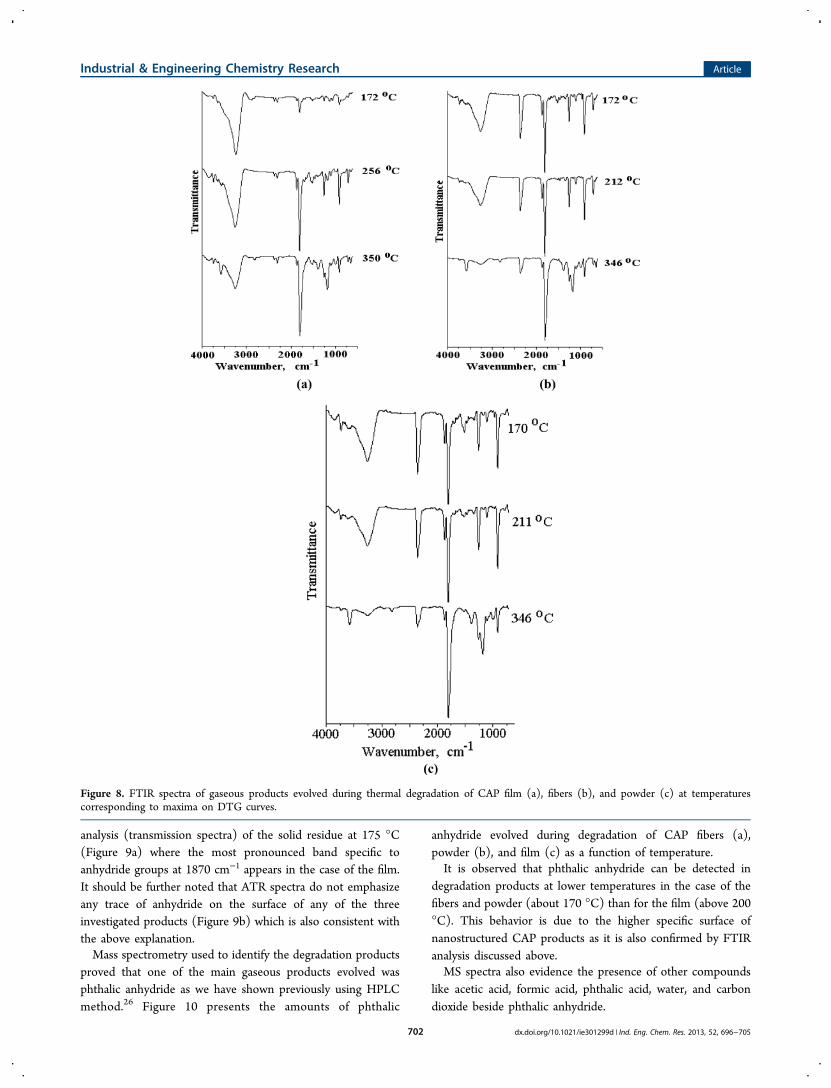
analysis (transmission spectra) of the solid residue at 175 °C(Figure 9a) where the most pronounced band specific toanhydride groups at 1870 cm−1 appears in the case of the film.It should be further noted that ATR spectra do not emphasizeany trace of anhydride on the surface of any of the threeinvestigated products (Figure 9b) which is also consistent withthe above explanation.Mass spectrometry used to identify the degradation products
proved that one of the main gaseous products evolved wasphthalic anhydride as we have shown previously using HPLCmethod.26 Figure 10 presents the amounts of phthalic
anhydride evolved during degradation of CAP fibers (a),powder (b), and film (c) as a function of temperature.It is observed that phthalic anhydride can be detected in
degradation products at lower temperatures in the case of thefibers and powder (about 170 °C) than for the film (above 200°C). This behavior is due to the higher specific surface ofnanostructured CAP products as it is also confirmed by FTIRanalysis discussed above.MS spectra also evidence the presence of other compounds
like acetic acid, formic acid, phthalic acid, water, and carbondioxide beside phthalic anhydride.
Figure 8. FTIR spectra of gaseous products evolved during thermal degradation of CAP film (a), fibers (b), and powder (c) at temperaturescorresponding to maxima on DTG curves.
Industrial & Engineering Chemistry Research Article
dx.doi.org/10.1021/ie301299d | Ind. Eng. Chem. Res. 2013, 52, 696−705702

3.3. Antimicrobial Activity of Nanostructured CAPs.The antimicrobial tests show evidence of the susceptibility ofthe two investigated microorganisms over the nanofibersamples and nanopowders of CAP as it is illustrated in Figures11 and 12 by the occurrence of an inhibition zone around thesample.For CAP powders, in Table 1 the antimicrobial activity is
expressed by the diameter of the inhibition zone, as in the caseof CAP films previously reported.31 Both nanopowders of CAPexhibit antibacterial activity, but this is slightly morepronounced in the case of powder obtained from solution in2-methoxyethanol−water mixture 85/15 (v/v) (P2 sample),
probably because of the lower particle size resulted in theseconditions.In the case of CAP nanofibers, the samples were soaked on
the surface of the culture medium and the inhibition zone inthis case may be associated with the area occupied by thesample and around it. The dark color of the sample area andthe halo around it suggest the inhibition of microorganismgrowth within these areas.
Figure 9. FTIR spectra for the solid residue of CAP at 175 °C as (a)transmission and (b) ATR.
Figure 10. MS analysis of phthalic anhydride produced by thermaldegradation of CAP fibers (a), powder (b), and film (c) as a functionof temperature.
Industrial & Engineering Chemistry Research Article
dx.doi.org/10.1021/ie301299d | Ind. Eng. Chem. Res. 2013, 52, 696−705703

■ CONCLUSIONS
Cellulose acetate phthalate (CAP) micro/nanoparticles ob-tained from 2-methoxyethanol at 10% (w/v) polymerconcentration and those obtained from 2-methoxyethanol−water mixture 85/15 (v/v) at 10% (w/v) polymer concen-tration exhibit lower width of their walls and lower diametersthan particles obtained from 2-methoxyethanol at 12.5% (w/v)polymer concentration previously reported. Also, polymersolution in the solvent mixture 2-methoxyethanol−water 85/15 (v/v) gives smaller particles with specific surface area higherthan of those produced from 2-methoxyethanol solution.CAP nanofiber mats obtained from 2-methoxyethanol−water
mixture 85/15 (v/v) at 30% (w/v) polymer concentrationappear more uniform and fibers diameters produced fromsolvent mixture are lower than those obtained from 2-methoxyethanol solvent.TG analysis reveals that CAP nanoproducts exhibit an
apparent lower stability than CAP films. Although TG/DTGcurves show a lower temperature of weight loss for the firststage of degradation for nanostructured products as comparedwith the film, FTIR investigations of the residual polymer andthe evolved degradation gas products as well as massspectrometry evidence the presence of phthalic anhydride asa main degradation product for all CAP samples. The higherporosity and specific surface areas of nanostructured CAPsallow a better diffusion of this degradation product which iseasier and continuously eliminated from these samples duringthe thermal treatment.
Nanostructured CAPs exhibit antimicrobial activity on bothStaphylococcus aureus and Escherichia coli.
■ AUTHOR INFORMATIONCorresponding Author*E-mail: [email protected] authors declare no competing financial interest.
■ REFERENCES(1) Fang, J.; Niu, H. T.; Lin, T.; Wang, X. G. Applications ofelectrospun nanofibers. Chin. Sci. Bull. 2008, 53, 2265.(2) Subbiah, T.; Bhat, G. S.; Tock, R. W.; Parameswaran, S.;Ramkumar, S. S. Electrospinning of nanofibers. J. Appl. Polym. Sci.2005, 96, 557.(3) Liu, H.; Hsieh, Y.-L. Ultrafine fibrous cellulose membranes fromelectrospinning of cellulose acetate. J. Polym. Sci., Part B: Polym. Phys.2002, 40, 2119.(4) Liu, H.; Tang, C. Electrospinning of Cellulose Acetate in SolventMixture N,N-Dimethylacetamide (DMAc)/Acetone. Polym. J. 2007,39, 65.(5) Han, D.; Gouma, P. I. Electrospun bioscaffolds that mimic thetopology of extracellular matrix. Nanomed.: NBM 2006, 2, 37.(6) Chen, L.; Bromberg, L.; Alan Hatton, T.; Rutledge, G. C.Electrospun cellulose acetate fibers containing chlorhexidine as abactericide. Polymer 2008, 49, 1266.(7) Son, W. K.; Youk, J. H.; Lee, T. S.; Park, W. H. Electrospinning ofultrafine cellulose acetate fibers: Studies of a new solvent system andacetylation of ultrafine cellulose acetate fibers. J. Polym. Sci., Part B:Polym. Phys. 2004, 42, 5.
Figure 11. Antimicrobial tests for CAP nanopowders obtained from solutions in 2-methoxyethanol (P1) and in 2-methoxyethanol−water mixture85/15 (v/v) (P2) at 10% (w/v) polymer concentration, against S. aureus (a) and E. coli (b).
Figure 12. Antimicrobial tests for CAP nanofibers obtained from solutions in 2-methoxyethanol (F1) and in 2-methoxyethanol−water mixture 85/15(v/v) (F2) at 10% (w/v) polymer concentration, against S. aureus (a) and E. coli (b).
Industrial & Engineering Chemistry Research Article
dx.doi.org/10.1021/ie301299d | Ind. Eng. Chem. Res. 2013, 52, 696−705704

(8) Zuwei, M.; Kotaki, M.; Ramakrishna, S. Electrospun cellulosenanofiber as affinity membrane. J. Membr. Sci. 2005, 265, 115.(9) Wu, X.; Wang, L.; Yu, H.; Huang, Y. Effect of solvent onmorphology of electrospinning ethyl cellulose fibers. J. Appl. Polym. Sci.2005, 97, 1292.(10) Park, J. Y.; Han, S. W.; Lee, I. H. Preparation of ElectrospunPorous Ethyl Cellulose Fiber by THF/DMAc Binary Solvent System.J. Ind. Eng. Chem. 2007, 13, 1002.(11) Jeun, J.-P.; Lim, Y.-M.; Choi, J.-H.; La, H.-S.; Kang, P.-H.; Nho,Y.-C. Preparation of Ethyl-Cellulose Nanofibers via An Electro-spinning. Solid State Phenom. 2007, 119, 255.(12) Frenot, A.; Walenius Henriksson, M.; Walkenstrom, P. Electro-spinning of cellulose-based nanofibers. J. Appl. Polym. Sci. 2007, 103,1473.(13) Sui, X.; Yuan, J.; Yuan, W.; Zhou, M. Preparation of CelluloseNanofibers/Nanoparticles via Electrospray. Chem. Lett. 2008, 37, 114.(14) Kim, C.-W.; Frey, M. W.; Marquez, M.; Joo, Y. L. Preparation ofsubmicron-scale, electrospun cellulose fibers via direct dissolution. J.Polym. Sci., Part B: Polym. Phys. 2005, 43, 1673.(15) Kulpinski, P. Cellulose nanofibers prepared by the N-methylmorpholine-N-oxide method. J. Appl. Polym. Sci. 2005, 98,1855.(16) Gyotoku, T.; Aurelian, L.; Neurath, A. R. Cellulose acetatephthalate (CAP): an ‘inactive’ pharmaceutical excipient with antiviralactivity in the mouse model of genital herpesvirus infection. Antiviral.Chem. Chemother. 1999, 10, 327.(17) Neurath, A. R.; Strick, N.; Li, Y. Y.; Debnath, A. K. Celluloseacetate phthalate, a common pharmaceutical excipient, inactivatesHIV-1 and blocks the coreceptor binding site on the virus envelopeglycoprotein gp 120. BMC Infect. Dis. 2001, 1, 17 ; http://www.biomedcentral.com/1471-2334/1/17.(18) Duncan, R. The dawning era of polymer therapeutics. Nat. Rev.Drug Discovery 2003, 2, 347.(19) Neurath, A. R.; Strick, N.; Li, Y. Y. Water dispersiblemicrobicidal cellulose acetate phthalate film. BMC Infect. Dis. 2003,3, 27 ; http://www.biomedcentral.com/1471-2334/3/27.(20) Neurath, A. R.; Strick, N.; Jiang, S.; Li, Y. Y.; Debnath, A. K.Anti-HIV-1 activity of cellulose acetate phthalate: Synergy with solubleCD4 and induction of “dead-end” gp41 six-helix bundles. BMC Infect.Dis. 2002, 2, 6 ; http://www.biomedcentral.com/1471-2334/2/6.(21) Manson, K. H.; Wyand, M. S.; Miller, C.; Neurath, A. R. Effectof a Cellulose Acetate Phthalate Topical Cream on VaginalTransmission of Simian Immunodeficiency Virus in Rhesus Monkeys.Antimicrob. Agents Chemother. 2000, 44, 3199.(22) Neurath, A. R.; Strick, N.; Li, Y. Y. Anti-HIV-1 activity ofanionic polymers: a comparative study of candidate microbicides. BMCInfect. Dis. 2002, 2, 27.(23) Olaru, N.; Olaru, L. Electrospinning of cellulose acetatephthalate from different solvent systems. Ind. Eng. Chem. Res. 2010, 49,1953.(24) Huang, C.; Soenen, S. J.; Van Gulck, E.; Vanham, G.; Rejman, J.;Van Calenbergh, S.; Vervaet, C.; Coenye, T.; Verstraelen, H.;Temmerman, M.; Demeester, J.; De Smedt, S. C. Electrospuncellulose acetate phthalate fibers for semen induced anti-HIV vaginaldrug delivery. Biomaterials 2012, 33, 962.(25) Roxin, P.; Karlsson, A.; Singh, S. K. Characterization ofCellulose Acetate Phthalate (CAP). Drug Dev. Ind. Pharm. 1998, 24,1025.(26) Olaru, N.; Olaru, L.; Leanca, M. On Thermal Degradation ofCellulose Acetate Phthalate. Cellul. Chem. Technol. 1995, 29, 253.(27) Olaru, N.; Olaru, L.; Andriescu, A.; Tudorachi, N. PartialHydrolysis of cellulose acetate in toluene/acetic acid/water system.Angew. Makromol. Chem. 1996, 241, 67.(28) Malm, C. J.; Genung, L. B.; Kuchmy, W. Analysis of PhthalicAcid Esters of Cellulose and of Polyvinyl Alcohol. Anal. Chem. 1953,25, 245.(29) Tanghe, L. J.; Genung, L. B.; Mench, J. W. ; Determination ofAcetyl Content and Degree of Substitution of Cellulose Acetate. In
Methods in Carbohydrate Chemistry; Whistler, R. L., Ed.; AcademicPress: New York, London, 1963; Vol. 3, 201−203.(30) Wistreich, G. A. Microbiology Laboratory; Prentice Hall: UpperSaddle River, NJ, 2000.(31) Necula, A. M.; Dunca, S.; Stoica, I.; Olaru, N.; Olaru, L.; Ioan, S.Morphological properties and antibacterial activity of nano-silvercontaining cellulose acetate phthalate films. Int. J. Polym. Anal. Charact.2010, 15, 341.
Industrial & Engineering Chemistry Research Article
dx.doi.org/10.1021/ie301299d | Ind. Eng. Chem. Res. 2013, 52, 696−705705


![THE ACETATE NEGATIVE SURVEY · using cellulose acetate.[1] Cellulose acetate is manufactured by combining cotton linters or wood pulp (the sources of the cellulose fibers) with acetic](https://static.fdocuments.in/doc/165x107/5e448d99bd61564bfe5016d9/the-acetate-negative-survey-using-cellulose-acetate1-cellulose-acetate-is-manufactured.jpg)