Nano Diamonds
11
Journal of Applied Pharmaceutical Science 01 (06); 2011: 29-39 I SSN: 223 1-3354 Received: 07-07-2011 Revised on: 17-07-2011 Accept ed: 0 2-08-2011 Khali d Mohame d El- S ay Depa rtme nt of Pharmace uti cs and I ndustri al Pharma cy F acult y o f Pharmacy A l-A z ha r U nive rs i ty Nas r ci ty, Ca i ro, Egy pt For Corr es po ndence : Dr.Khalid Mohamed El-Say, A ss oc i ate profes sor o f pharmace uti cs and i ndustri al pha rmacy Depa rtme nt of Pharmace uti cs and I ndustri al pha rmac y, F ac ult y o f Pharmac y, A l-A z ha r Uni ve rs i ty, Nasr C i ty, Cair o, Egypt. T e l. + 2-011218 188 2 Nanodiamond as a drug delivery system: Applications and prospective Khalid Mohamed El-Say ABSTRACT With the rapid development of nanoscience and nanotechnology, a wide variety of nanomaterials have been synthesized and discovered. Revolutionary particles called nanodiamonds (NDs) have been considered for use in several medical applications due to its unique mechanical, chemical, optical, and biological properties. It has also sensing, imaging, and drug delivery properties. The study associated with the interface between ND and life sciences which is important for development of effective drug delivery systems. Key word s: Nanotechnology, nanomaterial, nanodiamond, drug delivery system. INTRODUCTION In recent years, a number of synthetic methods for the preparation of nanocrystalline diamond, “Nanodiamond” (ND), in the form of films and powders, have been developed (Dolmatov, 2001; Shenderova et al., 2002). Particularly, detonation synthesis, from powerful explosive mixtures (Greiner et al., 1988; Kuznetsov et al., 1994) has made such ND powder commercially available in ton quantities which has enabled many engineering applications and has expanded the applicatio n scope of diamond (Dolmatov , 2001). ND powders prepared by explosive techniques present a novel class of nanomate rials possessing unique surface properties. Due to the very small particle size (2-10 nm), a larger percentage of atoms in NDs contribute to the defect sites on grain boundaries than in single crystal natural or microcrystalline synthetic diamonds. For example, in individual 4.3 nm spherical particles of ND comprising about 7200 carbon atoms, nearly 1100 atoms are located at the surface (Shenderova et al., 2002). For this reason, the surface modifications of the nanosize diamond grains can affect the bulk properties of this material more strongly than those of micro- and macroscale diamo nds. For example, ND powders can form good abrasive pastes and sus pensions for high-precision polishing; ND-polyme r composites are applied for manufacturing aircraft, cars and ships, as well as in hard and wear-resistant surface coatings. They are considered potential medical agents due to their high adsorption capacity, high specific surface area, and chemical inertness (Dolmatov, 2001; Shenderova et al., 2002). Applications of ND thin films have been demonstrated in the fabrication of cold cathodes, field emission displays (Choi et al., 1996; Ralchenko et al., 1999; Alimova et al., 1999; Jiang et al., 2002; Show et al., 2003), nanomechanical and nanoelectromechanical resonant structures (NEMS) (Wang et al., 2002; Sekaric et al., 2002; Philip et al., 2003), and were suggested for the design of biosensors as stable biologically active substrates after DNA-modification (Yang et al., 2002).
-
Upload
ravindrachary-niper -
Category
Documents
-
view
216 -
download
0
Transcript of Nano Diamonds

8/2/2019 Nano Diamonds
http://slidepdf.com/reader/full/nano-diamonds 1/11
Journal of Applied Pharmaceutical Science 01 (06); 2011: 29-39
I SSN: 2231-3354
Received: 07-07-2011
Revised on: 17-07-2011Accepted: 02-08-2011
Khalid Mohamed El- Say
Department of Pharmaceuti cs and
I ndustrial Pharmacy Faculty of Pharmacy
A l-A zhar U niversi ty Nasr ci ty, Cai ro, Egypt
For Corr espondence :
Dr.Khalid Mohamed El-Say,
Associ ateprofessor of pharmaceuti cs and industri al pharmacy
Department of Pharmaceuti cs and
I ndustrial pharmacy,Faculty of Pharmacy,
A l-A zhar Uni versi ty, Nasr Ci ty,Cairo, Egypt.
Tel. +2-0112181882
Nanodiamond as a drug delivery system:
Applications and prospective
Khalid Mohamed El-Say
ABSTRACT
With the rapid development of nanoscience and nanotechnology, a wide variety of nanomaterials have been synthesized and discovered. Revolutionary particles called nanodiamonds
(NDs) have been considered for use in several medical applications due to its unique mechanical,
chemical, optical, and biological properties. It has also sensing, imaging, and drug delivery
properties. The study associated with the interface between ND and life sciences which is important
for development of effective drug delivery systems.
Key words: Nanotechnology, nanomaterial, nanodiamond, drug delivery system.
INTRODUCTION
In recent years, a number of synthetic methods for the preparation of nanocrystalline
diamond, “Nanodiamond” (ND), in the form of films and powders, have been developed
(Dolmatov, 2001; Shenderova et al., 2002). Particularly, detonation synthesis, from powerful
explosive mixtures (Greiner et al., 1988; Kuznetsov et al., 1994) has made such ND powder
commercially available in ton quantities which has enabled many engineering applications and has
expanded the application scope of diamond (Dolmatov, 2001). ND powders prepared by explosive
techniques present a novel class of nanomaterials possessing unique surface properties. Due to the
very small particle size (2-10 nm), a larger percentage of atoms in NDs contribute to the defect
sites on grain boundaries than in single crystal natural or microcrystalline synthetic diamonds. For
example, in individual 4.3 nm spherical particles of ND comprising about 7200 carbon atoms,
nearly 1100 atoms are located at the surface (Shenderova et al., 2002). For this reason, the surface
modifications of the nanosize diamond grains can affect the bulk properties of this material morestrongly than those of micro- and macroscale diamonds. For example, ND powders can form good
abrasive pastes and suspensions for high-precision polishing; ND-polymer composites are applied
for manufacturing aircraft, cars and ships, as well as in hard and wear-resistant surface coatings.
They are considered potential medical agents due to their high adsorption capacity, high specific
surface area, and chemical inertness (Dolmatov, 2001; Shenderova et al., 2002). Applications of
ND thin films have been demonstrated in the fabrication of cold cathodes, field emission displays
(Choi et al., 1996; Ralchenko et al., 1999; Alimova et al., 1999; Jiang et al., 2002; Show et al.,
2003), nanomechanical and nanoelectromechanical resonant structures (NEMS) (Wang et al.,
2002; Sekaric et al., 2002; Philip et al., 2003), and were suggested for the design of biosensors as
stable biologically active substrates after DNA-modification (Yang et al., 2002).

8/2/2019 Nano Diamonds
http://slidepdf.com/reader/full/nano-diamonds 2/11
Journal of Applied Pharmaceutical Science 01 (06); 2011: 29-39
In order to minimize surface energy, individual ND
particles (crystallites) of 4-6 nm size structurally self-organize into
clusters or primary aggregates of 20-30 mm size. These, in turn,
form larger weakly bonded secondary aggregates ranging from
hundreds of nanometers to micron sizes. This agglomeration is
likely facilitated by surface functional groups, such as —OH, —
COOH, —SO3H, and —NH2, which are created along with otherfunctionalities by chemical treatment processing of detonation-
synthesized ND (Jiang et al., 1996; Shenderova et al., 2002) and
participate in the formation of hydrogen bonds between ND
clusters. However, for advanced applications of ND powder (e.g.,
in higher precision polishing compositions, nanoengineered
electronic devices, polymer and ceramic composites, and bio-
medical systems), the reduction of aggregate sizes to below 200
nm, and ultimately even to single clusters or particles, and the
availability of specific functional groups on the surface is highly
desirable. These functional groups can also serve as binding sites
for covalent integration of ND into polymer structures and provide
for improved solubility of ND powder in common solvents.
Surface modification of the ND powder particles through a
selective surface chemistry should be instrumental in approaching
these goals. In view of the foregoing, functionalized ND powder
will likely extend the utility of ND powder, and methods of making
such functionalized ND powder will be in great demand.
Nanodiamond Structure
ND is an allotrope of carbon. NDs are carbon-based
materials approximately 2 to 8 nanometers in diameter. Each ND's
surface possesses functional groups that allow a wide spectrum of
compounds to be attached to it, including chemotherapy agents.
The crystal structure of ND consists of two close packed
interpenetrating face centered cubic lattices; one lattice is shiftedwith respect to the other along the elemental cube space diagonal
by one-quarter of its length (Figure 1) (Iakoubovskii et al., 2000;
Iakoubovskii et al., 2008).
Fig 1: Crystalline structure of ND
The NDs also referred to as ultradisperse diamonds are
particles in the 2-8 nm size range. ND is often described as a
crystalline diamond core with a perfect diamond lattice surrounded
by an amorphous shell with a combination of sp2 /sp3 bonds or
onion-like graphite shell (Shengfu et al., 1998). NDs are clustered
carbon atoms with both graphitic (sp2) and diamondoid (sp
3)
bonds. The two types of bonds can be interchangeable, for
example, the stretched face of diamond is a graphene plane. In
reverse, the puckered graphene may become a diamond surface.
This interchangeability allows ND particles to be flexible
templates, particularly around the curved surface where electrons
are unstable (Krueger, 2010).
NANODIAMONDS SYNTHESIS
In the phase diagram of carbon (Figure 2) there is a region
at very high temperatures and pressure where diamond is stable.Hence, normally it is required to sustain according conditions for
its production (Krueger, 2010).
Fig 2: Carbon phase diagram.
NDs can be produced by different methods:
1. Detonation Nanodiamond
Detonation ND (DND), often also called ultradispersed
diamond (UDD), is diamond that originates from a detonation. A
ND can be created by detonating mixture of trinitrotoluene (TNT)
and hexogen (RDX) (Figure 3) and then gathering the remaining
soot.
Fig 3: Structural formulae of the two explosives most commonly employed for
detonation synthesis.
Fig 4: Detonation ND, TEM image
This idea is that pure ND can be produced by the
detonation of a diamond blend and will then form by chemical
purification. The soot left over actually contains tiny diamonds,
which measure four nanometers in size. However, in order for

8/2/2019 Nano Diamonds
http://slidepdf.com/reader/full/nano-diamonds 3/11
Journal of Applied Pharmaceutical Science 01 (06); 2011: 29-39
these diamonds to shine and look anything like diamonds they
must be exposed to a high-energy electron beam and then heated
800 degrees Celsius. The diamond yield after detonation crucially
depends on the synthesis condition and especially on the heat
capacity of the cooling medium in the detonation chamber (water,
air, CO2, etc.). The higher the cooling capacity, the larger the
diamonds yield, which can reach 90%. After the synthesis,diamond is extracted from the soot using high-temperature high-
pressure (autoclave) boiling in acid for a long period (ca. 1–2
days). The boiling removes most of the metal contamination,
originating from the chamber materials, and non-diamond carbon.
Various measurements, including X-ray diffraction (Iakoubovskii
et al., 2000) and high-resolution transmission electron microscopy
(Iakoubovskii et al., 2008) revealed that the size of the diamond
grains in the soot is distributed around 5 nm. The grains are
unstable with respect to aggregation and spontaneously form
micrometer-sized clusters (Figures 4 and 5). The adhesion is strong
and contacts between a few nano-grains can hold a micrometer-
sized cluster attached to a substrate (Iakoubovskii et al., 2008).
Fig 5: Detonation diamond as powder (a), as unstable suspension in water (center)
and as completely deagglomerated dispersion in water (b).
2. Ultrasonic Cavitation MethodDiamond nanocrystals can also be synthesized from a
suspension of graphite in organic liquid at atmospheric pressure
and room temperature using ultrasonic cavitation. The yield is
approximately 10%. The cost of NDs produced by this method is
estimated to be competitive with the HPHT process (Galimov et
al., 2004; Khachatryan et al., 2008).
3. Pulsed-laser irradiation:
An alternative synthesis technique is irradiation of
graphite by high-energy laser pulses. The structure and particle size
of the obtained diamond is rather similar to that obtained in
explosion. In particular, many particles exhibit multiple twinning
( Shengliang et al., 2008).
NANODIAMONDS PROPERTIES
ND has many unique physical characteristics just like its
bigger cousin, the full karat diamond stone. The following superior
properties of ND make it a special and promising material that can
be widely applied in numerous fields:
1. Hardness
Table 1 shows some physical properties of diamond
compared to Titanium and stainless steel. The hardness of diamond
is about 50 times of Titanium and stainless steel. The toughness of
diamond makes it suitable in applications in biomedical fields such
as implant, cutting tools for surgeries, etc.
Table 1: Comparison of properties of chemical vapor deposited (CVD) diamond,titanium and stainless steel (316).
Properties CVD
diamond
Titanium Stainless steel
Hardness (kg/mm2 10,000 230 210
Young's modulus (GPa) 1000 120.2 215.3Bulk modulus (GPa) 442 108.6 166
Thermal conductivity
(Watts/cm.°C)
20 0.21 0.16
3.8 (Cu)Thermal expansion
(×10-8
K-1
)
1.1 8.8 17.2
Coefficient of friction 0.05 0.1 (Graphite)
Refractive index
(in the near IR)
2.41- 2.44 3.42-3.5 (Si)
Source: From Ref. (Tang et al., 1995).
2. Chemical inertness is an important factor for ND to be applied
in biology, since the biological environment is corrosive. Alloy of
Ti6Al
4V coated with ND films show that the diamond films have a
very good chemical resistance to the corrosive liquid (Azevedo et
al., 2005).
3. Biocompatibility cannot be ignored when diamond is applied to
biology. Yu and coworkers investigated the biocompatibility of
fluorescent ND (FND) powder with size of 100nm in cell culture
and found
low cytotoxicity in kidney cells (Yu et al., 2005). Further, Schrand
and coworkers showed that ND with small size of 2-10nm are not
toxic to a variety of cells through mitochondrial function (MIT)
and luminescent ATP production assays (Schrand et al., 2007). It
was found that after the incubation of cells with NDs, cell
morphology is unaffected by the presence of NDs while NDs areseen surrounding the cell borders and attached to neurite
extensions.
4. Excellent optical property is necessary for diamond to be
applied as a biomarker or a biolabel. There are impurity sites
within core, defects in the diamond or sp2
clusters on the ND
surface. With the light excitation, the ND will emit light with
different frequency due to different type of impurity sites (Holt,
2007).
5. Chemical modification of diamond surface is essential for
diamond to be applied as potential biosensor or biochip (Guan et
al., 2006), or a substrate to immobilize biological molecules.
Diamond surface can be hydrogen-terminated by exposing the
surface to 13.5-MHz inductively coupled hydrogen plasma (15
torr) at 800°C (Thoms et al., 1994). With the hydrogen-terminated
nanocrystalline diamond, Yang and coworkers successfully
designed a chemical procedure to attach DNA onto the diamond
surface. Recently, ND with the size of 5-100nm in diameter was
carboxylated by Chang and Han, (2006). It was found that
carboxylated ND (Holt, 2007) has good physical absorption
properties including hydrophobic and hydrophilic interaction,
which can be used to immobilize biomolecules.

8/2/2019 Nano Diamonds
http://slidepdf.com/reader/full/nano-diamonds 4/11
Journal of Applied Pharmaceutical Science 01 (06); 2011: 29-39
6. Tiny size and ability to control the constancy of the nano-size
and the shape of agglutinates.
7. Large Surface area and high adsorption potential:
It has extremely large relative surface area. As a
result, its surface spontaneously attaches water and hydrocarbonmolecules from the ambient atmosphere ( Shengfu et al., 1998) and
also larger number of drugs can be placed on the particles.
However, clean ND surface can be obtained with appropriate
handling (Iakoubovskii et al., 2008).
8. Outstanding photoluminescence.
NANODIAMOND APPLICATIONS AND PROSPECTIVE
Owing to its properties, ND like the classical diamond is
an attractive material for many applications. They include the
preparation of composites and coatings, mechanical applications to
reduce friction or to modify surfaces, uses in electro-deposition or
biomedical applications.
1. Mechanical Applications
Super smooth polishing of gems, ceramics, glass, silicon
wafers, and surgical knives (Sung and Lin, 2010). So, it is
widely used in various polishing compositions (pastes, gels
and slurries) for obtaining especially smooth surfaces in
production of computer hard discs; lenses, prisms, mirrors, etc.
for various optical devices; high precision parts made of steel
and hard alloys; silicon wafers; and/or body implants.
Reinforcement of rubber, resins, plastic, PTFE, and metals
(Cu, Al) (Shenderova et al., 2002). NDs are used as activefillers in polymers providing improvement of various service
characteristics as strength, elasticity, and increasing heat
conductivity; and improving the optical characteristics of
polymers.
Lubrication of engine oil and machine grease: ND can reduce
significantly the frictional coefficient by coating the sliding
surface.
Electroplating of metal coating to provide markedly improved
mechanical properties of the metal (Sung and Lin, 2010) in
production of punches, dies, matrices and molds; screws,
check valves, pistons and spruces; metal cutting tools; and /or
food processing equipment.
2. Thermal Applications
The high thermal conductivity can be employed for ND
applications as well. ND is a suitable additive for coolants to
improve the thermal conductivity of cooling media. An addition of
only 0.3% to cooling oils for large transistors causes a 20% growth
of thermal conductivity. This effect serves to prevent the formation
of hot zones inside the coolant and the consequent destruction of
the transistor (Krueger, 2010). So, ND efficiently enhances
material's ability to dissipate heat. Consequently, using ND in
pastes, glues and substrates provides an exceptional opportunity of
avoiding burnout, increasing speed of active elements, reducing the
size of devices, and increasing their reliability and durability.
3. Electrochemical Applications
Due to its physicochemical stability, large electrochemical
potential window, and chemical sensitivity (Prado et al., 2002),
diamond is an excellent candidate for electrochemical applications.Diamond electrodes show the most stable response among
electrodes by far, and do not require extensive pretreatment to
regenerate the electroactive surface (Halpern et al., 2006). ND
electrodes/microelectrodes have been applied to biological system
as biosensors (Martinez-Huitle, 2007).
4. Cosmetic Applications
As ND is non-poisonous, a big area of application is ND-
impregnated cosmetics. So, ND can be formulated as a dental
filling, lotion, deodorant, toothpaste, shampoo, antibiotic, dermal
strip, skin cleanser, or exfoliant.
4.1. Dental care
ND may be formulated as a dental material. The dental
material can be formulated for use as a filling, veneer,
reconstruction, and the like. The ND particles can provide
additional mechanical strength, as well as an appearance that
approximates natural enamel when dry. Alternatively, the remedial
healthcare composition can be formulated as toothpaste including
an acceptable carrier and a plurality of ND particles. ND added
toothpaste has another advantage, as ND is known to cure gum
disease (Sung and Lin, 2010).
4.2. Skin Care
Owing to unique adsorption capabilities of ND, their
addition to skincare products enhances the effect of biologicallyactive ingredients and facilitates their penetration into deeper skin
layers. ND makes all biologically-active additives "work" at their
maximum efficiency. Also, due to their unique optical properties,
ND is an excellent agent for skin protecting from harmful UV
radiation.
The ND surface functional groups form powerful bonding
with water molecules to provide an all-day-long moisturizing effect
and to protect the skin from aging. At the same time, ND-base
creams are fully and rapidly absorbed by the skin.
ND compositions can be formulated for skin care products
such as lotions, facial tissue lotion, deodorant, dermal strip, skin
cleanser, soap, and exfoliant. The ND particles can be dispersed in
a biologically acceptable carrier and contacted with a biological
material such as organic oils, sebum, bacteria, epithelial cells,
amino acids, proteins, DNA, and combinations thereof. Once the
biological material is bonded to ND, the ND composition can be
removed from the surface or environment. The ND composition
can then be discarded or further treated to identify or otherwise
utilize the absorbed biological material.
The presence of ND particles can improve absorption of
oils and undesirable deposits from the skin without abrasiveness
associated with larger diamond particles. The NDs can be present
in the facial tissue lotion, deodorant, dermal strip, and skin
8/2/2019 Nano Diamonds
http://slidepdf.com/reader/full/nano-diamonds 5/11
Journal of Applied Pharmaceutical Science 01 (06); 2011: 29-39
cleanser. In addition, the skin cleanser can include additives such
as fragrance, colorants, vitamin E, herbal supplements, antibiotics,
UV absorbers, hydrating agents, sun-block agents, exfoliating
agents, and the like. Also, NDs in antibiotic and lotion
compositions can increase healing of skin and removal of damaged
skin such as with sunburns and scar tissue (Sung and Lin, 2010).
4.3. Hair and nail care
Shampoo can include an acceptable carrier and ND
particles. Suitable bubbling agents can be included to increase
contact of unsaturated NDs with a biological material. This can be
advantageous in maximizing the effect of NDs in skin cleansers,
deodorants, shampoos, soaps, toothpaste, etc...
Another cosmetic ND composition can be formulated as a nail
polish, eyeliner, lip-gloss, or exfoliant. ND particles can also
improve the durability of the applied nail compositions.
Specifically, NDs can provide increased resistance to chipping and
wear, e.g. typically a ND nail polish can last from about three to
ten time longer than typical nail lacquer formulations (Sung and
Lin, 2010).
5. Biological Applications
ND is the state-of-the-art material rapidly finding its way
into biotechnologies such as:
5.1. Nanodiamond in biomedical applications
Due to its hardness, chemical inertness, thermal
conductivity, and low cytotoxicity, ND could be applied as coating
materials of implants, other surgery tools, etc. in biomedical fields.
In 1995, for the first time, Zolyfinski and coworkers implanted
orthopedic screws, coated with nanocrystalline diamond film
(NCD) to a patient with a complex fracture of femoral bone
(Zolyfinski et al., 1996). After surgery, no ejection was observed,whereas the standard metal implants were rejected twice. As such,
diamond is ideal for use in medical applications, e.g. artificial
replacements (joint coatings, heart valves, etc.), and will not
deteriorate over time. Another implantation application of diamond
is that an endoprothesis of hip joint, coated with NCD film was
successfully implanted to a living organism (Mitura et al., 2006).
5.2. Immobilization of biomolecules
The chemical modification and physical absorption of
diamond surface hold promises for ND to be applied in
immobilization of protein and DNA for purification, separation,
and further analysis (Figure 6). Using detonation ND, Bondar and
coworkers (Bondar et al., 2004)
successfully separatedrecombinant Ca
2+-activated photoprotein apoobelin and
recombinant luciferase from bacterial cells of Escherichia coli
through physical absorption of proteins on ND. For traditional
purification by chromatographic means, it usually takes several
days. The procedures using ND, the whole process took 30-40 min
with a yield of 35-60%. Kong and coworker applied the same
principle to capture proteins and DNA for the matrix-assisted laser
desorption/ ionization (MALDI) time of fight (TOF) mass
spectrometry (MS).
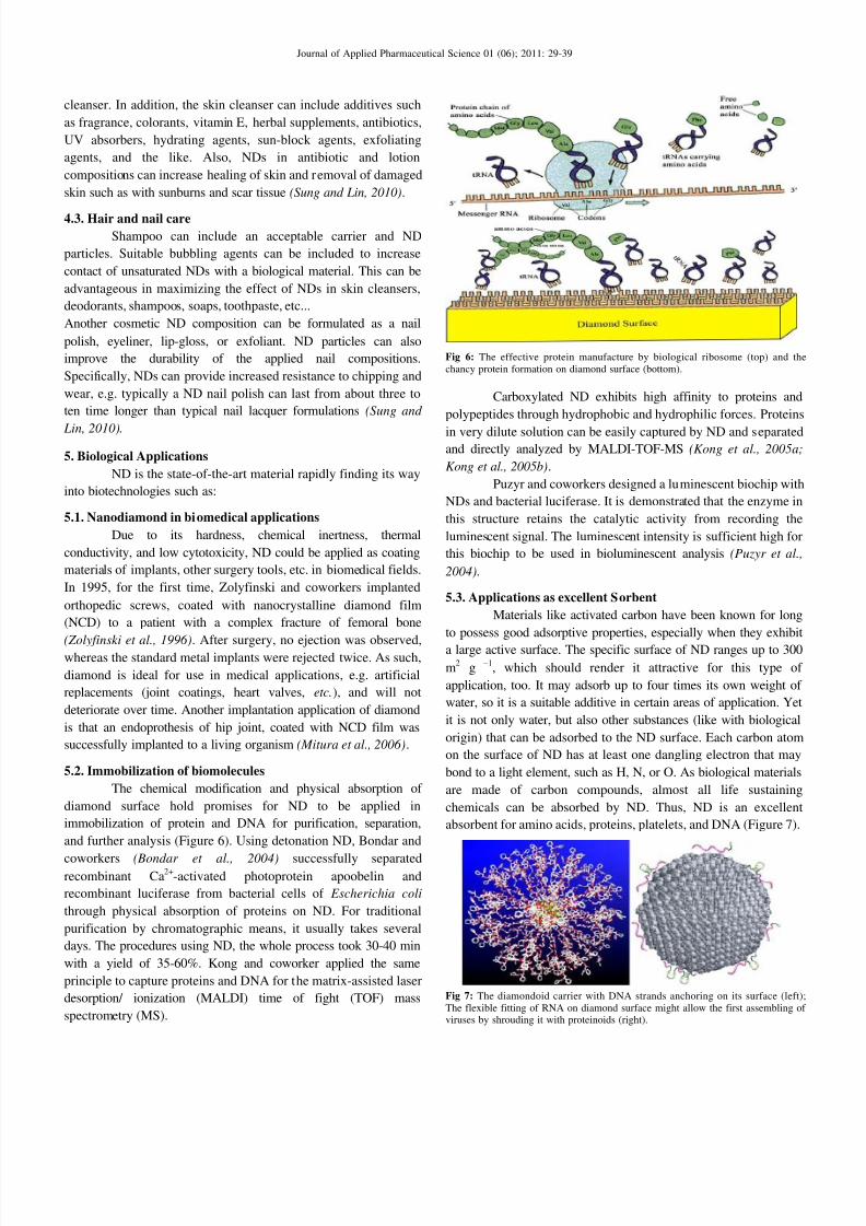
Fig 6: The effective protein manufacture by biological ribosome (top) and the
chancy protein formation on diamond surface (bottom).
Carboxylated ND exhibits high affinity to proteins and
polypeptides through hydrophobic and hydrophilic forces. Proteins
in very dilute solution can be easily captured by ND and separatedand directly analyzed by MALDI-TOF-MS (Kong et al., 2005a;
Kong et al., 2005b).
Puzyr and coworkers designed a luminescent biochip with
NDs and bacterial luciferase. It is demonstrated that the enzyme in
this structure retains the catalytic activity from recording the
luminescent signal. The luminescent intensity is sufficient high for
this biochip to be used in bioluminescent analysis (Puzyr et al.,
2004).
5.3. Applications as excellent Sorbent
Materials like activated carbon have been known for long
to possess good adsorptive properties, especially when they exhibit
a large active surface. The specific surface of ND ranges up to 300
m2
g −1
, which should render it attractive for this type of
application, too. It may adsorb up to four times its own weight of
water, so it is a suitable additive in certain areas of application. Yet
it is not only water, but also other substances (like with biological
origin) that can be adsorbed to the ND surface. Each carbon atom
on the surface of ND has at least one dangling electron that may
bond to a light element, such as H, N, or O. As biological materials
are made of carbon compounds, almost all life sustaining
chemicals can be absorbed by ND. Thus, ND is an excellent
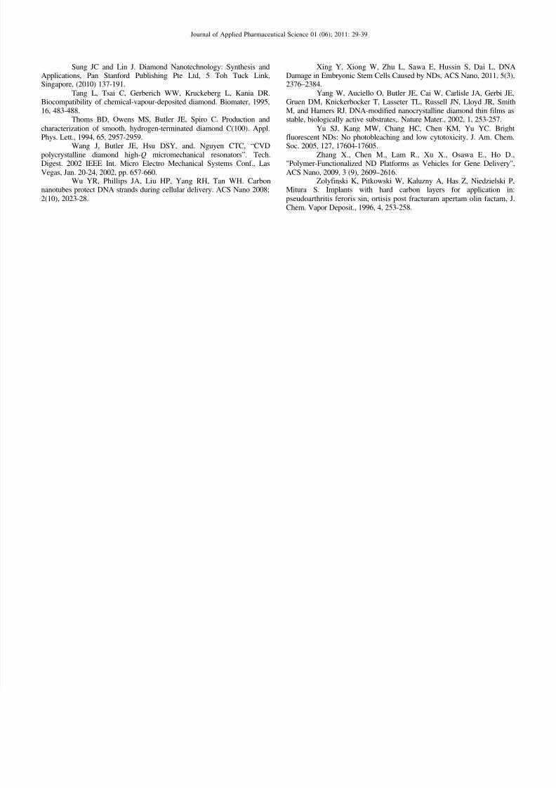
absorbent for amino acids, proteins, platelets, and DNA (Figure 7).
Fig 7: The diamondoid carrier with DNA strands anchoring on its surface (left);
The flexible fitting of RNA on diamond surface might allow the first assembling of viruses by shrouding it with proteinoids (right).

8/2/2019 Nano Diamonds
http://slidepdf.com/reader/full/nano-diamonds 6/11
Journal of Applied Pharmaceutical Science 01 (06); 2011: 29-39
Although diamond is highly stable, if the ND surface is
free of adsorbent or absorbent, i.e. clean, it is thought that carbon
atoms on the surface contain unpaired electrons that are highly
reactive. As a result, ND particles can readily bond to and
effectively absorb a variety of atomic species. For example, small
atoms such as H, B, C, N, O, and F can be readily adsorbed on the
ND surface, although other atoms can also be absorbed. Inaddition, those small atoms are building blocks, e.g. H, CO, OH,
COOH, N, CN and NO, of organic materials including biological
molecules. Consequently, ND particles can readily attach to amino
acids, proteins, cells, DNA, RNA, and other biological materials,
and ND particles can be used to remove skin oils, facial oils,
compounds that result in body odor, bacteria, etc.
Further, NDs are typically smaller than most viruses (10–
100 nm) and bacteria (10–100 µm). Therefore, ND can be used to
penetrate the outer layers of viruses and bacteria and then attach to
RNA, DNA or other groups within the organism to prevent the
virus or bacteria from functioning. Human body contains about
1/4 of carbon by weight. ND as carbon is non-poisonous.
Moreover, it is not only cancer inactive, but also a catalyst for
promoting drug effectiveness. For examples, ND has been used to
treat burning skin infections, food poisons, and intestine
malfunctions with good results. In comparison to tiny ND particles,
the human cells appear to be colossal (Figure 8). Much larger than
microbes, ND cannot harm normal cells. ND cannot penetrate the
cell’s membrane. On the contrary, ND can stick to the DNA of
bacteria or RNA of viruses. It is believable that ND may also be
effective in attaching genes and hence it is capable to kill drug
resistant viruses (e.g. HIV, SARS).
Fig 8: The relative scale of ND compared to microbes and cells.
ND surfaces may be modified by the termination of
various chemical radicals (Figure 9). If the termination is nonpolar
(e.g. H or F), the surface is hydrophobic. On the other hand, if
hydrogen bonds may be formed on the absorbents (e.g. O or S), thesurface is hydrophilic. The water repelling or wetting behavior is
important for dispersion in liquid. The surface modification also
allows the attachment of organic molecules, such as amine,
carboxyl, carbonyl, hydroxyl, amide, nitrile, sulfide, epoxyl,
phosphryl, sulfate, imide, etc.
For ND just formed, the surface contain ample C–O,
C=O, C–N, C=N, and OH. Although these radicals are wettable by
water, they tend to agglomerate. After boiling in sulfuric acid, the
dispersion in water improved, so is the dispersion. However, if an
organic polymer is used to as the matrix material, ND should be
heat treated beforehand in hydrogen, fluorine or chlorine to render
the surface hydrophobic (Sung and Lin, 2010).
Fig 9: The attachment of surface absorbents on ND (top diagram) and the major
surfactant radicals (bottom diagram).
5.4. Biomarkers and Biolabeling
ND has a very strong fluorescence at 700nm is emitted
after excited at 560nm (Yu et al., 2005) due to the nitrogen-
vacancy (N-V) center within diamonds. This is advantageous for
imaging in biological cells, as the background fluorescence in cell
is 300-400 nm (Holt, 2007). It was found that under the same
excitation conditions, the fluorescence of a single 35nm diamond is
significantly brighter than that of a single dye molecule such asAlexa Fluor 546. Fluorescent ND (FND) coated with poly-L-lysine
(PL) was used to study the interaction between DNA and FND on
an amine-terminated glass substrate (Fu et al., 2007). The PL was
used to facilitate the binding of DNA (fluorescently labeled with
TOTO-1 dye molecule) to FND. Due to the specific strong
fluorescence at 700nm of FND, the DNA molecule is wrapped
around the PL-coated FND particle. Fu and coworkers also
demonstrated that it is possible to conduct a single particle tracking
for a 35-nm FND in the cytoplasm of a live Hela cell. This tracking
method could be applied to drug delivery system of ND, where the
interaction between ND and cell could be monitored using
fluorescence microscopy. Raman spectrum of diamond exhibits asharp peak at 1332 cm
-1and the peak is isolated and the Raman
absorption cross section is large (Knight and White, 1989). This
peak can be used as an indicator of the location of ND. Also,
Cheng and co-workers tracked growth hormone factor in one
single cancer cell using ND growth hormone complex (cND-
rEaGH) as a specific probe (Cheng et al., 2007).
5.5. Nanodiamond could be used in disease diagnosis
Like ordinary diamonds, NDs have tiny holes, known as
vacancies, in which a nitrogen atom has replaced a carbon. High

8/2/2019 Nano Diamonds
http://slidepdf.com/reader/full/nano-diamonds 7/11
Journal of Applied Pharmaceutical Science 01 (06); 2011: 29-39
nitrogen content gives natural diamonds a yellowish tint, and the
NDs absorb yellow light and emit violet (Figure 10).
Fig 10: Fluorescent nanodiamond in diagnosis of disease.
NDs coated with sugars or proteins (such as dextran and
bovine serum albumin), and then introduced both coated and
uncoated types to transparent round worms (Caenorhabditis
elegans). The NDs were either fed to the worms as a colloidal
suspension or micro-injected into the gonads. It was found that the
plain NDs fed to the worms remained within the digestive tract,
coating its internal surfaces, while the coated NDs were able to
pass through the tract walls and into the worm’s body, collecting atseveral points. The location of the NDs was shown up as violet
light when yellow light was shone on the worms. When injected
into the gonads, the NDs were dispersed in the gonad and passed
on to the embryos and eventually to the hatched larvae. In addition,
all the worms in the study went on to live normal life spans with
normal reproduction, and none showed any sign of distress.
For this reason, the researchers showed NDs were safe,
stable, and non-toxic at cellular and organ levels. The NDs in the
study were coated with sugars or proteins, but the scientists said
almost any form of chemical could be used to coat them. The
coated NDs could then migrate to target cells such as pathogens,
immune system cells or cancer cells. This process could initially beused to map out cancers or other disease conditions, but could
eventually be used to deliver low doses of drugs directly to the
target tissues. Moreover NDs could be applied for imaging and
tracking human stem cells that might even be able to regenerate
entire organs if the research is successful (Mohan et al., 2010).
Fig 11: A Gd(III)-ND conjugate [Gd(III)-ND] was prepared and characterized,
enabling detection of NDs by MR imaging.
5.6. Nanodiamond improves image resolution of MRI
Magnetic resonance imaging (MRI) is a noninvasive
medical imaging technique that uses an intravenous contrast agent
to produce detailed images of internal structures in the body.
Contrast agents are used in MRI because they improve image
resolution. A Northwestern University study shows that coupling a
MRI contrast agent to a ND results in dramatically enhanced signal
intensity and thus bright image contrast. Gadolinium (Gd) is the
material most commonly used as an MRI contrast agent, but its
contrast efficacy can be improved by conjugation with ND. Gd(III)-ND complex (Figure 11) demonstrated a greater than 10-fold
increase in improvement of image resolution and, in turn, a
significant increase in contrast enhancement (Manus et al., 2010).
5.7. Drug Delivery Vehicles
Recently, the uptake of ND by living cell found in the
biological ND research facilitated the use of NDs as drug carriers
and delivery vehicles. NDs possess numerous hallmarks of an ideal
drug delivery system and are promising platforms for advancing
cancer therapy. They’re nontoxic, and the body’s immune system
doesn’t attack them. They can bind tightly to a variety of molecules
and deliver them right into a tumor. In 1995, Kossovsky and
coworkers used ND coated with cellobiose, a disaccharide, toimmobilize mussel adhesive protein (MAP) antigen (Kossovsky et
al., 1995). Then the diamond-cellobiose-MAP complex was
injected into NewZealand white rabbits which have their
specificity against MAP. The delivery of antigen caused a strong
and specific antibody response. Further experiments showed that
ND immobilization resulted in maintaining the protein
conformations allowing better antibody binding and hence a strong
immune response. Moreover, the drug-ND complexes had no
negative effect on the white blood cell count. This is especially
important for cancer treatment: if the white blood cell count drops
below a certain level, treatment is stopped due to the risk of major
complications
(Liu et al., 2007).For the first time, Huang and coworkers demonstrated that
NDs serve as efficient chemotherapeutic drug carriers. ND
materials can serve as highly versatile platforms for the controlled
functionalization and delivery of a wide spectrum of therapeutic
elements. In this work, doxorubicin hydrochloride (DOX), an
apoptosis-inducing drug widely used in chemotherapy, was
successfully applied toward the functionalization of ND and
introduced toward murine macrophages as well as human
colorectal carcinoma cells with preserved efficacy. The adsorption
of DOX onto the NDs and its reversible release were achieved by
regulating Cl-
ion concentration (Figure 12), and the NDs were
found to be able to efficiently transport the drug inside living cells
(Huang et al., 2007).
Fig 12: Nanodiamond hydrogels as efficient chemotherapeutic delivery vehicles.

8/2/2019 Nano Diamonds
http://slidepdf.com/reader/full/nano-diamonds 8/11
Journal of Applied Pharmaceutical Science 01 (06); 2011: 29-39
Measurement of pH dependent cancer therapeutic interactions
with ND carrier In this work, we have combined constant-pH molecular
dynamics simulations and experiments to provide a quantitative
analysis of pH dependent interactions between doxorubicin
hydrochloride (DOX) and faceted ND carriers.
This study suggests that when a mixture of faceted ND
and DOX is dissolved in a solvent, the pH of this solvent plays acontrolling role in the adsorption of DOX molecules on the ND.
We find that the binding of DOX molecules on ND occurs only athigh pH (Figure 13) and requires at least 10% of ND surface area
to be fully titrated for binding to occur. As such, this study reveals
important mechanistic insight underlying an ND-based pH-controlled therapeutic platform (Adnan et al., 2011).
Fig 13: Effect of pH on the adsorption of DOX on ND.
Nanodiamonds enhance the effectiveness of the anti-tumor
medicine
Chemotherapy, the therapeutic introduction of chemicaltoxins to cancerous tissue, can lose its effectiveness relativelyquickly. Due primarily to a strongly conserved, pumping
mechanism, known as cell efflux, chemo-resistant tumor cells areable to rapidly pump the anti-tumor chemicals out of the cell,
rendering the chemotherapy much less effective. Pumped-out
chemotoxins building up in extracellular tissue and sometimes
killing healthy cells. This adds both to the pain, and cost of chemotherapy. The key to enhancing the therapy lies in the
chemistry and the geometry of simple carbon crystals knownas NDs. A team of bio-medical engineers led by Dean Ho of
Northwestern University (Evanston, Illinois), are using NDs to
cleverly use a flaw in the cell’s pumping system and boost the
effectiveness of the anti-tumor medicine.The diamonds are all eight-sided carbon structures
“truncated octahedrals” measuring just a few billionths of a meter
in size (hence nano). These nanostructures likewise have facets,
with some facets carrying a negative charge and others beingelectrically neutral. This means that an additional chemical, such as
the chemotherapeutic agent doxorubicin , can be attached to them,and then later, inside the cell, be released and dispersed. NDs have
another useful property: their geometry doesn’t fit with the shapeof the proteins that make up the tumor cell’s efflux pump. Hence,
the diamonds stay inside the cell longer, boosting the effectivenessof the chemotherapy treatment, and inducing greater cell death.What’s more, the use of NDs as agents for delivery seems to
greatly reduce the toxic side effects of the therapy.The team administered two different treatments to two
groups of mice with liver tumors, the first treatment consisted of
the ND-doxorubicin combo (called NDX), and the second with
doxorubicin alone. After two days the liver tumors were biopsied
and analyzed. The results: mice treated with the ND compound hadcellular levels of doxorubicin ten times higher than those mice
treatment with the chemo drug alone. Further, tumor size was
reduced more and life span extended in mice receiving NDX. An
additional experiment was performed using the NDX preparationon mammary carcinoma models with similar positive results. Thus,ND-conjugated chemotherapy represents a promising,
biocompatible strategy for overcoming chemoresistance and
enhancing chemotherapy efficacy and safety (Chow et al., 2011).
Nanodiamonds as intracellular transporters of
chemotherapeutic drug
To explore the application of NDs as anticancer delivery
vehicles, firstly we examined the toxicity of NDs to three kinds of
mammalian cells. The studies indicated that serum proteins in cell
culture medium had significant effect (Figure 14).
Fig 14: Schematic illustration showing the different loading of HCPT and BSA on
NDs.
When cells were exposed to NDs dispersed in complete
cell culture medium, no cytotoxicity was detected. However,
severe cell death was found after exposed to NDs dispersed in
serum-free medium. Possible reasons for serum-dependent
cytotoxicity were discussed and the potential influence of serum on
the test of efficacy was pointed for anticancer delivery system
based on NDs. Then adsorption of anticancer 10-
hydroxycamptothecin (HCPT) by NDs was studied. Experiments
indicated that diluted NaOH solution could promote the loading of
HCPT on NDs and a slow release of HCPT from the HCPT-ND
complex was established in low pH media. Assessments of cell
viability and imaging with transmission electron microscopy
demonstrated a much higher efficacy of the HCPT-ND complex
compared with HCPT alone. On the basis of adsorption feature of
ND aggregates, new strategy for the design of targeting drug
delivery systems of NDs was proposed (Li et al., 2010).
Nanodiamond-insulin administration
One medical use for the nanoparticles is to administer
insulin, which acts as a growth hormone, into the body to help fight
infection after wounded. The NDs with insulin can then be put in
gels, ointments, and bandages. Since NDs tend to cluster naturally
after extraction, thus having a relatively large surface area, largeamounts of insulin can be placed on the NDs. When ND-insulin
clusters are in an environment with a slightly basic pH, the insulin
releases itself from the NDs. Because a bacterially infected wound
has a pH higher than the physiological pH of 7.4, the insulin will
only release where the infected area is. Since localized release of
therapeutic medicine is becoming more and more important (Wu et
al., 2008).
Safe gene therapy with NDs
Gene therapy holds promise in the treatment of a myriad
of diseases, including cancer, heart disease and diabetes, among

8/2/2019 Nano Diamonds
http://slidepdf.com/reader/full/nano-diamonds 9/11
Journal of Applied Pharmaceutical Science 01 (06); 2011: 29-39
many others. However, developing a scalable system for delivering
genes to cells both efficiently and safely has been challenging. The
power of NDs as a novel gene delivery technology that combines
key properties in one approach: enhanced delivery efficiency along
with outstanding biocompatibility. The application of NDs for
chemotherapeutic delivery and subsequently discovered that the
NDs also are extremely effective at delivering therapeutic proteins(Cui et al., 2005).
A research team engineered surface-modified
ND particles that successfully and efficiently delivered DNA into
mammalian cells (Figure 15).
Fig 15: Surface-modified ND with PEI800 delivered DNA into cells.
The delivery efficiency was 70 times greater than that of a
conventional standard for gene delivery. The new hybrid material
could impact many facets of nanomedicine (Zhang et al., 2009).
Functional NDs are rapidly emerging as promising carriers for
next-generation therapeutics with demonstrated potential. NDs
introduced as vectors for in vitro gene delivery via surface-
immobilization with 800 Da polyethyleneimine (PEI800) and
covalent conjugation with amine groups. PEI800-modified NDs
exhibited the high efficiency of high molecular weight PEI
(PEI25K), but without the high cytotoxicity inherent to PEI25K.
Additionally, it was demonstrated that the enhanced deliveryproperties were exclusively mediated by the hybrid ND−PEI800
material and not exhibited by any of the materials alone. This
platform approach represents an efficient avenue toward gene
delivery via DNA-functionalized NDs, and serves as a rapid,
scalable, and broadly applicable gene therapy strategy.
Nanodiamond-mediated delivery of water-insoluble
therapeutics
A broad array of water-insoluble compounds has
displayed therapeutically relevant properties toward a spectrum of
medical and physiological disorders, including cancer and
inflammation. However, the continued search for scalable, facile,
and biocompatible routes toward mediating the dispersal of these
compounds in water has limited their widespread application in
medicine. This study demonstrated a platform approach of water-
dispersible nanodiamond cluster-mediated interactions with several
therapeutics to enhance their dispersion in water and preserve their
functionality (Figure 16). These therapeutics include Purvalanol A,
a highly promising compound for hepatocarcinoma (liver cancer)
treatment, 4-hydroxytamoxifen (4-OHT), an emerging drug for the
treatment of breast cancer, as well as dexamethasone, a clinically
relevant anti-inflammatory that has addressed an entire spectrum of
diseases that span complications from blood and brain cancers to
rheumatic and renal disorders. Given the scalability of
nanodiamond processing and functionalization, this novel approach
serves as a facile, broadly impacting and significant route to
translate water-insoluble compounds toward treatment-relevant
scenarios (Chen et al., 2009).
Fig 16: ND cluster-mediated interaction with water insoluble drugs.
Nanodiamonds between biocompatibility and cytotoxicity
Because of their unique photoluminescence and magnetic
properties, NDs are promising for biomedical imaging and
therapeutic applications. However, these biomedical applications
will hardly be realized unless the potential hazards of NDs to
humans and other biological systems are ascertained. Previous
studies performed have demonstrated the excellent
biocompatibility of NDs in a variety of cell lines without
noticeable cytotoxicity. This paper reported the first genotoxicity
study on NDs. The results showed that incubation of embryonic
stem cells with NDs led to slightly increased expression of DNA
repair proteins, such as p53 and MOGG-1. Oxidized NDs (O-NDs)
were demonstrated to cause more DNA damage than the
pristine/raw NDs (R-NDs), showing the surface chemistry specificgenotoxicity. However, the DNA damages caused by either the O-
NDs or the R-NDs are much less severe than those caused by
multiwalled carbon nanotubes (MWNTs) observed. These findings
should have important implications for future applications of NDs
in biological applications (Xing et al., 2011).
CONCLUSIONS
The unique ND properties have demonstrated exceptional
performance in various fields. ND is the state-of-the-art material
widely used in polishing materials, polymers, lubricants and
electrolytes. ND is rapidly finding its way into biotechnologies,
thermal management in microelectronics, advanced compositematerials, field emission displays, and drug delivering and
targeting carriers and in other 21st
century applications.
Unfortunately, there is still an important factor that limits the
application of nanodiamond as a potential drug delivery tool. The
cytotoxicity of ND need to be further investigated, especially the
long-term effects on cell or an animal.
REFERENCES
Adnan A, Lam R, Chen H, Lee J, Schaffer DJ, Barnard AS,Schatz GC, Ho D, Liu WK, Atomistic Simulation and Measurement of pH

8/2/2019 Nano Diamonds
http://slidepdf.com/reader/full/nano-diamonds 10/11
Journal of Applied Pharmaceutical Science 01 (06); 2011: 29-39
Dependent Cancer Therapeutic Interactions with ND Carrier, Mol.Pharmaceutics. 2011, 8 (2), 368–374.
Alimova AN, Chubun NN, Belobrov PI, Detkov PY, Zhirnov
VV. J. Vac. Sci. Technol., 1999, v. B 17, 715.Azevedo AF, Corat EJ, Ferreira NG, Trava-Airoldi VJ.
"Wettability and corrosion tests of diamond films grown on Ti6Al4Valloy". Surf. Coatings Tech. 2005, 194, 271-275.
Bondar VS, Pozdnyakova IO, and Puzyr AP. Applications of
NDs for separation and purification of proteins, Physics of the SolidStates. 2004, 46, 737-739.
Chang HC, and Han CC. Diamond crystallites forbiotechnological applications. USA patent, US 2006/0154259 A1 (2006).
Chen M, Pierstorff ED, Lam R, Li S-Y, Huang H, Osawa E, HoD, Nanodiamond-Mediated Delivery of Water-Insoluble Therapeutics,ACS Nano, 2009, 3 (7), 2016–2022.
Cheng CY, Perevedentseva E, Tu JS, Chung PH, Cheng CL, LiuKK, Chao JI, Chen PH, and Chang CC. Direct and in vitro observation of
growth hormone receptor molecules in A549 human lung epithelial cellsby ND labeling,. Appl. Phys. Chem., 2007, 90, 163903-1-163903-3.
Choi WB, Cuomo, JJ, Zhirnov, VV, Myers, AF, Hren JJ, "Field
emission from silicon and molybdenum tips coated with diamond powderby dielectrophoresis". Appl. Phys. Lett. 1996, 68(5), 720-722.
Chow E K, Zhang X, Chen M, Lam R, Robinson E, Huang H,Schaffer D, Osawa E, Goga A, Ho D. ND Therapeutic Delivery Agents
Mediate Enhanced Chemoresistant Tumor Treatment. Sci Transl. Med.2011, 3(73), 73ra21.
Cui DX, Tian FR, Ozkan CS, Wang M, Gao HJ. Effect of single
wall NDs in gene therapy. Toxicol Lett. 2005, 155(1), 73-85.Dolmatov, "Detonation synthesis ultradispersed diamonds: properties andapplications". Russian Chemical Reviews. 2001, 70, 607.
Fu CC, Lee HY, Chen K, Lim TS, Wu HY, Lin PK, Wei PK,Tsao PH, Chang HC, and Fann W,. Characterization and application of
single fluorescent NDs as cellular biomarkers,. Proc. Natl. Acad. Soc.2007, 104, 727-732.
Galimov EM, Kudin AM, Skorobogatskii VN, et al.,"Experimental Corroboration of the Synthesis of Diamond in theCavitation Process". Doklady Physics. 2004, 49, 150-153.
Greiner NR, Phillips DS, Johnson JD, Volk F, "Diamonds indetonation soot". Nature. 1988, 333, 440.
Guan B, Wu LZ, Ren B, Zhi JF. An easy method for attaching
ND particles to amine glass-like carbon. Carbon, 2006, 44, 858-2860.Halpern JM, Xie S, Sutton GP, Higashikubo BT, Chestek CA,
Liu H, Chiel HJ, and Martin HB. Diamond electrodes for neurodynamicsstudies in Aplysia californica. Diamond and Related Materials. 2006, 15,183-187.
Holt KB. Diamond at the nanoscale: Applications of diamond
nanoparticles from cellular biomarkers to quantum computing. Phil. Trans.R. Soc. A. 2007, 365, 2845-2861.
Iakoubovskii K, Baidakova MV, Wouters BH, Stesmans A,
Adriaenssens GJ, Vul AY, Grobet PJ. "Structure and defects of detonationsynthesis ND", Diamond and Related Materials. 2000, 9, 861.
Iakoubovskii K, Mitsuishi K, Furuya K. "High-resolutionelectron microscopy of detonation ND". Nanotechnology. 2008, 19,
155705.Jiang T, Xu K, Ji S, "FTIR studies on the spectral changes of the
surface functional groups of ultradispersed diamond powder synthesizedby explosive detonation after treatment in hydrogen, nitrogen, methane
and air at different temperatures". J. Chem. Soc., Faraday Trans. 1996, 92,3401-6.
Jiang N, Eguchi K, Noguchi S, Inaoka T, Shintani Y, "Structuralcharacteristics and field electron emission properties of nano-diamond/carbon films". J. Cryst. Growth. 2002, 236 (4), 577-582.
Khachatryan AKh, Aloyan S., May PW, Sargsyan R,Khachatryan VA, and Baghdasaryan VS, "Graphite-to-diamondtransformation induced by ultrasonic cavitation" Diamond and Related
Materials. 2008, 17, 931.Knight DS and White W. Characterization of diamond films by
Raman spectroscopy. J. Mater. Res. 1989, 4, 385-393.
Kong XL, Haung LCL, Hsu CM, Chen WH, Han CC, ChangHC. "High-affinity capture of proteins by diamond nanoparticles for massspectrometric analysis". Anal. Chem., 2005a, 77, 259-265.
Kong XL, Huang LCL, Liau SCV, Han CC, Chang HC.Polylysine-coated diamond nanocrystals for MALDI-TOF mass analysisof DNA oligonucleotides. Anal. Chem., 2005b, 77, 4273-4277.
Kossovsky N, Gelman A, Hnatyszyn HJ, Rajguru S, Garrell RL,
Torbati S, Freitas SSF, and Chow GM. Surface-modified diamond
nanoparticles as antigen delivery vehicles. Bioconjugate Chem., 1995, 6,507-511.
Krueger A. Carbon Materials and Nanotechnology. WILEY-VCH Verlag GmbH & Co. KGaA, Weinheim. (2010) 321-386.
Kuznetsov V, Chuvilin A, Moroz E, Kolomiichuk V,Shaikhutdinov S, Butenko Y, Malkov I, "Effect of explosion conditions onthe structure of detonation soots: Ultradisperse diamond and onion
carbon", Carbon, 1994, 32 (5), 873-882.Li J, Zhu Y, Li W, Zhang X, Peng Y, Huang Q, NDs as
intracellular transporters of chemotherapeutic drug. Biomaterials. 2010,31, 8410-8418.
Liu KK, Cheng CL, Chang CC, Chao JI. Biocompatible and
detectable carboxylated ND on human cell. Nanotechnology, 2007,18(32), 325-327.
Manus LM, Mastarone DJ, Waters EA, Zhang X-Q, Schultz-Sikma EA, MacRenaris KW, Ho D, Meade TJ. Gd(III)-ND Conjugates for
MRI Contrast Enhancement. Nano Lett., 2010, 10 (2), 484–489.Martinez-Huitle CA. Diamond microelectrodes and their
applications in biological studies. Small, 2007, 3, 1474-1476.
Mitura S, Mitura K, Niedzielski P, Louda P, Danilenko V.Nanocrystalline diamond, its, synthesis, properties, and applications,.Journal of Archievements in Materials and Manufacturing Engineering,2006, 16, 9-16.
Mohan N, Chen C-S, hsieh H-H, Wu Y-C, Chang H-C. In Vivo
Imaging and Toxicity Assessments of Fluorescent NDs in Caenorhabditiselegans, Nano Lett. 2010, 10 (9), 3692-3699.
Philip J, Hess P, Feygelson T, Butler JE, Chattopadhyay S, ChenK, chen LC, "Elastic, mechanical, and thermal properties of nanocrystalline diamond films", Journal of Applied Physics, 2003, 93, 4,2164-2171.
Prado C, Flechsig GU, Gründler P, Foord JS, Marken F, andCompton RG. Electrochemical analysis of nucleic acids at boron-doped
diamond electrodes. Analyst, 2002, 127, 329-332.Puzyr AP, Pozdnyakova IO, and Bondar' VS. Design of a
luminescent biochip with NDs and bacterial luciferase. Phys. Solid State.2004, 46, 761-763.
Ralchenko V, Karabutov A, Vlasov I, Frolov V, Konov V,Gordeev S, Zhukov S and Dementjev A, "Diamond-carbon
nanocomposites: application for diamond film deposition and fieldelectron emission ", Diamond Relat. Mater, 1999, 8, 8-9, 1496-1501.
Schrand AM, Huang H, Carlson C, Schlager JJ, Osawa EN,
Hussain SM, Dai L. "Are diamond nanoparticles cytotoxic?", J. Phys.Chem. B, 2007, 111, 2-7.
Sekaric L, Parpia JM, Craighead HG, Feygelson T, Houston BH,and Butler JE. "Nanomechanical resonant structures in nanocrystalline
diamond". Appl. Phys. Lett. 81 (23), 2002, 4455-4457.Shenderova OA, Zhirnov VV, Brenner DW. Carbon
Nanostructures , Critical Reviews in Solid State and Materials Science. 2002, 27, 227 – 356.
Shengfu Ji, Tianlai Jiang, Kang Xu and Shuben Li. "FTIR studyof the adsorption of water on ultradispersed diamond powdersurface" Appl. Surf. Sci. 1998, 133, 231.
Shengliang Hu, Jing Sun, Xiwen Du, Fei Tian and Lei Jiang,"The formation of multiply twinning structure and photoluminescence of well-dispersed NDs produced by pulsed-laser irradiation" Diamond andRelated Materials, 2008, 17, 142.
Show Y, Witek MA, Sonthalia P, and Swain GM,
"Characterization and Electrochemical Responsiveness of Boron-DopedNanocrystalline Diamond Thin-Film Electrodes ", Chem. Mater., 2003, 15(4), 879-888.
8/2/2019 Nano Diamonds
http://slidepdf.com/reader/full/nano-diamonds 11/11
Journal of Applied Pharmaceutical Science 01 (06); 2011: 29-39
Sung JC and Lin J. Diamond Nanotechnology: Synthesis andApplications, Pan Stanford Publishing Pte Ltd, 5 Toh Tuck Link,Singapore, (2010) 137-191.
Tang L, Tsai C, Gerberich WW, Kruckeberg L, Kania DR.Biocompatibility of chemical-vapour-deposited diamond. Biomater, 1995,16, 483-488.
Thoms BD, Owens MS, Butler JE, Spiro C. Production and
characterization of smooth, hydrogen-terminated diamond C(100). Appl.
Phys. Lett., 1994, 65, 2957-2959.Wang J, Butler JE, Hsu DSY, and. Nguyen CTC, “CVD
polycrystalline diamond high-Q micromechanical resonators”. Tech.Digest. 2002 IEEE Int. Micro Electro Mechanical Systems Conf., Las
Vegas, Jan. 20-24, 2002, pp. 657-660.Wu YR, Phillips JA, Liu HP, Yang RH, Tan WH. Carbon
nanotubes protect DNA strands during cellular delivery. ACS Nano 2008;
2(10), 2023-28.
Xing Y, Xiong W, Zhu L, Sawa E, Hussin S, Dai L, DNADamage in Embryonic Stem Cells Caused by NDs, ACS Nano, 2011, 5(3),2376–2384.
Yang W, Auciello O, Butler JE, Cai W, Carlisle JA, Gerbi JE,Gruen DM, Knickerbocker T, Lasseter TL, Russell JN, Lloyd JR, SmithM, and Hamers RJ. DNA-modified nanocrystalline diamond thin films asstable, biologically active substrates,. Nature Mater., 2002, 1, 253-257.
Yu SJ, Kang MW, Chang HC, Chen KM, Yu YC. Bright
fluorescent NDs: No photobleaching and low cytotoxicity. J. Am. Chem.Soc. 2005, 127, 17604-17605.
Zhang X., Chen M., Lam R., Xu X., Osawa E., Ho D.,"Polymer-Functionalized ND Platforms as Vehicles for Gene Delivery",
ACS Nano, 2009, 3 (9), 2609–2616.Zolyfinski K, Pitkowski W, Kaluzny A, Has Z, Niedzielski P,
Mitura S. Implants with hard carbon layers for application in:
pseudoarthritis feroris sin, ortisis post fracturam apertam olin factam, J.Chem. Vapor Deposit., 1996, 4, 253-258.


![Lunar and Planetary Science XXXII (2001) 1631.pdf NANO ... · Nano-diamonds in chondritic IDPs. Dai et al. diamonds observed in meteorite residues [3]. In contrast to those in the](https://static.fdocuments.in/doc/165x107/6009d7410ab21811c105bc56/lunar-and-planetary-science-xxxii-2001-1631pdf-nano-nano-diamonds-in-chondritic.jpg)