Namma Kalvi XI BOTANY PRACTICAL MANUAL R1 · ¾ ey should observe microscopic slides, specimens and...
43
GOVERNMENT OF TAMILNADU State Council of Educational Research and Training, Chennai - 06 BOTANY HIGHER SECONDARY-FIRST YEAR PRACTICAL MANUAL Namma Kalvi www.nammakalvi.org
Transcript of Namma Kalvi XI BOTANY PRACTICAL MANUAL R1 · ¾ ey should observe microscopic slides, specimens and...
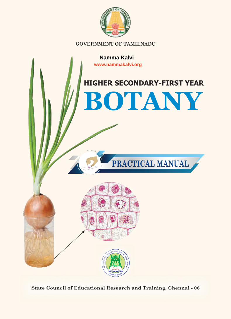
GOVERNMENT OF TAMILNADU
State Council of Educational Research and Training, Chennai - 06
BOTANYHIGHER SECONDARY-FIRST YEAR
PRACTICAL MANUAL
XI_BOTANY PRACTICAL MANUAL.indd 1 28-08-2018 11:36:56
Namma Kalviwww.nammakalvi.org
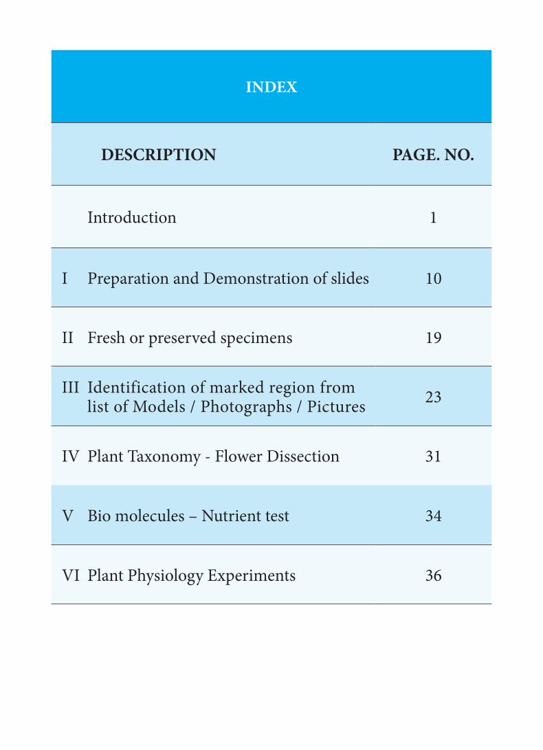
INDEX
DESCRIPTION PAGE. NO.
Introduction 1
I Preparation and Demonstration of slides 10
II Fresh or preserved specimens 19
III Identification of marked region from list of Models / Photographs / Pictures 23
IV Plant Taxonomy - Flower Dissection 31
V Bio molecules – Nutrient test 34
VI Plant Physiology Experiments 36
XI_BOTANY PRACTICAL MANUAL.indd 2 28-08-2018 11:36:56

1
Science learning is practical oriented and requires practical activities in the laboratory. As in any other science subject, practical have an important role in Botany too. The purpose of teaching botany is not only to acquaint the learner with terms, facts, concepts and principles but also to prepare them to understand these concepts by doing exercises relating to them. Practical work also gives students many opportunities to use their minds to discover laws and principles of science. It makes difficult and abstract concepts real, remove misconceptions, ignite, increase and sustain students interest in plant science through various practical activities. Self- experience not only eliminates doubts and misbeliefs in one’s mind but also generates an interest in the subject. Therefore, the students should be adequately taught through practical activities to acquire useful practical skills in concepts.
THE OBJECTIVES OF BIOLOGY PRACTICALSThe objectives of biology practicals are to:
develop practical skill for better understanding through first hand experience;
demonstrate the principles covered in the theory;
develop observational skill in the form of identifying and locating desired parts in specimen;
develop manipulative skills in arranging and handling the apparatus and instruments and taking reading on them;
collect material and to mount it to develop skill in preserving biological material and specimens;
draw, label and record experimental results and interpret them;
Through practical work, not only the theoretical concepts are tested but also it trains the student in scientific method of learning.
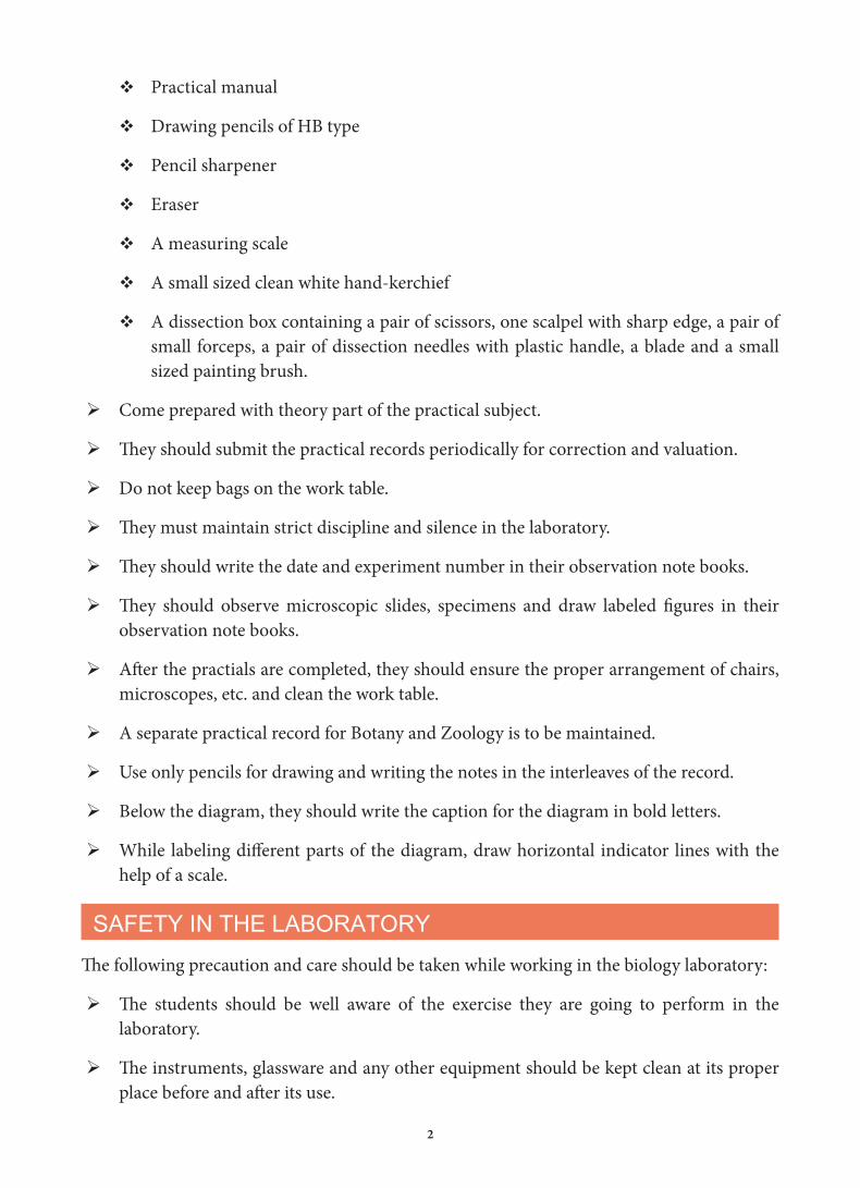
INSTRUCTIONS TO STUDENTSStudents must attend all the practical classes. They must also remember that there is a great degree of co-ordination between theory class and practicals.
The following are some of the items that they must bring to the Practical Classes.
Practical observation note book
Practical record
INTRODUCTION
XI_BOTANY PRACTICAL MANUAL.indd 1 28-08-2018 11:36:56
2
Practical manual
Drawing pencils of HB type
Pencil sharpener
Eraser
A measuring scale
A small sized clean white hand-kerchief
A dissection box containing a pair of scissors, one scalpel with sharp edge, a pair of small forceps, a pair of dissection needles with plastic handle, a blade and a small sized painting brush.
Come prepared with theory part of the practical subject.
They should submit the practical records periodically for correction and valuation.
Do not keep bags on the work table.
They must maintain strict discipline and silence in the laboratory.
They should write the date and experiment number in their observation note books.
They should observe microscopic slides, specimens and draw labeled figures in their observation note books.
After the practials are completed, they should ensure the proper arrangement of chairs, microscopes, etc. and clean the work table.
A separate practical record for Botany and Zoology is to be maintained.
Use only pencils for drawing and writing the notes in the interleaves of the record.
Below the diagram, they should write the caption for the diagram in bold letters.
While labeling different parts of the diagram, draw horizontal indicator lines with the help of a scale.
SAFETY IN THE LABORATORYThe following precaution and care should be taken while working in the biology laboratory:
The students should be well aware of the exercise they are going to perform in the laboratory.
The instruments, glassware and any other equipment should be kept clean at its proper place before and after its use.
XI_BOTANY PRACTICAL MANUAL.indd 2 28-08-2018 11:36:56

3
The microscope and other delicate instruments should be handled gently and properly and should be atleast 5 inches from the edge of the table to avoid its knocking off accidently.
Do not throw any broken glassware in the sink. It should be thrown in the dust bin.
Whenever working with the sharp instrument as blade / scalpel etc. be careful not to cut or puncture your skin.
Do not inhale, never taste or apply stain or any chemical as it may harm.
Never eat in the laboratory to avoid infection.
The steps involved in performing a practical are listed below in the chart to help students to do the practicals.
Read instruction carefully
Make observation
Complete worksheet
Note down all observations
Prepare a Record Book
Get it evaluated
1
Follow each step
2
3
45
6
7
XI_BOTANY PRACTICAL MANUAL.indd 3 28-08-2018 11:36:56

4
BASIC EQUIPMENTS USED IN BIOLOGY LABORATORY
Microscope:a. Dissection Microscope: Used to observe ground plan of T.S / L.S root, stem, leaf ovule and small organisms.
b. Compound Microscope: It consists of objective lens and ocular lens, which is used to magnify the object. The light entering into the microscope is adjusted by diaphragm.Specimen slide placed on the stage is illuminated by light. It is observed through low power or high power by changing objective lens. Using coarse and fine adjustment fine details of slide can be studied.
Glassware: Test tubes, Beakers, Flasks, Watch glass, Petri dishes, Slides, Cover slips, Reagent bottles, Pipette, Funnel and Graduated cylinder.
Tools for dissection:Scalpel, Forceps, Needle, Brushes, Blade
Fixatives: Formalin, F.A.A (Formalin-aceto-alcohol).), Ethanol and Acetone
Stains: Safranin (used to stain lignified and cutinised cells)Haematoxylin (used to stain nucleus)Iodine (used to find starch)Eosin (used to stain cytoplasm)Acetocarmine (used to stain chromosomes)Crystal Violet (used to stain bacteria)
Mounting agents:Glycerine and Canada balsam
Reagents and Solutions:Benedict’s reagent, Biuret reagent, Fehling’s solution, Starch solution, Iodine solution, and NaOH.
Indicators:pH paper
Temperature measurement: Thermometer
XI_BOTANY PRACTICAL MANUAL.indd 4 28-08-2018 11:36:56
5
PREPARATION OF SLIDE
Basic techniques used in biology laboratory during the preparation of micro slide and demonstration of experiments.
How to take peel?
step: 1. Remove an intact leaf epidermal layerstep: 2. Use needle and forceps to separate out peel from leafstep: 3. Keep the peeling on the slide, add drop of water or stainstep: 4. Observe through the microscope.
What is Smear?
A technique used to spread the cells uniformly on the slide from the sample or section.step: 1. Section placed in stainstep: 2. Crushed with help of scalpel or another slide step: 3. Slide gently heated over the flame, mounted with mounting mediumstep: 4. Cover the slide with cover slip and seal with melting wax.
How to take Sections?
A thin and transparent section is cut with the help of sharp razor or blade. Sections are basically two types: Transverse Sections (T.S) and Longitudinal sections (L.S) step: 1. Keep the material between thumb and first finger using pithstep: 2. Cut several thin sections using razor or bladestep: 3. Take out the section with the help of brush and place it in a watch glass containing water.step: 4. select a thin floating section, avoid oblique/incomplete section.
Fixation:
Fixation is the technique adopted to kill the cells and stop the cellular activities. It also protects the cells from drying and decaying.Some common fixatives: Formalin, Ethanol and FAA (Formalin-aceto-alcohol).
Procedures followed in Staining:
Staining is the technique used to view and differentiate the cells using specific dyes or Stains.Some common Stains: Safranin, Haematoxylin, Iodine, Eosin, Acetocarmine and Crystal Violet.
Mounting:
Technique adopted to preserve the sections for longer period of time and also protect the section from drying.
XI_BOTANY PRACTICAL MANUAL.indd 5 28-08-2018 11:36:56

6
Some common mounting media: Glycerine, DPX and Canada balsam.Step: 1. Pore a drop of mounting medium on the section over the slideStep: 2. Place the cover slip very gently over the slideStep: 3. Avoid air bubbles while mountingStep: 4. Wipe out excess mounting medium with blotting paper.
Know your Compound Microscope
Compound Microscope is an insdispensable instrument in a Biology laboratrory. Study the diagram of the microscope and compare it with an actual one in the laboratory.
Eye-Piece: Contains lenses to increase magni�cation
Body Tube: Holds lenses of eyepiece and objectivesat proper working distance from each other
Arm: Supports body tube and coarse adjustment.
Nose Piece: Permits interchange of low and high powered objects
Objective Lense: Contains lenses of di�erent magni�cation as 10X, 40X etc.Fine Adjustment: Permits exact focussing by moving stage or body tube up or down very slightly
Coarse Adjustment: Moves body tube up or down to the correct distance from the specimen for focussing the object
Stage: Supports slide over hole that admitslight from mirror below
Stage Clip: Hold slide �rmly in place
Mirror: Re�ects light upward through diaphgramand hole in stage
Base: Firm support bearing weight of microscope
Diaphragm: Regulates amount of light passing through the specimen
XI_BOTANY PRACTICAL MANUAL.indd 6 28-08-2018 11:36:57

7
BOTANY PRACTICAL MANUAL
MODEL QUESTION
I. Identify the given slide ‘A’ and give any two reasons
II. Identify the given specimen ‘B’ and give any two reasons
III. Identify the spot ‘C’ marked on the model/photograph/picture give any two reasons
IV. Identify the family, dissect and display the given flower ‘D’. Write the floral characters of essential parts, draw floral diagram and write floral formula.
V. Test the given sample solution ‘E’ for the presence of Reducing sugar (Glucose), Starch, Protein and Lipids Write the Principle and Tabulate the result.
VI. Write aim, procedure, observation and inference of the given plant physiological experiment setup ‘F’.
MARKS ALLOTMENT-PRACTICAL EXAMINATION
I. Identification- ½ Reasons (any two) – 1 (1 ½)
II. Identification – ½ Reasons (any two) – 1 (1 ½)
III. Identification – ½ Reasons (any two) – 1 (1 ½)
IV. Identification ½, Dissection – 1, Floral character – 1, Floral Diagram – 1, Floral formula – 1
(4 ½)
V. Principle – ½, Test - 1, Table - 1½ ( Procedure - ½, Observation -½, Inference - ½),
(3)
VI. Aim – ½, Procedure – 1, Observation – ½, Inference – 1 (3)
Total 15 marks
Record 3 marks
Skill 2 marks
Maximum marks 20 marks
XI_BOTANY PRACTICAL MANUAL.indd 7 28-08-2018 11:36:57

8
BOTANY PRACTICAL MANUAL
QUESTION No- I (A)Note: Teacher must prepare a temporary slide using fresh specimen for demonstration during the practical hours. (If temporary slide preparation is not possible, permanent slides are allowed only during Board practical examination)
Preparation and Demonstration of Slides
Exercise 1 Bacteria - Lactobacillus
Exercise 2 Fungi – Yeast and Rhizopus
Exercise 3 Algae - Chlamydomonas, Volvox, Spirogyra, Oedogonium
Exercise 4 Mitotic cell division Stages - Metaphase, Anaphase
Exercise 5 Anatomical structure – Dicot – Root, Stem, Leaf and Monocot – Root, Stem & Leaf
Exercise 6 Plasmolysis and Deplasmolysis
Exercise 7 Stomatal distribution
QUESTION No- II (B)
Fresh or preserved specimens
Exercise 8 Chara
Exercise 9 Agaricus – Basidiocarp
Exercise 10 Foliose Lichen
Exercise 11 Funaria-Habit
Exercise 12 Adiantum-Habit
Exercise 13 Phylloclade – Opuntia
Exercise 14 Special inflorescence – Cyathium
Exercise 15 Aggregate fruit – Polyalthia
QUESTION No- III (C)
Identification of marked region from list of Models / Photographs / Pictures
Exercise 16 Ultra-structure of a bacterial cell – Genophore, Plasmid, Polysome, Mesosome, Fimbriae
Exercise 17 Types of Stele- Actinostele, Plectostele, Dictyostele, Eustele
Exercise 18 Types of Venation- Pinnately reticulate, Palmately reticulate convergent, Pinnately Parallel, Palmately Parallel divergent
XI_BOTANY PRACTICAL MANUAL.indd 8 28-08-2018 11:36:57

9
Exercise 19 Types of Inflorescence - Panicle, Corymb, Simple dichasium, Polychasial cyme.
Exercise 20 Cell cycle stages – G1, S, G2 and M Phase
Exercise 21 Nitrogen bases – Adenine, Guanine, Cytosine, Thymine, Uracil
Exercise 22 Types of Tissue – Aerenchyma, Chlorenchyma, Angular Collenchyma, Brchysclereids, Xylem Tracheids, Xylem vessels and Sieve tube.
QUESTION No- IV (D)
Taxonomy - Flower Dissection
Exercise 23 Fabaceae - Clitoria ternatea
Exercise 24 Apocynaceae - Catharanthus roseus
Exercise 25 Solanaceae – Datura metal
Exercise 26 Euphorbiaceae – Ricinus communis
Exercise 27 Musaceae – Musa paradisiaca
QUESTION No- V (E)
Bio molecules – Nutrient test
Exercise 28 Test for reducing sugar-Benedict test
Exercise 29 Starch – Iodine test
Exercise 30 Protein –Biuret test
Exercise 31 Lipid –Saponification test
QUESTION No- VI (F)
Plant Physiology Experiments
Exercise 32 Potato Osmoscope
Exercise 33 Paper Chromatography
Exercise 34 Wilmott’s Bubbler
Exercise 35 Demonstration of production of CO2 during respiration
Exercise 36 Arc auxanometer
XI_BOTANY PRACTICAL MANUAL.indd 9 28-08-2018 11:36:57

1010
I - Preparation and Demonstration of SlidesNote: Teacher must prepare a temporary slide using fresh specimen for demonstration
during the practical hours. (If temporary slide preparation is not possible, permanent slides are allowed only during Board practical examination)
Aim: To study and identify the morphology of representative types of bacteria, fungi and Algae.
Principle: Morphology is the study of the characteristic features of the species. It could be a study of external or internal features. Morphological studies help in identifi cation and classifi cation of organisms.
Requirements: Buttermilk or curd, 100 ml sugar solution, crystals of yeast, bread mold, pond water, slide, cover slip to prepare temporary slides / Permanent slides of Bacteria, Yeast, Rhizopus, Chlamydomonas, Volvox, Spirogyra, Oedogonium , Compound microscope.
Exercise: 1
Bacteria (Lactobacillus)
Take sour buttermilk/curd and mount it on a slide to view lactobacillus.
Diagnostic Features• Unicellular, Prokaryotic, rod shape, Chemo organotrophic bacteria.• Absence of membrane bound organelles like mitochondria,
nucleus, golgi bodies, plastids, etc., • Mesosomes are present• Involved in lactic acid fermentation.
Exercise: 2
a. Fungi – Yeast
Add few crystals of Yeast to 100 ml sugar solution. Leave it for 2 to 3 hours. Later mount a drop of solution on a slide to view it under a microscope.
Figure 1: Bacteria
BOTANY PRACTICALS
XI_BOTANY PRACTICAL MANUAL.indd 10 28-08-2018 11:36:57

11
Diagnostic Features• Single celled, eukaryotic ascomycetes fungus • Cells are oval or spherical in shape and colourless.• Generally it reproduce by budding.• Strings of connected budding cells form pseudo mycelium.
b. Fungi – Rhizopus
Use bread mold. Th e surface of bread pieces is covered with white or colourless upright branches with black tips are developed. Pick up few threads with the help of forceps and needle. Stain by using safranin and put them on the slide in a drop of glycerin. Cover with the coverslips and observe under the microscope.
Diagnostic Features• Saprophytic fungus commonly grow on bread
(Zygomycetes)• Aseptate, coenocytic mycelium • Asexual spore producing structure called sporangium
which bears sporangiospores.
Algae
Collect the green pond water. Put 2 drops of water on a slide and mount it to see the algae.
Exercise: 3
a. Chlamydomonas
Diagnostic Features• Motile, unicellular green alga. • Presence of cup shaped chloroplast. Th e anterior side of the
chloroplast contains a tiny spot called stigma or eyespot.• Th e anterior part of thallus bears two whiplash fl agella. Each
fl agellum originates from a basal granule or blepharoplast.
Figure 2a: Yeast
Nucleus
Figure 2b: Rhizopus
Columella
Sporangium
Sporangiumspore
Sporangiophore
Rhizoids
Figure 3a: Chlamydomonas
Chloroplast
Pyrenoid
Flagella
XI_BOTANY PRACTICAL MANUAL.indd 11 28-08-2018 11:36:58

12
b. Volvox
Diagnostic Features• Motile and Colonial, green alga. • 500 to 5000 cells arranged to form hollow
sphere. This kind of habit is called Coenobium.• Each cell in the colony connected by thin strands
of cytoplasm.
c . Spirogyra
Diagnostic Features• Unbranched, filamentous green alga.• Spiral shaped Chloroplast• Cylindrical cells are arranged one above the other.• Nucleus is present at the centre of the cell.
d: Oedogonium
Diagnostic Features• Filamentous, unbranched, green alga.• Cells of the filament attached end to end form
uniseriate row.• Presence of reticulate chloroplast.• Presence of cap cells on the young dividing cells.• Three types of cells Basal cell (Hold fast), Middle cell
and Apical cell.
Figure 3b: Volvox
OosporesPlakea ofspormatozoids
Figure 3c: Spirogyra
Figure 3d: Oedogonium
Hold fast
Cap cellOogoniumEgg Nucleus
Supporting cell
Chain of Antheridia
XI_BOTANY PRACTICAL MANUAL.indd 12 28-08-2018 11:36:58

13
Exercise: 4
Mitosis in onion root tip
Aim: To study and identify the mitosis stages – Metaphase and Anaphase.
Principle: Somatic growth of both plants and animals takes place by increase in the number of cells. Th e cells divide mitotically wherein number of chromosomes remains unchanged in the daughter cells from that in the maternal cells. Cells from the growing root-tips and apex of shoot buds are suitable for mitotically dividing cells. In animals mitotically dividing cells can be easily scored from the bone marrow of a vertebrate. Th e cell from the epithelium of gills in fi shes and from the tail of growing tadpole larvae of frog are also good sources for scoring the mitotically dividing cells.
Requirements: Onion root, HCl, Safranin stain, slide, Coverslip, Permanent slides Compound microscope.
1. Cut the tip 5 to 8 mm from the tip of the freshly sprouted onion root. Discard the rest of the root.
2. Wash them in water on a clean microscope slide.
3. Place one drop of 1N HCl on the root tip and add 2-3 drops of Safranin/ Acetocarmine stain to the slide.
4. Warm the slide gently over the alcohol lamp for about one minute. (Do not allow the slide to get hot to the touch).
5. Carefully blot the excess stain with a blotting paper.
6. Aft er (10 to 20 seconds) put one or two drops of water and blot them carefully using blotting paper.
7. Again put a drop of water on the root tip and mount a cover slip on it avoiding air bubbles.
8. Squash the slide with your thumb using a fi rm and even pressure. (Avoid squashing with such force that the cover slip breaks or slides).
9. Observe it under a compound microscope in 10x objective. Scan and narrow down to a region containing dividing cells and switch to 40x for a better view.
XI_BOTANY PRACTICAL MANUAL.indd 13 28-08-2018 11:36:58

14
a. Mitosis – Stage : Metaphase
Diagnostic features: • The spindle fibres attached to the kinetochore
region of centromere of chromosomes• Chromosomes are arranged at the equator
region of the cell (metaphase plate)• Chromosomes are distinctly visible in this stage.
b. Mitosis – Stage : Anaphase
Diagnostic features: • Each chromosome splits and two daughter
chromatids begin to move towards opposite poles.
• Shortening of spindle fibre and longitudinal splitting of centromere creates a pull which divide the chromosomes.
Exercise: 5
Plant anatomical structures(Dicot-Root, Stem and Leaf, Monocot- Root, Stem and Leaf)
Figure 4a: Metaphase
Figure 4b: Anaphase
Spindlefibres
Chromatids
Cell wall
DaughterChromosomes
Spindlefibres
Cell wall
Aim: To study and identify the T.S of dicot root, dicot stem, dicot leaf, monocot root, monocot stem and monocot leaf.
Principle: A group of tissues performing a similar function, irrespective of its position in the plant body, is called a tissue system. The three types of tissue system in plants are: Epidermal tissue system, Ground tissue system and Vascular tissue system. In different parts of the plants, the various tissues are distributed in characteristics patterns. This is
XI_BOTANY PRACTICAL MANUAL.indd 14 28-08-2018 11:36:59

15
Start cutting transverse sections of material placing it in between pith. Select the thinnest section of the material with the help of a delicate brush. Take a clean watch glass with water, transfer thin sections of the material. Put a few drops of safranin stain in the watch glass with water. Leave it for 3-5 minutes. Drain off stain and wash with water if necessary. Put the thinnest section in the centre of the slide. Put a drop of glycerine over the material. Cover it with a coverslip with the help of needle. Observe it under a compound microscope aft er staining and mounting.
a. Dicot Root (T.S) b. Dicot Stem (T.S)
best understood by studying their internal structure by cutting sections either transverse or longitudinal or both of the part to be studied.
Requirements: Small twigs of locally available dicot and monocot plants, glycerine, safranin, slides, cover slip, brushes to prepare temporary slides and permanent slides of T.S. of Bean root, T.S. of Maize root, T.S. of Sunfl ower stem, T.S of Maize stem, T.S of Sunfl ower leaf, T.S of Grass leaf.
A sector enlarged
Ground plan
Root hairPiliferous layerCortexPhloem
MetaxylemConjunctive tissue
Root hair
Piliferous layer
Phloem
Pericycle
ProtoxylemConjunctive tissueMetaxylemCasparian stripPassage cell
Cortex
Endodermis
A sector enlarged
Ground plan
EpidermisHypodermisCortexEndodermis
Epidermal hair
Vascular bundle
Pith
Pericycle
Epidermal hairCuticle
Chlorenchyma
Resin duct
ParenchymaStarch sheathBundle capPrimary phloemCambiumMetaxylem
EpidermisCollenchyma
Protoxylem
PithPrimary medullary ray
Figure 5a: T. S of Dicot root (Bean root) Figure 5b: T.S of Dicot stem (Sun fl ower stem)
XI_BOTANY PRACTICAL MANUAL.indd 15 28-08-2018 11:36:59

16
Dicot Root (T.S) Dicot Stem (T.S) Diagnostic features: Diagnostic features
• Radial vascular bundle, exarch and tetrarch xylem.
• Cortex differentiated, hypodermis made up of collenchyma cells.
• Parenchymatous conjuctive tissue is present.
• Conjoint, Collateral and Open vascular bundle (Cambium present)
• Pith is absent. • Vascular bundle arranged like a ring, wedge shaped vascular bundle.
• Presence of pith and primary pith rays.
c. Dicot Leaf (T.S)
Diagnostic features• Conjoint, Collateral and closed vascular bundle.
• Mesophyll tissue differentiated into upper palisade parenchyma and lower spongy parenchyma. (Dorsiventral leaf)
• Stomata are more in number on the lower epidermis.
• Stomata surrounded by bean shaped guard cells.
CuticleUpper epidermisPalisade parenchymaProtoxylemMetaxylemSpongy parenchymaPhloemBundle sheathStomaEpidermal hairLower epidermisRespiratory cavity
Figure 5c: T.S of Dicot leaf (Sun flower leaf)
Diagnostic features: Diagnostic features• Radial vascular bundle, exarch and
Polyarch xylem.• Conjoint, Collateral and Closed vascular
bundle. (Cambium absent)• Pith is Present. • Skull shaped and scattered vascular bundle.
d. Monocot Root (T.S) e. Monocot Stem (T.S)
XI_BOTANY PRACTICAL MANUAL.indd 16 28-08-2018 11:37:00

17
f. Monocot Leaf (T.S)
A sector enlarged
Ground plan
Root hairPiliferous layerCortexEndodermis
Pith
Root hairPiliferous layer
Casparian strip
Endodermis
PericyclePassage cellPhloemProtoxylemMetaxylemPith
CortexPassage cell
EpidermisHypodermisGround tissueVascular bundles
CuticleEpidermisHypodermisChlorenchyma
Vascular bundle
Ground tissue
Crushed protophloemMetaphloemMetaxylemProtoxylemProtoxylem lacuna Sclerenchymatousbundle sheath
A sector enlarged
Ground plan
Figure 5d: T.S of Monocot root (Maize root) Figure 5e: T.S of Monocot stem (Maize Stem)
• Sclerenchymatous conjuctive tissue is present.
• Pith absent, homogenous ground tissue.
• Ground tissue is not differentiated into cortex and pith. Hypodermis made up of Sclerenchyma cells.
Figure 5f: T.S Monocot leaf(Grass leaf)
CuticleUpper epidermisSub-stomatal chamberMesophyIIBundle sheathXylemPhloemLower epidermis
Stoma
XI_BOTANY PRACTICAL MANUAL.indd 17 28-08-2018 11:37:00

18
Diagnostic features: • Conjoint, Collateral and closed vascular bundle.
• Mesophyll is not diff erentiated into Palisade and Spongy parenchyma. (Isobilateral leaf)
• Number of Stomata are more or less equal on both epidermis, Stomata surrounded by dumb-bell shaped guard cells.
Exercise: 6
Plasmolysis and Deplasmolysis
Aim: Study of plasmolysis in epidermal peel of leaf.Principal: Living cells are generally turgid due to the presence of water. When cells are immersed in hypertonic solution, shrinkage of protoplasm takes place with visible separation of plasma membrane from the cell walls. Th is is called plasmolysis and occurs due to exosmosis, a phenomenon in which water from the cells moves into the surrounding medium which is hypertonic, that is more concentrated than the cell sap.Requirements: Leaves of Tradescantia, 70% sugar solution, slide, cover slip, needle, petri dish / watch glass, microscope.
Peel off a small segment from lower epidermal surface of the Tradescantia leaf. Th is can be done by tearing the leaf obliquely with a single jerk or scraping it with blade. Dip it in 70% of sugar solution for 5 minutes. Later mount the peel on a slide to observe plasmolysis.Again dip the same peel in water for 5 minutes. Later mount it and observe it under the microscope for deplasmolysis.
Diagnostic features: Plasmolysis
• Cell membrane is pulled away from the cell wall.
• Cells becomes fl accid due to loss of water by exosmosis, when a plant cell is kept in a hypertonic solution.
Diagnostic features: Deplasmolysis• It is reverse of plasmolysis.• It is swelling of shrinked protoplasm to
regain its original unplasmolysed shape when cell is placed in hypotonic solution. It is a type of endosmosis.
Figure 6: Diff erent Stages of Plasmolysis and Deplasmolysis in a plant cell
XI_BOTANY PRACTICAL MANUAL.indd 18 28-08-2018 11:37:00
19
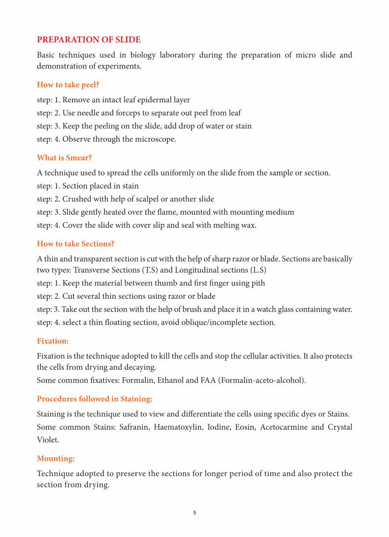
Exercise: 7
Stomatal Distribution
Aim: To study the distribution of stomata on the upper and lower surfaces of leaves.Principle: Stomata are tiny microscopic structures present in leaves of all fl owering plants. Number and distribution of stomata per unit area in variable in leaves of diff erent plants. A typical stoma consists of a pair of guard cells enclosing an aperture in the centre called the stomatal aperture. Stomata perform two important functions; that of, transpiration and exchange of gases.Requirements: Leaf samples – Hibiscus / Balsam / Bougainvillea / Petunia / Cassia / Solanum / any broad leaves of dicot and grass) microscope glass slides, cover slips, water, needle, brush, and petridish / watch glasses.
Peel a dicot leaf and monocot leaf. Dicot leaf to be peeled on both sides to observe upper and lower epidermis. Monocot leaf is to be peeled on any side. Mount the peel on a slide and observe under microscope.
Diagnostic features: • Stomata are more in number on lower
epidermis of dicot leaves and evenly distributed in monocot leaves.
• Stoma is guarded by two bean shaped guard cells.
II. Fresh or preserved specimensAim: To study and identify the morphology of representative types of Algae, Fungi, Lichen, Bryophytes and Pteridophytes.Principle: Morphology is the study of the characteristics features of the species. It could be a study of external or internal features. Morphological studies help in identifi cation and classifi cation of organisms.Requirements: Specimens of Chara, Basidiocarp of Agaricus, Foliose Lichen, Funaria, Adiantum.
Stomata
Guard Cells
Subsidiary Cells
Figure 7: Stomatal distribution
XI_BOTANY PRACTICAL MANUAL.indd 19 28-08-2018 11:37:01
20
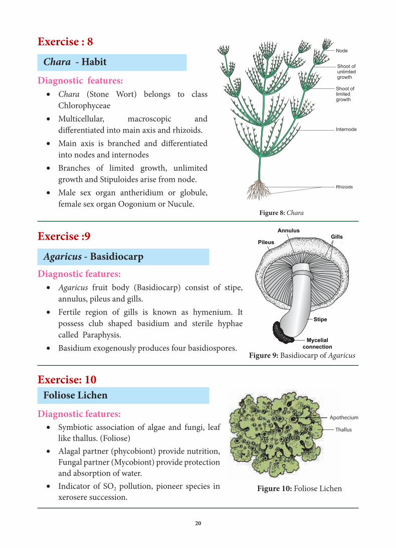
Exercise : 8
Chara - Habit
Diagnostic features:• Chara (Stone Wort) belongs to class
Chlorophyceae • Multicellular, macroscopic and
differentiated into main axis and rhizoids.• Main axis is branched and differentiated
into nodes and internodes• Branches of limited growth, unlimited
growth and Stipuloides arise from node.• Male sex organ antheridium or globule,
female sex organ Oogonium or Nucule.
Exercise :9
Agaricus - BasidiocarpDiagnostic features:
• Agaricus fruit body (Basidiocarp) consist of stipe, annulus, pileus and gills.
• Fertile region of gills is known as hymenium. It possess club shaped basidium and sterile hyphae called Paraphysis.
• Basidium exogenously produces four basidiospores.
Exercise: 10Foliose Lichen
Diagnostic features: • Symbiotic association of algae and fungi, leaf
like thallus. (Foliose)• Alagal partner (phycobiont) provide nutrition,
Fungal partner (Mycobiont) provide protection and absorption of water.
• Indicator of SO2 pollution, pioneer species in xerosere succession.
Figure 8: Chara
Pileus
AnnulusGills
Stipe
Mycelialconnection
Figure 9: Basidiocarp of Agaricus
Thallus
Apothecium
Figure 10: Foliose Lichen
XI_BOTANY PRACTICAL MANUAL.indd 20 28-08-2018 11:37:01

21
Exercise : 11Funaria - Habit
Diagnostic features: • Bryophyte- Funaria (cord moss) plant body gametophyte,
erect radial stem covered with small leaves.
• Attached to soil by multicellular rhizoids
• Antheridium and Archegonium formed in group in same thallus different branches (Monoecious).
• Sporophyte attached to gametophyte consist of foot, seta and capsule.
Exercise: 12Adiantum - Habit
Diagnostic features: • Pteridophyte - Adiantum (Maiden hair fern)
plant body is sporophyte differentiated into root, rhizome and leaves.
• Rhizome is covered with persistent leaf and hairy outgrowth called ramenta.
• Leaves are called fronds, pinnately compound, bears marginal sori covered by false indusium.
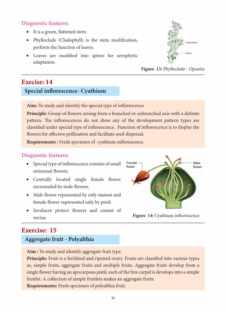
Exercise: 13Phylloclade - Opuntia
Aim : To study modifications of stemPrinciple: The stem is the central axis that provides supports to all the aerial parts of the plant. Besides,in some plants these also help in perennation, vegetative propagation, food storage, photosynthesis etc. through various modifications.Requirements: Specimen of Opuntia
Calyptra
Figure 2.5 Funaria
Rhizoid
Leaf
Figure 11: Funaria
Rachis
Rhizome
Petiole
Sorus
Pinnule
Figure 12: Adiantum
XI_BOTANY PRACTICAL MANUAL.indd 21 28-08-2018 11:37:01
22
Femaleflower
Maleflower
Figure 14: Cyathium inflorescence
Diagnostic features: • It is a green, flattened stem.
• Phylloclade (Cladophyll) is the stem modification, perform the function of leaves.
• Leaves are modified into spines for xerophytic adaptation.
Execise: 14 Special inflorescence- Cyathium
Aim: To study and identify the special type of inflorescencePrinciple: Group of flowers arising from a branched or unbranched axis with a definite pattern. The inflorescences do not show any of the development pattern types are classified under special type of inflorescence. Function of inflorescence is to display the flowers for effective pollination and facilitate seed dispersal. Requirements : Fresh specimen of cyathium inflorescence.
Diagnostic features: • Special type of inflorescence consists of small
unisexual flowers.
• Centrally located single female flower surrounded by male flowers.
• Male flower represented by only stamen and female flower represented only by pistil.
• Involucre protect flowers and consist of nectar.
Exercise: 15Aggregate fruit - Polyalthia
Aim : To study and identify aggregate fruit type.Principle: Fruit is a fertilized and ripened ovary. Fruits are classified into various types as, simple fruits, aggregate fruits and multiple fruits. Aggregate fruits develop from a single flower having an apocarpous pistil, each of the free carpel is develops into a simple fruitlet. A collection of simple fruitlets makes an aggregate fruits.Requirements: Fresh specimen of polyalthia fruit.
Pyylloclade opuntia
Spine
Scaly leaf
Cladode
Phylloclade
Cladode - AsparagasPyylloclade opuntia Cladode - AsparagasFigure 13: Phylloclade - Opuntia
XI_BOTANY PRACTICAL MANUAL.indd 22 28-08-2018 11:37:03

23
Diagnostic features:• Aggregate fruit develops from Single flower
multicarpellary and apocarpous ovary.
• A collection of simple fruitlets makes aggregate fruit.
III - Identify the marked region from the given model/ photograph / picture.
Exercise: 16Ultra structure of Bacterial cell.
Aim : To study and identify the ultra structure of bacterial cell.Principle : Bacteria are prokaryotic, unicellular ubiquitous, microscopic organism. Bacteria contain a well developed cell structure which is responsible for some of their unique biological structures and pathogenicity. Many structural features are unique to bacteria and are not found among archaea or eukaryotes. Because of the simplicity of bacteria relative to larger organisms and the ease with which they can be manipulated experimentally, the cell structure of bacteria has been well studied, revealing many biochemical principles that have been subsequently applied to other organisms.Requirements : Model / Photograph / picture of ultrastructure of bacterial cell.
Pedicel
Receptacle
Fruitlet
Figure 15: Aggregate fruit - Polyalthia
Pilus
Cytoplasm
Plasma membrane
Flagellum
Capsule
Nucleoid (DNA)
Plasmid
Cell wall
Polyribosome
Mesosome
Inclusion
Figure 1.12 Ultrastructure of a BacteriumFigure 16: Ultra structure of bacterial cell
XI_BOTANY PRACTICAL MANUAL.indd 23 28-08-2018 11:37:03
24
Diagnostic features:1. Genophore:
• Single, Circular bacterial chromosome.• Primitive nucleus (Nucleoid) without nuclear envelop and nucleolus.
2. Plasmid:• Double stranded, Circular, extra genomic DNA found in cytoplasm of bacteria.• It contains genes for fertility and antibiotic resistance.
3. Polysome:• Ribosomes are held together by mRNA and form Polyribosome or Polysome.• 70 S Ribosomes are attached mRNA in bacteria.
4. Mesosome:• Localized infoldings of bacterial plasma membrane.• It is found in the form of Vesicles, tubules and lamellae.• It helps in respiration and binary fission.
5. Fimbriae or Pili:• Fimbriae or Pili are hair like appendages found on the surface of the bacterial cell wall.• It helps in conjugation hence it is called as Sex pili.
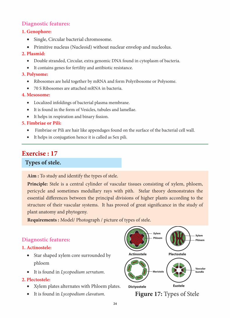
Exercise : 17Types of stele.
Aim : To study and identify the types of stele.Principle: Stele is a central cylinder of vascular tissues consisting of xylem, phloem, pericycle and sometimes medullary rays with pith. Stelar theory demonstrates the essential differences between the principal divisions of higher plants according to the structure of their vascular systems. It has proved of great significance in the study of plant anatomy and phytogeny.Requirements : Model/ Photograph / picture of types of stele.
Diagnostic features:1. Actinostele:
• Star shaped xylem core surrounded by phloem
• It is found in Lycopodium serratum.2. Plectostele:
• Xylem plates alternates with Phloem plates.• It is found in Lycopodium clavatum.
Actinostele Plectostele
Dictyostele Eustele
Xylem
PhloemXylem
Phloem
MeristeleVascular bundle
Figure 17: Types of Stele
XI_BOTANY PRACTICAL MANUAL.indd 24 28-08-2018 11:37:03

25
3. Dictyostele:• Stele is separated into several strands and each one is called meristele.• It is found in Adiantum.
4. Eustele:• Stele is split into distinct collateral vascular bundle around the pith.• It is found in dicot stem.
Exercise : 18Types of venation.
Aim: To study and identify the types of venationPrinciple: The arrangement of veins and veinlets on the leaf blade or lamina is called venation. Internally, the vein contains vascular tissues. Conventionally venation is classified into two types namely, Reticulate venation and Parallel venation.Requirements: Fresh specimen / photographs / pictures of pinnately reticulate, palmately reticulate convergent, palmetely reticulate divergent, pinnately parallel, palmatley parallel convergent and palmatley parallel divergent leaves.
Pinnately reticulate
Palmately reticulate divergent
Palmately reticulate convergent
Pinnately parallel
Palmately parallel convergent
Palmately parallel divergent
Diagnostic features:1. Pinnately reticulate:
• One midrib in the centre which forms many lateral branches to form a network.
• Example: Mangifera, Nerium, Ficus2. Palmately reticulate convergent:
• Two or more main veins arising from a single point, all veins converge at the apex of leaf.
• Example: Zizyphus, Cinnamomum.3. Palmately reticulate divergent:
• All principal veins originate from the base and diverge from one another towards the margin of the leaf.
• Example: Cucurbita, Carica.
4. Pinnately parallel:• Prominent midrib in the centre,
from which arise many veins perpendicularly and run parallel to each other.
• Example: Musa, Zinger, Canna.5. Palmately Parallel Convergent:
• All principal run parallal to each other from the base of the lamina and join at the apex
• Example: Bamboo, Rice6. Palmately Parallel Divergent:
• All principal veins originate from the base and diverge towards the margin as in Palm.
• Example: Borassus flabellifer.
Figure 18: Types of Venation
XI_BOTANY PRACTICAL MANUAL.indd 25 28-08-2018 11:37:03

26
Exercise: 19 Types of Inflorescence
Aim: To study and identify the types of inflorescencePrinciple: Group of flowers arising from a branched or unbranched axis with a definite pattern. Inflorescences may also have classified based on branching, number and arrangement of flowers, and some specialized structures. They are inderterminate inflorescence, determinate inflorescence, mixed inflorescence, special inflorescence. Function of inflorescence is to display the flowers for effective pollination and facilitate seed dispersal.Requirements: Fresh inflorescence of Mangifera, Neem (Panicle), Caesalpinia (Corymb), Jasminum (Simple dichasium) Nerium ( Polychasial Cyme). / Photograph/ picture.
Diagnostic features:1. Panicle
• A branched raceme is called panicle. It is also called compound raceme or raceme of racemes.
• Example: Mangifera, Neem.2. Corymb
• In corymb inflorescence the shorter pedicellate flowers at the top and longer pedicellate flowers at the bottom.
• All flowers appear at the same level to form convex or flat topped racemose inflorescence.
• Example: Caesalpinia.3. Simple dichasium (Biparous)
• A central axis ends in a terminal flower; further grows is produced by two lateral buds. • Each cymose unit consists of three flowers of which central one is old one.• Example: Jasminum
4. Polychasial cyme• In central axis ends with a flower. The lateral axes branches repeatedly.• Example: Nerium.
Polychasial cyme
Panicle
Old flower
Old flower
Young flower
Young flower
Simple dichasium
Corymb
Figure 19 : Types of Inflorescence (Diagrammatic representation)
XI_BOTANY PRACTICAL MANUAL.indd 26 28-08-2018 11:37:03

27
Exercise: 20
Cell cycle
Aim: To study and identify the stages of Cell cycle.Principle: A series of events leading to the formation of new cell is known as cell cycle. The phenomenonal changes leading to formation of new population take place in the cell cycle. The series of events include, G1, S, G2, and M.Requirements: Model / Photograph / picture of Cell cycle.
Diagnostic features:
1. G1 Phase: • G1 or first gap phase, cells are metabolically
active and produces proteins, lipids, carbohydrates, cell organelles and RNA.
• A non-dividing cell does not proceed beyond G 1 phase.
2. S Phase:• S Phase or synthesis phase duplication of
DNA takes place.
• The centrioles duplicate in the cytoplasm.
• DNA content increases from 2 C to 4 C.
3. G2 Phase:• G2 Phase or second gap phase cell grow continuously by protein and cell organelle
synthesis, mitochondria and chloroplast divide and DNA remain in 4 C.
• Tubulin, microtubules and Maturation Promoting Factor are formed.
• Spindle begins to form and nuclear divisions follows.
4. M Phase:• M Phase or Mitotic Phase is short, in this stage a single cell divided into two daughter
cells.
• The number of chromosomes in the parent and the daughter cells remain the same so it is also called as equational division.
• This phase is divided into Prophase, Metaphase, Anaphase and Telophase.
Figure 20: Cell cycle
Preparation for MitotisGrowth
Metaphase
Anaphase
M
G2G1
G0
S
DNA Replication
GrowthPreparationfor DNASynthesisInterphase
Prophase
Telophase
XI_BOTANY PRACTICAL MANUAL.indd 27 28-08-2018 11:37:04
28
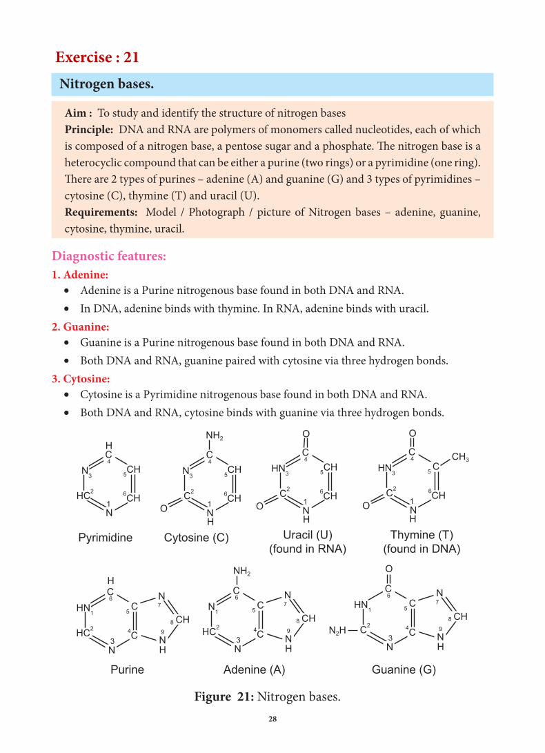
Exercise : 21 Nitrogen bases.
Aim : To study and identify the structure of nitrogen basesPrinciple: DNA and RNA are polymers of monomers called nucleotides, each of which is composed of a nitrogen base, a pentose sugar and a phosphate. The nitrogen base is a heterocyclic compound that can be either a purine (two rings) or a pyrimidine (one ring). There are 2 types of purines – adenine (A) and guanine (G) and 3 types of pyrimidines – cytosine (C), thymine (T) and uracil (U).Requirements: Model / Photograph / picture of Nitrogen bases – adenine, guanine, cytosine, thymine, uracil.
Diagnostic features:1. Adenine:
• Adenine is a Purine nitrogenous base found in both DNA and RNA.• In DNA, adenine binds with thymine. In RNA, adenine binds with uracil.
2. Guanine:• Guanine is a Purine nitrogenous base found in both DNA and RNA.• Both DNA and RNA, guanine paired with cytosine via three hydrogen bonds.
3. Cytosine:• Cytosine is a Pyrimidine nitrogenous base found in both DNA and RNA.• Both DNA and RNA, cytosine binds with guanine via three hydrogen bonds.
Figure 21: Nitrogen bases.
Guanine (G)Adenine (A)Purine
Thymine (T)(found in DNA)
Uracil (U)(found in RNA)
Cytosine (C)Pyrimidine
1N
HN3
C2 6CH
5CH
O
O
H
C4
1N
HN3
C2 6CH
5 C
O
CH3
O
H
C4
3N
HN1
C2 4C
5 C
CH
HN
N
N2H
O
C
9
8
76
3N
N1
HC2 4C
5 C
CH
HN
N
NH2
C
9
8
76
3N
HN1
HC2 4C
5 C
CH
HN
NHC
9
8
76
1N
N3
C2 6CH
5CH
O
NH2
H
C4
1N
N3
HC2 6CH
5CH
HC4
XI_BOTANY PRACTICAL MANUAL.indd 28 28-08-2018 11:37:04
29
4. Thymine:• Thymine is a Pyrimidine nitrogenous base found only in DNA.• In DNA, thymine pairs with adenine via two hydrogen bonds.
5. Uracil:• Uracil is a Pyrimidine nitrogenous base found only in RNA.• In RNA, uracil pairs with adenine via two hydrogen bonds.
Exercise :22
Types of tissues.
Aim : To study and identify the types of plant tissues and its diversity in shapes and sizes of plant cells.Principle: Tissue is a group of cells performing a common function. Tissue may be simple (parenchyma, collenchymas and sclerenchyma) i.e., containing only one type of cells or complex (xylem, phloem) that is containing more than one type of cells. The tissues are also classified into meristematic and permanent tissues. Cells of different types of tissues differ in their structure, shape, size, function and wall composition. Requirements: Model / photograph / pictures of aerenchyma, chlorenchyma, angular collenchyma, brachysclereid, xylem trachieds, xylem vessels, phloem sieve tube.
Diagnostic features:1. Aerenchyma:
• Parenchyma cells with large intercellular spaces and filled with air found in aquatic plants.
• It provides buoyancy to the plants in water.2. Chlorenchyma:
• Parenchyma cells with chloroplast found in green leaves and stem.• The main function is to perform photosynthesis.
3. Angular collenchyma:• Collenchyma cells have thickening at the angles where cells meet.• It is found in hypodermis of Datura and Nicotiana.
4. Brachysclereid:• Brachysclereids or stone cells are isodiametric with hard cell wall.• It is found in bark, pith, cortex, hard endosperm and pulp of pyrus.
XI_BOTANY PRACTICAL MANUAL.indd 29 28-08-2018 11:37:04

30
5. Xylem Tracheids:• Part of the xylem, elongated cells with tapering ends, dead cells with lignified secondary wall.• Imperforated cell with bordered pits, conduct water in gymnosperm and pteridophytes
and provide mechanical support to the plants.6. Xylem Vessels:
• Part of the xylem, elongated tube likes cell, arranged end to end.• Perforated, dead cells, wider lumen, water conducting elements in angiosperms.
7. Phloem Sieve tube:• Part of the Phloem, elongated cells, with strands of cytoplasm without nucleus.• Consist of sieve plate and helps to conduct food materials.
Stellate Parenchyma
Chloroplasts
Palisade Parenchyma
Spongy Parenchyma
Chlorenchyma
Brachy sclereids
Perforationplate
Xylem Tracheid
Xylemvessel
Pits
Phloem sieve t
Sieve plate
Sieve tube
Phloemparenchyma
Lumen cell
Thick cell wall
Angular Collenchyma
Vacuole
Thickened corners
Intercellular Space
Companian Cells
Figure 22: Types of tissues
XI_BOTANY PRACTICAL MANUAL.indd 30 28-08-2018 11:37:04
31
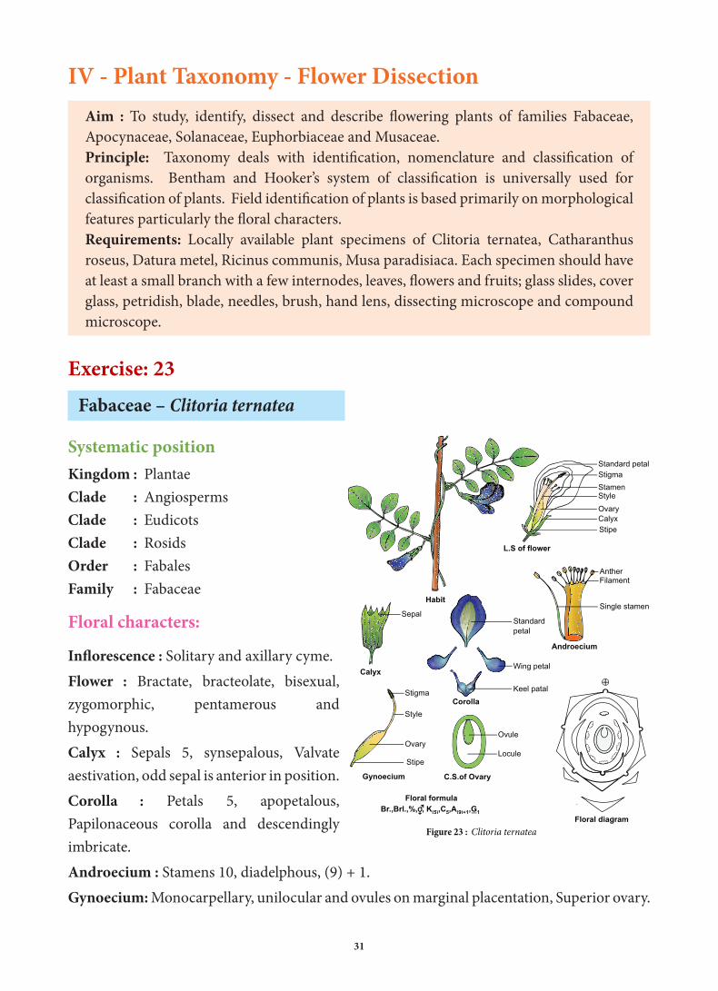
IV - Plant Taxonomy - Flower DissectionAim : To study, identify, dissect and describe flowering plants of families Fabaceae, Apocynaceae, Solanaceae, Euphorbiaceae and Musaceae.Principle: Taxonomy deals with identification, nomenclature and classification of organisms. Bentham and Hooker’s system of classification is universally used for classification of plants. Field identification of plants is based primarily on morphological features particularly the floral characters.Requirements: Locally available plant specimens of Clitoria ternatea, Catharanthus roseus, Datura metel, Ricinus communis, Musa paradisiaca. Each specimen should have at least a small branch with a few internodes, leaves, flowers and fruits; glass slides, cover glass, petridish, blade, needles, brush, hand lens, dissecting microscope and compound microscope.
Exercise: 23
Fabaceae – Clitoria ternatea
Systematic positionKingdom : PlantaeClade : AngiospermsClade : EudicotsClade : RosidsOrder : FabalesFamily : Fabaceae
Floral characters:
Inflorescence : Solitary and axillary cyme.Flower : Bractate, bracteolate, bisexual, zygomorphic, pentamerous and hypogynous.Calyx : Sepals 5, synsepalous, Valvate aestivation, odd sepal is anterior in position.Corolla : Petals 5, apopetalous, Papilonaceous corolla and descendingly imbricate.Androecium : Stamens 10, diadelphous, (9) + 1.Gynoecium: Monocarpellary, unilocular and ovules on marginal placentation, Superior ovary.
Figure 23 : Clitoria ternatea
Habit
L.S of flower
Calyx
Standard petal
Wing petal
Keel patal
Stigma
Ovary
Standard petal
StamenStyle
CalyxStipe
Sepal
AntherFilament
Single stamen
Androecium
Stigma
Ovary
Style
Stipe
Gynoecium C.S.of Ovary
Ovule
Locule
Floral diagram
Floral formulaBr.,Brl.,%, , K(5),C5,A(9)+1,G1
Corolla
XI_BOTANY PRACTICAL MANUAL.indd 31 28-08-2018 11:37:05

32
Exercise : 24Apocynaceae - Catharanthus roseus
Kingdom : PlantaeClade : AngiospermsClade : EudicotsClade : AsteridesOrder : GentianalesFamily : ApocynaceaeFloral characters:Inflorescence: Cymose and axillary pairs.Flower:Ebractate, ebracteolate, bisexual, actinomorphic, pentamerous and hypogynous.Calyx: Sepals 5, Synsepalous and valvate aestivation.Corolla: Petals 5, Synpetalous, twisted aestivation.Androecium: Stamens 5, epipetalous, inserted at the mouth of the corolla tube.Gynoecium: Bicarpellary, apocarpous, superior and marginal placentation.
Exercise: 25Solanaceae – Datura metel.
Kingdom : PlantaeClade : AngiospermsClade : EudicotsClade : AsteridesOrder : SolanalesFamily : SolanaceaeFloral charactersInflorescence: Solitary and axillary cyme.Flower: Bractate, ebracteolate, bisexual, actinomorphic, pentamerous and hypogynous.Calyx: Sepals 5, synsepalous, Valvate aestivation, persistent calyx and odd sepal posterior.Corolla: Petals 5, Synpetalous, twisted aestivation and plicate.Androecium: Stamens 5, epipetalous and alternipetalous.Gynoecium: Bicarpellary, syncarpous and superior ovary, bilocular due formation of false septum looks tetra locular.
Figure 24 : Catharanthus roseus
Habit
Flower entire
L.S. of the flower Gynoecium
C.S. of the ovary Fruit
Floral diagram
Floral formula
Ebr.,Ebrl., , ,K(5),C(5),A5,G2
A pair of elongated follicles
Calyx
Leaf
Fruit
Flower
Corolla
Calyx
Stigma
Style
Ovary
Corolla
EpipetalousstamensStigmaStyleOvary Scaly nectary
Scaly nectaryseedApocarpousovary
Sepal
Gynoecium
Floral Diagram
Corolla cut open
Calyx
Floral formula
Br.,Ebrl., , ,K(5),C(5),A5,G(2)
Figure 25 : Datura metel
Flower entireHabit
StamenCorolla
Calyx
StamenCorolla
Calyx
Leaf
Stigma
Style
Ovary
OvuleSwollenplacenta
Sepal
Corolla
Epipetalousstamens
Anther
C.S Ovary
XI_BOTANY PRACTICAL MANUAL.indd 32 28-08-2018 11:37:05

33
Exercise: 26Euphorbiaceae – Ricinus communis
Kingdom : PlantaeClade : AngiospermsClade : EudicotsClade : RosidsOrder : MalpighialesFamily : Euphorbiaceae
Floral characters:Inflorescence: Terminal racemeMale Flower: Bractate, ebracteolate, found in lower portion of inflorescence, actinomorphic, tepals 5, syntepalous and numerous stamens.Female flower: Bracteate, ebracteolate found in upper portion of inflorescence, actinomorphic, tepals 3, syntepalous, Tricarpellary, syncarpous, superior ovary and ovules on axile placentation.
Exercise: 27Musaceae – Musa paradisiaca.
Kingdom : PlantaeClade : AngiospermsClade : MonocotsClade : CommelinidsOrder : ZingiberalesFamily : MusaceaeFloral charactersInflorescence: Branched SpadixFlower: Bractate, ebracteolate, sessile, trimerous, zygomorphic and epigynous flowers.Perianth: Tepals 6, Three of the outer tepals and two inner tepals are fused by valvate aestivation and posterior inner tepal is free.Androecium: Stamens 6, three in 2 whorl, five stamens are fertile and one sterile staminode.Gynoecium: Tricarpellary, Syncarpous, trilocular, inferior ovary and ovules on axile placentation.
Male flower Polyadelphous stamens Stamen
Floral diagram of male flower Floral diagram of female flower
Stamen
Habit
Female flower
Male flower
Figure 26 : Ricinus communis
PerianthOvary
Style
Stigma
Female flower
Filament
AntherBranched filament
Perianth
Floral formula of male flower
Floral formula of female flower
Br.,Ebrl., , ,P(5),A ,G0
Br.,Ebrl., , ,P(3),A 0,G(3)
Stipitate phalanges
Crowded and connate stamen
Ovule
Stigma
Style
OvuleOvary
L.S.of female flowerC.S.of Ovary
Habit
Leaf
Pseudo stem
Figure 27 : Musa paradisiaca
Fruit
Branched spadix
Ovary
Labellum
AntherStigma
Inner tepalOuter tepal
Bisexual flower
Spathe
Flower
Spathe with flowersInflorescence
Stamens
Anther
Filament
Labellum
Abaxial lip
Fused tepals
C.S of ovary
LoculeOvuleAnterior median carpel
Gynoecium
Stigma
Style
Ovary
Floral diagram
Floral formula : Male flower :
Female flower :
Bisexual flower : Br.,Ebrl.,%, , P(3+2)+1,A3+3,G(3)
Br.,Ebrl.,%, , P(3+2)+1,A0,G(3)
Br.,Ebrl.,%, , P(3+2)+1,A3+3,G0
XI_BOTANY PRACTICAL MANUAL.indd 33 28-08-2018 11:37:06

34
V - Bio molecules-Nutrient testExercise: 28
Test for reducing sugar – Benedict reagent testAim:
To detect the presence of reducing sugar. Basic Principle: 1. Aldoses and Ketoses are reducing sugars. Glucose is the reducing sugar and sucrose is the
non-reducing sugar.2. When reducing sugar is heated with an alkaline solution of Copper (II) sulphate (Benedict’s
solution) reduces Cu2++ into Cu+ forming brick red precipitate of Copper (I) oxide.Requirements:
Test tube, test tube stand, test tube holder, Samples for test- Fruit juices of apples/ banana/ leaves of onion, sugar cane extract, milk etc., Benedict’s solution, spirit lamp, water bath. Procedure: 1. Take 1 ml of sample solution in a clean test tube2. Add 1 ml of Benedict’s solution3. Keep the test tube in the boiling water bath.4. Appearance of brick red colour depends on concentration of reducing sugar.
Table:
Procedure Observation Inference
1ml of sample solution + 1ml of Benedict’s solution,Heated
Appearance of brick red colour
Reducing sugar is present(Glucose is the reducing sugar)
Exercise: 29
Test for starch – Iodine test
Aim:To detect the presence of starch in the given sample solution.
Basic Principle:1. Starch is the storage polysaccharide of plants.2. It consist of two component a. amylose (linear, unbranched polymer, soluble in water)
b. amylopectin (a branched polymer)3. Amylose portion of starch react with Iodine (Potassium iodide) produces deep blue-black
colour.
XI_BOTANY PRACTICAL MANUAL.indd 34 28-08-2018 11:37:07
35
Requirements:Test tube, Iodine solution, Extract of sample foodstuff (potato, rice, wheat or maize grains).
Procedure:1. Take 1 ml of sample solution in a test tube.2. Add 1 ml of Iodine (Potassium iodide).3. Appearance of blue-black colour.Table:
Procedure Observation Inference
1ml of sample solution + 1ml of Iodine solution
Appearance of deep blue-black colour
Starch is present
Exercise: 30Test for protein – Biuret test
Aim :To detect the presence of proteins.
Basic Principle:1. Proteins are polymer of amino acids. (Polypeptide). 2. Amino group of one amino acid binds with carboxylic group of another amino acid to
form peptide bond. (NH-CO linkage)3. In alkaline medium CuSO4 reacts with peptide bond and gives a purple colour .4. All proteins do not contain the same amino acids, and hence they do not respond to all
colour reactions. ( Biuret test is for peptide bond in the molecule of a protein, xanthoproteic test is specific for protein containing aromatic amino acids).
Requirements:Test tube, NaoH, CuSO4 solution, milk/albumin of egg / gram seed extract.
Procedure:1. Take 2 ml of sample solution.2. Add 1 ml of sodium hydroxide solution.3. Add 1 or 2 drops of 1% copper (II) sulphate and mix it well.4. Appearance of Purple colour (Increase with increase in concentration)Table:
Procedure Observation Inference2 ml of sample solution + 1 ml of Sodium hydroxide +1 or 2 drops of 1% Copper (II) sulphate and mix it well.
Appearance of Purple colour Protein is present
XI_BOTANY PRACTICAL MANUAL.indd 35 28-08-2018 11:37:07
36
Exercise: 31Test for Lipids – Saponification test
Aim:To detect the presence of fats (lipid) in different plants and animal materials.
Basic Principle: 1. Lipids are esters of fatty acid and alcohol2. Lipids are not soluble in water and soluble in organic solvent like benzene, ether and
chloroform.3. Major groups of lipids are triglycerides, phospholipids, Steroids and Waxes.4. Soapy appearance due break down of ester bonds by NaOH.Requirements:
Test tubes, test tube stands, NaOH, oil/ghee/butter.Procedure:1. Take 1 ml of sample solution in a test tube.2. Add 1 ml of 5% NaOH and mix it well.3. Appearance of soapy solution.
Procedure Observation Inference
1 ml of sample solution + 1ml 5% NaOH solution and mix it well.
Appearance of Soapy solution Lipid is present
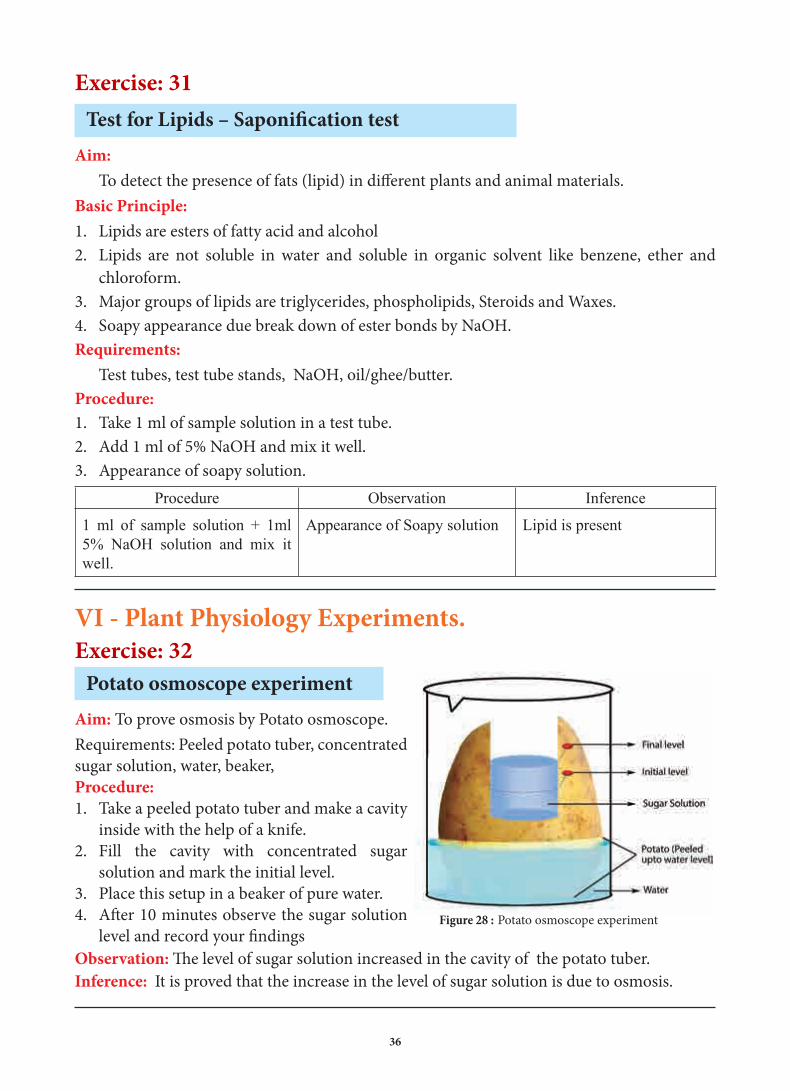
VI - Plant Physiology Experiments.Exercise: 32
Potato osmoscope experimentAim: To prove osmosis by Potato osmoscope.Requirements: Peeled potato tuber, concentrated sugar solution, water, beaker, Procedure:1. Take a peeled potato tuber and make a cavity
inside with the help of a knife. 2. Fill the cavity with concentrated sugar
solution and mark the initial level. 3. Place this setup in a beaker of pure water.4. After 10 minutes observe the sugar solution
level and record your findingsObservation: The level of sugar solution increased in the cavity of the potato tuber.Inference: It is proved that the increase in the level of sugar solution is due to osmosis.
Figure 28 : Potato osmoscope experiment
XI_BOTANY PRACTICAL MANUAL.indd 36 28-08-2018 11:37:07

37
Exercise: 33Paper chromatography experiment
Aim:To separate and study the photosynthetic
pigments (chloroplast pigments) by paper chromatography method.Requirements:
Fresh spinach leaves, chromatography paper (whatman No.1), a wide long test tube, a split cork, mortar & pestle, petroleum ether, acetone, funnel, beaker, filter paper, capillary tube, sand etc.,Procedure:1. Grind a few spinach leaves with little fine sand
and about 5 ml of acetone in a mortar and pestle. Filter it to get acetone extract of the leaf pigments.
2. Take a narrow strip of chromatographic paper (Whatman No.1). Cut one end of the strip into a narrow notch.
3. Put a drop of the pigment extract in the middle of the strip near the notch with the help of capillary tube. Allow the drop to dry and repeat till four or five drops are placed on the paper.
4. Take the test tube and pour about 5 ml of ether acetone solvent (9 ether : 1 acetone) in it. Now hang the pigment extract loaded chromatographic strip in the test tube with the help of a split cork, in such a way that the loading spot lies about 1 cm above the solvent level.
5. Make the cork air tight and place the test tube undisturbed for some time, when solvent rises about 3/4th of the strip, take out the strip carefully and let it dry.
Observation:
After one hour observe the chromatographic paper. The Photosynthetic pigments being separated into four distinct bands. Different leaf pigments can be identified by their colours.
Inference:
Photosynthetic pigments chlorophyll b, chlorophyll a, xanthophyll and carotenes are separated on the chromatographic paper. Presence of different photosynthetic pigments in chloroplast is proved.
Test tube
ChromatographyPaper
CarotenesXanthophyll
Chlrophyll a
Chlrophyll b
Ether acetone solvent
Yellow Orange
Carotene
Yellow
Xanthophyll
Bluish Green
Chlorophyll a
Greenish Yellow
Chlorophyll b
Figure 29 : Paper chromatography experiment
XI_BOTANY PRACTICAL MANUAL.indd 37 28-08-2018 11:37:07

38
Exercise: 34.
Wilmott’s bubbler experiment
Aim :
To determine rate of photosynthesis by Wilmott’s bubbler
Requirements :
Wilmott’s bubbler apparatus, Hydrilla twig, water.
Procedure :
1. Fill the bottle with water and insert Hydrilla twig into the wider part of the tube
2. Hydrilla plant should be cut inside the water to avoid entry of air bubbles
3. Fix the tube with jar which acts as water reservoir
4. Keep the apparatus in sunlight 5. Count the bubbles when they are in same size.6. Repeat the experiment in different light
intensity.Observation :
When there is an increase in photosynthesis, bubble count also increased.Inference :
Rate of photosynthesis increases with increase of light intensity is proved
Exercise: 35Experiment to demonstrate the production of CO2 in aerobic respiration.
Figure 30 : Wilmott's bubbler
Test tube
Water
Wax
Specimen tube
Hydrilla
Respiring Seeds
Water
Figure 31: Demonstration of production of CO2
during aerobic respiration
Potassium Hydroxide
Conical Flask
XI_BOTANY PRACTICAL MANUAL.indd 38 28-08-2018 11:37:08
39
Aim:
To prove carbon dioxide is released by germinating seeds during respiration.
Requirements:
A conical flask, cork, beaker, a twice bent glass tube, a small test tube, thread, water KOH, germinating seeds of bean / gram/ groundnut seeds.
Procedure :
1. Take a definite quantity (i.e 10 gm) of germinating seeds of bean/gram/groundnut in the conical flask and hang a small test tube containing Potassium hydroxide (KOH) crystal inside the flask with the help of a thread.
2. Introduce one end of the bent glass tube into the conical flask through the cork. Dip the free end of the tube in a beaker containing water.
3. Make the apparatus air tight and fix the apparatus with the help of a stand.
4. Note the initial level of water in the bent glass tube and keep the apparatus undisturbed.
Observation :
After two hours the level of water rises in the glass tube. Inference :
Carbon dioxide released by the germinating seeds is absorbed by KOH solution. It creates vacuum, to fill up the vacuum water raised in the tube. Liberation of carbon dioxide during respiration by germinating seeds is proved.
Exercise: 36
Arc auxanometer experiment
Aim:
To measure the growth of a plant in length by Arc auxanometerRequirements:
Arc auxanometer, potted plant, weight, thread, Procedure:
Arc auxanometer which consists of a small pulley to the axis of which is attached a long pointer sliding over a graduated arc.One end of a thread is tied to the stem tip and
Arc
PointerPulley
Weight
Potted plantStand
Figure 32 : Arc auxanometer experiment
XI_BOTANY PRACTICAL MANUAL.indd 39 28-08-2018 11:37:08

40
another end to a weight passes over the pulley tightly. Note down the initial reading of the pointer. Keep the set up for a week.
Observation :
The stem tip grows in length, the pulley moves, and the pointer slide over the graduated arc. The distance travelled by the pointer is noted down.Inference :
Actual growth in length is calculated with help of this formula.
Actual growth in length = Length of the pointer
Distance travelled by the pointer × radius of the pulley
XI_BOTANY PRACTICAL MANUAL.indd 40 28-08-2018 11:37:08

41
Botany - Class XIPractical Manual Developers
Content Developers Sathyavathi Sridhar P.G. Assistant in Botany Sri Sankara senior secondary school, Adyar, Chennai
U. Kalirajan P.G. Assistant in Botany GHSS (ADW), Meenampakkam, Kancheepuram.
D. Padma P.G. Assistant in Botany Prince Matriculation HSS, Madipakkam, Kancheepuram.
Experts Dr. D. Narashiman Professor and Head (Rtd.), Plant Biologly & BioTechnology, MCC College, Tambaram, Kancheepuram.
Dr. K.P. Girivasan Associate Professor of Botany, Govt. Arts & Science College, Nandanam, Chennai.
Dr. Renu Edwin Associate Professor of Botany, Presidency College, Chennai.
Academic Coordinators K. Manjula Lecturer in Botany, DIET, Triplicane, Chennai.
J. Radhamani Lecturer in Botany, DIET, Kaliyampoondi, Kancheepuram.
V. Kokiladevi P.G. Assistant in Botany, GHSS, Sunnambukulam, Thiruvallur.
Illustration Gandhirajan R Pakkirisamy A Santhosh kumar S Sagaya arasu R
Layout Kamatchi Balan Arumugam Madhan Raj Prasanth C Prasanth P Sagaya Arasu R
Wrapper Design Prasanth C
Co-ordination Ramesh Munisamy
Typist R. Chitra
Art and Design Team
XI_BOTANY PRACTICAL MANUAL.indd 41 28-08-2018 11:37:08