NAACCR Hospital Registry Webinar Series - … · Larynx Cancer Surveillance Data Collection 3/6/08...
36
Larynx Cancer Surveillance Data Collection 3/6/08 2007-2008 NAACCR Central Registry Webinar Series 1 NAACCR Hospital Registry Webinar Series Shannon Vann, CTR Jim Hofferkamp, CTR Abstracting Larynx Cancer Incidence & Treatment Data Estimated new cases and deaths from laryngeal cancer in the United States in 2008: New cases: 12,250 Deaths: 3,670 Source: National Cancer Institute www.cancer.org Histology Squamous Cell Carcinoma Keratinizing Non-keratinizing and well-differentiated to poorly differentiated grade. Non squamous cell carcinoma Source: National Cancer Institute www.cancer.org
Transcript of NAACCR Hospital Registry Webinar Series - … · Larynx Cancer Surveillance Data Collection 3/6/08...

Larynx Cancer Surveillance Data
Collection
3/6/08
2007-2008 NAACCR Central Registry
Webinar Series 1
NAACCR Hospital Registry Webinar Series
Shannon Vann, CTR
Jim Hofferkamp, CTR
Abstracting Larynx Cancer Incidence & Treatment Data
� Estimated new cases and deaths
from laryngeal cancer in the United States in 2008:
� New cases: 12,250
� Deaths: 3,670
Source: National Cancer Institute www.cancer.org
Histology
� Squamous Cell Carcinoma
� Keratinizing
� Non-keratinizing and well-differentiated to poorly differentiated grade.
� Non squamous cell carcinoma
Source: National Cancer Institute
www.cancer.org

Larynx Cancer Surveillance Data
Collection
3/6/08
2007-2008 NAACCR Central Registry
Webinar Series 2
Anatomy
Anatomy of the Larynx
Image Source: SEER Training Website

Larynx Cancer Surveillance Data
Collection
3/6/08
2007-2008 NAACCR Central Registry
Webinar Series 3
Credit line: Larynx. In: Greene, F.L., Compton, C.C., Fritz, A.G., et al., editors. AJCC Cancer Staging Atlas. New York: Springer, 2006: 41-52.
©American Joint Committee on Cancer.
Larynx
Used with the permission
of the American Joint
Committee on Cancer
(AJCC), Chicago, Illinois.
The original and primary
source for this information
is the AJCC Cancer
Staging Manual, Sixth
Edition (2002) published
by Springer-Verlag New
York. (For more
information, visit
www.cancerstaging.net.)
Any citation or quotation
of this material must be
credited to the AJCC as its
primary source. The
inclusion of this
information herein does
not authorize any reuse or
further distribution
without the expressed,
written permission of
Springer-Verlag New York,
Inc., on behalf of the
AJCC.
Credit line: Larynx. In: Greene, F.L., Compton, C.C., Fritz, A.G., et al., editors. AJCC Cancer Staging Atlas. New York: Springer, 2006: 41-52.
©American Joint Committee on Cancer.
Larynx
Used with the permission of
the American Joint
Committee on Cancer
(AJCC), Chicago, Illinois.
The original and primary
source for this information is
the AJCC Cancer Staging
Manual, Sixth Edition (2002)
published by Springer-
Verlag New York. (For more
information, visit
www.cancerstaging.net.)
Any citation or quotation of
this material must be
credited to the AJCC as its
primary source. The
inclusion of this information
herein does not authorize
any reuse or further
distribution without the
expressed, written
permission of Springer-
Verlag New York, Inc., on
behalf of the AJCC.
Head and Neck Lymph Node Levels and Groups

Larynx Cancer Surveillance Data
Collection
3/6/08
2007-2008 NAACCR Central Registry
Webinar Series 4
Credit line: Larynx. In: Greene, F.L., Compton, C.C., Fritz, A.G., et al., editors. AJCC Cancer Staging Atlas. New York: Springer, 2006: 41-52.
©American Joint Committee on Cancer.
Used with the permission
of the American Joint
Committee on Cancer
(AJCC), Chicago, Illinois.
The original and primary
source for this information
is the AJCC Cancer
Staging Manual, Sixth
Edition (2002) published
by Springer-Verlag New
York. (For more
information, visit
www.cancerstaging.net.)
Any citation or quotation
of this material must be
credited to the AJCC as its
primary source. The
inclusion of this
information herein does
not authorize any reuse or
further distribution
without the expressed,
written permission of
Springer-Verlag New York,
Inc., on behalf of the
AJCC.
Schematic diagram indicating the location of the lymph node levels in the neck
as described in the text.
Credit line: Larynx. In: Greene, F.L., Compton, C.C., Fritz, A.G., et al., editors. AJCC Cancer Staging Atlas. New York: Springer, 2006: 41-52.
©American Joint Committee on Cancer.
Location of parotid, buccal, retroauricular and occipital nodes.
Used with the permission of the
American Joint Committee on
Cancer (AJCC), Chicago, Illinois.
The original and primary source for
this information is the AJCC Cancer
Staging Manual, Sixth Edition (2002)
published by Springer-Verlag New
York. (For more information, visit
www.cancerstaging.net.)
Any citation or quotation of this
material must be credited to the
AJCC as its primary source. The
inclusion of this information herein
does not authorize any reuse or
further distribution without the
expressed, written permission of
Springer-Verlag New York, Inc., on
behalf of the AJCC.
Credit line: Larynx. In: Greene, F.L., Compton, C.C., Fritz, A.G., et al., editors. AJCC Cancer Staging Atlas. New York: Springer, 2006: 41-52.
©American Joint Committee on Cancer.
Introduction to Head and Neck Sites
Location of retropharyngeal nodes.
Used with the permission of the
American Joint Committee on
Cancer (AJCC), Chicago, Illinois.
The original and primary source for
this information is the AJCC Cancer
Staging Manual, Sixth Edition (2002)
published by Springer-Verlag New
York. (For more information, visit
www.cancerstaging.net.) Any
citation or quotation of this material
must be credited to the AJCC as its
primary source. The inclusion of this
information herein does not
authorize any reuse or further
distribution without the expressed,
written permission of Springer-
Verlag New York, Inc., on behalf of
the AJCC.

Larynx Cancer Surveillance Data
Collection
3/6/08
2007-2008 NAACCR Central Registry
Webinar Series 5
Diagnosing Larynx Cancer
� Physical exam
� Laryngoscope
� MRI/CT Scans
2007 Multiple Primary and Histology Rules
Coding Primary Site
1. Tumor Board
a. Specialty
b. General
2. Staging physician’s site assignment
a. AJCC staging form
b. TNM statement in medical record
3. If neither 1 or 2 available, based on
whether tumor was resected

Larynx Cancer Surveillance Data
Collection
3/6/08
2007-2008 NAACCR Central Registry
Webinar Series 6
Coding Primary Site
4. If total resection of primary tumor was
done, code based on:
a. Operative report – surgeon’s statement
b. Final diagnosis on pathology report
Coding Primary Site
5. If total resection was NOT done code
based on:
a. Endoscopy
b. Radiation oncologist
c. Diagnosing physician
d. Primary care physician
Continued on next slide
Coding Primary Site
e. Other physician
f. Diagnostic imaging
g. Physician statement based on clinical
examination

Larynx Cancer Surveillance Data
Collection
3/6/08
2007-2008 NAACCR Central Registry
Webinar Series 7
Default Site Codes
• Point of origin cannot be determined
– C02.8 Overlapping lesion of tongue
– C08.8 Overlapping lesion of major salivary glands
– C14.8 Overlapping lesion of lip, oral cavity, and pharynx.
Chart 1 – H&N Histology Groups and Specific Types
• Use this chart with the histology rules to code the most specific histologic term.
• The tree is arranged in descending order.
• Each branch is a histology group, starting with
the NOS or group terms and descending into the specific types for that group.
• As you follow the branch down, the terms become more specific
Papillary carcinoma (8050)Verrucous carcinoma (8051)Papillary squamous cell carcinoma; Papillary
epidermoid carcinoma (8052)
Large cell keratinizing; Keratinizing NOS (8071) Large cell nonkeratinizing;Nonkeratinizing
squamous cell carcinoma, NOS (8072) Small cell nonkeratinizing squamous cell carcinoma (8073)Sarcomatoid; Spindle cell squamous cell
carcinoma (8074)Acantholytic; Adenoid; Pseudoglandular
squamous cell carcinoma (8075)Squamous cell carcinoma with horn
formation (8078)
Lymphoepithelial carcinoma;Schmincke tumor (8082)
Basaloid squamous cell carcinoma (8083) Clear cell type squamous cell carcinoma (8084)
Squamous Carcinoma(8070)
Adenocarcinoma, NOS(8140)
Cancer/ Malignant
Neoplasm (8000-8001),Carcinoma, NOS
(8010)
Mucoepidermoidcarcinoma (8430)
Acinar carcinoma(8550)
Adenocarcinomawith mixed subtypes
(8255)
Adenocysticcarcinoma (8200)
Adenosquamous(8560)
UndifferentiatedCarcinoma (8020)

Larynx Cancer Surveillance Data
Collection
3/6/08
2007-2008 NAACCR Central Registry
Webinar Series 8
Multiple Primary Rules
Multiple Primary Rules
• Rule M1
– When it is not possible to determine if there is a single tumor or multiple tumors, opt for a
single tumor and abstract as a single primary.*
• Rule M2
– A single tumor is always a single primary. *
Multiple Tumors

Larynx Cancer Surveillance Data
Collection
3/6/08
2007-2008 NAACCR Central Registry
Webinar Series 9
Multiple Primary RulesMultiple Tumors
• Rule M3
– Tumors on the right side and the left side of a paired site are multiple primaries. **
• Rule M4
– Tumors on the upper lip (C000 or C003) and the lower lip (C001 or C004) are multiple primaries. **
• Rule M5
– Tumors on the upper gum (C030) and the lower gum (C031) are multiple primaries. **
Multiple Primary RulesMultiple Tumors
• Rule M6
– Tumors in the nasal cavity (C300) and the middle ear (C301) are multiple primaries. **
• Rule M7
– Tumors in sites with ICD-O-3 topography codes that are different at the second (Cxxx)
and/or third (Cxxx) character are multiple primaries. **
Multiple Primary RulesMultiple Tumors
• Rule M8
– An invasive tumor following an in situ tumor more than 60 days after diagnosis is a
multiple primary. **
• Rule M9
– Tumors diagnosed more than five (5) years
apart are multiple primaries. **

Larynx Cancer Surveillance Data
Collection
3/6/08
2007-2008 NAACCR Central Registry
Webinar Series 10
Multiple Primary RulesMultiple Tumors
• Rule M10
– Abstract as a single primary* when one tumor is:
• Cancer/malignant neoplasm, NOS (8000) and another is a specific histology or
• Carcinoma, NOS (8010) and another is a specific
carcinoma or
• Adenocarcinoma, NOS (8140) and another is a specific
adenocarcinoma or
• Squamous cell carcinoma, NOS (8070) and another is specific squamous cell carcinoma or
• Melanoma, NOS (8720) and another is a specific melanoma
• Sarcoma, NOS (8800) and another is a specific sarcoma
Multiple Primary RulesMultiple Tumors
• Rule M11
– Tumors with ICD-O-3 histology codes that are different at the first (xxxx), second (xxxx)
or third (xxxx) number are multiple primaries.
• Rule M12
– Tumors that do not meet any of the above
criteria are abstracted as a single primary.
Histology Rules
Larynx Cancer Surveillance Data
Collection
3/6/08
2007-2008 NAACCR Central Registry
Webinar Series 11
Single Tumor
Histology Rules
• Rule H1 – Code the histology documented by the physician
when there is no pathology/cytology specimen or the pathology/cytology report is not available.
• Rule H2 – Code the histology from a metastatic site when there
is no pathology/cytology specimen from the primary site.
• Rule H3 – Code the histology when only one histologic type is
identified.
Histology Rules
• Rule H4
– Code the invasive histologic type when a single tumor has invasive and in situ components.
• Rule H5
– Code the most specific histologic term using Chart 1 when there are multiple histologies within the same branch.
• Rule H6
– Code the histology with the numerically higher ICD-O-3 code.

Larynx Cancer Surveillance Data
Collection
3/6/08
2007-2008 NAACCR Central Registry
Webinar Series 12
Multiple Tumors Abstracted as a Single Primary
Histology Rules
• Rule H7
– Code the histology documented by the physician when there is no
pathology/cytology specimen or the pathology/cytology report is not available.
• Rule H8
– Code the histology from the metastatic site when there is no pathology/cytology specimen from the primary site.
Histology Rules
• Rule H9
– Code the histology when only one histologic type is identified.
• Rule H10
– Code the histology of the most invasive tumor.

Larynx Cancer Surveillance Data
Collection
3/6/08
2007-2008 NAACCR Central Registry
Webinar Series 13
Histology Rules• Rule H11 Code the most specific histologic
term using Chart 1 when there are multiple histologies within the same branch. Examples of histologies within the same branch are:– Cancer/malignant neoplasm, NOS (8000) and a more
specific histology or– Carcinoma, NOS (8010) and a more specific
carcinoma or– Squamous cell carcinoma, NOS (8070) and a more
specific squamous carcinoma or– Adenocarcinoma, NOS(8140) and a more specific
adenocarcinoma or– Melanoma, NOS (8720) and a more specific
melanoma or– Sarcoma, NOS (8800) and a more specific sarcoma
Histology Rules
• Rule H12
– Code the histology with the numerically higher ICD-O-3 code.
MP/H Task Force

Larynx Cancer Surveillance Data
Collection
3/6/08
2007-2008 NAACCR Central Registry
Webinar Series 14
Collaborative Staging
Larynx
Larynx
� Glottic, Larynx
� C32.0 Glottis
� Supraglottic
� C32.1 Supraglottis
� Subglottic
� C32.2 Subglottis
� Overlapping or Larynx, NOS
� C32.3 Laryngeal Cartilage
� C32.8 Overlapping lesion
� C32.9 Larynx, NOS
CS Tumor Size
� Use Standard Table

Larynx Cancer Surveillance Data
Collection
3/6/08
2007-2008 NAACCR Central Registry
Webinar Series 15
CS Extension
� Supraglottic Larynx
� 10-Invasive tumor with normal vocal cord mobility confined to: Supraglottis (one subsite)
� Glottic Larynx
� 10-Invasive tumor with normal mobility confined to glottis, NOS; Intrinsic larynx; laryngeal commissure (s) anterior, posterior; vocal cord (s), nos; true vocal cord (s), true cords.
CS Extension
� Subglottic Larynx
� 10-Invasive tumor with normal vocal cord mobility confined to the subglottis.
Credit line: Larynx. In: Greene, F.L., Compton, C.C., Fritz, A.G., et al., editors. AJCC Cancer Staging Atlas. New York: Springer, 2006: 41-52.
©American Joint Committee on Cancer.
Larynx
Anatomical sites and subsites of the three regions of the larynx: supraglottis, glottis, and subglottis. Supraglottis (C32.1) subsites include suprahyoid
epiglottis (i), aryepiglottic fold, laryngeal aspect (ii), infrahyoid epiglottis (iv), and ventricular bands or false cords (v).
Used with the permission of the
American Joint Committee on
Cancer (AJCC), Chicago, Illinois.
The original and primary source for
this information is the AJCC Cancer
Staging Manual, Sixth Edition (2002)
published by Springer-Verlag New
York. (For more information, visit
www.cancerstaging.net.) Any
citation or quotation of this material
must be credited to the AJCC as its
primary source. The inclusion of this
information herein does not
authorize any reuse or further
distribution without the expressed,
written permission of Springer-
Verlag New York, Inc., on behalf of
the AJCC.

Larynx Cancer Surveillance Data
Collection
3/6/08
2007-2008 NAACCR Central Registry
Webinar Series 16
Credit line: Larynx. In: Greene, F.L., Compton, C.C., Fritz, A.G., et al., editors. AJCC Cancer Staging Atlas. New York: Springer, 2006: 41-52.
©American Joint Committee on Cancer.
Anatomy of the Larynx
Image Source: SEER Training Website
Credit line: Larynx. In: Greene, F.L., Compton, C.C., Fritz, A.G., et al., editors. AJCC Cancer Staging Atlas. New York: Springer, 2006: 41-52.
©American Joint Committee on Cancer.
Larynx
Anatomical sites and subsites of the supraglottis and glottis. Supraglottis (C32.1) subsites include suprahyoid epiglottis (i), aryepiglottic fold, laryngeal aspect (ii), arytenoids (iii), and ventricular bands or false cords (v). Glottis (C32.0) subsites include vocal cords (i), anterior commissure (ii), and posterior commissure (iii).
Used with the permission of the
American Joint Committee on
Cancer (AJCC), Chicago, Illinois.
The original and primary source for
this information is the AJCC Cancer
Staging Manual, Sixth Edition (2002)
published by Springer-Verlag New
York. (For more information, visit
www.cancerstaging.net.) Any
citation or quotation of this material
must be credited to the AJCC as its
primary source. The inclusion of this
information herein does not
authorize any reuse or further
distribution without the expressed,
written permission of Springer-
Verlag New York, Inc., on behalf of
the AJCC.
Glottis

Larynx Cancer Surveillance Data
Collection
3/6/08
2007-2008 NAACCR Central Registry
Webinar Series 17
Credit line: Larynx. In: Greene, F.L., Compton, C.C., Fritz, A.G., et al., editors. AJCC Cancer Staging Atlas. New York: Springer, 2006: 41-52.
©American Joint Committee on Cancer.
Larynx
• T1 tumors of the glottis are limited to the vocal cord(s) with normal mobility (may involve anterior or posterior commissure).
• T1a tumors are limited to one vocal cord (top right) • T1b tumors involve both vocal cords (bottom right).
Used with the permission of the
American Joint Committee on
Cancer (AJCC), Chicago, Illinois.
The original and primary source for
this information is the AJCC Cancer
Staging Manual, Sixth Edition (2002)
published by Springer-Verlag New
York. (For more information, visit
www.cancerstaging.net.) Any
citation or quotation of this material
must be credited to the AJCC as its
primary source. The inclusion of this
information herein does not
authorize any reuse or further
distribution without the expressed,
written permission of Springer-
Verlag New York, Inc., on behalf of
the AJCC.
Credit line: Larynx. In: Greene, F.L., Compton, C.C., Fritz, A.G., et al., editors. AJCC Cancer Staging Atlas. New York: Springer, 2006: 41-52.
©American Joint Committee on Cancer.
Larynx
T2 tumors of the glottis extend to supraglottis and/or subglottis, or with impaired vocal cord mobility.
Used with the permission of
the American Joint
Committee on Cancer
(AJCC), Chicago, Illinois.
The original and primary
source for this information is
the AJCC Cancer Staging
Manual, Sixth Edition (2002)
published by Springer-
Verlag New York. (For more
information, visit
www.cancerstaging.net.)
Any citation or quotation of
this material must be
credited to the AJCC as its
primary source. The
inclusion of this information
herein does not authorize
any reuse or further
distribution without the
expressed, written
permission of Springer-
Verlag New York, Inc., on
behalf of the AJCC.
CS Ext
� 30-Tumor involves adjacent
regions(s) of larynx
� Subglottis
� Supraglottis
� False vocal cord(s)
� 35 Impaired vocal cord mobility

Larynx Cancer Surveillance Data
Collection
3/6/08
2007-2008 NAACCR Central Registry
Webinar Series 18
Credit line: Larynx. In: Greene, F.L., Compton, C.C., Fritz, A.G., et al., editors. AJCC Cancer Staging Atlas. New York: Springer, 2006: 41-52.
©American Joint Committee on Cancer.
Larynx
T3 tumors of the glottis are limited to the larynx with vocal cord fixation (shown), and/or invade paraglottic space, and/or minor thyroid cartilage erosion (e.g., inner cortex).
Used with the permission of
the American Joint
Committee on Cancer
(AJCC), Chicago, Illinois.
The original and primary
source for this information is
the AJCC Cancer Staging
Manual, Sixth Edition (2002)
published by Springer-
Verlag New York. (For more
information, visit
www.cancerstaging.net.)
Any citation or quotation of
this material must be
credited to the AJCC as its
primary source. The
inclusion of this information
herein does not authorize
any reuse or further
distribution without the
expressed, written
permission of Springer-
Verlag New York, Inc., on
behalf of the AJCC.
CS Ext� 40 Tumor limited to larynx WITH vocal
cord fixation� Involvement of intrinsic muscle(s):
� Aryepiglottic
� Corniculate tubercle
� Cuneform tubercule
� Arytenoid
� Cricoarytenoid
� Cricothyroid
� Thyroepiglottic
� Thyroarytenoid
� Vocalis
CS Ext
� 51 Paraglottic space
� 52 Minor thyroid cartilage erosion
(e.g., inner cortex)

Larynx Cancer Surveillance Data
Collection
3/6/08
2007-2008 NAACCR Central Registry
Webinar Series 19
Credit line: Larynx. In: Greene, F.L., Compton, C.C., Fritz, A.G., et al., editors. AJCC Cancer Staging Atlas. New York: Springer, 2006: 41-52.
©American Joint Committee on Cancer.
Credit line: Larynx. In: Greene, F.L., Compton, C.C., Fritz, A.G., et al., editors. AJCC Cancer Staging Atlas. New York: Springer, 2006: 41-52.
©American Joint Committee on Cancer.
Larynx
T4a tumors of the glottis invade through the thyroid cartilage and/or invade tissues beyond the larynx (e.g., trachea, soft tissues of neck including deep extrinsic muscle of the tongue, strap muscles, thyroid, or esophagus).
Used with the permission of
the American Joint Committee
on Cancer (AJCC), Chicago,
Illinois. The original and
primary source for this
information is the AJCC
Cancer Staging Manual, Sixth
Edition (2002) published by
Springer-Verlag New York.
(For more information, visit
www.cancerstaging.net.) Any
citation or quotation of this
material must be credited to
the AJCC as its primary
source. The inclusion of this
information herein does not
authorize any reuse or further
distribution without the
expressed, written permission
of Springer-Verlag New York,
Inc., on behalf of the AJCC.
CS Ext
� 60 Base of tongue� Hypopharynx, NOS� Pre-epiglottic tissues� Postcricoid area� Pyriform sinus� Vallecula
� 68 Extension to/through� Cricoid cartilage� Thyroid cartilage except minor erosion, see
code 52

Larynx Cancer Surveillance Data
Collection
3/6/08
2007-2008 NAACCR Central Registry
Webinar Series 20
CS Ext
� 70 Extension to/through tissues beyond larynx:� Extrinsic (strap) muscles� Omohyoid� Sternohyoid� Sternothyroid� Thyrohyoid� Oropharynx� Skin� Soft tissue of neck� Thyroid gland� Trachea
CS Ext
� 71 Cervical esophagus
� 73 Deep extrinsic muscle(s) of tongue
� 80 Further contiguous extension, including:
� Mediastinal structures
� Prevertebral space
� Tumor encases carotid artery
Lymph Nodes

Larynx Cancer Surveillance Data
Collection
3/6/08
2007-2008 NAACCR Central Registry
Webinar Series 21
CS Lymph Nodes
Note 1:
� For head and neck schemas, this field includes all lymph nodes defined as Levels I-VII and Other by AJCC. The complete definitions are provided in the General Instructions.
Note 2:
� For head and neck schemas, additional information about lymph nodes (size of involved nodes, extracapsular extension, and levels involved) is coded in Site-Specific Factors 1-6.
CS Lymph NodesNote 3: � If laterality of lymph nodes is not specified,
assume nodes are ipsilateral. Midline nodes are considered ipsilateral.
Note 4:� For head and neck cancers, if lymph nodes are
described only as "supraclavicular", try to determine if they are in Level IV (deep to the sternocleidomastoid muscle, in the lower jugular chain) or Level V (in the posterior triangle, inferior to the transverse cervical artery) and code appropriately. If the specific level cannot be determined, consider them as Level V nodes
CS Lymph Nodes
Code 10
� Single positive ipsilateral regional node:
� Level II
� Level III
� Level IV
� Level VI
� Cervical, NOS
� Deep cervical, NOS
� Internal jugular NOS:
� Regional lymph node, NOS
� Stated as N1, NOS

Larynx Cancer Surveillance Data
Collection
3/6/08
2007-2008 NAACCR Central Registry
Webinar Series 22
CS Lymph Nodes
Code 11
� Single positive ipsilateral regional node:
� Level I
� Other groups
� Retropharyngeal
� Mandibular, NOS
CS Lymph Nodes
Code 12
� Single positive ipsilateral regional node:
� Level V node
� Level VII node
� Upper mediastinum (for other mediastinal
nodes see CS Mets at DX)
� Other groups
� Supraclavicular, NOS (See Note 4)
CS Lymph Node
� 30 Regional lymph nodes as listed in code 10:
� Positive ipsilateral node(s), not stated if single or multiple
� 31 Regional lymph nodes as listed in code 11:
� Positive ipsilateral node(s), not stated if single or multiple
� 32 Regional lymph nodes as listed in code 12:
� Positive ipsilateral node(s), not stated if single or multiple

Larynx Cancer Surveillance Data
Collection
3/6/08
2007-2008 NAACCR Central Registry
Webinar Series 23
CS Lymph Nodes
� 40 Regional lymph nodes as listed in code 10:
� Positive bilateral or contralateral nodes
� 41 Regional lymph nodes as listed in code 11:
� Positive bilateral or contralateral nodes
� 42 Regional lymph nodes as listed in code 12:
� Positive bilateral or contralateral nodes
CS Lymph Nodes
� 50 Regional lymph nodes as listed in code 10:� Positive node(s), not stated if ipsilateral, or bilateral,
or contralateral, AND not stated if single or multiple
� 51 Regional lymph nodes as listed in code 11:� Positive node(s), not stated if ipsilateral, or bilateral,
or contralateral, AND not stated if single or multiple
� 52 Regional lymph nodes as listed in code 12:� Positive node(s), not stated if ipsilateral, or bilateral,
or contralateral, AND not stated if single or multiple
Credit line: Larynx. In: Greene, F.L., Compton, C.C., Fritz, A.G., et al., editors. AJCC Cancer Staging Atlas. New York: Springer, 2006: 41-52.
©American Joint Committee on Cancer.
Introduction to Head and Neck Sites
Schematic diagram indicating the location of the lymph node levels in the neck as described in the text.
Used with the permission of the
American Joint Committee on
Cancer (AJCC), Chicago, Illinois.
The original and primary source for
this information is the AJCC Cancer
Staging Manual, Sixth Edition (2002)
published by Springer-Verlag New
York. (For more information, visit
www.cancerstaging.net.) Any
citation or quotation of this material
must be credited to the AJCC as its
primary source. The inclusion of this
information herein does not
authorize any reuse or further
distribution without the expressed,
written permission of Springer-
Verlag New York, Inc., on behalf of
the AJCC.

Larynx Cancer Surveillance Data
Collection
3/6/08
2007-2008 NAACCR Central Registry
Webinar Series 24
Credit line: Larynx. In: Greene, F.L., Compton, C.C., Fritz, A.G., et al., editors. AJCC Cancer Staging Atlas. New York: Springer, 2006: 41-52.
©American Joint Committee on Cancer.
Introduction to Head and Neck Sites
Location of parotid, buccal, retroauricular and occipital nodes.
Used with the permission of the
American Joint Committee on
Cancer (AJCC), Chicago, Illinois.
The original and primary source for
this information is the AJCC Cancer
Staging Manual, Sixth Edition (2002)
published by Springer-Verlag New
York. (For more information, visit
www.cancerstaging.net.)
Any citation or quotation of this
material must be credited to the
AJCC as its primary source. The
inclusion of this information herein
does not authorize any reuse or
further distribution without the
expressed, written permission of
Springer-Verlag New York, Inc., on
behalf of the AJCC.
Credit line: Larynx. In: Greene, F.L., Compton, C.C., Fritz, A.G., et al., editors. AJCC Cancer Staging Atlas. New York: Springer, 2006: 41-52.
©American Joint Committee on Cancer.
Introduction to Head and Neck Sites
Location of retropharyngeal nodes.
Used with the permission of the
American Joint Committee on
Cancer (AJCC), Chicago, Illinois.
The original and primary source for
this information is the AJCC Cancer
Staging Manual, Sixth Edition (2002)
published by Springer-Verlag New
York. (For more information, visit
www.cancerstaging.net.) Any
citation or quotation of this material
must be credited to the AJCC as its
primary source. The inclusion of this
information herein does not
authorize any reuse or further
distribution without the expressed,
written permission of Springer-
Verlag New York, Inc., on behalf of
the AJCC.
Credit line: Larynx. In: Greene, F.L., Compton, C.C., Fritz, A.G., et al., editors. AJCC Cancer Staging Atlas. New York: Springer, 2006: 41-52.
©American Joint Committee on Cancer.
Introduction to Head and Neck Sites
Regional lymph node (N) classification for all head and neck cancer sites except nasopharynx and thyroid cancers.
Used with the permission of
the American Joint
Committee on Cancer
(AJCC), Chicago, Illinois.
The original and primary
source for this information
is the AJCC Cancer Staging
Manual, Sixth Edition
(2002) published by
Springer-Verlag New York.
(For more information, visit
www.cancerstaging.net.)
Any citation or quotation of
this material must be
credited to the AJCC as its
primary source. The
inclusion of this information
herein does not authorize
any reuse or further
distribution without the
expressed, written
permission of Springer-
Verlag New York, Inc., on
behalf of the AJCC.

Larynx Cancer Surveillance Data
Collection
3/6/08
2007-2008 NAACCR Central Registry
Webinar Series 25
Distant Mets
Distant Mets
� Common only for patients who have
bulky regional lymphadenopathy
� Lung is the most common site
� Skeletal and Hepatic less often
� Mediastinal lymph nodes are considered distant mets
CS Site-Specific Factor 1Size of Lymph Nodes
Code Description
000 No involved regional nodes
001-988 Exact size in millimeters
989 989 mm or larger
990 Microscopic focus
991 Described as less than 1 cm
992 Described as less than 2 cm or greater than 1 cm or between 1 cm and 2 cm

Larynx Cancer Surveillance Data
Collection
3/6/08
2007-2008 NAACCR Central Registry
Webinar Series 26
CS Site-Specific Factor 1Size of Lymph Nodes
Code Description
993 Described as less than 3 cm or greater than 2 cm or between 2 cm and 3 cm
994 Described as less than 4 cm or greater than 3 cm or
between 3 cm and 4 cm
CS Site-Specific Factor 1Size of Lymph Nodes
Code Description995 Described as less than 5 cm
or greater than 4 cm or between 4 cm and 5 cm
996 Described as less than 6 cm or greater than 5 cm or between 5 cm and 6 cm
997 Described as more than 6 cm999 Unknown
CS Site-Specific Factor 2Extracapsular Extension
CodeDescription
000 No extracapsular extension
001 Extracapsular extension clinically
005 Extracapsular extension pathologically
888 Not applicable; no lymph node involvement
999 Unknown

Larynx Cancer Surveillance Data
Collection
3/6/08
2007-2008 NAACCR Central Registry
Webinar Series 27
CS Site-Specific Factors 3-6
� One digit represents lymph nodes of
a single level� 0 = lymph nodes not involved
� 1 = lymph nodes involved
� 9 = unknown
� Code unknown lymph node as 999
� Code regional nodes, NOS, as 000
CS Site-Specific Factor 3
� Record involvement or non-
involvement of levels I, II, and III lymph nodes
_______ _______ _______
I II III
CS Site-Specific Factor 4
� Record involvement or non-
involvement of levels IV, V, and retropharyngeal (RP) lymph nodes
_______ _______ _______
IV V RP
Larynx Cancer Surveillance Data
Collection
3/6/08
2007-2008 NAACCR Central Registry
Webinar Series 28
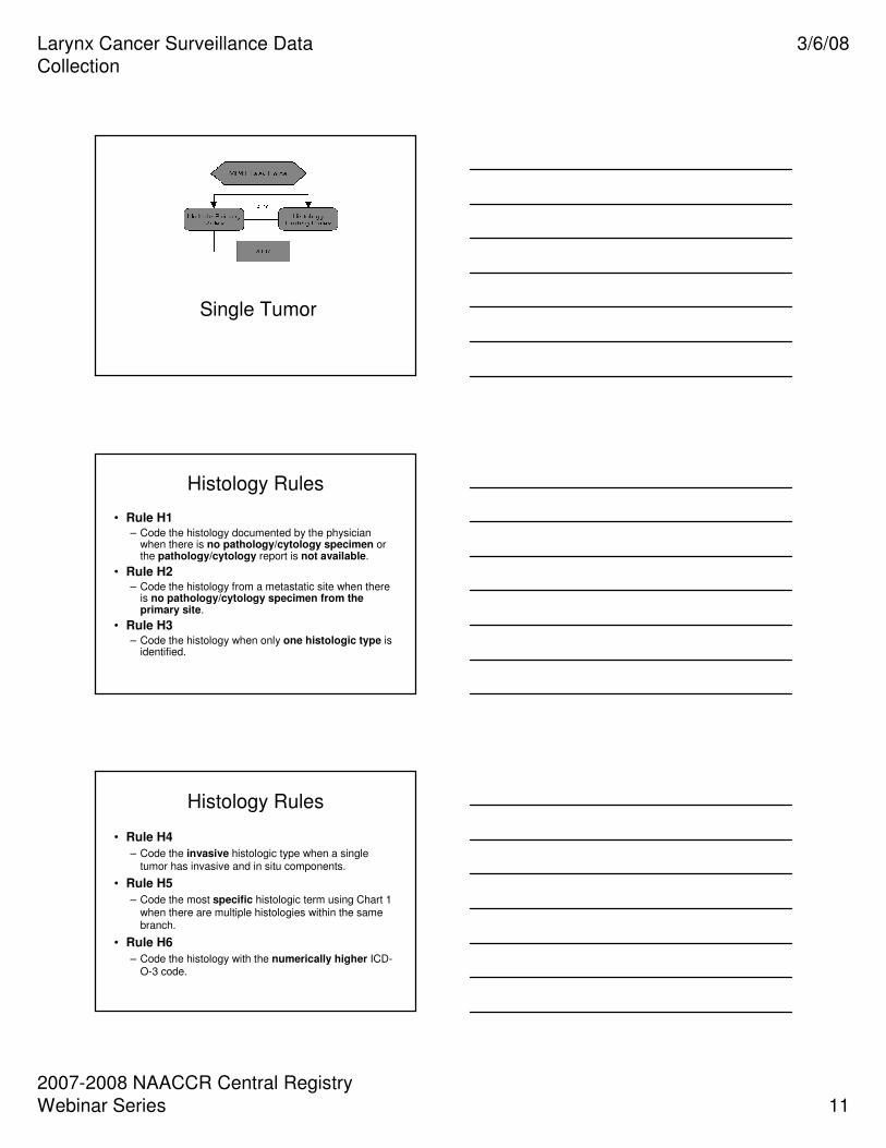
CS Site-Specific Factor 5
� Record involvement or non-
involvement of levels VI, VII, and facial (F) lymph nodes
_______ _______ _______
VI VII F
CS Site-Specific Factor 6
� Record involvement or non-
involvement of parapharyngeal (PP), parotid (PA), and suboccipital (S)
lymph nodes
_______ _______ _______
PP PA S
CS Site-Specific Factors 1-6
Example 11: Path from radical neck dissection for parotid gland primary: 2 metastatic submandibular nodes with extracapsular extension to one node; 3 metastatic posterior cervical nodes, largest malignant node 2.5 cm in diameter.
Primary site: C07.9 Parotid gland
SSF1: 025 SSF4: 010
SSF2: 005 SSF5: 000
SSF3: 100 SSF6: 000

Larynx Cancer Surveillance Data
Collection
3/6/08
2007-2008 NAACCR Central Registry
Webinar Series 29
CS Site-Specific Factors 1-6
Example 12: Tonsillectomy path – 1 cm squamous cell carcinoma of tonsillar fossa. CT scan head/neck – swelling to cervical nodes, probably malignant, less than 2 cm in size.
Primary site: C09.0 Tonsillar fossa
SSF1: 992 SSF4: 000
SSF2: 000 SSF5: 000
SSF3: 000 SSF6: 000
CS Site-Specific Factors 3-6
� One digit represents lymph nodes of
a single level� 0 = lymph nodes not involved
� 1 = lymph nodes involved
� 9 = unknown
� Code unknown lymph node as 999
� Code regional nodes, NOS, as 000
CS Site-Specific Factor 3
� Record involvement or non-
involvement of levels I, II, and III lymph nodes
_______ _______ _______
I II III

Larynx Cancer Surveillance Data
Collection
3/6/08
2007-2008 NAACCR Central Registry
Webinar Series 30
Credit line: Larynx. In: Greene, F.L., Compton, C.C., Fritz, A.G., et al., editors. AJCC Cancer Staging Atlas. New York: Springer, 2006: 41-52.
©American Joint Committee on Cancer.
Introduction to Head and Neck Sites
Schematic diagram indicating the location of the lymph node levels in the neck
as described in the text.
Used with the permission of the
American Joint Committee on
Cancer (AJCC), Chicago, Illinois.
The original and primary source for
this information is the AJCC Cancer
Staging Manual, Sixth Edition (2002)
published by Springer-Verlag New
York. (For more information, visit
www.cancerstaging.net.) Any
citation or quotation of this material
must be credited to the AJCC as its
primary source. The inclusion of this
information herein does not
authorize any reuse or further
distribution without the expressed,
written permission of Springer-
Verlag New York, Inc., on behalf of
the AJCC.
CS Site-Specific Factor 4
� Record involvement or non-
involvement of levels IV, V, and retropharyngeal (RP) lymph nodes
_______ _______ _______
IV V RP
Credit line: Larynx. In: Greene, F.L., Compton, C.C., Fritz, A.G., et al., editors. AJCC Cancer Staging Atlas. New York: Springer, 2006: 41-52.
©American Joint Committee on Cancer.
Introduction to Head and Neck Sites
Schematic diagram indicating the location of the lymph node levels in the neck as described in the text.
Used with the permission of the
American Joint Committee on
Cancer (AJCC), Chicago, Illinois.
The original and primary source for
this information is the AJCC Cancer
Staging Manual, Sixth Edition (2002)
published by Springer-Verlag New
York. (For more information, visit
www.cancerstaging.net.) Any
citation or quotation of this material
must be credited to the AJCC as its
primary source. The inclusion of this
information herein does not
authorize any reuse or further
distribution without the expressed,
written permission of Springer-
Verlag New York, Inc., on behalf of
the AJCC.

Larynx Cancer Surveillance Data
Collection
3/6/08
2007-2008 NAACCR Central Registry
Webinar Series 31
Credit line: Larynx. In: Greene, F.L., Compton, C.C., Fritz, A.G., et al., editors. AJCC Cancer Staging Atlas. New York: Springer, 2006: 41-52.
©American Joint Committee on Cancer.
Introduction to Head and Neck Sites
Location of retropharyngeal nodes.
Used with the permission of the
American Joint Committee on
Cancer (AJCC), Chicago, Illinois.
The original and primary source for
this information is the AJCC Cancer
Staging Manual, Sixth Edition (2002)
published by Springer-Verlag New
York. (For more information, visit
www.cancerstaging.net.) Any
citation or quotation of this material
must be credited to the AJCC as its
primary source. The inclusion of this
information herein does not
authorize any reuse or further
distribution without the expressed,
written permission of Springer-
Verlag New York, Inc., on behalf of
the AJCC.
CS Site-Specific Factor 5
� Record involvement or non-
involvement of levels VI, VII, and facial (F) lymph nodes
_______ _______ _______
VI VII F
Credit line: Larynx. In: Greene, F.L., Compton, C.C., Fritz, A.G., et al., editors. AJCC Cancer Staging Atlas. New York: Springer, 2006: 41-52.
©American Joint Committee on Cancer.
Introduction to Head and Neck Sites
Schematic diagram indicating the location of the lymph node levels in the neck as described in the text.
Used with the permission of the
American Joint Committee on
Cancer (AJCC), Chicago, Illinois.
The original and primary source for
this information is the AJCC Cancer
Staging Manual, Sixth Edition (2002)
published by Springer-Verlag New
York. (For more information, visit
www.cancerstaging.net.) Any
citation or quotation of this material
must be credited to the AJCC as its
primary source. The inclusion of this
information herein does not
authorize any reuse or further
distribution without the expressed,
written permission of Springer-
Verlag New York, Inc., on behalf of
the AJCC.

Larynx Cancer Surveillance Data
Collection
3/6/08
2007-2008 NAACCR Central Registry
Webinar Series 32
CS Site-Specific Factor 6
� Record involvement or non-
involvement of parapharyngeal (PP), parotid (PA), and suboccipital (S)
lymph nodes
_______ _______ _______
PP PA S
Credit line: Larynx. In: Greene, F.L., Compton, C.C., Fritz, A.G., et al., editors. AJCC Cancer Staging Atlas. New York: Springer, 2006: 41-52.
©American Joint Committee on Cancer.
Introduction to Head and Neck Sites
Location of parotid, buccal, retroauricular and occipital nodes.
Used with the permission of the
American Joint Committee on
Cancer (AJCC), Chicago, Illinois.
The original and primary source for
this information is the AJCC Cancer
Staging Manual, Sixth Edition (2002)
published by Springer-Verlag New
York. (For more information, visit
www.cancerstaging.net.)
Any citation or quotation of this
material must be credited to the
AJCC as its primary source. The
inclusion of this information herein
does not authorize any reuse or
further distribution without the
expressed, written permission of
Springer-Verlag New York, Inc., on
behalf of the AJCC.
First Course Treatment
Larynx Gland

Larynx Cancer Surveillance Data
Collection
3/6/08
2007-2008 NAACCR Central Registry
Webinar Series 33
First Course Treatment
� Intended to affect tumor by
� Modification
� Control
� Removal
� Destruction
� Includes curative and palliative
treatment
Treatment: Stage I
� Supraglottis � External-beam radiation therapy alone.
� Supraglottic laryngectomy.
� Glottis� Radiation therapy� Cordectomy
� Partial or hemilaryngectomy or total laryngectomy� Laser excision
� Subglottis� Radiation therapy � Surgery
Source: National Cancer Institute
www.cancer.org
Treatment: Stage II� Supraglottis
� External-beam radiation therapy � Supraglottic laryngectomy or total
laryngectomy, � Postoperative radiation therapy is indicated for
positive or close surgical margins.
� Glottis � Radiation therapy� Partial or hemilaryngectomy or total
laryngectomy� Laser microsurgery
� Subglottis � Radiation therapy
Source: National Cancer Institute
www.cancer.org

Larynx Cancer Surveillance Data
Collection
3/6/08
2007-2008 NAACCR Central Registry
Webinar Series 34
Treatment: Stage III
� Supraglottis � Surgery with or without postoperative
radiation therapy.
� Definitive radiation therapy with surgery for salvage of radiation failures.
� Chemotherapy administered concomitantly with radiation therapy
� Laryngectomy
Source: National Cancer Institute
www.cancer.org
Treatment: Stage III
� Glottis
� Surgery with or without postoperative radiation therapy
� Definitive radiation therapy with surgery for salvage of radiation failures
� Chemotherapy administered concomitantly with radiation therapy
Laryngectomy
Source: National Cancer Institute
www.cancer.org
Treatment: Stage III
� Subglottis
� Laryngectomy plus isolated thyroidectomy and tracheoesophageal
node dissection usually followed by postoperative radiation therapy.
� Radiation therapy alone
Source: National Cancer Institute
www.cancer.org

Larynx Cancer Surveillance Data
Collection
3/6/08
2007-2008 NAACCR Central Registry
Webinar Series 35
Treatment: Stage IV
� Supraglottis
� Total laryngectomy with postoperative radiation therapy.
� Definitive radiation therapy with surgery for salvage of radiation failures.
� Chemotherapy administered concomitantly with radiation therapy
Source: National Cancer Institute
www.cancer.org
Treatment: Stage IV
� Glottis
� Total laryngectomy with postoperative radiation therapy.
� Definitive radiation therapy with surgery for salvage of radiation failures.
� Chemotherapy administered concomitantly with radiation therapy
Source: National Cancer Institute
www.cancer.org
Treatment: Stage IV
� Subglottis
� Laryngectomy plus total thyroidectomy and bilateral tracheoesophageal node
dissection usually followed by postoperative radiation therapy.
� Treatment by radiation therapy alone is indicated for patients who are not candidates for surgery.
Source: National Cancer Institute
www.cancer.org

Larynx Cancer Surveillance Data
Collection
3/6/08
2007-2008 NAACCR Central Registry
Webinar Series 36
Surgery
� Hemilaryngectomy (30)� Left or right half of larynx including thyroid cartilage,
false cord, ventricle, and true vocal cord.
� Partial laryngectomy (30)� Part of thyroid cartilage and corresponding portions of
laryngeal mucosa.
� Supraglottic laryngectomy (33)� Part of larynx superior to the true vocal cord
(transection through the ventricles).
� Total laryngectomy (41)� Entire larynx.
Treatment
� Radiation Therapy
� Beam Radiation
� Preferred therapy for early stage disease
� Chemotherapy
� Hormone Therapy
� Other Therapy
Questions?