NA-MIC National Alliance for Medical Image Computing NA-MIC Ron Kikinis, M.D., Professor of...
14
NA-MIC National Alliance for Medical Image Computing http://na-mic.org NA-MIC Ron Kikinis, M.D., Professor of Radiology, Harvard Medical School, Director, Surgical Planning Laboratory, Brigham and Women’s Hospital [email protected] Founding Director, Surgical Planning Laboratory, Brigham and Women’s Hospital Principal Investigator, the National Alliance for Medical Image Computing, and the Neuroimage Analysis Center Research Director, National Center for Image Guided Therapy
-
Upload
katherine-willis -
Category
Documents
-
view
226 -
download
0
Transcript of NA-MIC National Alliance for Medical Image Computing NA-MIC Ron Kikinis, M.D., Professor of...
NA-MICNational Alliance for Medical Image Computing http://na-mic.org
NA-MIC
Ron Kikinis, M.D.,
Professor of Radiology, Harvard Medical School, Director, Surgical Planning Laboratory, Brigham and Women’s [email protected]
Founding Director, Surgical Planning Laboratory, Brigham and Women’s HospitalPrincipal Investigator, the National Alliance for Medical Image Computing, and the Neuroimage Analysis CenterResearch Director, National Center for Image Guided Therapy

National Alliance for Medical Image Computing http://na-mic.org
National Alliance
http://wiki.na-mic.org/Wiki/index.php/Leadership:Main
My research-our research

3National Alliance for Medical Image Computing http://na-mic.org
Medical Image Computing• More image data, more complexity
• MIC: Extract relevant information• Algorithms, Tools, Applications
Provided by Odonnell, et al.Provided by Odonnell, et al. Provided by Kindlmann, et al.Provided by Kindlmann, et al.Golby, Archip et al.Golby, Archip et al.

National Alliance for Medical Image Computing http://na-mic.org
NA-MIC Algorithm Science
• Mathematical sciences and technologies applied to image analysis– Statistical methods– Differential algebra and geometry– Particle methods– Adaptive filtering techniques
National Alliance for Medical Image Computing http://na-mic.org
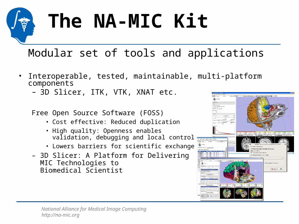
The NA-MIC Kit
Modular set of tools and applications
• Interoperable, tested, maintainable, multi-platform components– 3D Slicer, ITK, VTK, XNAT etc.
Free Open Source Software (FOSS) • Cost effective: Reduced duplication
• High quality: Openness enables validation, debugging and local control
• Lowers barriers for scientific exchange
– 3D Slicer: A Platform for Delivering MIC Technologies to Biomedical Scientist
National Alliance for Medical Image Computing http://na-mic.org
6
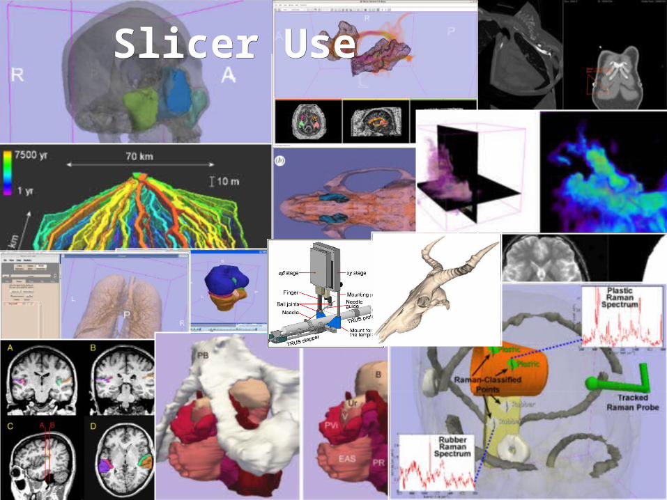
Image GallerySlicer UseSlicer Use

National Alliance for Medical Image Computing http://na-mic.org
Driving Biological Projects I
• 2004-2007– Dartmouth/Indiana
• Examines DW-MRI and fMRI data in patients with schizophrenia to determine association with brain activation during memory tasks
– Harvard • Uses structural MRI, diffusion-weighted MRI, and fMRI to
study the neural bases of schizophrenia and related psychiatric disorders.
– UCI• Investigate the connections between neuroanatomy and
schizophrenia.
– Toronto• Investigate genetic links in schizophrenia.

National Alliance for Medical Image Computing http://na-mic.org
Driving Biological Projects II
• 2007-2010– Harvard
• Collect high-res DTI, structural and fMRI data from patients with VCFS and use NAMIC tools to analyse the data.
– JHU / Queens• Developing novel systems and procedures for prostate
cancer interventions, such as biopsy and needle-based local therapies.
– Mind• Evaluation of existing tools and the development new tools
within SLICER for the time series analysis of brain lesions in lupus.
– UNC• Longitudinal study of early brain development by cortical
thickness in autistic children and controls (2 years with follow-up at 4 years).

National Alliance for Medical Image Computing http://na-mic.org
Outreach
Website
Self-Training
2006
2005
2007
Hands-on Workshops
National Alliance for Medical Image Computing http://na-mic.org
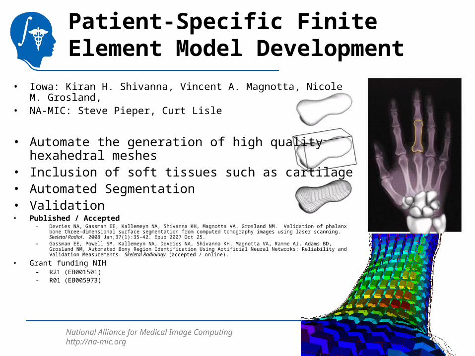
Patient-Specific Finite Element Model Development
• Iowa: Kiran H. Shivanna, Vincent A. Magnotta, Nicole M. Grosland, • NA-MIC: Steve Pieper, Curt Lisle
• Automate the generation of high quality hexahedral meshes
• Inclusion of soft tissues such as cartilage• Automated Segmentation• Validation• Published / Accepted
– Devries NA, Gassman EE, Kallemeyn NA, Shivanna KH, Magnotta VA, Grosland NM. Validation of phalanx bone three-dimensional surface segmentation from computed tomography images using laser scanning. Skeletal Radiol. 2008 Jan;37(1):35-42. Epub 2007 Oct 25.
– Gassman EE, Powell SM, Kallemeyn NA, DeVries NA, Shivanna KH, Magnotta VA, Ramme AJ, Adams BD, Grosland NM, Automated Bony Region Identification Using Artificial Neural Networks: Reliability and Validation Measurements. Skeletal Radiology (accepted / online).
• Grant funding NIH– R21 (EB001501)– R01 (EB005973)

National Alliance for Medical Image Computing http://na-mic.org
Measuring Alcohol and Stress Interactions with Structural and Perfusion MRI in Monkeys
• Virginia Tech: Ch. Wyatt, Wake Forrest: J. Daunais • NA-MIC: Kilian Pohl, W. Wells
• Implement and validate algorithms for:– brain extraction – white-gray matter segmentation– subcortical structure segmentation
• Grant funding NIH– R01AA016748

National Alliance for Medical Image Computing http://na-mic.org
NA-MIC NCBC Collaboration:An Integrated System for Image-Guided Radiofrequency Ablation of Liver Tumors
• Georgetown: Enrique Campos-Nanez, Patrick (Peng) Cheng, Kevin Cleary, Ziv Yaniv
• NA-MIC: Nobuhiko Hata
• Implement and validate algorithms for:– brain extraction – white-gray matter segmentation– subcortical structure segmentation
• Grant funding NIH– R01CA124377

National Alliance for Medical Image Computing http://na-mic.org
External Collaborations• Funded through a variety of mechanisms• PAR-05-057: BRAINS Morphology and Image Analysis
– This project is a funded under a Continued Development and Maintenance of Software grant to PIs Vincent Magnotta, Hans Johnson, Jeremy Bockholt, and Nancy Andreasen at the University of Iowa. The goal of this project is to update the BRAINS image analysis software developed at the University of Iowa.
• Vascular Modeling Toolkit– Collaboration with Luca Antiga of the Mario Negri Institute, Italy.
• Children's Pediatric Cardiology Collaboration with SCI/SPL/Northeastern– Collaboration with John Triedman, Matt Jolley, Dana Brooks, SCI.
• NA-MIC Collaboration with NITRC– The NA-MIC Project is working to make NA-MIC neuroimaging software available through the NITRC web site.
Supplemental support is helping to create the Slicer3 Loadable Modules project so that slicer plugins can be hosted on NITRC, allowing greater scalability for developers and users of Slicer.
• NA-MIC Collaboration with NAC– NAC, the neuroimage analysis center, is a national resource center. NAC is relying on the NA-MIC kit for its
general software environment. The mission of NAC is to develop novel concepts for the analysis of images of the brain and develop and disseminate tools based on those concepts.
• NA-MIC Collaboration with NCIGT– The National Center for Image Guided Therapy is using the NA-MIC kit as the platform for its software tool
development. • NA-MIC Collaboration with the Japanese Research and Development Project on Intelligent
Surgical Instruments– Intelligent Surgical Instruments Projects uses Open-source software engineering tools developed by NA-MIC,
and leverage it to surgical robotics, funded by the Japanese Government

National Alliance for Medical Image Computing http://na-mic.org
BWH CWMCWM
Toward real-time image guided neurosurgery using distributed and grid computing (with Andriy Fedorov, Andriy Kot, Neculai Archip, Peter Black, Olivier Clatz, Alexandra Golby, Ron Kikinis, and Simon K. Warfield. In Proceedings of the 2006 ACM/IEEE Conference on Supercomputing, Tampa, Florida, November 11- 17, 2006.
Example: Non-rigid Deformation
(*) Non-rigid alignment of preoperative MRI, fMRI, DT-MRI, with intra-operative MRI for enhanced visualization and navigation In image-guided neurosurgery (with N. Archip, O. Clatz, A. Fedorov, A. Kot, S. Whalen, D. Kacher, F. Jolesz, A. Golby, P.Black, S. Warfield) in NeuroImage, 35(2):609-624, 2007.