N J FROM GUANGXI PROVINCE CHINA - GIA
8
NOTES & NEW TECHNIQUES NEPHRITE J ADE FROM GUANGXI PROVINCE, CHINA Zuowei Yin, Cui Jiang, M. Santosh, Yiming Chen, Yi Bao, and Quanli Chen Nephrite jade with high market value and production potential from the Chinese city of Hechi, in Guangxi Province, was tested by standard gemological methods, polariz- ing microscopy, scanning electron micros- copy (SEM), X-ray diffraction (XRD), Raman spectroscopy, and energy-dispersive spec- trometry (EDS). The three samples were mainly white and gray-white (with areas of gray, gray-green, dark green, and black) and had a greasy to waxy luster. Within each of these color varieties, the samples exhibited either “band” or dendritic patterns. The band pattern, composed of tremolite, varied in shape and was either transparent or opaque. Its color, transparency, and distri- bution were different from the unpatterned areas. The dendritic patterns, which had a brownish yellow and dark brown to black color, were composed of chlorite that formed during metamorphism. N ephrite is favored by Chinese collectors for its color and luster, as well as its fine texture and outstanding toughness. Primary nephrite deposits are found in more than 20 countries, including China, Russia, Canada, Australia, New Zealand, and South Korea. Chinese deposits are mainly distributed in the areas of Xinjiang, Qinghai, Guizhou, and Liaoning (Liao et al., 2005; Zhang et al., 2011). Research shows that nephrite’s excellent toughness is a product of its fine-grained interlocking structure (Bradt et al., 1973; Dorling and Zussman, 1985; Yang, 2011). Its green color is due mainly to the substitution of Cr 3+ and Fe 2+ for Mg 2+ (Flint, 1990). In principle, nephrite can form in two different ways. One is by contact meta- somatism between intermediate-acidic intrusive See end of article for About the Authors and Acknowledgments. GEMS & GEMOLOGY , Vol. 50, No. 3, pp. 228–235, http://dx.doi.org/10.5741/GEMS.50.3.228. © 2014 Gemological Institute of America rocks and dolomite and/or limestone. The other is by contact metasomatism between serpentine or ser- pentinizing peridotite and more silicic rocks (Harlow and Sorensen, 2005; Siqin et al., 2012). The Chinese nephrite samples in this study were recovered in 2012 from Dahua County, in the city of Hechi. There are several mines in the county, worked by independent miners who also farm the area. One farmer estimated production of more than 1,000 tons per year. The local government controls the mining, and the market for this nephrite is growing in Guangxi and Guangdong. In 2013, the pendant shown in figure 1 sold for US$300, while the bangles in figure 5 brought US$3,000 each. This paper introduces the mine at Hechi and char- acterizes the patterns in these nephrites. GEOLOGIC BACKGROUND AND LOCATION The nephrite mining area at Hechi, shown in figure 2, belongs to the west wing of the NNE-striking (15°– 30°) Yangshan-Damingshan anticline. Regional strata dip steeply, and almost all main structural features strike NNE. The wall rocks are marine carbonate rocks deposited from the Upper Devonian to Lower Permian periods (362–290 Ma). Basic intrusive and volcanic rocks were emplaced during the Permian period (approximately 295–250 Ma). The heat gener- Figure 1. This nephrite pendant, from a mine at Hechi in China’s Guangxi Province, features a dendritic pat- tern. Photo by Yiming Chen. 228 NOTES & NEW TECHNIQUES GEMS & GEMOLOGY FALL 2014
Transcript of N J FROM GUANGXI PROVINCE CHINA - GIA
NOTES & NEW TECHNIQUES
NEPHRITE JADE FROM GUANGXI PROVINCE, CHINA Zuowei Yin, Cui Jiang, M. Santosh, Yiming Chen, Yi Bao, and Quanli Chen
Nephrite jade with high market value and production potential from the Chinese city of Hechi, in Guangxi Province, was tested by standard gemological methods, polariz-ing microscopy, scanning electron micros-copy (SEM), X-ray diffraction (XRD), Raman spectroscopy, and energy-dispersive spec-trometry (EDS). The three samples were mainly white and gray-white (with areas of gray, gray-green, dark green, and black) and had a greasy to waxy luster. Within each of these color varieties, the samples exhibited either “band” or dendritic patterns. The band pattern, composed of tremolite, varied in shape and was either transparent or opaque. Its color, transparency, and distri-bution were different from the unpatterned areas. The dendritic patterns, which had a brownish yellow and dark brown to black color, were composed of chlorite that formed during metamorphism.
Nephrite is favored by Chinese collectors for its color and luster, as well as its fine texture and
outstanding toughness. Primary nephrite deposits are found in more than 20 countries, including China, Russia, Canada, Australia, New Zealand, and South Korea. Chinese deposits are mainly distributed in the areas of Xinjiang, Qinghai, Guizhou, and Liaoning (Liao et al., 2005; Zhang et al., 2011). Research shows that nephrite’s excellent toughness is a product of its fine-grained interlocking structure (Bradt et al., 1973; Dorling and Zussman, 1985; Yang, 2011). Its green color is due mainly to the substitution of Cr3+ and Fe2+ for Mg2+ (Flint, 1990). In principle, nephrite can form in two different ways. One is by contact meta-somatism between intermediate-acidic intrusive
See end of article for About the Authors and Acknowledgments. GEMS & GEMOLOGY, Vol. 50, No. 3, pp. 228–235, http://dx.doi.org/10.5741/GEMS.50.3.228. © 2014 Gemological Institute of America
rocks and dolomite and/or limestone. The other is by contact metasomatism between serpentine or ser-pentinizing peridotite and more silicic rocks (Harlow and Sorensen, 2005; Siqin et al., 2012).
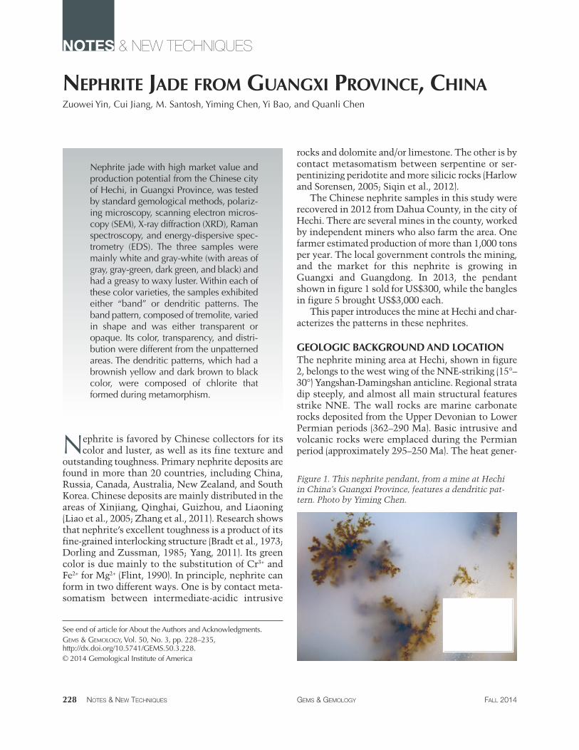
The Chinese nephrite samples in this study were recovered in 2012 from Dahua County, in the city of Hechi. There are several mines in the county, worked by independent miners who also farm the area. One farmer estimated production of more than 1,000 tons per year. The local government controls the mining, and the market for this nephrite is growing in Guangxi and Guangdong. In 2013, the pendant shown in figure 1 sold for US$300, while the bangles in figure 5 brought US$3,000 each.
This paper introduces the mine at Hechi and char-acterizes the patterns in these nephrites.
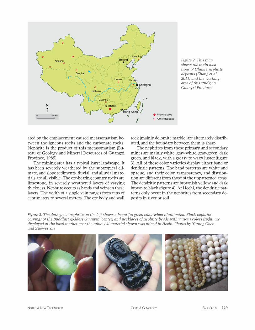
GEOLOGIC BACKGROUND AND LOCATION The nephrite mining area at Hechi, shown in figure 2, belongs to the west wing of the NNE-striking (15°– 30°) Yangshan-Damingshan anticline. Regional strata dip steeply, and almost all main structural features strike NNE. The wall rocks are marine carbonate rocks deposited from the Upper Devonian to Lower Permian periods (362–290 Ma). Basic intrusive and volcanic rocks were emplaced during the Permian period (approximately 295–250 Ma). The heat gener-
Figure 1. This nephrite pendant, from a mine at Hechi in China’s Guangxi Province, features a dendritic pat-tern. Photo by Yiming Chen.
228 NOTES & NEW TECHNIQUES GEMS & GEMOLOGY FALL 2014
0
0
0 _/~------,, ---/----
t
0
0 ! • 0
N
0 800 km
Xinjiang
Sichuan
Qinghai
Liaoning
GUANGXI
Henan Jianasu
Beijing
Shanghai
Hong Kong
Guizhou
Working area
Other deposits
ated by the emplacement caused metasomatism be-tween the igneous rocks and the carbonate rocks. Nephrite is the product of this metasomatism (Bu-reau of Geology and Mineral Resources of Guangxi Province, 1985).
The mining area has a typical karst landscape. It has been severely weathered by the subtropical cli-mate, and slope sediments, fluvial, and alluvial mate-rials are all visible. The ore-bearing country rocks are limestone, in severely weathered layers of varying thickness. Nephrite occurs as bands and veins in these layers. The width of a single vein ranges from tens of centimeters to several meters. The ore body and wall
Figure 2. This map shows the main loca-tions of China’s nephrite deposits (Zhang et al., 2011) and the working area of this study, in Guangxi Province.
rock (mainly dolomite marble) are alternately distrib-uted, and the boundary between them is sharp.
The nephrites from these primary and secondary mines are mainly white, gray-white, gray-green, dark green, and black, with a greasy to waxy luster (figure 3). All of these color varieties display either band or dendritic patterns. The band patterns are white and opaque, and their color, transparency, and distribu-tion are different from those of the unpatterned areas. The dendritic patterns are brownish yellow and dark brown to black (figure 4). At Hechi, the dendritic pat-terns only occur in the nephrites from secondary de-posits in river or soil.
Figure 3. The dark green nephrite on the left shows a beautiful green color when illuminated. Black nephrite carvings of the Buddhist goddess Guanyin (center) and necklaces of nephrite beads with various colors (right) are displayed at the local market near the mine. All material shown was mined in Hechi. Photos by Yiming Chen and Zuowei Yin.
NOTES & NEW TECHNIQUES GEMS & GEMOLOGY FALL 2014 229
A B
C D
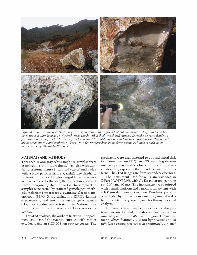
Figure 4. A: In the hills near Hechi, nephrite is found in shallow ground, about one meter underground, and be-longs to secondary deposits. B: Grayish green rough with a thick weathered surface. C: Nephrites with dendritic patterns and country rock. The country rock is dolomitic marble that has undergone metasomatism. The bound-ary between marble and nephrite is sharp. D: In the primary deposit, nephrite occurs as bands of dark green, white, and gray. Photos by Yiming Chen.
MATERIALS AND METHODS Three white and gray-white nephrite samples were examined for this study: the two bangles with den-dritic patterns (figure 5, left and center) and a slab with a band pattern (figure 5, right). The dendritic patterns in the two bangles ranged from brownish yellow to black. In the slab, the banded area showed lower transparency than the rest of the sample. The samples were tested by standard gemological meth-ods, polarizing microscopy, scanning electron mi-croscopy (SEM), X-ray diffraction (XRD), Raman spectroscopy, and energy-dispersive spectrometry (EDS). We conducted the tests at the National Key Lab of the China University of Geosciences in Wuhan.
For SEM analysis, the authors fractured the speci-mens and coated the fracture surfaces with carbon powders using an SCD-005 ion sputter coater. The
specimens were then fastened to a round metal disk for observation. An FEI Quanta 200 scanning electron microscope was used to observe the nephrites’ mi-crostructure, especially their dendritic and band pat-terns. The SEM images are from secondary electrons.
The instrument used for XRD analysis was an X’Pert PRO DY 2198 with Cu Kα radiation operating at 40 kV and 40 mA. The instrument was equipped with a small platform and a monocapillary lens with a 100 μm diameter micro-zone. Dendritic patterns were tested by the micro-area method, since it is dif-ficult to detect very small particles through normal analysis.
To detect the mineral composition of the pat-terns, we used a Bruker Senterra scanning Raman microscope in the 40–3650 cm–1 region. The instru-ment, which features a 785 nm light source and 50 mW laser energy, was set to approximately 3.5 cm–1
230 NOTES & NEW TECHNIQUES GEMS & GEMOLOGY FALL 2014
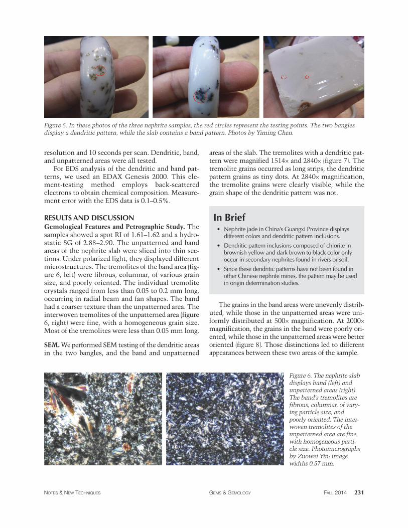
Figure 5. In these photos of the three nephrite samples, the red circles represent the testing points. The two bangles display a dendritic pattern, while the slab contains a band pattern. Photos by Yiming Chen.
resolution and 10 seconds per scan. Dendritic, band, and unpatterned areas were all tested.
For EDS analysis of the dendritic and band pat-terns, we used an EDAX Genesis 2000. This ele-ment-testing method employs back-scattered electrons to obtain chemical composition. Measure-ment error with the EDS data is 0.1–0.5%.
RESULTS AND DISCUSSION Gemological Features and Petrographic Study. The samples showed a spot RI of 1.61–1.62 and a hydro-static SG of 2.88–2.90. The unpatterned and band areas of the nephrite slab were sliced into thin sec-tions. Under polarized light, they displayed different microstructures. The tremolites of the band area (fig-ure 6, left) were fibrous, columnar, of various grain size, and poorly oriented. The individual tremolite crystals ranged from less than 0.05 to 0.2 mm long, occurring in radial beam and fan shapes. The band had a coarser texture than the unpatterned area. The interwoven tremolites of the unpatterned area (figure 6, right) were fine, with a homogeneous grain size. Most of the tremolites were less than 0.05 mm long.
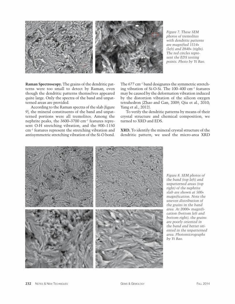
SEM. We performed SEM testing of the dendritic areas in the two bangles, and the band and unpatterned
areas of the slab. The tremolites with a dendritic pat-tern were magnified 1514× and 2840× (figure 7). The tremolite grains occurred as long strips, the dendritic pattern grains as tiny dots. At 2840× magnification, the tremolite grains were clearly visible, while the grain shape of the dendritic pattern was not.
In Brief • Nephrite jade in China’s Guangxi Province displays
different colors and dendritic pattern inclusions.
• Dendritic pattern inclusions composed of chlorite in brownish yellow and dark brown to black color only occur in secondary nephrites found in rivers or soil.
• Since these dendritic patterns have not been found in other Chinese nephrite mines, the pattern may be used in origin determination studies.
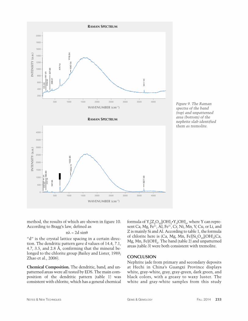
The grains in the band areas were unevenly distrib-uted, while those in the unpatterned areas were uni-formly distributed at 500× magnification. At 2000× magnification, the grains in the band were poorly ori-ented, while those in the unpatterned areas were better oriented (figure 8). Those distinctions led to different appearances between these two areas of the sample.
Figure 6. The nephrite slab displays band (left) and unpatterned areas (right). The band’s tremolites are fibrous, columnar, of vary-ing particle size, and poorly oriented. The inter-woven tremolites of the unpatterned area are fine, with homogeneous parti-cle size. Photomicrographs by Zuowei Yin; image widths 0.57 mm.
NOTES & NEW TECHNIQUES GEMS & GEMOLOGY FALL 2014 231
Raman Spectroscopy. The grains of the dendritic pat-terns were too small to detect by Raman, even though the dendritic patterns themselves appeared quite large. Only the spectra of the band and unpat-terned areas are provided.
According to the Raman spectra of the slab (figure 9), the mineral constituents of the band and unpat-terned portions were all tremolites. Among the nephrite peaks, the 3600–3700 cm–1 features repre-sent O-H stretching vibration, and the 900–1150 cm–1 features represent the stretching vibration and antisymmetric stretching vibration of the Si-O bond.
Figure 7. These SEM photos of tremolites with dendritic patterns are magnified 1514× (left) and 2840× (right). The red circles repre-sent the EDS testing points. Photo by Yi Bao.
The 677 cm–1 band designates the symmetric stretch-ing vibration of Si-O-Si. The 100–400 cm–1 features may be caused by the deformation vibration induced by the distortion vibration of the silicon oxygen tetrahedron (Zhao and Gan, 2009; Qiu et al., 2010; Yang et al., 2012).
To verify the dendritic patterns by means of their crystal structure and chemical composition, we turned to XRD and EDS.
XRD. To identify the mineral crystal structure of the dendritic pattern, we used the micro-area XRD
Figure 8. SEM photos of the band (top left) and unpatterned areas (top right) of the nephrite slab are shown at 500× magnification. Note the uneven distribution of the grains in the band area. At 2000× magnifi-cation (bottom left and bottom right), the grains are poorly oriented in the band and better ori-ented in the unpatterned area. Photomicrographs by Yi Bao.
232 NOTES & NEW TECHNIQUES GEMS & GEMOLOGY FALL 2014
0
125.00
182.49
229.65
327.60
398.01
676.54
1016
.64
1062
.04
3677
.30
83.40
122.87
180.83
227.79
397.86
676.56
1032
.85
1064.15
3677
.40
1200
1400
1600
1800
2000
2500
3000
3500
4000
500 1000 1500 2000 2500 3000 3500 4000
200
400
600
800
1000
500 1000 1500 2000 2500 3000 3500 4000
500
1000
1500
2000
125.00
182.49
229.65
327.60
398.01
676.54
101
1062
.04
3677
.30
83.40
122.87
180.83
227.79
397.86
676.56
1032
.85
1064.15
3677
.40
RAMAN SPECTRUM
RAMAN SPECTRUM
WAVENUMBER (cm–1)
IN
TEN
SITY
(a.
u.)
INTE
NSI
TY (
a.u.
)
WAVENUMBER (cm–1)
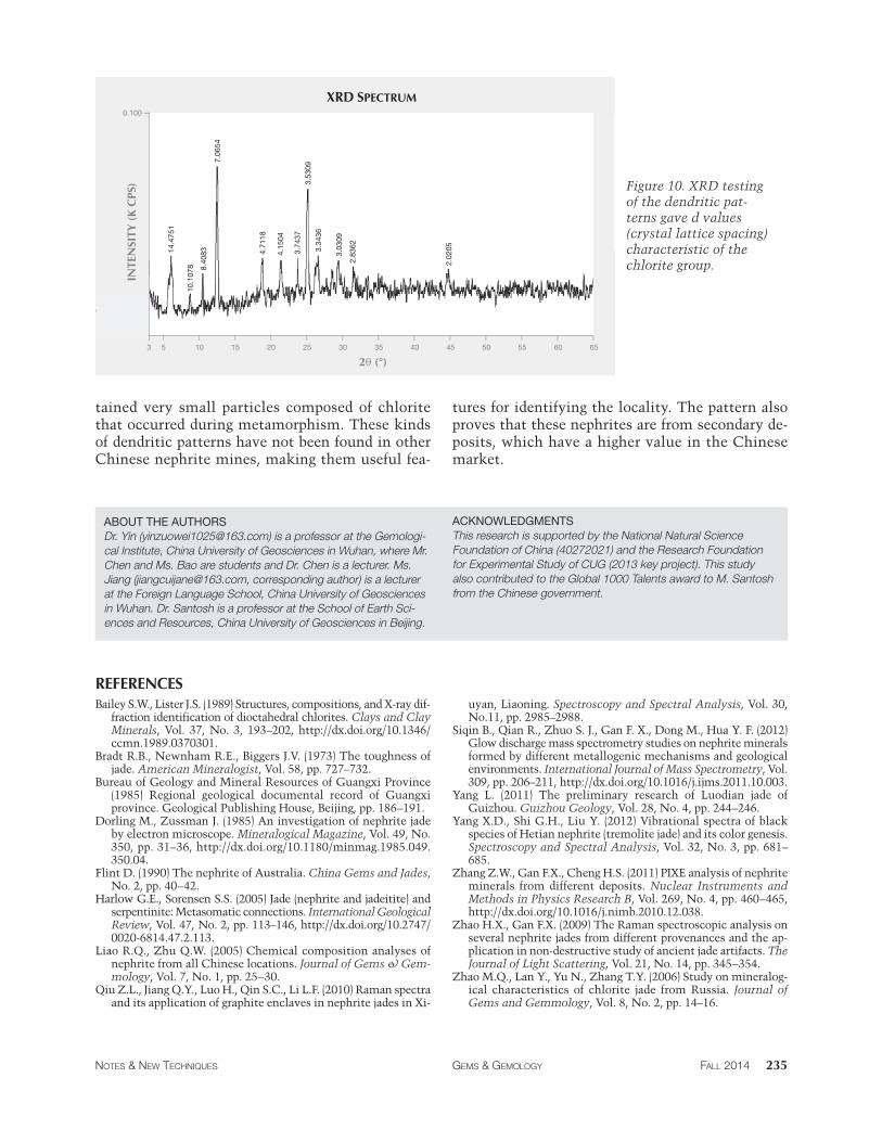
method, the results of which are shown in figure 10. According to Bragg’s law, defined as
nλ = 2d sinθ
“d” is the crystal lattice spacing in a certain direc-tion. The dendritic pattern gave d values of 14.4, 7.1, 4.7, 3.5, and 2.8 Å, confirming that the mineral be-longed to the chlorite group (Bailey and Lister, 1989; Zhao et al., 2006).
Chemical Composition. The dendritic, band, and un-patterned areas were all tested by EDS. The main com-position of the dendritic pattern (table 1) was consistent with chlorite, which has a general chemical
Figure 9. The Raman spectra of the band (top) and unpatterned area (bottom) of the nephrite slab identified them as tremolite.
formula of Y [Z O ](OH) •Y (OH) , where Y can repre-3 4 10 2 3 6 sent Ca, Mg, Fe2+, Al, Fe3+, Cr, Ni, Mn, V, Cu, or Li, and Z is mainly Si and Al. According to table 1, the formula of chlorite here is (Ca, Mg, Mn, Fe)[Si4O10](OH)2(Ca, Mg, Mn, Fe)(OH)6. The band (table 2) and unpatterned areas (table 3) were both consistent with tremolite.
CONCLUSION Nephrite jade from primary and secondary deposits at Hechi in China’s Guangxi Province displays white, gray-white, gray, gray-green, dark green, and black colors, with a greasy to waxy luster. The white and gray-white samples from this study
NOTES & NEW TECHNIQUES GEMS & GEMOLOGY FALL 2014 233
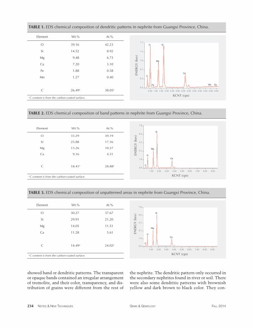
TABLE 1. EDS chemical composition of dendritic patterns in nephrite from Guangxi Province, China.
Element Wt.% At.%
O 39.16 42.23
Si 14.52 8.92
Mg 9.48 6.73
Ca 7.20 3.10
Fe 1.88 0.58
Mn 1.27 0.40
C 26.49a 38.05a
a C content is from the carbon-coated surface.
0.50 1.00 1.50 2.00 2.50 3.00 3.50 4.00 4.50 5.00 5.50 6.00 6.50
0.0
0.3
0.7
1.0
1.4
1.7
C
O
Fe
Mg
Si
Ca
Mn Fe
KCNT (cps)
ENER
GY
(ke
v)
TABLE 2. EDS chemical composition of band patterns in nephrite from Guangxi Province, China.
Element Wt.% At.%
O 33.29 39.19
Si 25.88 17.36
Mg 13.26 10.27
Ca 9.16 4.31
C 18.41a 28.88a
a C content is from the carbon-coated surface.
1.00 2.00 3.00 4.00 5.00 6.00 7.00 8.00 9.00
0.0
1.6
3.1
4.7
6.3
7.8
C
O
Mg
Si
Ca
KCNT (cps)
ENER
GY
(ke
v)
TABLE 3. EDS chemical composition of unpatterned areas in nephrite from Guangxi Province, China.
Element Wt.% At.%
O 30.27 37.67
Si 29.91 21.20
Mg 14.05 11.51
Ca 11.28 5.61
C 14.49a 24.02a
a C content is from the carbon-coated surface.
1.00 2.00 3.00 4.00 5.00 6.00 7.00 8.00 9.00
0.0
1.6
3.1
4.7
6.3
7.8
C
O
Mg
Si
Ca
KCNT (cps)
ENER
GY
(ke
v)
showed band or dendritic patterns. The transparent the nephrite. The dendritic pattern only occurred in or opaque bands contained an irregular arrangement the secondary nephrites found in river or soil. There of tremolite, and their color, transparency, and dis- were also some dendritic patterns with brownish tribution of grains were different from the rest of yellow and dark brown to black color. They con-
234 NOTES & NEW TECHNIQUES GEMS & GEMOLOGY FALL 2014
3 5 10 15 20 25 30 35 40 45 50 55 60 65
14.4
751
10.1
078 8.
4083
7.06
54
4.71
18
3.74
37
4.15
04
3.53
093.
3436
3.03
09
2.83
62
2.02
05
0.100
XRD SPECTRUM
2θ (°)
INTE
NSI
TY (
K C
PS)
tained very small particles composed of chlorite that occurred during metamorphism. These kinds of dendritic patterns have not been found in other Chinese nephrite mines, making them useful fea-
ABOUT THE AUTHORS Dr. Yin ([email protected]) is a professor at the Gemologi-cal Institute, China University of Geosciences in Wuhan, where Mr. Chen and Ms. Bao are students and Dr. Chen is a lecturer. Ms. Jiang ([email protected], corresponding author) is a lecturer at the Foreign Language School, China University of Geosciences in Wuhan. Dr. Santosh is a professor at the School of Earth Sci-ences and Resources, China University of Geosciences in Beijing.
REFERENCES Bailey S.W., Lister J.S. (1989) Structures, compositions, and X-ray dif-
fraction identification of dioctahedral chlorites. Clays and Clay Minerals, Vol. 37, No. 3, 193–202, http://dx.doi.org/10.1346/ ccmn.1989.0370301.
Bradt R.B., Newnham R.E., Biggers J.V. (1973) The toughness of jade. American Mineralogist, Vol. 58, pp. 727–732.
Bureau of Geology and Mineral Resources of Guangxi Province (1985) Regional geological documental record of Guangxi province. Geological Publishing House, Beijing, pp. 186–191.
Dorling M., Zussman J. (1985) An investigation of nephrite jade by electron microscope. Mineralogical Magazine, Vol. 49, No. 350, pp. 31–36, http://dx.doi.org/10.1180/minmag.1985.049. 350.04.
Flint D. (1990) The nephrite of Australia. China Gems and Jades, No. 2, pp. 40–42.
Harlow G.E., Sorensen S.S. (2005) Jade (nephrite and jadeitite) and serpentinite: Metasomatic connections. International Geological Review, Vol. 47, No. 2, pp. 113–146, http://dx.doi.org/10.2747/ 0020-6814.47.2.113.
Liao R.Q., Zhu Q.W. (2005) Chemical composition analyses of nephrite from all Chinese locations. Journal of Gems & Gem-mology, Vol. 7, No. 1, pp. 25–30.
Qiu Z.L., Jiang Q.Y., Luo H., Qin S.C., Li L.F. (2010) Raman spectra and its application of graphite enclaves in nephrite jades in Xi-
Figure 10. XRD testing of the dendritic pat-terns gave d values (crystal lattice spacing) characteristic of the chlorite group.
tures for identifying the locality. The pattern also proves that these nephrites are from secondary de-posits, which have a higher value in the Chinese market.
ACKNOWLEDGMENTS This research is supported by the National Natural Science Foundation of China (40272021) and the Research Foundation for Experimental Study of CUG (2013 key project). This study also contributed to the Global 1000 Talents award to M. Santosh from the Chinese government.
uyan, Liaoning. Spectroscopy and Spectral Analysis, Vol. 30, No.11, pp. 2985–2988.
Siqin B., Qian R., Zhuo S. J., Gan F. X., Dong M., Hua Y. F. (2012) Glow discharge mass spectrometry studies on nephrite minerals formed by different metallogenic mechanisms and geological environments. International Journal of Mass Spectrometry, Vol. 309, pp. 206–211, http://dx.doi.org/10.1016/j.ijms.2011.10.003.
Yang L. (2011) The preliminary research of Luodian jade of Guizhou. Guizhou Geology, Vol. 28, No. 4, pp. 244–246.
Yang X.D., Shi G.H., Liu Y. (2012) Vibrational spectra of black species of Hetian nephrite (tremolite jade) and its color genesis. Spectroscopy and Spectral Analysis, Vol. 32, No. 3, pp. 681– 685.
Zhang Z.W., Gan F.X., Cheng H.S. (2011) PIXE analysis of nephrite minerals from different deposits. Nuclear Instruments and Methods in Physics Research B, Vol. 269, No. 4, pp. 460–465, http://dx.doi.org/10.1016/j.nimb.2010.12.038.
Zhao H.X., Gan F.X. (2009) The Raman spectroscopic analysis on several nephrite jades from different provenances and the ap-plication in non-destructive study of ancient jade artifacts. The Journal of Light Scattering, Vol. 21, No. 14, pp. 345–354.
Zhao M.Q., Lan Y., Yu N., Zhang T.Y. (2006) Study on mineralog-ical characteristics of chlorite jade from Russia. Journal of Gems and Gemmology, Vol. 8, No. 2, pp. 14–16.
NOTES & NEW TECHNIQUES GEMS & GEMOLOGY FALL 2014 235