Selective synthesis of formamides, 1,2-bis(N-heterocyclic ...

N-hydroxypyrazolyl glycine derivatives as selectiveN-methyl-D-aspartic acid receptor LigandsRasmus P. Clausen, University of CopenhagenCaspar Christensen, University of CopenhagenKasper B. Hansen, Emory UniversityJeremy R. Greenwood, University of CopenhagenLars Jørgensen, University of CopenhagenNicola Micale, University of CopenhagenJens Christian Madsen, H. Lundbeck A/SBirgitte Nielsen, University of CopenhagenJan Egebjerg, H. Lundbeck A/SHans Bräuner-Osborne, University of Copenhagen
Only first 10 authors above; see publication for full author list.
Journal Title: Journal of Medicinal ChemistryVolume: Volume 51, Number 14Publisher: American Chemical Society | 2008-07-24, Pages 4179-4187Type of Work: Article | Post-print: After Peer ReviewPublisher DOI: 10.1021/jm800025ePermanent URL: https://pid.emory.edu/ark:/25593/rpp75
Final published version: http://dx.doi.org/10.1021/jm800025e
Copyright information:© 2008 American Chemical Society.
Accessed October 11, 2021 11:55 AM EDT

N-Hydroxypyrazolyl glycine derivatives as selective N-methyl-D-aspartic acid receptor ligands
Rasmus P. Clausen*,†, Caspar Christensen†, Kasper B. Hansen†,‡,¤, Jeremy R. Greenwood†, Lars Jørgensen†, Nicola Micale†, Jens Christian Madsen§, Birgitte Nielsen†, Jan Egebjerg¤, Hans Bräuner-Osborne†, Stephen F. Traynelis‡, and Jesper L. Kristensen†
†Department of Medicinal Chemistry, Faculty of Pharmaceutical Sciences, University of Copenhagen, 2 Universitetsparken, DK-2100 Copenhagen, Denmark
‡Department of Pharmacology, Emory University School of Medicine, Rollins Research Center, Atlanta, Georgia, USA
§Department of Medicinal Chemistry, H. Lundbeck A/S, 9 Ottiliavej, DK-2500 Valby, Denmark
¤Department of Molecular Neurobiology, H. Lundbeck A/S, 9 Ottiliavej, DK-2500 Valby, Denmark
Abstract
A series of analogues based on N-hydroxypyrazole as a bioisostere for the distal carboxylate group
of aspartate have been designed, synthesized and pharmacologically characterized. Affinity studies
on the major glutamate receptor subgroups show that these 4-substituted N-hydroxypyrazol-5-yl
glycine (NHP5G) derivatives are selectively recognized by N-methyl-D-aspartic acid (NMDA)
receptors and that the (R)-enantiomers are preferred. Moreover, several of the compounds are able
to discriminate between individual subtypes among the NMDA receptors, providing new
pharmacological tools. For example, 4-propyl NHP5G is an antagonist at the NR1/NR2A subtype,
but an agonist at the NR1/NR2D subtype. Molecular docking studies indicate that the substituent
protrudes into a region that may be further exploited to improve subtype-selectivity, thereby
opening up a design strategy for ligands which can differentiate individual NMDA receptor
subtypes.
Graphical Abstract
Correspondence: Associate Professor Rasmus P. Clausen, Department of Medicinal Chemistry, Faculty of Pharmaceutical Sciences, University of Copenhagen, 2 Universitetsparken, DK-2100 Copenhagen, Denmark, Phone: +45 35 33 65 66, Fax: +45 35 33 60 40, [email protected].
Supporting Information Available: routine NMR spectra, Routine NMR data, preparation of Mosher amides, CD spectra, HPLC traces, and table of purity data. This material is available free of charge via the Internet at http://pubs.acs.org.
HHS Public AccessAuthor manuscriptJ Med Chem. Author manuscript; available in PMC 2016 April 28.
Published in final edited form as:J Med Chem. 2008 July 24; 51(14): 4179–4187. doi:10.1021/jm800025e.
Author M
anuscriptA
uthor Manuscript
Author M
anuscriptA
uthor Manuscript

Keywords
NMDA receptor; N-hydroxypyrazole-glycine; glutamate; molecular modeling; subtype selectivity
Introduction
The glutamate receptors (GluRs) are a group of receptors with a prominent role in
neurotransmission, since most of the excitatory signals in the brain are mediated by glutamic
acid (Glu, Figure 1).1–3 These receptors are involved in key processes of the central nervous
system (CNS) such as learning and memory,4 but are also implicated in several
neuropathological conditions, such as ischemia, epilepsy, schizophrenia, chronic pain, and
Alzheimer’s disease.5 Therefore, the GluRs have been the target of extensive research for
more than three decades, and one of the goals has been to develop compounds that can alter
glutamatergic neurotransmission in a selective manner. Such compounds have been and
continue to be important tools in discovering new therapeutic possibilities and to gain a
functional understanding of this group of receptors.6
Two different signaling pathways divide the GluRs: metabotropic glutamate receptors
(mGluRs) are G-protein-coupled receptors that mediate slow modulatory responses, and
ionotropic glutamate receptors (iGluRs) are cation-conducting ion channels that mediate fast
neurotransmission via depolarization of the membrane potential. Based on activation by
selective ligands and cDNA homology, the iGluRs are divided into N-methyl-D-aspartic acid
(NMDA), (S)-2-amino-3-(3-hydroxy-5-methyl-4-isoxazolyl)propionic acid (AMPA) and
kainic acid (KA) receptors. The iGluRs are homo- or heteromeric assemblies of four
subunits. Seven NMDA subunits (NR1, NR2A-D, NR3A, NR3B), four AMPA (GluR1-4)
and five KA (GluR5-7 and KA1-2) preferring subunits have been identified.1 NMDA
glutamate receptors are assembled from two NR1 subunits and two NR2 subunits, and are
activated by the simultaneous binding of glycine and glutamate to the NR1 and NR2
subunits, respectively. NR3 subunits also can co-assemble with NMDA receptors, although
the stoichiometry is unclear.
The iGluR subunit consists of an intracellular C-terminal domain, a transmembrane domain
that forms the ion channel pore, and a pair of extracellular domains: the agonist binding
Clausen et al. Page 2
J Med Chem. Author manuscript; available in PMC 2016 April 28.
Author M
anuscriptA
uthor Manuscript
Author M
anuscriptA
uthor Manuscript
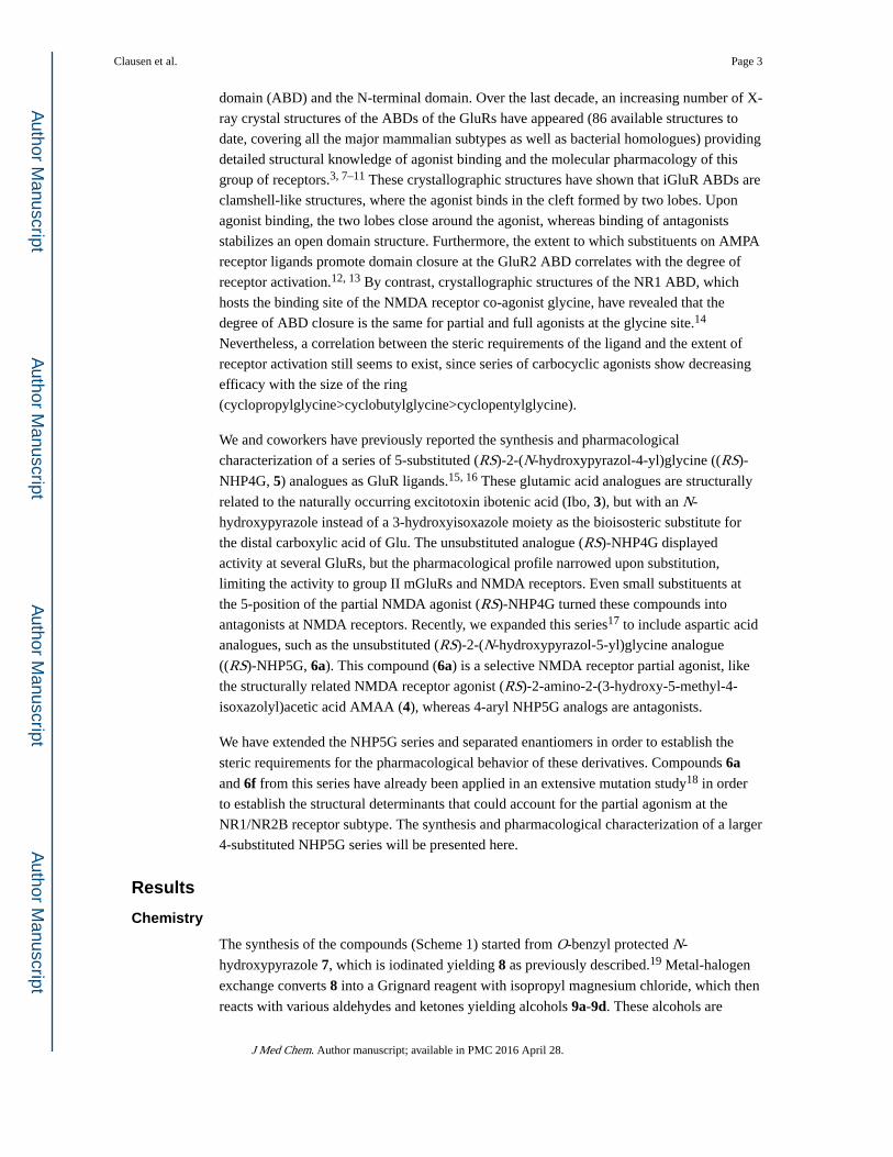
domain (ABD) and the N-terminal domain. Over the last decade, an increasing number of X-
ray crystal structures of the ABDs of the GluRs have appeared (86 available structures to
date, covering all the major mammalian subtypes as well as bacterial homologues) providing
detailed structural knowledge of agonist binding and the molecular pharmacology of this
group of receptors.3, 7–11 These crystallographic structures have shown that iGluR ABDs are
clamshell-like structures, where the agonist binds in the cleft formed by two lobes. Upon
agonist binding, the two lobes close around the agonist, whereas binding of antagonists
stabilizes an open domain structure. Furthermore, the extent to which substituents on AMPA
receptor ligands promote domain closure at the GluR2 ABD correlates with the degree of
receptor activation.12, 13 By contrast, crystallographic structures of the NR1 ABD, which
hosts the binding site of the NMDA receptor co-agonist glycine, have revealed that the
degree of ABD closure is the same for partial and full agonists at the glycine site.14
Nevertheless, a correlation between the steric requirements of the ligand and the extent of
receptor activation still seems to exist, since series of carbocyclic agonists show decreasing
efficacy with the size of the ring
(cyclopropylglycine>cyclobutylglycine>cyclopentylglycine).
We and coworkers have previously reported the synthesis and pharmacological
characterization of a series of 5-substituted (RS)-2-(N-hydroxypyrazol-4-yl)glycine ((RS)-
NHP4G, 5) analogues as GluR ligands.15, 16 These glutamic acid analogues are structurally
related to the naturally occurring excitotoxin ibotenic acid (Ibo, 3), but with an N-
hydroxypyrazole instead of a 3-hydroxyisoxazole moiety as the bioisosteric substitute for
the distal carboxylic acid of Glu. The unsubstituted analogue (RS)-NHP4G displayed
activity at several GluRs, but the pharmacological profile narrowed upon substitution,
limiting the activity to group II mGluRs and NMDA receptors. Even small substituents at
the 5-position of the partial NMDA agonist (RS)-NHP4G turned these compounds into
antagonists at NMDA receptors. Recently, we expanded this series17 to include aspartic acid
analogues, such as the unsubstituted (RS)-2-(N-hydroxypyrazol-5-yl)glycine analogue
((RS)-NHP5G, 6a). This compound (6a) is a selective NMDA receptor partial agonist, like
the structurally related NMDA receptor agonist (RS)-2-amino-2-(3-hydroxy-5-methyl-4-
isoxazolyl)acetic acid AMAA (4), whereas 4-aryl NHP5G analogs are antagonists.
We have extended the NHP5G series and separated enantiomers in order to establish the
steric requirements for the pharmacological behavior of these derivatives. Compounds 6a and 6f from this series have already been applied in an extensive mutation study18 in order
to establish the structural determinants that could account for the partial agonism at the
NR1/NR2B receptor subtype. The synthesis and pharmacological characterization of a larger
4-substituted NHP5G series will be presented here.
Results
Chemistry
The synthesis of the compounds (Scheme 1) started from O-benzyl protected N-
hydroxypyrazole 7, which is iodinated yielding 8 as previously described.19 Metal-halogen
exchange converts 8 into a Grignard reagent with isopropyl magnesium chloride, which then
reacts with various aldehydes and ketones yielding alcohols 9a-9d. These alcohols are
Clausen et al. Page 3
J Med Chem. Author manuscript; available in PMC 2016 April 28.
Author M
anuscriptA
uthor Manuscript
Author M
anuscriptA
uthor Manuscript

deoxygenated by treatment with TFA and triethylsilane giving 4-substituted
hydroxypyrazoles 10a-10d. The chloro substituted compound 10e is obtained directly from
7 by treatment with sulfuryl chloride. Selective lithiation followed by treatment with the
amino acid synthon diethyl N-Boc iminomalonate yields protected amino diesters 11a-e,
which are debenzylated by hydrogenation yielding 12a-e. Ester hydrolysis followed by
acidic removal of the Boc group and concomitant spontaneous decarboxylation yields
racemic amino acids 6b-f. The enantiomers could be separated by chiral HPLC thus
enabling a preparative resolvation of the compounds including NHP5G. Since the mobile
phase was diluted aqueous TFA, the enantiomers were isolated as TFA salts, containing
varying amounts of TFA. The amount of TFA was determined by NMR spectroscopy using
the ERETIC technique,20 where an electronically generated signal serves as a quantification
reference. For TFA, the ERETIC technique must be applied to 19F NMR spectroscopy, since
the absence of non-labile protons makes 1H NMR unsuitable in this case. However, the
compounds studied here are void of fluorine nuclei, so quantification relative to TFA
necessitates the use of an ERETIC signal in both the 1H and the 19F NMR spectra. The
relative strength of the 1H and 19F ERETIC signals must then be calibrated using a
compound containing both protons and fluorine. For this purpose we used a sample of
citalopram oxalate. In general, the content of TFA was less than 1 equivalent.
Determination of Absolute Configuration
We were unable to obtain crystals suitable for X-ray structure determination of any of the
compounds and therefore we turned to a number of other methods in order to determine the
absolute configuration. Firstly, the elution order on the chiral HPLC column clearly
indicated the absolute stereochemistry for all the compounds (Figure 2). Thus, the L-form of
α-amino acids generally elutes before the D-form on a CR(−) Crownpak chiral column,
corresponding to the (S)- and (R)-enantiomer, respectively.21, 22
Secondly, we turned to the Electronic Circular Dichroism (ECD) spectra. Traditionally, an
empirical correlation has been noted between the S-isomer of aryl amino acids and
observation of a positive Cotton Effect in the ECD.23 However, in recent years, prediction of
the ECD spectrum has become tractable from first principles using methods such as Time-
Dependent Density Functional Theory (TD-DFT), reducing the reliance on empiricism for
interpretation. Thus, to obtain further evidence for the absolute configuration, CD spectra
(Figure 3) of at least one enantiomer of 6b-6f were therefore recorded and in the case of (S)-
Cl-NHP5G (6f) compared with that calculated using TD-DFT for eight low energy
conformers in dilute acidic aqueous solution (Figure 3). Although the first major peak
according to the Boltzmann average lies approximately 20 nm lower in energy than
observed, the magnitude was in good agreement with experiment, and indicative of the same
correlation as usually seen. Although the exact shape of the predicted peak depends on the
weighting of the conformers, and is thus highly sensitive to the accuracy of conformational
energies, the small experimental negative peak at around 230 nm for (S)-6f was also visible
in two of the four lowest energy conformers and in agreement with that previously observed
for phenylglycine.23 Comparing (S)-6b with (S)-6f, showed that there was no significant
qualitative difference between the calculated spectra or between any of the experimental
spectra, and thus it is reasonable to expect the correlation to hold throughout this series.
Clausen et al. Page 4
J Med Chem. Author manuscript; available in PMC 2016 April 28.
Author M
anuscriptA
uthor Manuscript
Author M
anuscriptA
uthor Manuscript

Finally, we prepared the Mosher amide of (S)-6c and compared the NMR spectra of the two
diastereomers with the isotropic magnetic shielding values calculated for the hydrogen
nuclei using DFT. Since these amide derivatives have several rotatable bonds, spectra were
calculated for the hundred lowest energy conformers of both (S,S) and (R,S), according to a
conformational search with OPLS2005. Although qualitative agreement was noted, the
precise differences in the calculated shifts between the diastereomers again proved highly
sensitive to conformational energies when using a Boltzmann weighted average. We
therefore calculated regression coefficients between the shifts in the two experimental proton
spectra and the two hundred calculated spectra. For a majority of the calculated (R,S)
conformers, the calculated spectrum was a better match for (R,S) than (S,S), and for a
majority of the calculated (S,S) conformers, the calculated spectrum was a better match for
experimental (S,S). Moreover, for the single conformers of (R,S) and (S,S) with the highest
regression coefficients, the calculated (R,S) spectrum was a better match for experimental
(R,S) spectrum (R2 = 0.9993) and vice versa for (S,S) (R2 = 0.9985). The absolute
configurations are therefore assigned on the basis of three independent types of experiment,
interpreted by a combination of empirical and first-principles correlations.
Pharmacology
First we investigated the affinity of the compounds for the major iGluR subgroups. As
shown in Table 1, all the analogues are selective NMDA receptor ligands with affinity in the
low micromolar range. Furthermore, the affinity was found to reside with the (R)-enantiomer
of the compounds, the most potent being (R)-6a, (R)-6b and (R)-6f.
The affinity at NMDA receptors prompted functional characterization at recombinant
NMDA receptors expressed in Xenopus laevis oocytes to determine whether the compounds
could discriminate between individual subtypes (Table 2, Figures 4 and 5).
Compounds (RS)-6c, (RS)-6d, and (RS)-6f acted as partial agonists on four NMDA receptor
subunit combinations (NR1/NR2A, NR1/NR2B, NR1/NR2C, and NR1/NR2D), but with
highly varying potency and agonist efficacy at the different subtypes. At NR2A-containing
receptors, the ethyl substituted (RS)-6c and chloro substituted (RS)-6f are low-efficacy
partial agonists, but (RS)-6c is an order of magnitude more potent than (RS)-6f. (RS)-6d displayed antagonist activity at NR1/NR2A (n=5), but was an agonist on all other subtypes
(Figure 5), and is thus the compound showing the largest difference among subtypes in this
series. At NR1/NR2B (RS)-6c and (RS)-6f were approximately equipotent and had higher
agonist efficacies than at NR1/NR2A, but the efficacies varied more. By contrast,
compounds (RS)-6c and (RS)-6f showed similar agonist efficacy relative to Glu around 0.5
at NR2C and 0.7 at NR2D, and again compound (RS)-6c was more potent than (RS)-6f.
In our previous study including compounds (R)-6a and (R)-6f, ligand variation was matched
against corresponding mutations in the receptor, leading to the conclusion that the degree of
agonist efficacy relative to Glu correlates inversely with the steric bulk introduced into the
agonist binding pocket, either by the ligand or in the receptor by mutation. It was therefore
interesting to see whether this dependence would manifest in the present series too. The R-
enantiomer of compounds 6a-d and 6f was therefore characterized at NR1/NR2B (Table 3
and Figure 6; See also supplemental data). However, the relative agonist efficacy of the
Clausen et al. Page 5
J Med Chem. Author manuscript; available in PMC 2016 April 28.
Author M
anuscriptA
uthor Manuscript
Author M
anuscriptA
uthor Manuscript

compounds did not show a simple correlation with the nature of the substituents. Thus, the
unsubstituted NHP5G (6a) induced a fraction of 0.61 of the Glu-induced current and
compounds with methyl- (6b) and n-propyl-substituents (6d) displayed markedly lower
relative efficacies of 0.23 and 0.09, respectively. The chloro substituted compound (6f) was
slightly more efficacious (0.33) than the methyl substituted compounds, but the ethyl
substituted compound (6c) induced a 0.46 current relative to the Glu current, which is twice
that of the methyl substituted compound, and which is notable since an ethyl substituent is
more bulky than a methyl substituent. The iso-propyl substituted compound (6e) was an
antagonist at NR1/NR2B (See Supplemental data).
Molecular Modeling
To understand the unexpected trend in the agonist efficacies of this series of 4-substituted
NHP5G we docked the compounds in a homology model of the ABD of NR2B using the
recently published X-ray structure of the ABD of NR2A (PDB code 2A5S) as a template.25
In our recent mutation study the homology model was based on a NR1 crystallographic
structure (PDB code 1PB7), but the new X-ray structure of NR2A in complex with
glutamate enabled the inclusion of two water molecules in the homology model that are
involved in the binding of the charged groups of glutamate into the homology model. The
compounds were submitted to a conformational search in Macromodel 9.0 in the tri-ionised
state and the resulting minimized structures were then docked flexibly into a homology
model of NR2B. This resulted in similar binding modes for all the compounds, with the
amino acid moiety overlapping with that of Glu as seen in our previous study18 and the
deprotonated N-hydroxy group receiving an H-bond from the water molecule involved in
binding the distal acidic group of Glu. The substituents point like a wedge towards residues
H486 and V686, which form a crevice between the upper and lower domains of the
clamshell-like ABD structure (Figure 7A). While the ethyl substituent is accommodated by
the crevice, the n-propyl is in close proximity with residues Lys485 (Figure 7B) and Tyr731
(not shown). In the X-ray crystal structure of NR2A, several water molecules are present in
this region and two of them are shown in Figure 7. Furthermore, the n-propyl substituent
reaches a less conserved region of the ABD. Thus, in the X-ray structure of the ABD of
NR2A, Lys485 is in close proximity to the backbone carbonyl of Arg711, which is followed
by Gly712. This two-peptide sequence corresponds to Arg712-Gly713 in NR2B, but
Arg722-Ser723 in NR2C and Pro736-Arg737 in NR2D. This sequence difference could
explain the observed difference in pharmacology of Pr-NHP5G (6d) and designing ligands
with substituents protruding into this region may lead to NMDA receptor ligands with
increased subtype-selectivity as suggested previously.26
Discussion
We have synthesized and pharmacologically characterized a series of 4-substituted NHP5G
derivatives to identify ligands suitable for investigating the mechanisms governing partial
agonism at the NMDA receptor. This series of NHP5G compounds displays several
remarkable properties. All of the compounds are partial agonists at the NMDA receptors
with varying degrees of receptor activation. Furthermore the compounds show marked
variation at the various NMDA receptor subtypes both in terms of potency and efficacy.
Clausen et al. Page 6
J Med Chem. Author manuscript; available in PMC 2016 April 28.
Author M
anuscriptA
uthor Manuscript
Author M
anuscriptA
uthor Manuscript

Thus, these compounds highlight the importance of introducing substituents in NMDA
receptor ligands that can reach less conserved regions of the receptor. We were able to
separate the enantiomers of the racemic mixtures, and have been able to determine to a high
degree of confidence the absolute configuration from several lines of evidence, including the
HPLC elution order, the good agreement between calculated and measured ECD spectra and
the similarity between calculated and experimental NMR spectra of Mosher amide
diastereomers of the ethyl compounds. The separation of enantiomers is important since
apparent partial agonism can arise from mixed agonistic and antagonistic behavior of
opposing enantiomers.27 However, our data showed that the activity could be attributed to
the (R)-enantiomers in this series. The unsubstituted (R)-6a and chloro substituted (R)-6f have already been employed in a mutagenic study, where it was concluded that steric clashes
between the substituents and residues His486 and Val686 were important determinants for
the agonist efficacy of the compounds relative to Glu. These two residues form a crevice in
the ligand binding domain separating upper and lower domains in the clamshell-like ABD
structure, and introducing steric bulk in this region was expected to prevent closure of the
ligand binding domain and concomitant receptor activation.
Completion of the entire series now reveals that the compounds display a trend with respect
to substituents that was unexpected on the basis of the mutagenesis study, since the efficacy
does not completely follow inversely the steric bulk added to the ligands. In particular, the
ethyl substituted compound is more efficacious than the methyl substituted compound but
still less efficacious compared to the unsubstituted compound, which is surprising
considering the increased steric bulk imposed by the ethyl group. Docking the compounds
into a homology model of the ABD of NR2B based on the X-ray crystallographic structure
of the highly homologous NR2A ABD construct shows that the ethyl group reaches a region
where several water molecules are present. Compared with the methyl and chloro groups,
displacement of some of these water molecules by the ethyl group could thermodynamically
favor a conformation of the ABD that permits receptor activation, offering a plausible
explanation for the unusual trend. By contrast, n-propyl and isopropyl substituents are in
close contact with residues in the receptor, which again is in agreement with steric inhibition
of the domain closure.
In conclusion, we have identified a series of analogous selective NMDA receptor ligands
which induce various degrees of receptor activation. These compounds may constitute
valuable pharmacological tools for studying the mechanisms governing partial agonism at
the NMDA receptor. This series shows that factors other than steric hindrance may
determine the degree of activation of NMDA receptors. In particular, the n-propyl-
substituted compound in this series is interesting since it significantly differentiates between
NMDA receptors containing different NR2 subunits and therefore offers a promising
pharmacological tool for investigating differences in the functional properties of NMDA
receptor subtypes at a molecular and cellular level. This could have relevance in several CNS
disorders, including schizophrenia. Finally, modeling studies suggest a strategy for
designing novel ligands that may be able to discriminate between NMDA receptor subtypes.
Clausen et al. Page 7
J Med Chem. Author manuscript; available in PMC 2016 April 28.
Author M
anuscriptA
uthor Manuscript
Author M
anuscriptA
uthor Manuscript

Experimental
Chemistry
General Methods—All reactions involving air-sensitive reagents were performed under
N2 using syringe-septum cap technique and all glassware was flame-dried prior to use. Flash
chromatography (FC) was performed using silica Merck 60 (230–400 mesh). 1H (300
MHz), 19F (282 MHz) and 13C (75 MHz) NMR were recorded on a Varian instrument using
TMS and citalopram oxalate as internal standard. HRMS was performed on a JEOL
Hx110/110 mass spectrometer. All solvents and reagents were analytical grade purchased
from Aldrich or Fluka and used without further purification, unless otherwise stated. THF
was distilled from Na/benzophenone under N2. DMF was stored over 3Å molecular sieves.
n-Buli28 and i-PrMgCl29 solutions were titrated prior to use. Preparative chiral HPLC was
performed using a Crownpak CR(+) column (10 × 150 mm, Daicel) equipped with a CR(+)
guard column (4.0 × 10 mm, Daicel) connected to a HPLC system consisting of a Jasco
880PU pump, a Rheodyne 7125 injector, a TSP UV100 detector set at 234 nm, and a Merck-
Hitachi D-2000 Chromato-Integrator. The column was eluted at rt with 1.5 ml/min of
aqueous TFA, pH 2. Upon resolvation the solutions containing the amino acid was collected
and lyophilized. Chiral HPLC analyses of the resolved stereoisomers were performed using
a Crownpak CR(−) column (4.0 × 150 mm, Daicel) equipped with a water-jacket and
generally the enantiomeric purities of the isolated amino acids were >95% unless otherwise
indicated. The column was eluted with aqueous HClO4 (pH 1.5 or pH 2.0) at 0.4 ml/min and
the temperature was controlled by a Hetofrig thermostat at 20, 10, or 1 °C. A TSP HPLC
system consisting of a P2000 pump, an AS3000 autoinjector, and an SM5000 PDA detector
was used for the chiral HPLC analyses.
General Procedure for Metal-Halogen-Exchange and Reaction with Aldehyde—8 (1 mmol) in THF (20 ml) was added to a flame-dried flask and the resulting solution
was cooled to 0 °C. Isopropyl magnesium chloride (1.2 mmol, 2 M in THF) was added
dropwise over a period of 5 min and the resulting mixture was stirred at 0 °C for 1 hr. The
aldehyde or ketone (1.2 mmol) dissolved in THF (2 ml) was added and the resulting solution
was stirred for an additional 30 min at 0 °C before allowing to warm to rt over 30 min, and
quenched with NH4Cl (sat. aq., 10 ml). The aqueous phase was extracted with EtOAc (3 ×
20 ml) and the combined organic phases were dried over MgSO4 and evaporated in vacuo.
FC (PE:EtOAc) afforded the title compound.
1-(1-Benzyloxy-pyrazol-4-yl)-ethanol (9b): From 8 (3.0 g, 10 mmol) and acetaldehyde (0.8
ml, 12 mmol). FC (PE;EtOAc 2:1) afforded 1.47 g (72%) of the title compound.
1-(1-Benzyloxy-pyrazol-4-yl)-propanol (9c): From 8 (3.0 g, 10 mmol) and propanal (1.1
ml, 12 mmol). FC (PE;EtOAc 2:1 – 1:1) afforded 2.13 g (92%) of the title compound.
2-(1-Benzyloxy-pyrazol-4-yl)-propan-2-ol (9d): From 8 (3.0 g, 10 mmol) and acetone
(0.87 ml, 12 mmol). FC (PE;EtOAc 2:1 – 1:1) afforded 2.14 g (92%) of the title compound.
General Procedure for Dehydroxylation—To a solution of the alcohol (9a-9d) (1
mmol) and triethylsilane (2 mmol) in CHCl3 (10 ml), trifluoroacetic acid (5 ml) was added
Clausen et al. Page 8
J Med Chem. Author manuscript; available in PMC 2016 April 28.
Author M
anuscriptA
uthor Manuscript
Author M
anuscriptA
uthor Manuscript

dropwise at room temperature and the resulting solution was stirred at reflux for 2 hr. The
mixture was cooled, water (10 ml) was added and the aqueous phase was extracted with
diethyl ether (3 × 10 ml). The combined organic phases were dried over MgSO4 and
evaporated in vacuo. FC (PE:EtOAc) afforded the title compound.
1-Benzyloxy-4-methylpyrazole (10a): From 9a (2 g, 9.8 mmol). FC (PE:EtOAc 8:1)
afforded 1.45 g (79%) of the title compound.
1-Benzyloxy-4-ethylpyrazole (10b): From 9b (1.3 g, 6 mmol). FC (PE:EtOAc 8:1) afforded
1.13 g (95%) of the title compound.
1-Benzyloxy-4-propylpyrazole (10c): From 9c (2.14 g, 9.2 mmol). FC (PE:EtOAc 6:1)
afforded 1.71 g (87%) of the title compound.
1-Benzyloxy-4-isopropylpyrazole (10d): From 9d (2.32 g, 10.8 mmol). FC (PE:EtOAc 6:1)
afforded 1.64 g (82%) of the title compound.
1-Benzyloxy-4-chloropyrazole (10e): 1-Benzyloxypyrazole30 (3 g, 17 mmol) was dissolved
in diethyl ether (50 ml) and cooled to −15°C. Sulfuryl chloride (5 ml, 62 mmol) was added
dropwise over 30 min and the resulting solution stirred for an additional 30 min at 0 °C.
Water (20 ml) was slowly added and the aqueous phase extracted with diethyl ether (3 × 25
ml). The combined organic phases was washed with NaHCO3 (sat. aq., 3 × 25 ml), brine (1
× 25 ml), dried over MgSO4 and evaporated in vacuo to yield 3.51 g of the title compound,
which crystallised upon standing in a refrigerator.
General Procedure for the Synthesis of the Protected Amino Acid—To a stirred
solution of the benzyloxypyrazole (10a-e) (1 mmol) in THF (10 ml) at −78 °C, n-BuLi (0.75
ml, 1.6 M in hexane, 1.2 mmol) was added dropwise over app. 2 min. After 5 min, a solution
of diethyl N-Boc iminomalonate (382 mg, 1.4 mmol) in THF (1 ml) was added and the
resulting mixture was stirred at −78 °C for 3 hr, before quenching with water. The solution
was allowed to warm to room temperature, NH4Cl (sat. aq., 10 ml) was added, the organic
phase was separated and the water phase was extracted with EtOAc (3 × 10 ml). The
combined organic layers were dried over MgSO4 and evaporated in vacuo to afford a crude
residue, which was purified by FC (PE:EtOAc) to give the title compound containing minor
amounts of co-eluting diethyl N-Boc-iminimalonate. This material was used without further
purification.
Diethyl 2-(1-benzyloxy-4-methyl-pyrazol-5-yl)-2-tert-butyloxycarbonylaminomalonate (11a): From 10a (0.90 g, 4.8 mmol). FC (PE:EtOAc 7:1) afforded 0.91 g (41%) of 11a as a
colorless oil.
Diethyl 2-(1-benzyloxy-4-ethyl-pyrazol-5-yl)-2-tert-butyloxycarbonylaminomalonate (11b): From 10b (0.92 g, 4.8 mmol). FC (PE:EtOAc 7:1) afforded 1.08 g 50%) of 11b as a
white solid.
Clausen et al. Page 9
J Med Chem. Author manuscript; available in PMC 2016 April 28.
Author M
anuscriptA
uthor Manuscript
Author M
anuscriptA
uthor Manuscript
Diethyl 2-(1-benzyloxy-4-propyl-pyrazol-5-yl)-2-tert-butyloxycarbonylaminomalonate (11c): From 10c (1.1 g, 5.1 mmol). FC (PE:EtOAc 4:1) afforded 1.83 g (73%) of 11c as a
white solid.
Diethyl 2-(1-benzyloxy-4-isopropyl-pyrazol-5-yl)-2-tert-butyloxycarbonylaminomalonate (11d): From 10d (0.96 g, 4.4 mmol). FC (PE:EtOAc
4:1) afforded 1.14 g (53%) of 11d as a colorless oil.
Diethyl 2-(1-benzyloxy-4-chloro-pyrazol-5-yl)-2-tert-butyloxycarbonylaminomalonate (11e): From 10e (1 g, 5.7 mmol). FC (PE:EtOAc 6:1 - 4:1) afforded 2.1 g (76%) of 11e as a
colorless oil.
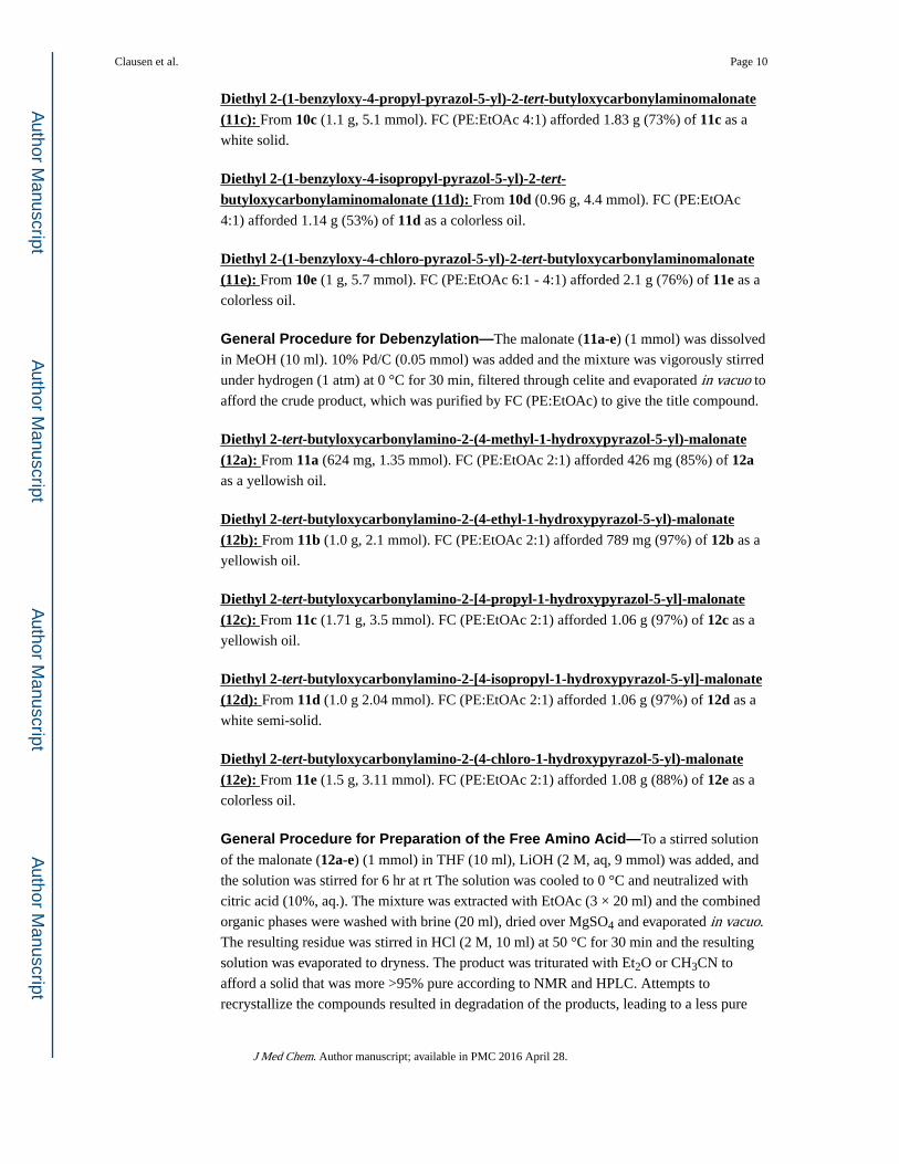
General Procedure for Debenzylation—The malonate (11a-e) (1 mmol) was dissolved
in MeOH (10 ml). 10% Pd/C (0.05 mmol) was added and the mixture was vigorously stirred
under hydrogen (1 atm) at 0 °C for 30 min, filtered through celite and evaporated in vacuo to
afford the crude product, which was purified by FC (PE:EtOAc) to give the title compound.
Diethyl 2-tert-butyloxycarbonylamino-2-(4-methyl-1-hydroxypyrazol-5-yl)-malonate (12a): From 11a (624 mg, 1.35 mmol). FC (PE:EtOAc 2:1) afforded 426 mg (85%) of 12a as a yellowish oil.
Diethyl 2-tert-butyloxycarbonylamino-2-(4-ethyl-1-hydroxypyrazol-5-yl)-malonate (12b): From 11b (1.0 g, 2.1 mmol). FC (PE:EtOAc 2:1) afforded 789 mg (97%) of 12b as a
yellowish oil.
Diethyl 2-tert-butyloxycarbonylamino-2-[4-propyl-1-hydroxypyrazol-5-yl]-malonate (12c): From 11c (1.71 g, 3.5 mmol). FC (PE:EtOAc 2:1) afforded 1.06 g (97%) of 12c as a
yellowish oil.
Diethyl 2-tert-butyloxycarbonylamino-2-[4-isopropyl-1-hydroxypyrazol-5-yl]-malonate (12d): From 11d (1.0 g 2.04 mmol). FC (PE:EtOAc 2:1) afforded 1.06 g (97%) of 12d as a
white semi-solid.
Diethyl 2-tert-butyloxycarbonylamino-2-(4-chloro-1-hydroxypyrazol-5-yl)-malonate (12e): From 11e (1.5 g, 3.11 mmol). FC (PE:EtOAc 2:1) afforded 1.08 g (88%) of 12e as a
colorless oil.
General Procedure for Preparation of the Free Amino Acid—To a stirred solution
of the malonate (12a-e) (1 mmol) in THF (10 ml), LiOH (2 M, aq, 9 mmol) was added, and
the solution was stirred for 6 hr at rt The solution was cooled to 0 °C and neutralized with
citric acid (10%, aq.). The mixture was extracted with EtOAc (3 × 20 ml) and the combined
organic phases were washed with brine (20 ml), dried over MgSO4 and evaporated in vacuo.
The resulting residue was stirred in HCl (2 M, 10 ml) at 50 °C for 30 min and the resulting
solution was evaporated to dryness. The product was triturated with Et2O or CH3CN to
afford a solid that was more >95% pure according to NMR and HPLC. Attempts to
recrystallize the compounds resulted in degradation of the products, leading to a less pure
Clausen et al. Page 10
J Med Chem. Author manuscript; available in PMC 2016 April 28.
Author M
anuscriptA
uthor Manuscript
Author M
anuscriptA
uthor Manuscript
material. All the amino acids were separated on a Crownpack CR(+) column with the (R)-
form eluting first.
Amino-(1-hydroxy-4-methyl-pyrazol-3-yl)-acetic acid hydrochloride (6b): From 12a (281 mg, 0.76 mmol) afforded 153 mg (97%) of 6b as a white solid.
Amino-(1-hydroxy-4-ethyl-pyrazol-3-yl)-acetic acid hydrochloride (6c): From 12b (759
mg, 1.97 mmol) afforded 247 mg (60%) of 6c as a pale yellow solid.
Amino-(1-hydroxy-4-propyl-pyrazol-3-yl)-acetic acid hydrochloride (6d): From 12c (1.06 mg, 2.65 mmol) afforded 450 mg (72%) of 6d as a white solid.
Amino-(1-hydroxy-4-isopropyl-pyrazol-3-yl)-acetic acid hydrochloride (6e): From 12d (685 mg, 1.71 mmol) afforded 285 mg (71%) of 6e as a white solid.
Amino-(1-hydroxy-4-chloro-pyrazol-3-yl)-acetic acid hydrochloride (6f): From 12e (1.07 g, 2.7 mmol) afforded 0.536 mg (86%) of 6f as a white solid.
Molecular Modeling and Ligand—Protein Docking—A homology model of the
agonized state of NR2B was constructed based on the soluble NR2A-ABD construct in
complex with glutamate (PDB code 2A5S)11 following the procedure for the NR2B
homology model based on NR1 (1PB7). The crystal structure of the soluble NR2A-S1S2
(PDB code 2A5S)11 was used as a template for the ligand binding domain of NR2B. The
sequence of NR2B was aligned with NR2A, truncated, and the GT linker added to form a
virtual NR2B-ABD construct containing residues 404–540 followed by the GT linker and
residues 662–802. Residues are numbered according to the sequence of total wild type
NR2B, including the signal peptide. Using the NR2B construct a homology model was
created using Prime (Schrödinger, Portland, OR) with standard parameters. Van der Waals
and electrostatic grids within a (14Å +14Å)3 box around the ligand position were calculated
on this model with the docking code Glide 2.5 (Schrödinger, Portland, OR); default
parameters were used, apart from the scaling of non-polar atoms of the receptor that was set
to 0.9. These grids were then used for ligand docking.
The ligands (R)-Et-NHP5G, and (R)-Pr-NHP5G were submitted to Monte Carlo analysis in
tri-ionised forms using the MMFFs forcefield31,32 including GB-SA treatment of aqueous
solvation in Macromodel 8.1 (Schrödinger, Portland, OR). The global minima of
conformations without intramolecular hydrogen bonds of the compounds were flexibly
docked with Glide 3.5 to the agonist binding site of the NR2B-S1S2 model. Default
parameters were used, apart from the scaling factors of the radii of the non-polar receptor
and ligand atoms, both set to 0.9.
Computational Prediction of Spectra
Initial geometries for the low pH monocationic states of (S)-6b and (S)-6f were calculated
using OPLS2005 with GB-SA aqueous solvation, and for the Mosher amide of (S)-6c with
chloroform solvation, in Macromodel 9.0 (Schrödinger 2006). For TD-DFT calculations, the
conformers were first optimised at B3LYP/6-311+G(d,p), and excitations calculated at the
Clausen et al. Page 11
J Med Chem. Author manuscript; available in PMC 2016 April 28.
Author M
anuscriptA
uthor Manuscript
Author M
anuscriptA
uthor Manuscript
same level of theory, with IEF-PCM treatment of aqueous solvation, in Gaussian’03
(Gaussian 2003). Gaussian line broadening with a halfwidth of 0.2 eV was used to transform
rotatory velocities to simulated spectra. NMR calculations were conducted on the optimised
geometries at B3LYP/6-311+G(d,p) with solvation by chloroform (PB-SCRF model) in
Jaguar 7.0 (Schrödinger 2006), with magnetic shielding tensors calculated in gas phase at
the same level of theory. Correlation coefficients were calculated between experimental and
calculated shifts of all protons relative to TMS at the same level of theory, neglecting the five
phenyl protons which are of little value in distinguishing the diastereomers.
In Vitro Pharmacology
Receptor Binding Assays—Affinities for native AMPA, KA and NMDA receptors in rat
cortical synaptosomes were determined using 5 nM [3H]AMPA (45.5 Ci/mmol),33 5 nM
[3H]KA (47.0 Ci/mmol)34 and 2 nM [3H]CGP 39653 (Kd = 6 nM, 50.0 Ci/mmol)35
respectively, with minor modifications as previously described.36 Rat brain membrane
preparations used in these receptor binding experiments were prepared according to a
method previously described.37
Two-Electrode Voltage-Clamp Electrophysiology
For expression in Xenopus oocytes, rat NR1 (GenBank U11418) cDNA was subcloned into
pCI-IRES-neo38 or pGEMHE vectors. The open reading frames of rat NR2A (GenBank
D13211), NR2B (M91562), NR2C (D13212), and NR2D (D13214) cDNAs were subcloned
into pCI-IRES-bla38 or pCI-neo (NR2A), pBluescript (NR2B), SP6 (NR2C-D) vectors.
Xenopus oocytes and cRNA for injection were prepared as previously described.39 Two-
electrode voltage-clamp recordings were performed essentially as previously described.39
During recordings, the oocytes were voltage-clamped at −60 mV to −40 mV and
continuously perfused at 23 °C with extracellular solution containing (in mM) 90 NaCl, 0.5,
BaCl2, 1 KCl, 0.01 EDTA, and 10 HEPES (pH 7.6). 20 μM glycine was included in the
extracellular solution at all times.
Agonist concentration–response data for individual oocytes were fitted to the Hill equation.
The logEC50 and nH from the individual oocytes were used to calculate the mean ± SEM.
For graphical presentation, datasets from individual oocytes were normalized to the maximal
response evoked by glutamate in the same recording. The mean ± SEM was calculated for
each of the normalized data points. The averaged data points were then fitted to the Hill
equation and plotted together with the resulting curve. Relative Imax was calculated from a
full concentration-response measurement as Imax, agonist/Imax, glutamate, where Imax, agonist is
the fitted value according to the Hill equation and Imax, glutamate is the maximal response
evoked by glutamate in the same recording.
Acknowledgments
Lars Skov and Michael Gajhede are acknowledged for their help with CD measurements. We thank Kimberly Haustein and Phuong Le for excellent technical assistance. We thank Drs. S. Heinemann (Salk Institute), S. Nakanishi (Kyoto University), and P. Seeburg (University of Heidelberg) for sharing cDNA encoding the NMDA receptor subunits. Tommy Liljefors is acknowledged for fruitful discussions on modeling and design. This work was supported by NIH (SFT), the Carlsberg Foundation (CC), the Alfred Benzon Foundation (KBH), Villum Kann
Clausen et al. Page 12
J Med Chem. Author manuscript; available in PMC 2016 April 28.
Author M
anuscriptA
uthor Manuscript
Author M
anuscriptA
uthor Manuscript

Rasmussen Foundation (KBH), the Lundbeck Foundation (KBH), the Drug Research Academy (NM) and the Augustinus Foundation (HBO).
Nonstandard abbreviations
NHP5G 2-(N-hydroxypyrazol-5-yl)glycine, 2-(N-hydroxypyrazol-4-yl)glycine
NMDA N-methyl-D-aspartic acid
GluRs glutamate receptors
Glu glutamate
CNS central nervous system
mGluRs metabotropic glutamate receptors
iGluRs ionotropic glutamate receptors
AMPA (S)-2-amino-3-(3-hydroxy-5-methyl-4-isoxazolyl)propionic acid
KA kainic acid
ABD agonist binding domain
AMAA (RS)-2-amino-2-(3-hydroxy-5-methyl-4-isoxazolyl)acetic acid
References
1. Bräuner-Osborne H, Egebjerg J, Nielsen EØ, Madsen U, Krogsgaard-Larsen P. Ligands for glutamate receptors: Design and therapeutic prospects. J Med Chem. 2000; 43:2609–2645. [PubMed: 10893301]
2. Dingledine R, Borges K, Bowie D, Traynelis SF. The Glutamate Receptor Ion Channels. Pharmacol Rev. 1999; 51:7–62. [PubMed: 10049997]
3. Erreger K, Chen PE, Wyllie DJ, Traynelis SF. Glutamate receptor gating. Crit Rev Neurobiol. 2004; 16:187–224. [PubMed: 15701057]
4. Riedel G, Platt B, Micheau J. Glutamate receptor function in learning and memory. Behav Brain Res. 2003; 140:1–47. [PubMed: 12644276]
5. Javitt DC. Glutamate as a therapeutic target in psychiatric disorders. Mol Psychiatry. 2004; 9:984–997. [PubMed: 15278097]
6. Parsons CG, Danysz W, Quack G. Glutamate in CNS disorders as a target for drug development: an update. Drug News Perspect. 1998; 11:523–569. [PubMed: 15616669]
7. Armstrong N, Gouaux E. Mechanisms for activation and antagonism of an AMPA-sensitive glutamate receptor: crystal structures of the GluR2 ligand binding core. Neuron. 2000; 28:165–181. [PubMed: 11086992]
8. Armstrong N, Sun Y, Chen GQ, Gouaux E. Structure of a glutamate-receptor ligand-binding core in complex with kainate. Nature. 1998; 395:913–917. [PubMed: 9804426]
9. Mayer ML. Crystal structures of the GluR5 and GluR6 ligand binding cores: molecular mechanisms underlying kainate receptor selectivity. Neuron. 2005; 45:539–552. [PubMed: 15721240]
10. Naur P, Vestergaard B, Skov LK, Egebjerg J, Gajhede M, Kastrup JS. Crystal structure of the kainate receptor GluR5 ligand-binding core in complex with (S)-glutamate. FEBS Lett. 2005; 579:1154–1160. [PubMed: 15710405]
11. Furukawa H, Gouaux E. Mechanisms of activation, inhibition and specificity: crystal structures of the NMDA receptor NR1 ligand-binding core. EMBO J. 2003; 22:2873–2885. [PubMed: 12805203]
12. Jin R, Banke TG, Mayer ML, Traynelis SF, Gouaux E. Structural basis for partial agonist action at ionotropic glutamate receptors. Nat Neurosci. 2003; 6:803–810. [PubMed: 12872125]
Clausen et al. Page 13
J Med Chem. Author manuscript; available in PMC 2016 April 28.
Author M
anuscriptA
uthor Manuscript
Author M
anuscriptA
uthor Manuscript

13. Frandsen A, Pickering DS, Vestergaard B, Kasper C, Nielsen BB, Greenwood JR, Campiani G, Fattorusso C, Gajhede M, Schousboe A, Kastrup JS. Tyr702 is an important determinant of agonist binding and domain closure of the ligand-binding core of GluR2. Mol Pharmacol. 2005; 67:703–713. [PubMed: 15591246]
14. Inanobe A, Furukawa H, Gouaux E. Mechanism of partial agonist action at the NR1 subunit of NMDA receptors. Neuron. 2005; 47:71–84. [PubMed: 15996549]
15. Cali P, Begtrup M. Synthesis of 1-hydroxypyrazole glycine derivatives. Tetrahedron. 2002; 58:1595–1605.
16. Clausen RP, Hansen KB, Cali P, Nielsen B, Greenwood JR, Begtrup M, Egebjerg J, Bräuner-Osborne H. The respective N-hydroxypyrazole analogues of the classical glutamate receptor ligands ibotenic acid and (RS)-2-amino-2-(3-hydroxy-5-methyl-4-isoxazolyl)acetic acid. Eur J Pharmacol. 2004; 499:35–44. [PubMed: 15363949]
17. Jørgensen CG, Bräuner-Osborne H, Nielsen B, Kehler J, Clausen RP, Krogsgaard-Larsen P, Madsen U. Novel 5-substituted 1-pyrazolol analogues of ibotenic acid: synthesis and pharmacology at glutamate receptors. Bioorg Med Chem. 2007; 15:3524–3538. [PubMed: 17376693]
18. Hansen KB, Clausen RP, Bjerrum EJ, Bechmann C, Greenwood JR, Christensen C, Kristensen JL, Egebjerg J, Brauner-Osborne H. Tweaking Agonist Efficacy at N-Methyl-D-aspartate Receptors by Site-Directed Mutagenesis. Mol Pharmacol. 2005; 68:1510–1523. [PubMed: 16131614]
19. Felding J, Kristensen J, Bjerregaard T, Sander L, Vedsø P, Begtrup M. Synthesis of 4-Substituted 1-(Benzyloxy)pyrazoles via Iodine-Magnesium Exchange of 1-(Benzyloxy)-4-iodopyrazole. J Org Chem. 1999; 64:4196–4198.
20. Akoka S, Barantin L, Trierweiler M. Concentration measurement by proton NMR using the ERETIC method. Anal Chem. 1999; 71:2554–2557. [PubMed: 21662801]
21. Shinbo T, Yamaguchi T, Nishimura K, Sugiura M. Chromatographic-Separation of Racemic Amino-Acids by Use of Chiral Crown Ether-Coated Reversed-Phase Packings. J Chromatogr. 1987; 405:145–153. [PubMed: 3693463]
22. de Leon CAP, Sutton KL, Caruso JA, Uden PC. Chiral speciation of selenoamino acids and selenium enriched samples using HPLC coupled to ICP-MS. J Anal At Spectrom. 2000; 15:1103–1107.
23. Hawkins CL, Lawrance GA. Circular dichroism spectra of N,N-Dimethyl-L-amino acids. Aust J Chem. 1973; 26:1801–1803.
24. Christopoulos A. Assessing the distribution of parameters in models of ligand-receptor interaction: to log or not to log. Trends Pharmacol Sci. 1998; 19:351–357. [PubMed: 9786022]
25. Furukawa H, Singh SK, Mancusso R, Gouaux E. Subunit arrangement and function in NMDA receptors. Nature. 2005; 438:185–192. [PubMed: 16281028]
26. Kinarsky L, Feng BH, Skifter DA, Morley RM, Sherman S, Jane DE, Monaghan DT. Identification of subunit- and antagonist-specific amino acid residues in the N-methyl-D-aspartate receptor glutamate-binding pocket. J Pharmacol Exp Ther. 2005; 313:1066–1074. [PubMed: 15743930]
27. Ebert B, Lenz S, Brehm L, Bregnedal P, Hansen JJ, Frederiksen K, Bøgesø KP, Krogsgaard-Larsen P. Resolution, absolute stereochemistry, and pharmacology of the S-(+)- and R-(−)-isomers of the apparent partial AMPA receptor agonist (R,S)-2-amino-3-(3-hydroxy-5-phenylisoxazol-4-yl)propionic acid [(R,S)-APPA]. J Med Chem. 1994; 37:878–884. [PubMed: 7512140]
28. Suffert J. Simple Direct Titration of Organo-Lithium Reagents Using N-Pivaloyl-Ortho-Toluidine and Or N-Pivaloyl-Ortho-Benzylaniline. J Org Chem. 1989; 54:509–510.
29. Lin HS, Paquette LA. A Convenient Method for Determining the Concentration of Grignard-Reagents. Synth Commun. 1994; 24:2503–2506.
30. Vedsø P, Begtrup M. Synthesis of 5-Substituted 1-Hydroxypyrazoles Through Directed Lithiation of 1-(Benzyloxy)Pyrazole. J Org Chem. 1995; 60:4995–4998.
31. Halgren TA. MMFF VI. MMFF94s option for energy minimization studies. J Comp Chem. 1999; 20:720–729.
32. Halgren TA. MMFF VII. Characterization of MMFF94, MMFF94s, and other widely available force fields for conformational energies and for intermolecular-interaction energies and geometries. J Comp Chem. 1999; 20:730–748.
Clausen et al. Page 14
J Med Chem. Author manuscript; available in PMC 2016 April 28.
Author M
anuscriptA
uthor Manuscript
Author M
anuscriptA
uthor Manuscript
33. Honore T, Nielsen M. Complex structure of quisqualate-sensitive glutamate receptors in rat cortex. Neurosci Lett. 1985; 54:27–32. [PubMed: 2858071]
34. Braitman DJ, Coyle JT. Inhibition of [3H]kainic acid receptor binding by divalent cations correlates with ion affinity for the calcium channel. Neuropharmacology. 1987; 26:1247–1251. [PubMed: 2444898]
35. Sills MA, Fagg D, Pozza M, Angst C, Brundish DE, Hurt SD, Wilusz EJ, Williams M. [3H]CGP 39653: a new N-methyl-D-aspartate antagonist radioligand with low nanomolar affnity in rat brain. Eur J Pharmacol. 1991; 192:19–24. [PubMed: 1674916]
36. Hermit MB, Greenwood JR, Nielsen B, Bunch L, Jørgensen CG, Vestergaard HT, Stensbøl TB, Sanchez C, Krogsgaard-Larsen P, Madsen U, Bräuner-Osborne H. Ibotenic acid and thioibotenic acid: a remarkable difference in activity at group III metabotropic glutamate receptors. Eur J Pharmacol. 2004; 486:241–250. [PubMed: 14985045]
37. Ransom RW, Stec NL. Cooperative modulation of [3H]MK801 binding to the N-methyl-D-aspartate receptor ion channel complex by L-glutamate, glycine and polyamines. J Neurochem. 1988; 51:830–836. [PubMed: 2457653]
38. Hansen KB, Bräuner-Osborne H, Egebjerg J. Pharmacological characterization of ligands at recombinant NMDA receptor subtypes by electrophysiological recordings intracellular calcium measurements. Comb Chem High Throughput Screen. 2008 In Press.
39. Traynelis SF, Burgess MF, Zheng F, Lyuboslavsky P, Powers JL. Control of voltage-independent zinc inhibition of NMDA receptors by the NR1 subunit. J Neurosci. 1998; 18:6163–6175. [PubMed: 9698310]
Clausen et al. Page 15
J Med Chem. Author manuscript; available in PMC 2016 April 28.
Author M
anuscriptA
uthor Manuscript
Author M
anuscriptA
uthor Manuscript
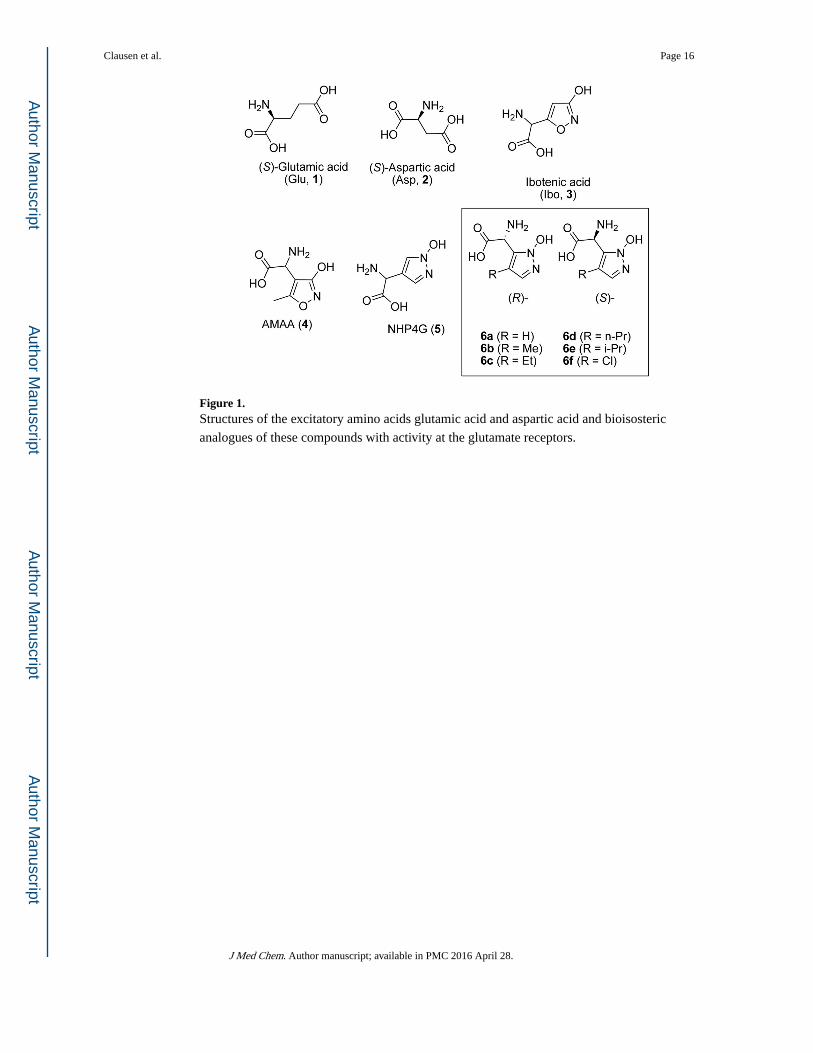
Figure 1. Structures of the excitatory amino acids glutamic acid and aspartic acid and bioisosteric
analogues of these compounds with activity at the glutamate receptors.
Clausen et al. Page 16
J Med Chem. Author manuscript; available in PMC 2016 April 28.
Author M
anuscriptA
uthor Manuscript
Author M
anuscriptA
uthor Manuscript

Figure 2. Two chiral HPLC chromatograms of (S)- and (R)-6d, respectively, showing the elution order
on an analytical Crownpak CR(−) column. The order is reversed on a preparative Crownpak
CR(+) column.
Clausen et al. Page 17
J Med Chem. Author manuscript; available in PMC 2016 April 28.
Author M
anuscriptA
uthor Manuscript
Author M
anuscriptA
uthor Manuscript

Figure 3. Electronic Circular Dichroism spectra (A) of compounds (R)- and (S)-6f at a concentration
of 0.325 mg/mL in H2O compared with a TD-DFT simulated spectra (B) of a weighted
average of eight low energy conformers as well as a single low energy conformer of (S)-6f in dilute acid.
Clausen et al. Page 18
J Med Chem. Author manuscript; available in PMC 2016 April 28.
Author M
anuscriptA
uthor Manuscript
Author M
anuscriptA
uthor Manuscript
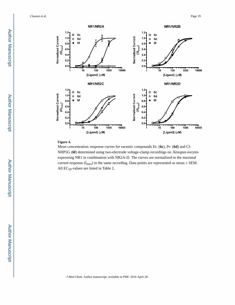
Figure 4. Mean concentration–response curves for racemic compounds Et- (6c), Pr- (6d) and Cl-
NHP5G (6f) determined using two-electrode voltage-clamp recordings on Xenopus oocytes
expressing NR1 in combination with NR2A-D. The curves are normalized to the maximal
current response (Imax) in the same recording. Data points are represented as mean ± SEM.
All EC50-values are listed in Table 2.
Clausen et al. Page 19
J Med Chem. Author manuscript; available in PMC 2016 April 28.
Author M
anuscriptA
uthor Manuscript
Author M
anuscriptA
uthor Manuscript
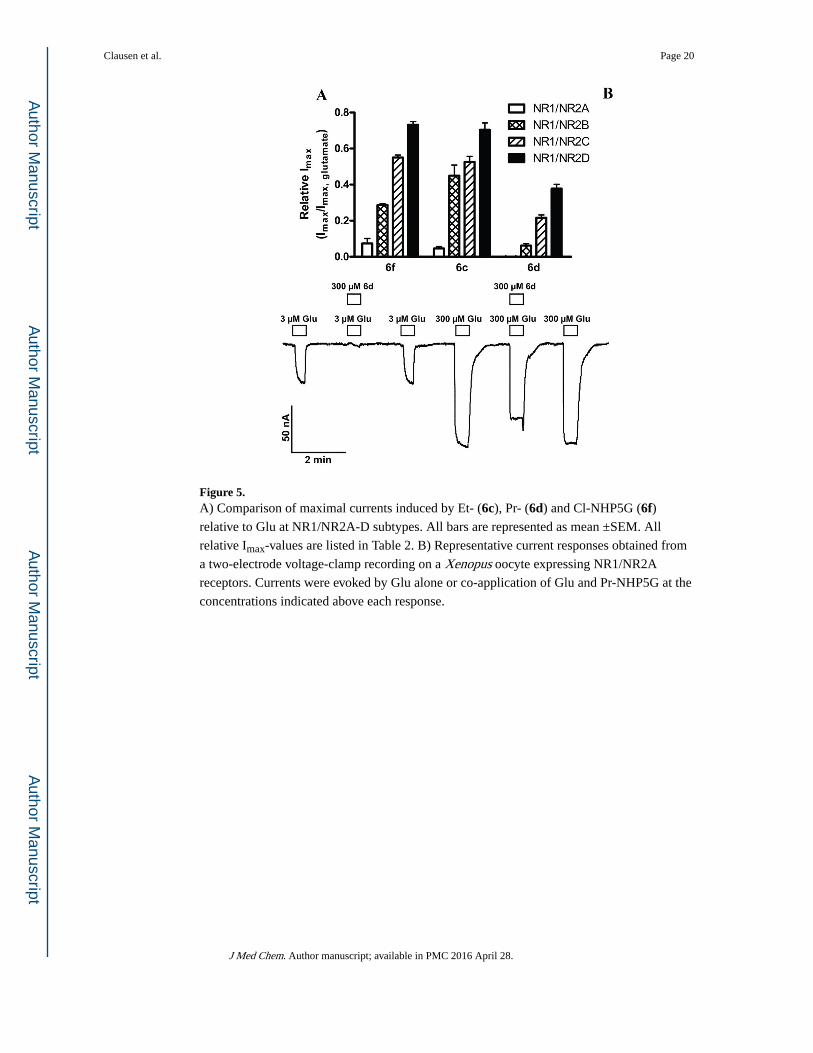
Figure 5. A) Comparison of maximal currents induced by Et- (6c), Pr- (6d) and Cl-NHP5G (6f) relative to Glu at NR1/NR2A-D subtypes. All bars are represented as mean ±SEM. All
relative Imax-values are listed in Table 2. B) Representative current responses obtained from
a two-electrode voltage-clamp recording on a Xenopus oocyte expressing NR1/NR2A
receptors. Currents were evoked by Glu alone or co-application of Glu and Pr-NHP5G at the
concentrations indicated above each response.
Clausen et al. Page 20
J Med Chem. Author manuscript; available in PMC 2016 April 28.
Author M
anuscriptA
uthor Manuscript
Author M
anuscriptA
uthor Manuscript
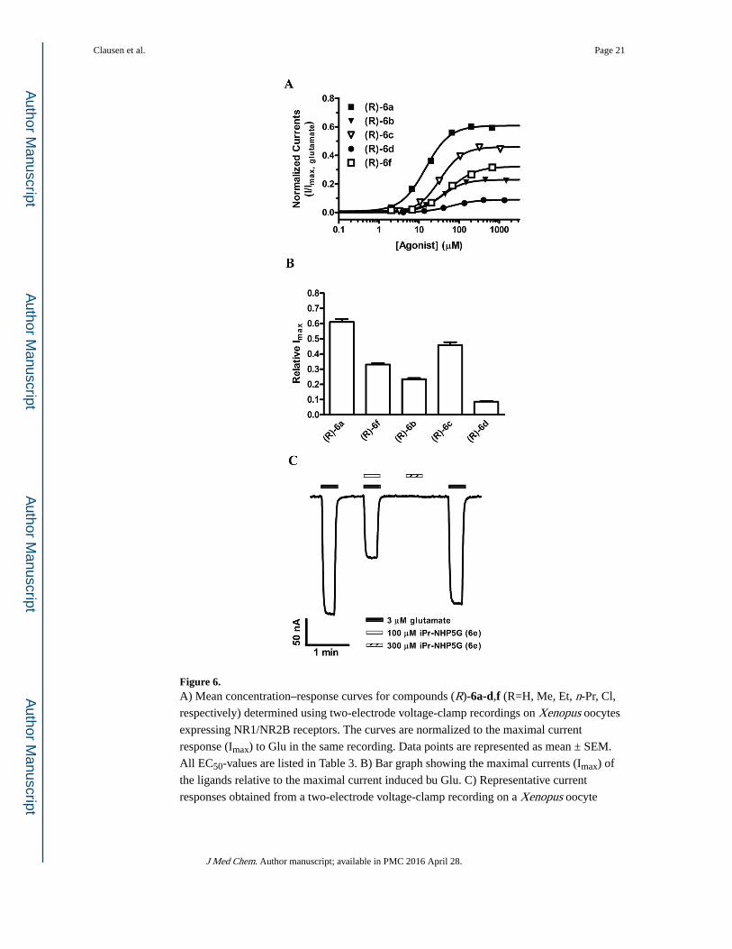
Figure 6. A) Mean concentration–response curves for compounds (R)-6a-d,f (R=H, Me, Et, n-Pr, Cl,
respectively) determined using two-electrode voltage-clamp recordings on Xenopus oocytes
expressing NR1/NR2B receptors. The curves are normalized to the maximal current
response (Imax) to Glu in the same recording. Data points are represented as mean ± SEM.
All EC50-values are listed in Table 3. B) Bar graph showing the maximal currents (Imax) of
the ligands relative to the maximal current induced bu Glu. C) Representative current
responses obtained from a two-electrode voltage-clamp recording on a Xenopus oocyte
Clausen et al. Page 21
J Med Chem. Author manuscript; available in PMC 2016 April 28.
Author M
anuscriptA
uthor Manuscript
Author M
anuscriptA
uthor Manuscript
expressing NR1/NR2B receptors. Currents were evoked by Glu alone or co-application of
Glu and (R)-6e at the concentrations indicated above each response.
Clausen et al. Page 22
J Med Chem. Author manuscript; available in PMC 2016 April 28.
Author M
anuscriptA
uthor Manuscript
Author M
anuscriptA
uthor Manuscript

Figure 7. A) Structure of a homology model of the ABD of NR2B (cyan) containing n-Pr-NHP5G
((R)-6d, blue) showing the residues (red) lining a crevice to the agonist binding site.
Previous studies have shown these residues to be important determinants of agonist
efficacies of several compounds.18 B) Et-NHP5G ((R)-6c, pink) and n-Pr-NHP5G ((R)-6d,
blue) docked in the homology model. The distances from the amino group of Lys485 to the
n-propyl group and the peptide carbonyl between Arg712 and Gly713 are indicated in
Ångstroms. Water molecules are shown as red asterisks.
Clausen et al. Page 23
J Med Chem. Author manuscript; available in PMC 2016 April 28.
Author M
anuscriptA
uthor Manuscript
Author M
anuscriptA
uthor Manuscript
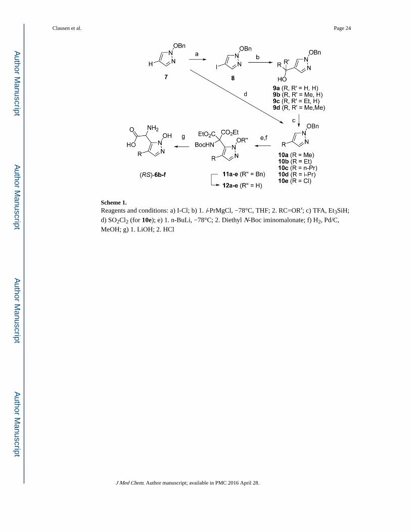
Scheme 1. Reagents and conditions: a) I-Cl; b) 1. i-PrMgCl, −78°C, THF; 2. RC=OR′; c) TFA, Et3SiH;
d) SO2Cl2 (for 10e); e) 1. n-BuLi, −78°C; 2. Diethyl N-Boc iminomalonate; f) H2, Pd/C,
MeOH; g) 1. LiOH; 2. HCl
Clausen et al. Page 24
J Med Chem. Author manuscript; available in PMC 2016 April 28.
Author M
anuscriptA
uthor Manuscript
Author M
anuscriptA
uthor Manuscript

Author M
anuscriptA
uthor Manuscript
Author M
anuscriptA
uthor Manuscript
Clausen et al. Page 25
Table 1
Receptor binding affinities of compounds 6a-6f at three major groups of iGluRs in rat cortical synaptosome
assay. The numbers in brackets [min, max] indicate mean ± SEM according to a logarithmic distribution.24
Compd R
IC50 (μM) Ki (μM)
[3H]AMPA [3H]KAIN [3H]CGP39653
Glu 0.34a 0.38a 0.20a
(RS)-6a H > 100 > 100 10 [9;11]a
(R)-6a > 100 > 100 2.2 [2.0;2.4]
(S)-6a > 100 > 100 > 100
(RS)-6b Me > 100 > 100 22 [20;24]
(R)-6b > 100 > 100 2.2 [2.1;2.4]
(S)-6b > 100 > 100 > 100
(RS)-6c Et > 100 > 100 13 [11;15]
(R)-6c > 100 > 100 4.4 [3.3;5.7]
(S)-6c > 100 > 100 > 100
(RS)-6d n-Pr > 100 > 100 14 [14;15]
(R)-6d > 100 > 100 6.9 [5.9;8.2]
(S)-6d > 100 > 100 > 100
(RS)-6e i-Pr > 100 > 100 20 [19;21]
(R)-6e > 100 > 100 13 [11;16]
(S)-6e > 100 > 100 > 100
(RS)-6f Cl > 100 > 100 2.9 [2.7;3.2]
(R)-6f nd nd 2.9 [2.6;3.2]
(S)-6f > 100 > 100 25 [24;26]
nd ~ not determined
aRef. 16
J Med Chem. Author manuscript; available in PMC 2016 April 28.

Author M
anuscriptA
uthor Manuscript
Author M
anuscriptA
uthor Manuscript
Clausen et al. Page 26
Tab
le 2
Pote
ncy
and
effi
cacy
rel
ativ
e to
Glu
of
com
poun
ds 6
a, 6
c, 6
d an
d 6f
at r
ecom
bina
nt N
R1/
NR
2A-D
rec
epto
rs e
xpre
ssed
in X
enop
us o
ocyt
es. D
ata
are
from
3–5
ooc
ytes
and
the
num
bers
in b
rack
ets
[min
, max
] in
dica
te m
ean
± S
EM
acc
ordi
ng to
a lo
gari
thm
ic d
istr
ibut
ion.
24
Com
poun
dR
Subt
ype
EC
50 (
μM)
Rel
. Im
axn H
ill
Glu
a-
NR
1/N
R2A
2.9
11.
5
NR
1/N
R2B
1.8
11.
7
NR
1/N
R2C
1.0
11.
3
NR
1/N
R2D
0.45
11.
6
NM
DA
a-
NR
1/N
R2A
750.
90±
0.04
1.5
NR
1/N
R2B
220.
77±
0.01
1.4
NR
1/N
R2C
230.
73±
0.02
1.4
NR
1/N
R2D
8.3
0.80
±0.
021.
6
(RS)
-6ab
HN
R1/
NR
2A82
[75
, 90]
0.45
±0.
031.
3
NR
1/N
R2B
48 [
47, 4
9]0.
58±
0.02
1.6
NR
1/N
R2C
54 [
50, 5
7]0.
52±
0.01
1.4
NR
1/N
R2D
ND
ND
ND
(RS)
-6c
Et
NR
1/N
R2A
47[3
9;57
]0.
05±
0.01
2.4
NR
1/N
R2B
68[6
4;72
]0.
45±
0.06
1.2
NR
1/N
R2C
91[8
2;10
1]0.
52±
0.03
1.3
NR
1/N
R2D
43[4
2;44
]0.
70±
0.03
1.5
(RS)
-6d
n-Pr
NR
1/N
R2A
NA
NA
NA
NR
1/N
R2B
105[
95;1
16]
0.06
±0.
011.
9
NR
1/N
R2C
429[
367;
500]
0.22
±0.
021.
1
NR
1/N
R2D
153[
142;
165]
0.37
±0.
021.
6
(RS)
-6f
Cl
NR
1/N
R2A
565[
464;
687]
0.07
±0.
033.
2
NR
1/N
R2B
149[
139;
160]
0.29
±0.
011.
2
NR
1/N
R2C
255[
250;
260]
0.55
±0.
011.
4
NR
1/N
R2D
152[
149;
155]
0.73
±0.
021.
4
NA
~ n
o ac
tivity
a Ref
. 38
J Med Chem. Author manuscript; available in PMC 2016 April 28.
Author M
anuscriptA
uthor Manuscript
Author M
anuscriptA
uthor Manuscript
Clausen et al. Page 27b R
ef. 1
6
J Med Chem. Author manuscript; available in PMC 2016 April 28.

Author M
anuscriptA
uthor Manuscript
Author M
anuscriptA
uthor Manuscript
Clausen et al. Page 28
Table 3
Potency and efficacy relative to Glu of the (R)-form of 6a-d and 6f at recombinant NR1/NR2B receptors
expressed in Xenopus oocytes. Data are from 4–6 oocytes and the numbers in brackets [min, max] indicate
mean ± SEM according to a logarithmic distribution.24
Compd R EC50 (μM) Rel. Imax nHill
(R)-6a H 14[14;15] 0.61±0.02 1.4
(R)-6b Me 36[35;38] 0.23±0.01 1.6
(R)-6c Et 34[32;35] 0.46±0.02 1.6
(R)-6d n-Pr 63[59;68] 0.09±0.004 1.4
(R)-6f Cl 58[55;62] 0.33±0.01 1.2
J Med Chem. Author manuscript; available in PMC 2016 April 28.