Myringosclerosis with Chronic Safe Suppurative Otitis ... · Myringosclerosis is a sequela seen...
4
Int Adv Otol 2014; 10(3): 260-3 • DOI: 10.5152/iao.2014.467 Original Article Myringosclerosis with Chronic Safe Suppurative Otitis Media: Hearing Outcome after Surgical Management According to Myringosclerotic Patch Site and Inclusion in the Surgery Hatem S. Badran, Hazem M. Abdel Tawab, Basim M. Wahba, Khairy M. Abulnasr Department of Otorhinolaryngology, Cairo University, Cairo, Egypt (HSB, HMA, BMW) Hearing and Speech Institute, Cairo, Egypt (KMA) OBJECTIVE: Compare hearing outcomes between sparing of the myringosclerotic patch and removing it as a step in myringoplasty for chronic tubotympanic chronic otitis media with central perforation and myringosclerotic patch. MATERIALS and METHODS: A total of 94 patients having chronic suppurative otitis media (inactive stage) with central drum perforation and a myringosclerotic patch in the same ear were included in the study. They were divided into four groups according to the site of the myringosclerotic patch, and each group was randomly divided into two subgroups: subgroup A, where myringoplasty was done after removing the tympanosclerotic patch regardless of site, and subgroup B, where myringoplasty was done, leaving the tympanosclerotic patch in its place. Pure tone audiometry was done preoperatively and compared with that done 10 weeks postoperatively. RESULTS: A non significantly higher net hearing gain was noticed in subgroup A, irrespective of the site of the myringosclerotic patch. Significant results were seen only in cases where the myringosclerotic patch was connecting the handle of the malleus to the tympanic annulus in subgroup A compared to subgroup B. CONCLUSION: The site of the myringosclerotic patch and its inclusion as a step in the surgery largely affect the improvement of hearing after my- ringoplasty. KEY WORDS: Myringosclerosis, myringoplasty, otitis media INTRODUCTION Tympanosclerosis is described as an irreversible, though not immutable, end result of any unresolved specific or nonspecific in- flammatory disease of the middle ear characterized by anatomical distortion resulting in functional impairment that is conductive hearing loss [1] . Myringosclerosis is a sequela seen after otitis media with effusion, chronic otitis media, and sometimes after multiple acute epi- sodes of acute otitis media [2] . It is also related to treatment by ventilation tube insertion or myringotomy [3, 4] . Tympanosclerosis is a disorder of healing in which large amounts of collagenous fibrous tissue are laid down in relation to the mid- dle ear. Thickening and fusion of the collagen may follow with the formation of a homogeneous hyaline mass in which deposition of scattered intracellular and extracellular calcium and phosphate crystals may ultimately take place. The process occurs in the sub- mucosal connective tissue layer that covers the auditory ossicles, lines the bony walls of the tympanic cavity, and forms the middle fibrous layer of the tympanic membrane [5] . Clinically, myringosclerotic lesions are seen as whitish, sclerotic plaques (chalk patches) in the tympanic membrane (TM) [6] . The myringosclerotic mass may assume clinical importance by interfering with the transmission of sound vibrations across the middle ear structures [5] . It is frequently associated with marked conductive hearing loss. In contrast, myringosclerosis may be as- ymptomatic, as the effect on hearing is insignificant in most instances [7] . According to some epidemiological investigations on non-selected populations, the prevalence of sclerotic plaques in the ear drum varies from 1.7% to 22.4% [8, 9] . Corresponding Address: Hazem M. Abdel Tawab, Department of Otorhinolaryngology, Cairo University, Cairo, Egypt Phone: +96891128430; E-mail: [email protected] Submitted: 11.11.2014 Accepted: 12.11.2014 Copyright 2014 © The Mediterranean Society of Otology and Audiology 260
Transcript of Myringosclerosis with Chronic Safe Suppurative Otitis ... · Myringosclerosis is a sequela seen...

Int Adv Otol 2014; 10(3): 260-3 • DOI: 10.5152/iao.2014.467
Original Article
Myringosclerosis with Chronic Safe Suppurative Otitis Media: Hearing Outcome after Surgical Management According to Myringosclerotic Patch Site and Inclusion in the Surgery
Hatem S. Badran, Hazem M. Abdel Tawab, Basim M. Wahba, Khairy M. AbulnasrDepartment of Otorhinolaryngology, Cairo University, Cairo, Egypt (HSB, HMA, BMW)Hearing and Speech Institute, Cairo, Egypt (KMA)
OBJECTIVE: Compare hearing outcomes between sparing of the myringosclerotic patch and removing it as a step in myringoplasty for chronic tubotympanic chronic otitis media with central perforation and myringosclerotic patch.
MATERIALS and METHODS: A total of 94 patients having chronic suppurative otitis media (inactive stage) with central drum perforation and a myringosclerotic patch in the same ear were included in the study. They were divided into four groups according to the site of the myringosclerotic patch, and each group was randomly divided into two subgroups: subgroup A, where myringoplasty was done after removing the tympanosclerotic patch regardless of site, and subgroup B, where myringoplasty was done, leaving the tympanosclerotic patch in its place. Pure tone audiometry was done preoperatively and compared with that done 10 weeks postoperatively.
RESULTS: A non significantly higher net hearing gain was noticed in subgroup A, irrespective of the site of the myringosclerotic patch. Significant results were seen only in cases where the myringosclerotic patch was connecting the handle of the malleus to the tympanic annulus in subgroup A compared to subgroup B.
CONCLUSION: The site of the myringosclerotic patch and its inclusion as a step in the surgery largely affect the improvement of hearing after my-ringoplasty.
KEY WORDS: Myringosclerosis, myringoplasty, otitis media
INTRODUCTIONTympanosclerosis is described as an irreversible, though not immutable, end result of any unresolved specific or nonspecific in-flammatory disease of the middle ear characterized by anatomical distortion resulting in functional impairment that is conductive hearing loss [1].
Myringosclerosis is a sequela seen after otitis media with effusion, chronic otitis media, and sometimes after multiple acute epi-sodes of acute otitis media [2]. It is also related to treatment by ventilation tube insertion or myringotomy [3, 4].
Tympanosclerosis is a disorder of healing in which large amounts of collagenous fibrous tissue are laid down in relation to the mid-dle ear. Thickening and fusion of the collagen may follow with the formation of a homogeneous hyaline mass in which deposition of scattered intracellular and extracellular calcium and phosphate crystals may ultimately take place. The process occurs in the sub-mucosal connective tissue layer that covers the auditory ossicles, lines the bony walls of the tympanic cavity, and forms the middle fibrous layer of the tympanic membrane [5].
Clinically, myringosclerotic lesions are seen as whitish, sclerotic plaques (chalk patches) in the tympanic membrane (TM) [6].
The myringosclerotic mass may assume clinical importance by interfering with the transmission of sound vibrations across the middle ear structures [5]. It is frequently associated with marked conductive hearing loss. In contrast, myringosclerosis may be as-ymptomatic, as the effect on hearing is insignificant in most instances [7].
According to some epidemiological investigations on non-selected populations, the prevalence of sclerotic plaques in the ear drum varies from 1.7% to 22.4% [8, 9].
Corresponding Address:Hazem M. Abdel Tawab, Department of Otorhinolaryngology, Cairo University, Cairo, EgyptPhone: +96891128430; E-mail: [email protected] Submitted: 11.11.2014 Accepted: 12.11.2014 Copyright 2014 © The Mediterranean Society of Otology and Audiology260

Intratympanic tympanosclerosis has been subdivided into “open” and “closed” varieties, depending on the integrity of the tympanic membrane [10, 11].
By far, the commonest otological finding in ears with tympanoscle-rosis is a central perforation (54%). The next most common finding is an intact tympanic membrane [1].
The management of hearing loss due to tympanosclerosis has re-mained the center of considerable controversy [5].
The aim of this study is to compare the hearing outcomes after surgical management of myringosclerosis associated with chronic tubotympanic suppurative otitis media (CSOM) and central perfora-tion in cases of sparing of the myringosclerotic patch and its removal.
MATERIALS and METHODSThis prospective longitudinal study was done from April 2010 to July 2013 at Department of Otorhinolaryngology and Head and Neck Surgery, Kasr El-Aini Faculty of Medicine, Cairo University, Egypt and Saudi German Hospital in Jeddah, Saudi Arabia. Informed consents were taken from all patients after explanation of the procedure.
Ethical committee approval was obtained from Kasr El-Aini Faculty of Medicine and Saudi German Hospital.
A total of 94 patients, aged over 12 years and of either sex, who were admitted to our clinics having chronic tubotympanic otitis media (inactive stage) with central drum perforation and myringosclerotic patch in the same ear and already planned for myringoplasty were included in this study.
Exclusion Criteria1- CSOM (tubotympanic) in the active stage2- CSOM (atticoantral) type3- Sensorineural hearing loss4- Revision myringoplasty
All patients were subjected to a full history taking and otoscopic and microscopic examination to confirm the diagnosis and the absence of all exclusion criteria.
During the examination, we noticed the different sites of myringo-sclerotic patches in the tympanic membrane. We arranged the pa-tients into four groups according to the site of the myringosclerotic patch.
Group 1: The myringosclerotic patch was adherent to the tympanic annulus.Group 2: The patch was adherent to the handle or umbo of the mal-leus.Group 3: The patch was not attached to the annulus or umbo or han-dle of the malleus. Group 4: The myringosclerotic patch connected the annulus to either the umbo or handle of the malleus.
Each of the groups above was randomly divided into two equal sub-groups: subgroup A, where the patients were managed with myrin-
goplasty after removing the myringosclerotic patch, and subgroup B, which included patients who were managed with myringoplasty with sparing of the myringosclerotic patch. The method of random-ization was to allow the patients to randomly withdraw a paper with the subgroup number from a box.
Grafting was done using the temporalis fascia after apostauricular approach. Myringoplasty using the underlay technique was used in all patients.
All patients were discharged on the second day of operation on (amoxicillin clavulanic acid; GlaxoSmithKline plc (GSK), New York, USA) 1 gm tablets every 12 hours for 1 week. Postoperative instruc-tions were given and removal of the ear pack was carried out on the 10th day after the operation.
Preoperative hearing assessment in the form of pure tone audiom-etry was done for all patients preoperatively and was repeated 10 weeks after the operation.
Only values at 500, 1000, and 2000 Hz were considered in this study.
Statistical AnalysisData were represented as mean±standard deviation (SD) for numeric data and as frequency (number and percent) for non-numeric data. They were compared using independent t-test. The correlation was done using Pearson correlation coefficient for numeric data and cross-tables chi-square test for non-numeric data. Two-tailed signifi-cant values were considered when p<0.05. Data were analyzed using SPSS for Windows, version 16 (IBM Company, Chicago, USA).
RESULTSThis prospective study included 94 patients with chronic tubotympan-ic otitis media with central drum perforation and a myringosclerotic patch in the same ear. The patients were admitted in Kasr El-Aini Hos-pital, Faculty of Medicine, Cairo University and Saudi German Hospital, Saudi Arabia, and were subjected to underlay myringoplasty.
According to our observations, there were four unequal groups: group 1 included 18 patients (19.2%), group 2 included 20 patients (21.3%), group 3 included 32 patients (34.1%), and group 4 included 24 patients (25.4%).
The average age of patients was 29.22±8.42 years, and the age range was 16 to 47 years (Table 1). According to gender, there were 45 males (47.9%) and 49 females (52.1%), as shown in Table 2.
The overall hearing results are shown in Table 3. Regarding group 1, a net hearing gain of 18 dB was observed in subgroup A compared
Mean±SD Minimum Maximum
Group 1 (n=18) 29.72±8.51 17 46
Group 2 (n=20) 27.80±8.87 20 43
Group 3 (n=32) 25.71±7.65 16 40
Group 4 (n=24) 30.75±8.87 16 47
Total 29.22±8.42 16 47
Table 1. Age distribution of study groups (years)
261
Badran et al. Surgical Outcome of Myringosclerosis
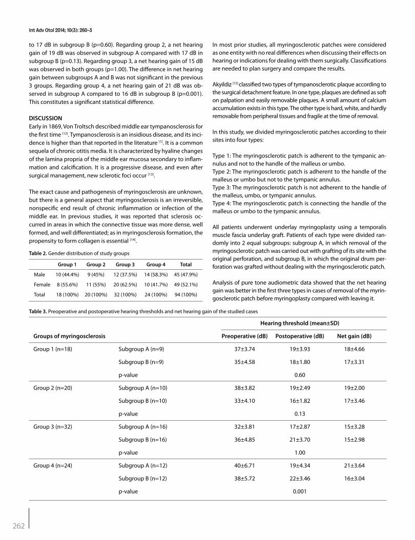
to 17 dB in subgroup B (p=0.60). Regarding group 2, a net hearing gain of 19 dB was observed in subgroup A compared with 17 dB in subgroup B (p=0.13). Regarding group 3, a net hearing gain of 15 dB was observed in both groups (p=1.00). The difference in net hearing gain between subgroups A and B was not significant in the previous 3 groups. Regarding group 4, a net hearing gain of 21 dB was ob-served in subgroup A compared to 16 dB in subgroup B (p=0.001). This constitutes a significant statistical difference.
DISCUSSIONEarly in 1869, Von Troltsch described middle ear tympanosclerosis for the first time [12]. Tympanosclerosis is an insidious disease, and its inci-dence is higher than that reported in the literature [1]. It is a common sequela of chronic otitis media. It is characterized by hyaline changes of the lamina propria of the middle ear mucosa secondary to inflam-mation and calcification. It is a progressive disease, and even after surgical management, new sclerotic foci occur [13].
The exact cause and pathogenesis of myringosclerosis are unknown, but there is a general aspect that myringosclerosis is an irreversible, nonspecific end result of chronic inflammation or infection of the middle ear. In previous studies, it was reported that sclerosis oc-curred in areas in which the connective tissue was more dense, well formed, and well differentiated; as in myringosclerosis formation, the propensity to form collagen is essential [14].
In most prior studies, all myringosclerotic patches were considered as one entity with no real differences when discussing their effects on hearing or indications for dealing with them surgically. Classifications are needed to plan surgery and compare the results.
Akyildiz [15] classified two types of tympanosclerotic plaque according to the surgical detachment feature. In one type, plaques are defined as soft on palpation and easily removable plaques. A small amount of calcium accumulation exists in this type. The other type is hard, white, and hardly removable from peripheral tissues and fragile at the time of removal.
In this study, we divided myringosclerotic patches according to their sites into four types:
Type 1: The myringosclerotic patch is adherent to the tympanic an-nulus and not to the handle of the malleus or umbo.Type 2: The myringosclerotic patch is adherent to the handle of the malleus or umbo but not to the tympanic annulus.Type 3: The myringosclerotic patch is not adherent to the handle of the malleus, umbo, or tympanic annulus.Type 4: The myringosclerotic patch is connecting the handle of the malleus or umbo to the tympanic annulus.
All patients underwent underlay myringoplasty using a temporalis muscle fascia underlay graft. Patients of each type were divided ran-domly into 2 equal subgroups: subgroup A, in which removal of the myringosclerotic patch was carried out with grafting of its site with the original perforation, and subgroup B, in which the original drum per-foration was grafted without dealing with the myringosclerotic patch.
Analysis of pure tone audiometric data showed that the net hearing gain was better in the first three types in cases of removal of the myrin-gosclerotic patch before myringoplasty compared with leaving it.
Group 1 Group 2 Group 3 Group 4 Total
Male 10 (44.4%) 9 (45%) 12 (37.5%) 14 (58.3%) 45 (47.9%)
Female 8 (55.6%) 11 (55%) 20 (62.5%) 10 (41.7%) 49 (52.1%)
Total 18 (100%) 20 (100%) 32 (100%) 24 (100%) 94 (100%)
Table 2. Gender distribution of study groups
Hearing threshold (mean±SD)
Groups of myringosclerosis Preoperative (dB) Postoperative (dB) Net gain (dB)
Group 1 (n=18) Subgroup A (n=9) 37±3.74 19±3.93 18±4.66
Subgroup B (n=9) 35±4.58 18±1.80 17±3.31
p-value 0.60
Group 2 (n=20) Subgroup A (n=10) 38±3.82 19±2.49 19±2.00
Subgroup B (n=10) 33±4.10 16±1.82 17±3.46
p-value 0.13
Group 3 (n=32) Subgroup A (n=16) 32±3.81 17±2.87 15±3.28
Subgroup B (n=16) 36±4.85 21±3.70 15±2.98
p-value 1.00
Group 4 (n=24) Subgroup A (n=12) 40±6.71 19±4.34 21±3.64
Subgroup B (n=12) 38±5.72 22±3.46 16±3.04
p-value 0.001
Table 3. Preoperative and postoperative hearing thresholds and net hearing gain of the studied cases
262
Int Adv Otol 2014; 10(3): 260-3
Group 4 patients, where the myringosclerotic patch was the largest in size and connecting the annulus with the umbo or handle of the mal-leus, showed more hearing affection. In this particular group, removal of the myringosclerotic patch before myringoplasty gave a significant net hearing gain when compared with sparing of this patch.
Based on these results, we found that myringosclerosis can affect hearing mainly if it is connecting the handle of the malleus or umbo to the tympanic annulus. Welling et al., in 1993, stated that the mobil-ity of the tympanic membrane is greatly reduced, leading to marked hearing loss, when tympanosclerotic plaques had involved a large area of the tympanic membrane or were adherent to the bony annu-lus, the handle of the malleus, or promontory [16].
Prasad et al. [17], in 2009, compared the hearing results of ears in which tympanosclerotic plaques were completely removed to the hearing results of ears in which tympanosclerotic plaques were either partial-ly removed or left as such. The ears with complete removal of tym-panosclerotic plaques had an average postoperative hearing gain of 12.31 dB, whereas in ears with partial removal of tympanosclerotic plaques or tympanosclerotic plaques left as such had an average postoperative hearing gain of 13.6 dB. They did not note a significant statistical difference, as in this study.
With the application of the suggested types of myringosclerosis ac-cording to their sites, we can expect hearing affection and a degree of improvement prior to surgery. Surgical treatment of myringoscle-rosis with central drum perforation leads to better hearing results if the myringosclerotic patch is removed and not spared. A significant hearing gain would result after surgery in cases where the myringo-sclerotic patch is connecting the handle of the malleus or umbo to the tympanic annulus and removed during surgery.
Ethics Committee Approval: Ethics comittee approval was received for this study from the ethics committee of Kasr El-Aini Faculty of Medicine and Saudi German Hospital.
Informed Consent: Written informed consent was obtained from the patients who participated in this study.
Peer-review: Externally peer-reviewed.
Author Contributions: Concept - H.S.B., H.M.A.; Design - H.S.B., H.M.A.; Super-vision - H.S.B., H.M.A; Materials - H.S.B., H.M.A.; Data Collection and/or Process-ing - H.M.A., B.M.W.; Analysis and/or Interpretation - H.S.B., B.M.W., K.M.A.; Lit-erature Review - H.S.B., H.M.A.; Writer - H.S.B., H.M.A., K.M.A.; Critical Review - H.M.A., B.M.W.; Other - H.S.B., H.M.A.
Conflict of Interest: No conflict of interest was declared by the authors.
Financial Disclosure: The authors declared that this study has received no financial support.
REFERENCES1. Pal I, Sengupta A. Clinicopathological and audiological study of tympa-
nosclerosis. Indian J Otolaryngol Head Neck Surg 2005; 57: 235-9.2. Friedman EM, Sprecher RC, Simon S, Dunn JK. Quantitation and preva-
lence of tympanosclerosis in a pediatric otolaryngology clinic. Int J Pedi-atr Otorhinolaryngol 2001; 60: 205-11. [CrossRef]
3. Tos M, Bonding P, Poulsen G. Tympanosclerosis of the drum in secretory otitis after insertion of grommets: A prospective, comparative study. J Laryngol Otol 1983; 97: 489-96. [CrossRef]
4. Maw AR. Development of tympanosclerosis in children with otitis media with effusion and ventilation tubes. J Laryngol Otol 1991; 105: 614-7. [CrossRef]
5. Gibb A, Pang YT. Current considerations in the etiology and diagnosis of tympanosclerosis. Eur Arch Otorhinolaryngol 1994; 251: 439-51. [CrossRef]
6. Raustyte G, Hermansson A. Development of myringosclerosis during acute otitis media caused by Streptococcus pneumoniae and non-typea-ble Haemophilus influenzae: a clinical otomicroscopical study using the rat model. Medicina 2005; 41: 661-7.
7. Tos M, Stangerup SE, Holm-Jensen S, Sorensen CH. Spontaneous course of secretory otitis and changes of the eardrum. Arch Otolaryngol 1984; 110: 281-9. [CrossRef]
8. Raustyte G, Kinduris V. Myringosclerosis in children population. In: Pa-paspyrou S, editor. Proceedings 5th European Congress of Otorhino-laryngology Head and Neck Surgery; 2004 Sept 11-16; Rodos, Greece: Medimond. 89-92.
9. Stangerup SE, Schwer S, Pedersen K, Brofeldt S, Niebuhr M. Prevalence of eardrum pathology in a cohort born in 1955. J Laryngol Otol 1995; 109: 281-5. [CrossRef]
10. Ferlito A. Histopathogenesis of tympanosclerosis. J Laryngol Otol 1979; 93: 25-37. [CrossRef]
11. Gibb A. Tympanosclerosis. Acta Otorhinolaryngol Belg 1971; 25: 956-964.
12. Ho KY, Tsai SM, Chai CY, Wang HM. Clinical analysis of intratympanic tym-panosclerosis: etiology, ossicular chain findings, and hearing results of surgery. Acta Otolaryngol 2010; 130: 370-4. [CrossRef]
13. Selcuk A, Ensari S, Sargin AK, Can B, Dere H. Histopathological classifica-tion of tympanosclerotic plaques. Eur Arch Otorhinolaryngol 2008; 265: 409-13. [CrossRef]
14. Bhaya M, Schachern PA, Morizono T, Paparella MM. Pathogenesis of tym-panosclerosis. Otolaryngol Head Neck Surg 1993; 109: 413-20.
15. Akyıldız N. Kulak hastalıkları ve mikrocerrahisi 1: Otitis media. Timpa-noskleroz. Bilimsel Tıp Yayınevi. Ankara 1998; Sayfa 461-72.
16. Wielinga EWJ, Kerr AG. Tympanosclerosis. Clin Otolaryngol Allied Sci 1993; 18: 341-9. [CrossRef]
17. Prasad P, Bhattarai H. Influence of Tympanosclerosis on Graft Uptake and Hearing Status in Patients Undergoing Underlay Myringoplasty. J Nepal Health Res Counc 2009; 7: 116-9.
263
Badran et al. Surgical Outcome of Myringosclerosis


![Acute and Chronic Otitis Media[1]](https://static.fdocuments.in/doc/165x107/577d2ca91a28ab4e1eac8be8/acute-and-chronic-otitis-media1.jpg)