Myosin Light Chain Kinase MYLK1: Anatomy, Interactions ...
22
This review is intended to consolidate, analyze, and summarize molecular and cell biology data on myosin light chain kinase (MLCK) including the most recent data that have not been reviewed previously [1-10]. MLCK was discovered approximately 40 years ago as a skeletal and smooth muscle enzyme [11, 12]. Ever since, significant progress has been made. In vertebrates, at least three MLCK-encoding genes have been identi- fied – mylk2 and mylk3 encode for tissue-specific skeletal muscle and cardiac MLCKs, respectively [13, 14]. In contrast to mylk2 and mylk3, mylk1 has a complex struc- ture [15, 16], and multiple protein products of the mylk1 gene are expressed in most if not all cell types. This review deals with the mylk1 gene and its protein products – mul- tiple MLCK isoforms and noncatalytic KRP/telokin pro- tein. Previously, phosphorylation of 20-kDa myosin regu- latory light chains (RLC) was thought to be an exclusive MLCK function. Subsequent studies revealed that in some cases MLCK contributes to cellular responses by its scaffolding activity to recruit macromolecular complexes. A balance between MLCK catalytic and scaffolding activ- ities, especially for high molecular weight MLCK iso- form, is an urgent research issue that promises to eluci- date roles of MLCK in cell (patho)physiology. MLCK is a substrate for other protein kinases; more- over, MLCK is acetylated and methylated in vivo. Modern proteomic analysis methods have identified over 50 post- translational modifications (PTM) of MLCK; many of these are found in vivo (http://www.phosphosite.org [17]). However, functional significance and the modifying enzymes are identified for only a few PTMs. Since PTMs often exert regulatory effects on a protein in vivo, it seems necessary to elucidate the roles of MLCK PTMs in the context of cellular functions. Here we provide a systemat- ic review of the reported MLCK PTMs and suggest direc- tions for further studies on this critical issue. The involvement of MLCK in pathological process- es is not the major theme of this review; nevertheless, ten- dencies in translation of fundamental MLCK studies to technologies for practical medicine are given in a separate section along with estimations of perspectives of MLCK as a molecular target for novel drug discovery efforts. ISSN 0006-2979, Biochemistry (Moscow), 2016, Vol. 81, No. 13, pp. 1676-1697. © Pleiades Publishing, Ltd., 2016. Original Russian Text © A. Y. Khapchaev, V. P. Shirinsky, 2016, published in Uspekhi Biologicheskoi Khimii, 2016, Vol. 56, pp. 211-258. REVIEW 1676 Abbreviations: CaM, calmodulin; Erk1/2, extracellular regulat- ed kinase-1/2; IFNγ, interferon gamma; IL-1β, interleukin- 1beta; KRP, kinase-related protein; MAP-kinase, mitogen- activated protein kinase; MLCK, myosin light chain kinase; MLCP, myosin light chain phosphatase; MYPT1, myosin phosphatase target subunit 1; PKA, cyclic AMP-dependent protein kinase, protein kinase A; PTM, posttranslational mod- ification; RLC, regulatory light chain; ROCK, Rho-associated protein kinase; TNFα, tissue necrosis factor alpha; ZIPK, zip- per-interacting protein kinase. * To whom correspondence should be addressed. Myosin Light Chain Kinase MYLK1: Anatomy, Interactions, Functions, and Regulation A. Y. Khapchaev 1,2 * and V. P. Shirinsky 1 1 Russian Cardiology Research and Production Center, 121552 Moscow, Russia; E-mail: [email protected] 2 Lomonosov Moscow State University, Faculty of Fundamental Medicine, 119192, Moscow, Russia Received September 12, 2016 Abstract—This review discusses and summarizes the results of molecular and cellular investigations of myosin light chain kinase (MLCK, MYLK1), the key regulator of cell motility. The structure and regulation of a complex mylk1 gene and the domain organization of its products is presented. The interactions of the mylk1 gene protein products with other proteins and posttranslational modifications of the mylk1 gene protein products are reviewed, which altogether might determine the role and place of MLCK in physiological and pathological reactions of cells and entire organisms. Translational potential of MLCK as a drug target is evaluated. DOI: 10.1134/S000629791613006X Keywords: myosin light chain kinase, KRP/telokin, mylk1 gene organization and regulation, posttranslational modifica- tions, protein–protein interactions, MLCK-dependent cellular reactions, MLCK in pathology
Transcript of Myosin Light Chain Kinase MYLK1: Anatomy, Interactions ...

This review is intended to consolidate, analyze, and
summarize molecular and cell biology data on myosin
light chain kinase (MLCK) including the most recent
data that have not been reviewed previously [1-10].
MLCK was discovered approximately 40 years ago as
a skeletal and smooth muscle enzyme [11, 12]. Ever
since, significant progress has been made. In vertebrates,
at least three MLCK-encoding genes have been identi-
fied – mylk2 and mylk3 encode for tissue-specific skeletal
muscle and cardiac MLCKs, respectively [13, 14]. In
contrast to mylk2 and mylk3, mylk1 has a complex struc-
ture [15, 16], and multiple protein products of the mylk1
gene are expressed in most if not all cell types. This review
deals with the mylk1 gene and its protein products – mul-
tiple MLCK isoforms and noncatalytic KRP/telokin pro-
tein.
Previously, phosphorylation of 20-kDa myosin regu-
latory light chains (RLC) was thought to be an exclusive
MLCK function. Subsequent studies revealed that in
some cases MLCK contributes to cellular responses by its
scaffolding activity to recruit macromolecular complexes.
A balance between MLCK catalytic and scaffolding activ-
ities, especially for high molecular weight MLCK iso-
form, is an urgent research issue that promises to eluci-
date roles of MLCK in cell (patho)physiology.
MLCK is a substrate for other protein kinases; more-
over, MLCK is acetylated and methylated in vivo. Modern
proteomic analysis methods have identified over 50 post-
translational modifications (PTM) of MLCK; many of
these are found in vivo (http://www.phosphosite.org [17]).
However, functional significance and the modifying
enzymes are identified for only a few PTMs. Since PTMs
often exert regulatory effects on a protein in vivo, it seems
necessary to elucidate the roles of MLCK PTMs in the
context of cellular functions. Here we provide a systemat-
ic review of the reported MLCK PTMs and suggest direc-
tions for further studies on this critical issue.
The involvement of MLCK in pathological process-
es is not the major theme of this review; nevertheless, ten-
dencies in translation of fundamental MLCK studies to
technologies for practical medicine are given in a separate
section along with estimations of perspectives of MLCK
as a molecular target for novel drug discovery efforts.
ISSN 0006-2979, Biochemistry (Moscow), 2016, Vol. 81, No. 13, pp. 1676-1697. © Pleiades Publishing, Ltd., 2016.
Original Russian Text © A. Y. Khapchaev, V. P. Shirinsky, 2016, published in Uspekhi Biologicheskoi Khimii, 2016, Vol. 56, pp. 211-258.
REVIEW
1676
Abbreviations: CaM, calmodulin; Erk1/2, extracellular regulat-
ed kinase-1/2; IFNγ, interferon gamma; IL-1β, interleukin-
1beta; KRP, kinase-related protein; MAP-kinase, mitogen-
activated protein kinase; MLCK, myosin light chain kinase;
MLCP, myosin light chain phosphatase; MYPT1, myosin
phosphatase target subunit 1; PKA, cyclic AMP-dependent
protein kinase, protein kinase A; PTM, posttranslational mod-
ification; RLC, regulatory light chain; ROCK, Rho-associated
protein kinase; TNFα, tissue necrosis factor alpha; ZIPK, zip-
per-interacting protein kinase.
* To whom correspondence should be addressed.
Myosin Light Chain Kinase MYLK1:
Anatomy, Interactions, Functions, and Regulation
A. Y. Khapchaev1,2* and V. P. Shirinsky1
1Russian Cardiology Research and Production Center, 121552 Moscow, Russia; E-mail: [email protected] Moscow State University, Faculty of Fundamental Medicine, 119192, Moscow, Russia
Received September 12, 2016
Abstract—This review discusses and summarizes the results of molecular and cellular investigations of myosin light chain
kinase (MLCK, MYLK1), the key regulator of cell motility. The structure and regulation of a complex mylk1 gene and the
domain organization of its products is presented. The interactions of the mylk1 gene protein products with other proteins
and posttranslational modifications of the mylk1 gene protein products are reviewed, which altogether might determine the
role and place of MLCK in physiological and pathological reactions of cells and entire organisms. Translational potential of
MLCK as a drug target is evaluated.
DOI: 10.1134/S000629791613006X
Keywords: myosin light chain kinase, KRP/telokin, mylk1 gene organization and regulation, posttranslational modifica-
tions, protein–protein interactions, MLCK-dependent cellular reactions, MLCK in pathology

MYOSIN LIGHT CHAIN KINASE: A REVIEW 1677
BIOCHEMISTRY (Moscow) Vol. 81 No. 13 2016
NOMENCLATURE AND DESIGNATIONS
Originally, MLCK has been isolated from skeletal
muscle [12]. Next, a smooth muscle MLCK isoform was
isolated [11], and a high molecular weight MLCK con-
taining the whole smooth muscle MLCK sequence and a
unique N-terminal extension was identified [18].
Moreover, the C-terminal fragment of smooth muscle
MLCK is expressed as an independent non-kinase pro-
tein KRP/telokin (see below) [19]. These proteins (two
MLCK isoforms and KRP/telokin) are encoded by a
complex mylk gene initially described as a genetic locus
[18]. In the literature, the designation mylk1 is often
used to discriminate skeletal muscle and cardiac
MLCKs. In this review, the numbering of exons is pro-
vided as outlined in a pioneering characterization of the
mylk1 gene [15]; the numbering of amino acid residues
corresponds to human high molecular weight MLCK,
which contains 1914 amino acid residues. In the human
genome, the mylk1 gene resides at 3qcen-q21 [16] and
spans approximately 270 kb of DNA [20, 21]. Skeletal
muscle MLCK (skMLCK) is encoded by the mylk2 gene
[14]; mylk3 encodes a specific cardiac muscle MLCK
(caMLCK) [13]. Moreover, a calmodulin (CaM)-inde-
pendent cardiac MLCK isoform has recently been
described [22].
In the literature, there are confusing designations for
mylk1-derived proteins. Specifically, “non-muscle”,
“heavy”, and “long” are synonyms of 210-220 kDa “high
molecular weight” MLCK; for a 108-155 kDa low molec-
ular weight MLCK, the terms “smooth muscle”, “light”,
“small”, or “short” are applied. From our point of view,
the terms “long MLCK” (L-MLCK) and “short MLCK”
(S-MLCK) fit best to the current knowledge on the
organization and tissue-specific expression of the MLCK
isoforms encoded by the mylk1 gene. A 17-kDa non-
kinase C-terminal fragment of MLCK [19] is known as
KRP (Kinase-Related Protein) or telokin (telos +
kinase). Considering that the term “KRP” is used also to
designate Kinesin-Related Proteins, in this review we use
the term “KRP/telokin”.
THE mylk1 GENE AND TRANSCRIPTIONAL
CONTROL
Three mRNA classes (e.g. for avian species – 9.0,
5.5, and 2.7 kb) correspond to L-MLCK, S-MLCK, and
KRP/telokin, respectively [15, 18, 23] (Fig. 1a). In mam-
mals, the corresponding mRNA sizes differ slightly [24].
Each mRNA derives from an independent promoter.
Thus, both S-MLCK and KRP/telokin promoters are
located in introns of the mylk1 gene resulting in tran-
scription of unique 5′-untranslated mRNA sequences
from the corresponding introns, i.e. the starting exons
utilized by either S-MLCK or KRP/telokin mRNAs con-
tain unique 5′-upstream extensions not present in the
larger transcripts [25-27].
KRP/telokin promoter. The KRP/telokin promoter
(Fig. 1b) is active only in smooth muscles of the adult
organism [29]. In mice, a major driver of KRP/telokin
expression is a short 30-bp sequence containing A/T-rich
and CC(AT)6GG (CArG-box) cis-acting elements. The
CArG-box binds serum response factor (SRF) [30] and is
indispensable for KRP/telokin expression independently
from S-MLCK expression [31]. Activation of the
KRP/telokin promoter by SRF is enhanced by a cofactor,
myocardin [32].
Additional transcription factors seem to play a sec-
ondary role [33, 34]. All identified KRP/telokin promot-
er regulators bind near the SRF binding site and either
promote or inhibit the SRF/myocardin complex. Effects
of these factors may be responsible for varying
KRP/telokin expression levels in different smooth muscle
types. The A/T-rich region attracts several enhancer fac-
tors like Hoxa10-1 [33], thyrotroph embryonic factor
(TEF) α and β isoforms [34], and Foxf1 [35], as well as
Foxq1 [36] and Hoxb8 [33] repressor factors. There are
two additional Hoxb8 binding sites located downstream
from the transcription initiation site; Hoxa10-1 competes
for the one located more upstream. While Hoxa10-1
seems to be important for controlling the KRP/telokin
expression in some smooth muscles, i.e. in uterus and
intestine, silencing effects of Hoxa10-2 and Hoxb8 may
be involved in the KRP/telokin promoter regulation in
non-muscle tissues [33].
In the KRP/telokin promoter, adjacent to the
CArG-box, there are binding sites for GATA-6 [37] and
Elk-1 [38]. Both these factors compete with myocardin
for SRF binding and exert a negative effect on the pro-
moter activity. Interestingly, in the homologous region of
human KRP/telokin promoter, there is a replacement in
the conventional GATA-6 site (TATC to CATC). Effects
of this replacement are unclear; however, a potential lack
of GATA-6 effects in humans might be compensated by
other repressor factors and/or might explain a trace
KRP/telokin expression in the human heart [39]. While
thymine DNA glycosylase (TDG) [40] and HMG2L1
[41] are competitive inhibitors of the SRF/myocardin
complex, binding sites for these factors have not been
identified within the KRP/telokin promoter. A negative
influence of TEA Domain Transcription Factor-1
(TEAD1) on activity of the KRP/telokin promoter is
expected based on the finding that several smooth mus-
cle-specific genes including S-MLCK are inhibited by
TEAD1, which interferes with the SRF/myocardin com-
plex [42].
S-MLCK promoter. The S-MLCK promoter
(Fig. 1c) has no conventional TATA-box and shares some
common properties with the KRP/telokin promoter. A
minimal S-MLCK promoter is located in a short (432 bp)
intron, which separates exons 14 and 15. In the S-MLCK

1678 KHAPCHAEV, SHIRINSKY
BIOCHEMISTRY (Moscow) Vol. 81 No. 13 2016
Fig. 1. Schematic structure of the mylk1 gene and regulation of its multiple promoters. a) Three mRNA classes are transcribed from the mylk1
gene. They encode KRP/telokin, S-MLCK, and L-MLCK isoforms. KRP/telokin utilizes the larger exon 29A instead of exon 29 used by
both S-MLCK and L-MLCK [15, 26]. S-MLCK utilizes a larger exon versus the exon 15 used by L-MLCK [25]. L-MLCK2 lacks exon 11,
which is present in L-MLCK1 [28]. In more recent reports, exon 9 has been designated as exon 11 due to identification of additional 5′-
untranslated exons [5]. Exon and intron sizes and distances here and below are not to scale. b) Regulation of KRP/telokin promoter.
Position +1 corresponds to homologous human telokin mRNA start (chr3 reverse strand: 123,620,607). Nucleotide numbering refers to the
human mylk1 sequence and is based on the reports describing KRP/telokin promoter in rabbit, mouse, and rat. Transcription activators and
enhancers are coded by green range colors, whereas inhibitors and silencers are shown in red to purple range colors. Green arrows, stimulat-
ing activity; red lines with round cap, inhibitory action. c) Regulation of S-MLCK promoter. Homologous human mylk1 nucleotide number-
ing is based on extensive studies of mouse S-MLCK promoter. Position +1 corresponds to the homologous human S-MLCK mRNA start
(chr3 reverse strand: 123,701,003). Dash line indicates that myocardin effect is much weaker compared to its effect on the KRP/telokin pro-
moter. Foxf1 and TEF factors are shown bound to S-MLCK promoter based on their effect, although binding sites have not been defined. AR,
androgen receptor. d) Regulation of L-MLCK promoter. Locations of homologous human cis-acting elements are indicated; two different +1
positions are indicated, which correspond to the transcription initiation sites driven by the two distinct promoters: human mRNA NM_053025
(chr3 reverse strand: 123,831,670) and NM_001321309 (chr3 reverse strand: 123,884,253). See text for details.
a
b
c
d
5.6 kb S-MLCK mRNA
MYOSIN LIGHT CHAIN KINASE: A REVIEW 1679
BIOCHEMISTRY (Moscow) Vol. 81 No. 13 2016
promoter, a CArG-box binds SRF, provides for the basal
promoter activity, and is required for S-MLCK expres-
sion. The S-MLCK promoter is less dependent on
myocardin compared to the KRP/telokin promoter [25,
32]. Interestingly, spontaneously hypertensive rats con-
tain a 12-bp insertion adjacent to the CArG-box. This
insertion promotes SRF binding to the CArG-box, i.e.
positively regulates the S-MLCK promoter [43]. In mice,
GATA-6 binds and suppresses the S-MLCK promoter
[25]. It is plausible to suggest that in humans the effects of
GATA-6 on the S-MLCK promoter might be compro-
mised due to a replacement (TATC to CATC) found in
the homologous GATA-6 binding site, like it is in the
KRP/telokin promoter. While Foxf1 activates the S-
MLCK promoter, Foxq1 has no effect [35]. Considering
that in the KRP/telokin promoter Foxf1 and Foxq1 com-
pete for a common cis-acting element, lack of the Foxq1
effect on the S-MLCK promoter activity may be
explained by the action of unidentified transcription fac-
tors. The same explanation may be applied to the finding
that the KRP/telokin promoter activators, TEFα and
TEFβ, exert opposite effects on KRP/telokin activity:
TEFα suppresses, but TEFβ activates S-MLCK expres-
sion in A10 smooth muscle cells [34].
In the first intron of the S-MLCK gene, there is an
additional CArG-box that has no significant influence
on S-MLCK expression in A10 smooth muscle cells and
10T1/2 fibroblasts [25]. However, deletion of an intronic
region including this CArG-box selectively downregulat-
ed S-MLCK in smooth muscles of transgenic mice while
it had no effect on either L-MLCK or KRP/telokin
expression [44].
In mice, apart from regulation of the S-MLCK pro-
moter activity by the SRF/myocardin complex, there is a
conservative CTGGGAA cis-acting element responsible
for the S-MLCK promoter activation by the Notch1
receptor complex. Decreased expression of S-MLCK in
smooth muscles of transgenic Notch1-defective mice
supports the involvement of the Notch1-signaling in S-
MLCK expression [45]. The Notch1 receptor complex
induces Hairy Related Transcription factor-2 (HRT2),
which represses both myocardin- and Notch1-induced S-
MLCK promoter activities. Notch1 dose escalation over-
whelms the inhibitory effects of HRT2, indicating that
the Notch1 effect on the S-MLCK promoter activity is
autoregulatory and depends on intensity of the Notch1
signal [46].
L-MLCK promoter. The L-MLCK promoter
(Fig. 1d) has no conventional TATA-box; however, a
downstream promoter element (DPE) and other conven-
tional regulatory cis-acting elements, a CAAT-box, and
several E-boxes have been mapped. In epithelial cells,
basal activity of the L-MLCK promoter is controlled
independently by a p53 binding element [20] and two
cooperative Sp1 binding sites [47]. In epithelial cells,
inflammatory factors such as interferon gamma (IFNγ),
tissue necrosis factor alpha (TNFα), or interleukin-1 beta
(IL-1β) induce L-MLCK expression [20, 48] via NF-κB
independently of p53 [20] or Sp1 [47]. It has been shown
that a silencing effect of the NF-κB p50/p50 homodimer
is antagonized by NF-κB p50/p65 heterodimer binding
and activation of the L-MLCK promoter in response to
stimulation with either TNFα [49] or IL-1β [50]. In
epithelial cells, a TNFα-dependent pathway activates the
p50/p65 heterodimer via NIK and IKKα protein kinases
[51]. Additionally, IL-1β-dependent activation of Erk1/2
induces Elk-1, which directly interacts and activates the
L-MLCK promoter [52], probably in cooperation with
NF-κB p65. Simultaneously, IL-1β evokes p38 mitogen-
activated protein (MAP)-kinase-dependent activation of
Activating Transcription Factor-2 (ATF-2) [53].
Independent inhibition of either Elk-1 or ATF-2 blocks
the activating effect of IL-1β, indicating that Elk-1 and
ATF-2 may act cooperatively [52, 53]. More distant 5′-
upstream regions of the L-MLCK promoter contain addi-
tional enhancer elements – two NF-κB binding sites and
three Ap-1 binding sites. Similarly, there are experimental
data consistent with unidentified silencing elements pres-
ent in the same upstream region [47]. Complex multicom-
ponent regulation of the L-MLCK promoter may explain
both the lack of pharmacological NF-κB inhibition on the
TNFα-dependent induction of L-MLCK in epithelial
cells [48] as well as an attenuation of NF-κB and concur-
rent intensification of Ap-1 effects on induction of L-
MLCK during epithelial cell differentiation [47].
Synergistic stimulation with TNFα and IFNγ significant-
ly enhances L-MLCK induction compared to individual
effects of these agents [47], confirming the complex pat-
tern of L-MLCK expression regulation. Hypoxia
Inducible Factor-1α (HIF-1α) and its antagonist, Factor
Inhibiting HIF (FIH), may represent a convergence point
for the IFNγ- and TNFα-activated pathways. Direct
interaction of HIF-1α with the L-MLCK promoter has
not been demonstrated, but HIF-1α activation either by
hypoxia or in response to stimulation of epithelial cells
with IFNγ or TNFα correlates with increased L-MLCK
expression; moreover, specific inhibition of HIF-1α nega-
tively affects L-MLCK expression [54, 55].
In endothelial cells, approximately 52 kb upstream
from the regulatory elements described above, an alterna-
tive “minimal promoter” and transcription initiation site
have been identified [21]. The discrepancy is probably
due to alternative transcription of the L-MLCK gene in
epithelial and endothelial cells resulting in mRNAs that
differ in 5′-untranslated regions [21, 47]. Thus, in the
shorter “epithelial” mRNA, exon 1A corresponds to exon
3 (mRNA NM_053025) reported in endothelial cells
[21]. The latter study demonstrated that L-MLCK
expression in human endothelial cells is sensitive to
VEGF stimulation and depends on the binding of Sp1
transcription factor within –450 to –331 bp upstream of
the most distant transcription initiation site.

1680 KHAPCHAEV, SHIRINSKY
BIOCHEMISTRY (Moscow) Vol. 81 No. 13 2016
Additionally, in A10 smooth muscle cells, GATA-6
enhances L-MLCK promoter activity in contrast to its
silencing effects on both the S-MLCK and KRP/telokin
promoters [25]. Finally, androgens have been implicated
in regulation of the mylk1 gene. In prostate cancer cells,
which express both L-MLCK and S-MLCK, androgens
downregulated the expression of both MLCK isoforms
[56].
Posttranscriptional control of mylk1 gene expression.
MicroRNAs are short endogenous noncoding RNA
species contributing to the control of many mRNAs. For
endothelial cells, several microRNAs, including miR-
374a, miR-374b, miR-1290, miR-520c-3p, and miR-
155, have been shown to additively downregulate L-
MLCK expression [57, 58]. These microRNAs are com-
plementary to the 3′-untranslated region of L-MLCK
mRNA, suggesting they might control the level of both S-
MLCK and KRP/telokin mRNAs. Indeed, in cardiomyo-
cytes of miR-1–/– mice, expression of smooth muscle
myocardin and KRP/telokin is induced, but S-MLCK
expression is not altered [59]. These different responses
are probably due to a greater dependence of KRP/telokin
expression on myocardin (see above), but the influence of
additional regulatory factors cannot be excluded. In
endothelial HUVEC cell line, oxidized low-density
lipoprotein downregulated miR-1 and upregulated L-
MLCK, indicating that in endothelial cells, miR-1, at
least in part, may be responsible for the low expression
level of L-MLCK [60]. In several cancer cell lines and in
breast cancer patients, a decrease in miR-200c level cor-
relates with increased L-MLCK expression and higher
invasive behavior of the cancer cells. Moreover, in the 3′-
untranslated region of L-MLCK mRNA, there are miR-
200c binding sites [61] that may recruit miR-200c to
downregulate L-MLCK mRNA and, probably, other
mylk1-encoded transcripts.
Polymorphism of mylk1 gene transcripts. As noted
above, the mylk1 gene encodes for three mRNA classes,
for L-MLCK, S-MLCK, and KRP/telokin, respectively.
Moreover, for L-MLCK, two distinct independent pro-
moters and two alternative, probably tissue-specific, tran-
scription initiation sites determine the length and alterna-
tive splicing of the 5′-untranslated exons [21, 47]. Splice
variants of coding exons of L-MLCK and KRP/telokin
mRNAs have been described. Alternative splicing of exons
1 and 2 of the KRP/telokin mRNA results in insertion of
Glu29 in one of the KRP/telokin isoforms [39]. For
human L-MLCK, several splice variants have been identi-
fied, but only two protein products are confirmed, the full-
length L-MLCK1 and L-MLCK2 lacking residues 437-
505 [28]. In addition, many SNPs have been reported in
the human mylk1 gene, including those that result in
amino acid replacements, e.g. Pro21His, Pro147Ser,
Val261Ala, Ser1341Pro, Arg1450Gln, Ser1759Pro, and
premature polypeptide chain termination (Arg1480X), or
modulate the expression level [62-66].
KRP/telokin is characterized by significant hetero-
geneity. In tissue-purified avian KRP/telokin, three
acetylated N-termini corresponding to Ala2, Met3, and
Ser8 were found, with the latter form being predominant;
moreover, one to six C-terminal glutamyl residues were
missing [67]. Considering that the C-terminal region of
KRP/telokin is involved in binding to the myosin rod, the
C-terminal heterogeneity may result in KRP/telokin
species with varying affinity to myosin and may modulate
the ability of KRP/telokin to compete with S-MLCK for
myosin binding (see below).
In the antisense strand of the mylk1 gene, there are
two genes for noncoding RNAs [68]: MYLK-AS1, which
comprises four exons, and a shorter MYLK-AS2, which
comprises three exons. Interestingly, one of the MYLK-
AS1 exons overlaps the coding sequence of the mylk1
gene, suggesting a role for MYLK-AS1 in posttranscrip-
tional regulation of both S-MLCK and L-MLCK.
Tissue-specific expression of mylk1 gene-derived pro-
teins. Tissue-specific expression of L-MLCK, S-MLCK,
and KRP/telokin is controlled by independent regulation
of the corresponding promoters as specified above and,
apparently, additional still unidentified regulatory signals.
KRP/telokin is predominantly expressed in smooth
muscle tissues with much higher levels found in phasic
smooth muscles [15, 27, 69]. Trace amounts of
KRP/telokin are present in human heart [39].
S-MLCK is expressed in most adult tissues [70]. In
embryonic tissues, the S-MLCK levels are low and are
upregulated after birth, in contrast to a decrease in L-
MLCK expression [15, 71-73]. In many tissues and cell
types, S-MLCK coexists with L-MLCK, e.g. this is a case
for avian aorta, which was used as a source for the first-
ever L-MLCK isolation [74, 75]. L-MLCK seems to be
unique to neutrophils [76] and is a predominant form in
endothelium, epithelium, monocytes, and T-cells [77-
79]. Platelets contain S-MLCK and traces of L-MLCK
[80].
Western blot analysis with anti-MLCK antibodies
reveals that the L-MLCK levels are higher in non-muscle
cell-enriched tissues with a relatively low content of
smooth muscle cells (lung, liver, kidney, or spleen) [73,
75]. Upon cultivation of primary cells, L-MLCK expres-
sion increases and displaces S-MLCK either totally or
partially [70, 71, 73]. As a rule, both MLCK isoforms
coexist in cell lines, e.g. in A10, A7r5, and GbaSM-4 cells
of smooth muscle origin [74, 81] or in non-muscle cell
lines like HeLa, COS-7, or 3T3 [73, 82, 83].
Transformation to cancer cells is associated with upregu-
lation of MLCK expression in some cell types [84, 85]
and downregulation in others [61, 86]. Considering the
complex regulation of the mylk1 gene, it is reasonable to
suggest that a shifted balance of regulatory factors con-
tributes to the developmental and tissue-specific differ-
ences in MLCK expression as well as its alterations in
transformed cells and during primary cell cultivation.
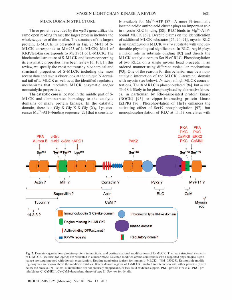
MYOSIN LIGHT CHAIN KINASE: A REVIEW 1681
BIOCHEMISTRY (Moscow) Vol. 81 No. 13 2016
MLCK DOMAIN STRUCTURE
Three proteins encoded by the mylk1 gene utilize the
same open reading frame; the larger protein includes the
whole sequence of the smaller. The structure of the largest
protein, L-MLCK, is presented in Fig. 2; Met1 of S-
MLCK corresponds to Met923 of L-MLCK; Met1 of
KRP/telokin corresponds to Met1761 of L-MLCK. The
biochemical structure of S-MLCK and issues concerning
its enzymatic properties have been review [6, 10]. In this
review, we specify the most noteworthy biochemical and
structural properties of S-MLCK including the most
recent data and take a closer look at the unique N-termi-
nal tail of L-MLCK as well as at the identified regulatory
mechanisms that modulate MLCK enzymatic and/or
noncatalytic properties.
The catalytic core is located in the middle part of S-
MLCK and demonstrates homology to the catalytic
domains of many protein kinases. In the catalytic
domain, there is a Gly-X-Gly-X-X-Gly-(X)14-Lys con-
sensus Mg2+-ATP-binding sequence [23] that is constant-
ly available for Mg2+-ATP [87]. A more N-terminally
located acidic amino acid cluster plays an important role
in myosin RLC binding [88]. RLC binds to Mg2+-ATP-
bound MLCK [89]. Despite claims on the identification
of additional MLCK substrates [76, 90, 91], myosin RLC
is an unambiguous MLCK in vivo substrate with unques-
tionable physiological significance. In RLC, Arg16 plays
a major role in substrate binding [92] and directs the
MLCK catalytic core to Ser19 of RLC. Phosphorylation
of two RLCs on a single myosin head proceeds in an
ordered manner using different molecular mechanisms
[93]. One of the reasons for this behavior may be a non-
catalytic interaction of the MLCK C-terminal domain
with myosin (see below). In vitro, at high MLCK concen-
trations, Thr18 of RLC is phosphorylated [94], but in vivo
Thr18 is likely to be phosphorylated by alternative kinas-
es, in particular, by Rho-associated protein kinase
(ROCK) [95] or zipper-interacting protein kinase
(ZIPK) [96]. Phosphorylation of Thr18 enhances the
activating effect of Ser19 phosphorylation [97], but
monophosphorylation of RLC at Thr18 correlates with
Fig. 2. Domain organization, protein–protein interactions, and posttranslational modifications of L-MLCK. The main structural elements
of L-MLCK (see inset for legend) are presented in a linear mode. Selected modified amino acid residues with suggested physiological signif-
icance are superimposed with domain organization. Residue numbering is given for human L-MLCK1 (NM_053025). Responsible modify-
ing enzymes are shown above the modified residues. Braces denote regions of L-MLCK involved in interaction with other proteins (listed
below the braces). (?) – site(s) of interaction are not precisely mapped and/or lack solid evidence support. PKG, protein kinase G; PKC, pro-
tein kinase C; CaMKII, Ca-CaM-dependent kinase of type II. See text for details.

1682 KHAPCHAEV, SHIRINSKY
BIOCHEMISTRY (Moscow) Vol. 81 No. 13 2016
smooth muscle relaxation [98], suggesting Thr18
monophosphorylation may not be sufficient for myosin II
Mg2+-ATPase activation in non-muscle cells.
The regulatory segment of MLCK flanks the catalyt-
ic core at its C-terminus and consists of partially overlap-
ping autoinhibitory and Ca2+/CaM-binding sites.
Initially, a pseudosubstrate model was proposed based on
similarity of the disposition of basic amino acids in the
autoinhibitory region and near the RLC phosphorylation
site [99]. However, the adjacent hydrophobic Tyr-Met-
Ala cluster appeared as the most important for MLCK
autoinhibition [100], indicating that the autoinhibitory
mechanism is not restricted to imitation of the RLC
sequence. Based on the MLCK autoinhibitory sequence,
Lukas et al. developed the Peptide Inhibitor of Kinase
(PIK) [101], which could penetrate through biological
membranes [102, 103]. More recently, several proteoly-
sis-resistant PIK analogs have been developed for use in
in vitro and in vivo studies [102, 104, 105].
MLCK is a Ca2+/CaM-dependent enzyme. Even at
basal intracellular Ca2+ concentrations, the C-terminal
domain of CaM seems to be constantly bound to MLCK
[106]. An increase in the Ca2+ level and subsequent Ca2+
binding at the N-terminal domain of CaM induces
rearrangements in both CaM and MLCK and makes the
MLCK active site available for RLC binding [107]. These
data suggest that an increase in Ca2+ and release of the
autoinhibitory segment are prerequisites for effective
binding of RLC-imitating MLCK peptide inhibitors (e.g.
peptide 11-19 [108]).
Finally, it should be noted that in vitro, the L-MLCK
substrate specificity, activation by Ca2+/CaM, and enzy-
matic properties are identical to those of S-MLCK [74].
The myosin-binding site, which is distinct from the
catalytic center, resides at the very C-terminus of the
MLCK molecule and is common for L-MLCK, S-
MLCK, and KRP/telokin. The acidic C-terminus of
KRP/telokin plays a major role in KRP/telokin binding
to myosin in vitro [109] to induce the myosin II unfolding
and filament formation [110]. Moreover, the KRP/
telokin C-terminus is responsible for competitive inhibi-
tion of RLC phosphorylation by MLCK [111] and alter-
native myosin II protein kinases [112].
IgG- and Fn-like domains. Adjacent to the N-termi-
nal end of the catalytic domain is a fibronectin type III
(Fn-like) domain of unknown function. Additionally,
in S-MLCK, there are three immunoglobulin G (IgG)
C2-type domains (IgG-like), each comprising approxi-
mately 100 amino acid residues. While two IgG-like
domains reside N-terminally to the Fn-like domain, the
third IgG-like domain resides at the C-terminus of the
MLCK molecule. The C-terminal IgG-like domain with
short N- and C-terminal flanking sequences is expressed
as an independent KRP/telokin. In the unique N-termi-
nal extension of L-MLCK, six additional IgG-like
domains are found [18, 28]. Functional roles of these
domains remain unclear, but they have been suggested to
contribute to protein–protein interactions of L-MLCK
directly or, by analogy with IgG-like domains of titin
[113], they may unfold reversibly, thereby increasing the
linear dimensions of L-MLCK and/or disclosing binding
sites for L-MLCK protein partners (see below).
KPV/A repeats are found in mammalian MLCKs
and reside more N-terminally to the first IgG-like
domain of S-MLCK. These are 12-amino acid repeats
containing conserved Lys-Pro-Val (KPV) and Lys-Pro-
Ala (KPA) triplets. The number of KPV/A repeats is
species-dependent and contributes to variation of the
MLCK molecular mass among species. The physiological
role of the KPV/A repeats remains unestablished.
The actin-binding domain of S-MLCK is located at
the very N-terminus of the molecule and consists of three
28-amino acid repeats with internal DFRxxL motifs play-
ing a leading role in interaction with actin [114]. Each
DFRxxL motif is believed to bind to an individual actin
monomer; thus, by three DFRxxL motifs, the S-MLCK
N-terminal region can cross-link individual actin fila-
ments in vitro [115]. The alternate binding/dissociation of
individual DFRxxL motifs has been suggested to provide
for migration of S-MLCK along actin filaments to expand
the effective area of the kinase [116]. On an actin filament,
sites of interaction with DFRxxL motifs are distinct from
the binding sites for myosin, tropomyosin, caldesmon,
and calponin [117]; thus, there are no steric obstructions
for the lateral migration of S-MLCK. CaM might influ-
ence the association of MLCK with actin through the sec-
ond CaM-binding site, which, according to in vitro stud-
ies, overlaps with the DFRxxL-containing region [118,
119]. Because of a relatively weak binding of DFRxxL
motifs to actin, in non-muscle cells, which are less “satu-
rated” with actomyosin structures, S-MLCK shows pre-
dominantly diffuse distribution and is readily washed out
upon cell permeabilization [115]. In the unique N-termi-
nus of L-MLCK, two additional DF/VRxxL motifs
enhance the affinity of L-MLCK to actin [74]. However,
an internal deletion or the five DFRxxL motifs did not
induce L-MLCK dissociation from the cytoskeleton
[115], indicating there is(are) an additional actin-binding
site(s). Indeed, an actin-binding site structurally different
from the DFRxxL-containing domain has been mapped
to the two first IgG-like domains [120, 121].
PROTEIN–PROTEIN INTERACTIONS
OF mylk1 GENE PRODUCTS
Actin, myosin, and CaM are unquestionable MLCK
partners with well-established physiological significance.
However, experimental data indicate there are additional
protein partners of MLCK (Fig. 2).
Tubulin, a structural component of microtubules,
interacts with L-MLCK in vitro and colocalizes with L-

MYOSIN LIGHT CHAIN KINASE: A REVIEW 1683
BIOCHEMISTRY (Moscow) Vol. 81 No. 13 2016
MLCK in transfected CV-1 cells [122]. Via its interaction
with tubulin, L-MLCK may provide an integration link
between the actin and tubulin cytoskeleton structures.
Supervillin is a scaffold membrane-bound protein
and interacts with actin, myosin, and several cytoskeletal
proteins. The supervillin N-terminal fragment (residues
1-174) interacts with the L-MLCK unique N-terminal
tail in vitro and colocalizes with activated myosin on the
cell periphery [123]. It was suggested that an inhibitory
effect of supervillin on cell spreading is brought about by
myosin activation and myosin filament formation in the
submembrane region of a cell. Inhibition of MLCK activ-
ity reverted the effects of supervillin.
Cortactin, an actin-binding cytoskeletal protein,
interacts with L-MLCK in endothelial cells. Antibody
against L-MLCK immunoprecipitated cortactin from
endothelial cells; moreover, stimulation of endothelial
cell tyrosine kinases increased the amount of precipitated
cortactin [124]. Phosphorylation of either cortactin or the
full-length L-MLCK (L-MLCK1) by c-Src tyrosine
kinase enhanced the interaction of cortactin and L-
MLCK1. Functionally, cortactin did not affect the activ-
ity of either MLCK isoform in vitro and, similarly, MLCK
did not affect the actin-binding properties of cortactin but
inhibited cortactin-stimulated activation of Arp2/3 [125].
While the cortactin SH3 domain is responsible for inter-
action with MLCK isoforms, putative cortactin-binding
sites are located within the DFRxxL domain of MLCK
[126] suggesting that the interaction might be regulated
by Ca2+/CaM, which binds to the same MLCK region
and may modulate MLCK interaction with actin. The
unique N-terminal tail of L-MLCK might contain addi-
tional cortactin-binding sites. A L-MLCK-dependent
increase in cortactin tyrosine phosphorylation in
endothelial cells may be due to recruiting of non-receptor
tyrosine kinases, in particular c-Src, via L-MLCK scaf-
folding activity [124] and/or due to L-MLCK-dependent
conformational changes in cortactin that facilitate its
interaction with c-Src. Involvement of L-MLCK in acti-
vation of tyrosine kinases was observed in neutrophils as
well [76].
Pyk2, a FAK-family cytoplasmic tyrosine kinase, is
recruited to β2-integrins and contributes to an adequate
integrin activation and neutrophil adhesion. In murine L-
MLCK–/– neutrophils, both Pyk2 activation and interac-
tion with β2-integrins were impaired. Direct interaction
of Pyk2 and L-MLCK is supported by coimmunoprecip-
itation studies and in vitro binding of purified Pyk2 and
the recombinant MLCK catalytic domain. Interestingly,
in neutrophils, the inhibition of L-MLCK enzymatic
activity attenuated Pyk2 activation; moreover, the MLCK
catalytic domain phosphorylated and activated Pyk2 in
vitro. These data suggest both enzymatic and noncatalyt-
ic activities of MLCK contribute to Pyk2 activation [76].
MIF (macrophage migration inhibition factor) has
been identified as an MLCK partner by the yeast two-
hybrid assay system. Interaction between the two proteins
was confirmed by coimmunoprecipitation of GST-tagged
MLCK fragment from BPAEC cell lysates and indirect
immunofluorescence data. Biochemical studies with
truncated mutants show that the L-MLCK N-terminal
515 amino acids were sufficient for MIF binding and sug-
gest that one MIF binding site on L-MLCK1 may reside
between amino acids 437-505, a sequence that is spliced
out in L-MLCK2. However, the functional significance
of the MIF/MLCK complex is unclear, because MIF did
not compete with c-Src-dependent L-MLCK phospho-
rylation within amino acids 415-500 and had no effect on
the interaction of L-MLCK with actin or cortactin [127].
14-3-3 scaffold. In the L-MLCK regions flanking
the first IgG-like domain, there are putative binding sites
for 14-3-3 proteins. Interaction of L-MLCK with 14-3-3
proteins is supported by an enrichment of two 14-3-3 iso-
forms (24 and 27 kDa) in the immunoprecipitate of the
FLAG-tagged L-MLCK fragment and ability of a 14-3-3
antagonist to abolish the attenuation of L-MLCK phos-
phorylation at Tyr464 [128]. Unfortunately, the putative
interaction of L-MLCK with 14-3-3 is based only on
indirect data and has not been confirmed by further stud-
ies.
Myosin light chain phosphatase (MLCP) is a dedicat-
ed MLCK antagonist and seems to be a protein partner of
mylk1 gene-derived proteins. Based on copurification of
MLCK and MLCP from smooth muscle, early reports
suggested that MLCK and MLCP exist in a complex
[129]. More recently, these indirect data gained support
from several experimental findings in KRP/telokin–/–
mice indicating a positive modulation of MLCP by
KRP/telokin [31]. Because in a myosin II molecule,
KRP/telokin shields RLC from activating protein kinases
[111, 112] and KRP/telokin does not bind to phosphory-
lated myosin [130], it has been suggested that modulation
of myosin dephosphorylation by KRP/telokin is due to its
direct or indirect effects on MLCP [31]. Recent physio-
logical and biochemical data are in favor of a hypothesis
that direct interaction of KRP/telokin with regulatory
MLCP subunit, MYPT1, abolishes the effect of inhibito-
ry MYPT1 phosphorylation [131]. The interaction can
probably be explained by some degree of similarity
between KRP/telokin and a small MLCP subunit (M20)
of unidentified function.
Using the yeast two-hybrid assay, coimmunoprecipi-
tation, or peptide matrices, large-scale screening studies
identified a score of putative MLCK protein partners;
however, additional efforts are needed to confirm the
interactions and identify the physiological significance of
the confirmed ones.
Enzymes that use MLCK isoforms and/or KRP/
telokin as substrates, e.g. c-Src tyrosine kinase [125],
could be, with certain limitations, considered as
MLCK/KRP protein partners. Because posttranslational
modifications (PTM) play an important role in regulation

1684 KHAPCHAEV, SHIRINSKY
BIOCHEMISTRY (Moscow) Vol. 81 No. 13 2016
of most if not all human proteins, below are summarized
the most noteworthy PTMs that have been identified in
the mylk1-derived proteins.
POSTTRANSLATIONAL MODIFICATIONS
OF mylk1 GENE PRODUCTS
According to the PhosphositePlus database
(http://www.phosphosite.org) [17], there are over 10
acetylation and 40 phosphorylation sites in L-MLCK. In
contrast, few methylation and ubiquitinylation sites have
been identified. For the great majority of L-MLCK
PTMs, the physiological significance remains unestab-
lished, and there are few reports on this issue.
Acetylation-dependent modulation of mylk1 gene-
encoded proteins. One of the earliest identified PTMs in
KRP/telokin was a constitutive acetylation of N-termini
of KRP/telokin isoforms that differed in the length of
their N-terminal ends [67]. The Met1 residue of S-
MLCK is acetylated as well [132]. These modifications
seem to protect KRP/telokin and S-MLCK from degra-
dation. Recently, hARD1 acetyltransferase was shown to
modulate L-MLCK activity [133]. It was demonstrated
that hARD1 binds within the L-MLCK region spanning
the fourth and fifth IgG-like domains and acetylates the
epsilon amino group of Lys608 (Fig. 2). In transfected
cells, hARD1 downregulated both RLC phosphorylation
and MLCK-dependent invasion and migration of
HT1080 cancer cells. Moreover, the Lys608Arg mutation
in L-MLCK completely abolished the effect of hARD1
on L-MLCK activity. Interestingly, the interaction of
hARD1 with L-MLCK required a prior activation of L-
MLCK by Ca2+/CaM, suggesting that there are complex
interactions between L-MLCK domains that are located
far from each other in the primary structure of the kinase.
Phosphorylation-dependent modulation of mylk1
gene-encoded proteins. Among the PTMs of the mylk1
gene-encoded proteins, phosphorylation-dependent
modulation has been much more extensively studied.
Proteomic techniques validated several in vivo phospho-
rylation sites of MLCK that partially matched the sites
identified in earlier in vitro studies (Fig. 2). Among these
is a functionally important MLCK phosphorylation in
the CaM-binding domain (designated the phosphoryla-
tion site A) [134]. In site A, phosphorylation at Ser1760
by protein kinases A, G, C (PKA, PKG, and PKC,
respectively), or Ca2+/CaM-dependent protein kinase II
(CaMKII) results in inhibition of both CaM binding and
RLC access to the MLCK catalytic center, which remains
closed by the MLCK autoinhibitory region [134-136].
The p21-activated protein kinase-2 (PAK2) phosphory-
lates the adjacent Ser1759 and brings about the same
inhibitory effect [137]. There is a commercial anti-
(P)Ser1760 antibody that allows monitoring the level of
Ser1760 phosphorylation and the corresponding level
of MLCK inhibition, but in isolated studies, e.g. in [83],
a positive reaction with this antibody is interpreted as
being associated with MLCK activation. Conclusions
drawn in reports of this kind should be considered with
caution.
In L-MLCK, several phosphorylation sites have
been identified that reside outside the catalytic domain
but affect the activity of the enzyme. In the unique N-
terminal tail of L-MLCK, c-Src-dependent phosphory-
lation at Tyr464 and Tyr471 in vitro increases the enzy-
matic activity of L-MLCK 2-3-fold [138]. These data
indicate that L-MLCK may adopt a “folded” tertiary
structure with its N- and C-terminal moieties coming
into contact. This hypothesis is supported by the above-
mentioned effects of Lys608 acetylation on L-MLCK
activity [133]. Unfortunately, no crystal structure is avail-
able for the full-length L-MLCK that would reflect the
intramolecular interactions of distantly located L-MLCK
domains.
Interestingly, both Tyr464 and Tyr471 are encoded
by a single exon that is spliced out in L-MLCK2 [28].
Correspondingly, c-Src does not phosphorylate and regu-
late L-MLCK2. In vitro, c-Abl tyrosine kinase phospho-
rylates up to 10 tyrosine residues in L-MLCK1, including
Tyr464, and it increases both L-MLCK1 catalytic activi-
ty and affinity to cortactin, as in the case with c-Src [139].
The same study reported autophosphorylation of L-
MLCK at 19 serine and threonine residues. In this regard,
it seems impossible to correlate the phosphorylation at a
given residue with changes in L-MLCK catalytic activity.
Moreover, phosphorylation of multiple residues may
result from nonspecific modification due to excess of the
protein kinase in in vitro experiments. This assumption is
favored by the fact that only two out of nine c-Abl in vitro
phosphorylation sites have been validated in cancer cells
by mass-spectrometry (see http://www.phosphosite.org).
In chicken L-MLCK, Ser149Asp mutation (homol-
ogous to Ser154 in human L-MLCK and Ser149 in
murine L-MLCK and identified as an in vivo phospho-
site) attenuated the binding of the actin-binding N-ter-
minal L-MLCK fragment to cytoskeletal structures in
transfected HeLa cells [120]. In vitro, Ser149 in avian L-
MLCK could be phosphorylated by PKA and Aurora B,
which show similar substrate specificity. The latter kinase
is active in mitosis and phosphorylates serine residues
within the N-terminal tail of L-MLCK in mitotic HeLa
cells [140]. In contrast, PKA may phosphorylate L-
MLCK in interphase cells. Thus, PKA and Aurora B
team up to represent a minimal set of protein kinases that
can modulate actin-binding properties of L-MLCK in
cells.
Most studies reporting L-MLCK phosphorylation in
cells and tissues have not established links between phos-
phorylation at individual L-MLCK amino acid residues
and alterations in cellular responses. Phosphorylation of
L-MLCK at tyrosine residues has been demonstrated in

MYOSIN LIGHT CHAIN KINASE: A REVIEW 1685
BIOCHEMISTRY (Moscow) Vol. 81 No. 13 2016
endothelial cells following stimulation with diperoxy-
vanadate, a potent tyrosine kinase activator and tyrosine
phosphatase inhibitor [124], and in fibroblasts following
transformation with constitutively activated mutant epi-
dermal growth factor receptor (v-ErbB) [141]. In both
these studies, the authors reported alterations in the acti-
vation status of contractile proteins in cells.
Phosphorylation of MLCK by MAP-kinases has been
associated with MLCK activation, an increase in myosin
RLC phosphorylation, and acceleration of cell migration
[142]. Because MAP-kinase consensus sites are located
outside the catalytic domain of MLCK, it seems likely
that the MAP-kinase-dependent activation of MLCK
required an interaction between distantly located MLCK
domains, much as suggested for the phosphorylation of
Tyr464 and Tyr471 and acetylation of Lys608 in the N-
terminal tail of L-MLCK.
Both KRP/telokin and the corresponding domain
within MLCK are multiply phosphorylated (see
http://www.phosphosite.org). Within the unstructured
N-terminus of KRP/telokin, eight of ten Ser/Thr
residues are phosphorylated, suggesting that this region
has an important regulatory role. However, there are no
indications on the functional properties that are regulat-
ed by phosphorylation of the KRP/telokin domain except
a report suggesting that PKA-dependent phosphorylation
in this domain intensifies the inhibitory effect of the site
A phosphorylation [136]. In KRP/telokin, PKA- and/or
MAP-kinase-dependent phosphorylation of the homolo-
gous site does not alter the inhibitory effects of
KRP/telokin on myosin phosphorylation and smooth
muscle contraction [112]. In isolated phasic smooth
muscle, KRP/telokin phosphorylation is increased dur-
ing contraction and slightly varies during the subsequent
relaxation [75]. Quantitative analysis of the phosphoryla-
tion dynamics at individual sites demonstrated that dur-
ing smooth muscle contraction, phosphorylation at Ser13
of KRP/telokin increases from basal 20 to 100%; howev-
er, the level of KRP/telokin phosphorylation at Ser19 did
not show significant changes during the contraction/
relaxation cycle and amounted to about 25% [143].
According to the model suggested by A. Somlyo’s group,
KRP/telokin activates MLCP and, in this way, con-
tributes to smooth muscle relaxation. Moreover, the
effect is augmented by PKG-dependent KRP/telokin
phosphorylation at Ser13 [144, 145] because phospho-
KRP/ telokin binds to and activates MYPT1 by overrid-
ing the preexisting inhibitory phosphorylation of MYPT1
[131]. It should be noted, however, that direct interaction
with MYPT1 in vitro was shown for the phospho-mim-
iсking KRP/telokin mutant (Ser13Asp), whereas the
behavior of KRP/telokin phosphorylated at Ser13 was
not studied.
In summary, all proteins that derive from the mylk1
gene are extensively labeled by multiple PTMs. Isolated
PTMs have been shown to regulate MLCK enzymatic
activity, which affects the activation status of myosin II
ATPase and, respectively, parameters of cell motility. For
most PTMs that are found in MLCK and/or KRP/
telokin, the functional significance remains unclear.
ROLE OF MLCK/KRP IN PHYSIOLOGICAL
CELLULAR RESPONSES
The roles of MLCK in physiological processes are
predominantly associated with myosin II activation,
which drives a great variety of cellular responses. Thus,
MLCK contributes to a variety of apparently unrelated
processes, but all these depend on the motility apparatus
of the cell. MLCK is a Ca2+/CaM-dependent myosin II
activator and is activated by an increase in intracellular
Ca2+ level that, in turn, is evoked by Ca2+-mobilizing
agents like histamine, thrombin, bradykinin, bombesin,
cholecystokinin, acetylcholine, catecholamines, fMet-
Leu-Phe, oxidants, etc. Apart from MLCK protein
kinase activity, interactions of MLCK with its protein
partners (see section “Protein–Protein Interactions of
mylk1 Gene Products”) may contribute to the MLCK-
dependent functional responses of the cell.
Cell adhesion and migration. Adhesion and migration
are fundamental features of substrate-dependent cells.
MLCK is involved in both these cellular processes.
Conjointly with focal adhesion kinase, protein kinase c-
Src, and extracellular-regulated kinases (Erk), MLCK
participates in both formation and disassembly of focal
adhesions at the cell front and provides for onward migra-
tion [146]. Pharmacological inhibition of MLCK and
CaM changes fibroblast-populated collagen lattice con-
traction, cell migration, focal adhesion formation, and
wound contraction [147]. The Erk pathway regulates F9
parietal endoderm cell migration by affecting the forma-
tion of focal adhesions and lamellipodia via modulating
MLCK activity [148].
In fibroblasts, MLCK-dependent focal adhesion for-
mation proceeds at the cell periphery; in contrast,
ROCK-dependent focal adhesions are located more cen-
trally. Selective inhibition of these protein kinases results
in disassembly of adhesions either in the center or at the
periphery and alters the migration pattern of cells.
MLCK-inhibited cells generated membrane protrusions
all around the cell, turned more frequently, and migrated
less effectively compared to ROCK-inhibited cells, which
moved faster and straighter [149]. Other studies in differ-
ent cell models support the conclusion regarding the spa-
tial segregation of MLCK- and ROCK-dependent activa-
tion of myosin II [150-153].
On the other hand, accumulating data suggest that
noncatalytic properties of MLCK, e.g. interaction with
actin and other protein partners, play an important role in
migrating cells [76, 81, 154]. Indeed, as noted above,
MLCK may act as a scaffold and/or integrate cytoskele-

1686 KHAPCHAEV, SHIRINSKY
BIOCHEMISTRY (Moscow) Vol. 81 No. 13 2016
tal structures [122-124]. In isolated model systems, e.g. in
L-MLCK–/– neutrophils, an assay of neutrophil transmi-
gration across endothelial monolayer revealed that an
interaction between L-MLCK and c-Src and Pyk2 tyro-
sine kinases and subsequent recruitment of Pyk2 to β2-
integrins was a sufficient MLCK-dependent event, while
ROCK-dependent myosin activation assured motile
activity [76]. Meanwhile, data from experiments with
“substitution” of the MLCK catalytic activity by its
actin-binding activity by overexpression of kinase-dead
MLCK or its individual domains [81, 154] should be con-
sidered with caution. Ectopic overexpression of MLCK
actin-binding domains would obviously stabilize the actin
cytoskeleton even when the MLCK catalytic activity is
lacking [122], and the motile component would be pro-
vided by alternative myosin-activating kinases like
ROCK, ZIPK, citron kinase, etc.
Proliferation. MLCK takes part in the control of cell
division. In proliferating rodent hepatocytes, inhibition
of MLCK catalytic activity or downregulation of MLCK
expression revealed a role for MLCK in the control of cell
transition from late G1 to S phase involving Erk2-
dependent p70S6 kinase activation [155]. Similarly,
downregulation of MLCK expression or inhibition of
MLCK catalytic activity slows the proliferation of various
cancer cells [156-158]. In contrast, MLCK downregula-
tion in GbaSM-4 smooth muscle cells increased the pro-
liferation rate and shortened the doubling period of the
population [159]. Effects of MLCK on mitosis seem to
depend on the cytoskeleton status, activity of intracellu-
lar signaling pathways that affect MLCK activity, and the
levels of MLCK protein partners. These parameters may
vary significantly in normal versus tumor cells.
In mitotic HeLa cells, L-MLCK is concentrated in
cortical cytoplasm during metaphase and in the cleavage
furrow during anaphase and telophase [160]. Targeting of
L-MLCK to the cleavage furrow required both the actin-
binding DFRxxL motifs, which are located in the central
part of the MLCK molecule, and additional sequences
from the L-MLCK N-terminus. L-MLCK targeting to
the cleavage furrow seems obvious, because the cleavage
furrow is a typical actomyosin structure. Both L-MLCK
catalytic and noncatalytic domains may contribute to
cleavage furrow formation and contraction. Additionally,
in the same study, the L-MLCK N-terminus disrupted
normal spindle morphology during mitosis. The authors
suggested the identification of a novel regulatory role of
L-MLCK during mitosis, but there should be considered
possible artefact consequences of overexpression of the L-
MLCK N-terminus, which interacts with tubulin in vitro
and microtubules in cells [122].
Endocytosis. Convincing experimental evidence
has accumulated in support of active involvement of
MLCK in endocytosis. Membrane internalization
occurs for volume recovery in response to osmotic
swelling. MLCK and myosin II localized with actin to
swelling-induced membrane blebs just before retraction;
inhibition of MLCK induced persistent blebbing and
attenuated cell volume recovery. At the sites of mem-
brane internalization, MLCK localized to dynamic
actin-coated rings and patches, which contained also c-
Src, cortactin, and dynamin; inhibition of either MLCK
or c-Src altered the lifetimes of these actin-coated
structures [161].
Endocytosis of synaptic vesicles recycles vesicle
membranes to maintain synaptic transmission. The enzy-
matic activity of MLCK is essential in this process.
MLCK inhibition or downregulation of MLCK expres-
sion slowed vesicle endocytosis and prevented depolariza-
tion-induced phosphorylation of RLC in rat hippocam-
pal boutons. Similarly, inhibition of myosin II impaired
endocytosis; moreover, the effect was shared by blockers
of Ca2+-channels and CaM [162]. Earlier, a similar effect
was observed at the calyx of Held synapse in rats [163].
The authors assumed that MLCK-dependent myosin
phosphorylation and activation of synaptic vesicle endo-
cytosis is a general neuronal process that facilitates recy-
cling of vesicle membranes under enhanced neuronal
activity.
Exocytosis. It is widely accepted that MLCK plays a
role in exocytosis. In 1994, the essential role of MLCK in
ATP-dependent initiation of Ca2+-induced release of cat-
echolamines from adrenal chromaffin cells was reported
[164]. Afterwards, involvement of MLCK in exocytosis/
secretion was demonstrated in other cell types. In pancre-
atic β-cells, via activation of non-muscle myosin IIA,
MLCK modulates the translocation of secretory granules
and enhances insulin release [165]. In agonist-stimulated
platelets, MLCK cooperates with ROCK to activate
myosin II and induce platelet contraction and dense
granule secretion [166].
In primary alveolar type II cells, L-MLCK and
ROCK translocate to actin-coated plasma membrane-
fused lamellar bodies and activate myosin II on actin
coats. For efficient actin coat contraction, cofilin-1 and
α-actinin are recruited to the surface of secretory gran-
ules to promote active extrusion of cargo [167]. While
cofilin-1 regulates actin depolymerization on the contact
surface of a lamellar granule with the plasma membrane
to promote fusion, MLCK- and ROCK-dependent con-
traction of other parts of the actin coat probably pro-
motes extrusion of cargo (surfactant components) via the
pore.
The outlined scheme of molecular events during the
late phases of exocytosis is in concert with experimental
data and, thus, may be true regardless the exocytosis
type – whether it is a release of catecholamines or insulin,
externalization of receptors or glucose transporters,
extrusion of Weibel–Palade or lamellar bodies, etc. In
every instance, MLCK seems to activate myosin on the
granules to be extruded, but an effect of scaffolding activ-
ity of MLCK cannot be excluded.

MYOSIN LIGHT CHAIN KINASE: A REVIEW 1687
BIOCHEMISTRY (Moscow) Vol. 81 No. 13 2016
Regulation of mechanical properties of cells. Cells are
characterized by a certain degree of stiffness, which under
normoosmotic conditions depends presumably on the
cytoskeleton and its remodeling activity. In fibroblasts
overexpressing constitutively active S-MLCK, cytoskele-
tal stiffness was increased two-fold and balanced by
microfilament depolymerization with cytochalasin D
[168]. Recent study in L-MLCK–/– mice demonstrated a
reduced flow-mediated NO-dependent dilation of small
arteries that had L-MLCK-deficient endothelium. In
wild-type mice, an inhibitor of MLCK produced a simi-
lar effect [169]. Thus, MLCK downregulation/inhibition
impaired the sensitivity of endothelial NO-synthase to
shear stress.
Using atomic force microscopy (AFM), Dudek et al.
provided evidence supporting an important role of the c-
Abl/cortactin/L-MLCK protein complex in both reorga-
nization of cortical actin in endothelial cells and in a
sphingosine-1-phosphate-induced 30% increase in cell
stiffness [139]. This study did not show that L-MLCK
directly regulates endothelial cell stiffness, but further
studies supported this hypothesis. Indeed, AFM revealed
that wild-type endothelial cells are stiffer compared to
endothelial cells from L-MLCK–/– mice. Moreover,
ROCK cooperates with L-MLCK to maintain endothe-
lial cell stiffness [170, 171]. It is to be established whether
noncatalytic domains of L-MLCK play a role in control-
ling cell stiffness. Potential cross-linking properties of L-
MLCK, which are realized via distantly positioned actin-
binding sites including those subject to phosphorylation-
dependent modulation [120], may affect the cytoskeleton
stiffness and the cell elastic modulus.
Epithelial and endothelial barriers. Epithelial and
endothelial cells form tight semipermeable monolayers to
allow passage of low molecular weight substances while
preventing leakage of proteins [172]. The cytoplasmic
domains of intercellular contact proteins interact with the
actin cytoskeleton and meet with the actomyosin-
induced drag force. Many natural stressors (histamine,
thrombin, VEGF, TNFα, bacterial lipopolysaccharide,
reactive oxygen species, excessive mechanical stress, etc.)
evoke a disassembly of intercellular contacts in endothe-
lium and epithelium and activate actomyosin-dependent
contraction. In a number of situations, actomyosin acti-
vation in epithelium and endothelium is mediated by L-
MLCK catalytic activity [1, 173]. Contraction drives the
adjacent cells apart and hampers rapid contact recovery,
which is achieved via cell spreading. Moreover, MLCK
and myosin II accumulation in lamellipodia decreased
lamellipodial lifetime and size, and stimulated retraction
[153]. Thus, in endothelium and epithelium, L-MLCK is
engaged in cell barrier destabilization and hyperperme-
ability.
On the other hand, in a complex with c-Abl tyrosine
kinase and cortactin, L-MLCK is engaged in remodeling
of the cortical actin in endothelium in response to sphin-
gosine-1-phosphate, which is a platelet-derived barrier
protective agent [139]. In this case, noncatalytic scaffold-
ing and cross-linking activities of L-MLCK may play a
role in the barrier protection, but focused studies are
lacking. The L-MLCK–/– mice were less susceptible to a
stress-induced endothelial and epithelial barrier dysfunc-
tion [102, 105, 174, 175]; conversely, overexpression of L-
MLCK in murine endothelium enhanced its permeabili-
ty to macromolecules [176].
Smooth muscle contraction. In contrast to S-MLCK,
which initiates smooth muscle contraction via activation
of myosin II, KRP/telokin seems to play an antagonistic
role. Through the binding to myosin heads next to the
RLC-binding site, KRP/telokin hinders RLC phospho-
rylation by myosin-activating protein kinases [112].
Additionally, KRP/telokin may be involved in MLCP
activation. Intestinal smooth muscle of KRP/telokin–/–
mice exhibited an approximately 30% decrease in MLCP
activity accompanied by an increase in RLC phosphory-
lation [31]. Thus, in smooth muscle, independently of the
molecular mechanism, KRP/telokin attenuates myosin
activation and promotes relaxation. It remains to be
established whether the KRP/telokin domain of MLCK
plays a similar role.
ROLE OF MLCK IN PATHOLOGICAL PROCESSES
Endothelial/epithelial barrier dysfunction. Dysfunc-
tion of endothelial/epithelial barrier occurs in multiple
pathological states in humans, including sepsis, myocar-
dial infarction, stroke, wet macular degeneration, trau-
matic brain injury, and pancreatitis. Alterations in vascu-
lar permeability and lung and brain edema are complica-
tions of aggressive treatment for cancer, poisoning with
pulmonotoxic substances, thermal burns of the body, tis-
sue crashing, high altitude hypoxic exposure, and other
conditions. Alterations of intestinal epithelium perme-
ability are found in Crohn disease, celiac disease, autoim-
mune and inflammatory processes in the gut, infections,
intestinal obstruction, and poisoning.
In mice, TNFα activates intestinal epithelial
MLCK, which initiates caveolin-dependent endocytosis
of occludin, the principal component of tight junctions in
the gut epithelium [177]. Occludin endocytosis could be
one of the mechanisms increasing intestinal epithelium
permeability in response to various stress factors that
increase TNFα levels in the organism.
A MLCK-dependent mechanism is apparently
involved in the penetration of commensal bacteria in
intestinal enterocytes in the presence of IFNγ. In such
conditions, myosin II phosphorylation is increased in the
terminal web, and so-called arches are formed. The fan-
ning of brush border takes place, which is followed by
transcytosis of bacterial cells. Introduction of an MLCK
inhibitor into intestinal lumen or the use of anti-IFNγ

1688 KHAPCHAEV, SHIRINSKY
BIOCHEMISTRY (Moscow) Vol. 81 No. 13 2016
antibodies blocks the penetration of bacteria into entero-
cytes. IFNγ-induced bacterial translocation is not
observed in L-MLCK–/– mice, confirming the participa-
tion of MLCK in this process [178].
In several studies that used L-MLCK–/– mice and
MLCK inhibitors (small organic molecules and/or cell-
penetrating peptides), suppression of MLCK activity
improved the integrity of the endothelial/epithelial barri-
er, reduced the transmigration of immune cells in tissues,
and minimized clinical signs of pathology [1, 76, 104,
105, 174, 179-182].
Polymorphisms in the mylk1 gene are shown to asso-
ciate with predisposition of various human populations
(African Americans, African Caribbeans, Caucasians,
Spanish descent families) to the development of acute
lung injury, sepsis, and asthma [5]. These genetic varia-
tions may alter expression levels of MLCK in endotheli-
um and epithelium of the lung and could be responsible
for the increased content of this protein in the airway
smooth muscles of asthmatic patients [183]. Several poly-
morphisms in the mylk1 gene are associated with aortic
dissection and familial thoracic aortic aneurysm in
humans [66].
Atherosclerosis. In ApoE–/–/L-MLCK–/– mice fed
high fat diet, the size of aortic lesions is reduced as well as
the content of lipids and macrophages in these lesions
[77]. The authors link the alterations to the reduced per-
meability of aortic endothelium in these animals and
confirm the suggestion by direct experiments. At the same
time, they notice a decrease in c-Src phosphorylation in
L-MLCK–/– endothelium following stimulation with
thrombin [77] and relate the effect to noncatalytic activi-
ty of MLCK. Confirming the earlier report [76], they
demonstrated a slower transmigration rate of L-MLCK-
deficient monocytes through the endothelium and attrib-
uted the effect to the scaffold activity of L-MLCK.
Insulin resistance and diabetes. MLCK plays an
important role in insulin signaling, the development of
insulin resistance, and in diabetes. Inhibition of myosin II
and MLCK decreased glucose-stimulated insulin secre-
tion from pancreatic beta cells, which was associated with
shortening of peripheral actin stress fibers, and reduced
numbers of focal adhesions and insulin granules near the
basal membrane [165]. Thus, MLCK catalytic activity is
required for the modulation of insulin secretion by the
pancreas.
Interaction of insulin with insulin receptors on the
surface of skeletal muscle and adipose cells assumes the
traverse of insulin from the bloodstream into tissue
through the endothelial barrier. Transport of insulin
across microvascular endothelium is accomplished by
transcytosis using clathrin-coated vesicles [184]. It is
known that myosin II participates in exocytosis of such
vesicles [185], and MCLK may regulate myosin activity.
Limited insulin granule exocytosis by endothelial cells
due to alterations in the MLCK–myosin II system may
therefore be a factor leading to insulin resistance.
Decreased insulin concentration at the surface of the glu-
cose-accumulating cells will result in hyperglycemia and
reactive hyperinsulinemia.
Under physiological conditions, insulin stimulates
the incorporation of GLUT4 glucose transporters in plas-
ma membranes of adipocytes and skeletal myocytes. This
process is a form of exocytosis; it depends on myosin IIA
and myosin-activating protein kinases such as MLCK.
Active (acto)myosin is required for the fusion of GLUT4-
loaded vesicles with plasma membrane and for further
maintenance of GLUT4 activity [83, 186, 187]. A
decrease in efficiency of GLUT4 in adipocytes leads to
hyperglycemia and the development of insulin resistance.
Persisting hyperglycemia typical for diabetes causes
the accumulation of so-called advanced glycation end-
products (AGE) such as albumin and hemoglobin that are
non-enzymatically modified by glucose. AGE bind to
their receptors (RAGE) on endothelium and induce its
hyperpermeability. Because MLCK is involved in
endothelial barrier dysfunction, inhibition of catalytic
activity/expression of MLCK attenuates edemagenic
effects of AGE and retards the development of diabetic
vasculopathies [188].
Additionally, hyperglycemia and hyperlipidemia
increase the expression of MLCK and the accumulation
of phosphorylated (inhibited) MYPT1 in aortic endothe-
lium of rats with streptozotocin-induced diabetes. These
alterations favor the contractile phenotype and hyperper-
meability of endothelium. These effects are largely
reversed by melatonin administered to the animals [189].
In contrast, insulin restores the depressed expression of
MLCK in the stomach and in the intestine of streptozo-
tocin-diabetic rats. The decreased MLCK content in
these organs is a possible cause of the delayed gastroin-
testinal passage of food typical for these animals [190].
Finally, a recent report states that MLCK concentra-
tion in the serum of patients with type 2 diabetes is signif-
icantly higher than in the serum of healthy donors. Thus,
MLCK may soon become a novel biomarker of type 2
diabetes [191]. Perhaps MLCK immunoreactivity is
increased in diabetic patients because of persistent dam-
age of vascular endothelium due to oxidative stress. The
latter condition might be augmented by endothelial L-
MLCK through the modulation of cortactin–p47(phox)
interaction, which is required for the assembly and acti-
vation of endothelial NADPH oxidase [192].
Cancer. Since MLCK is actively involved in physio-
logical processes of cell adhesion, migration, and prolif-
eration, alterations of its expression and activity regula-
tion during oncogenic transformation of cells may be
principal determinants of migratory and invasive poten-
tial of these cells, thereby affecting the dynamics of the
disease and tactics of treatment. Studies show that
MLCK is associated with various types of cancer, includ-
ing breast, lung, prostate, colon cancer, etc. [5].

MYOSIN LIGHT CHAIN KINASE: A REVIEW 1689
BIOCHEMISTRY (Moscow) Vol. 81 No. 13 2016
Decreasing MLCK content in normal breast epithe-
lial cells leads to alterations in cell–cell interactions and
activates invasive behavior of these cells. MLCK content
in invasive breast cancer cells is lower than in noninvasive
breast cancer cells, and low levels of MLCK correlate
with poor prognosis of HER2-positive breast cancer
patients [85]. At the same time, other reports demon-
strate an opposite relationship between the content/activ-
ity of MLCK and the invasiveness of cancer cells [61,
193] that may reflect specific features of cells studied by
various groups. Gene expression profiles in various cancer
cells, even those derived from the same organ, may differ
significantly; accordingly, cellular reactions may differ as
well. Acetylation of Lys608 in L-MLCK by acetyltrans-
ferase hARD1 inactivates L-MLCK and leads to
decreased myosin RLC phosphorylation in cancer cells
and inhibition of cell migration and invasion [133].
MLCK is activated locally and activates actomyosin
contraction in vascular endothelium, through which can-
cer cells invade the vasculature. Inhibition of myosin
phosphorylation in endothelial cells attenuates
transendothelial but not paracellular invasion of cancer
cells [194].
MLCK as a molecular target for drug development.
Due to the involvement of MLCK in major pathologies,
many researchers consider this protein as a promising
molecular target for the development of new drugs with
wide application potential. Still, progress in this direction
may encounter significant problems associated with
potential negative influence of these compounds on phys-
iological processes regulated by MLCK. In particular,
inhibition of catalytic activity of L-MLCK in endothe-
lial/epithelial cells could suppress the activity of smooth
muscle S-MLCK and, possibly, other MLC kinases
(skeletal, cardiac) in the absence of technologies for tar-
geted drug delivery. Selective modulation of MLCK
expression in a particular cell type will help understand-
ing the tissue-specific regulation of the mylk1 gene multi-
ple promoters. Focused manipulation of noncatalytic
activities of MLCK will require detailed analysis of
MLCK interaction with partner proteins and will critical-
ly depend on effective methods of drug targeting.
Concluding remarks. For several decades, MLCK has
been attracting persistent interest, which highlights a
widely appreciated biomedical role of the enzyme and
indicates that many elusive issues are still to be discov-
ered. These concern the organization and regulation of
the mylk1 gene, structural and functional properties of the
RNA and protein products that originate from the gene,
and the role of these products in (patho)physiology of the
cell and organism. We now know quite a lot about the dif-
ferential regulation of the mylk1 gene, but there are only
a few studies available concerning the links between the
mylk1 gene polymorphisms and liability to diseases.
Moreover, the list of mylk1 polymorphism-associated
pathologies seems far from being complete.
Still, the initial steps have been made in identifica-
tion of protein–protein interactions that may affect enzy-
matic/noncatalytic properties of L-MLCK, S-MLCK,
and KRP/telokin. The unique N-terminus may target L-
MLCK to specific intracellular domains and govern the
participation of the kinase in various cellular reactions.
Considering the confirmed involvement of L-MLCK in
many physiological processes in cells and tissues, identi-
fication of L-MLCK protein partners and deciphering
the functional role and regulation of these interactions
becomes an urgent fundamental issue with promising per-
spectives in applied fields.
Posttranslational modifications are well known to
affect protein structure and function, but for the mylk1
gene products, a regulatory role of PTMs is far from being
completely elucidated; moreover, further questions are
raised by reports that describe the relationships between
particular PTMs and alterations of mylk1 protein fea-
tures. In part, this is due to incomplete understanding of
the L-MLCK structure and interactions with other pro-
teins. Mapping of intramolecular interaction sites and
sites of interaction with protein partners in the MLCK
sequence is a clue to understanding the roles of particular
PTMs. Conversely, localization of PTMs within the
MLCK structure may reflect the sites essential for various
interactions and regulation in vivo. Thus, solving a “PTM
problem” is an actual issue for further studies of L-
MLCK to expand our knowledge about the role of the
enzyme in (patho)physiology of the cell. Progress in this
direction depends mainly on gaining new insights into
MLCK molecular anatomy and identification of partner
binding sites.
A range of MLCK-dependent cellular and whole
organism reactions has been outlined by studies utilizing
molecular genetics and pharmacological methods for con-
trolling either expression or activity of MLCK. This range,
predictably, has included various aspects of cell motility.
Later, alterations in cellular motility, which underlie car-
diovascular, oncological, infectious, and other diseases,
diabetes, and trauma, have been often associated with
impairment in the “MLCK/partners” molecular system.
These findings suggest MLCK as a promising molecular
target for development of novel drugs. The primary steps
have already been made in this direction. However, there
are still many objective problems due to ubiquitous
expression of MLCK and involvement of the enzyme in
important physiological processes. In this regard, a critical
issue for success is an understanding of the mechanisms
that govern tissue-specific expression of the mylk1 gene,
MLCK interactions and PTMs, as well as the develop-
ment of targeted delivery of MLCK-selective agents (syn-
thetic inhibitors, interfering peptides, molecular genetic
constructs, etc.) in vivo. Future studies along these
avenues of research should facilitate translation of the fun-
damental knowledge about MLCK into technologies and
products urgently needed by clinical medicine.

1690 KHAPCHAEV, SHIRINSKY
BIOCHEMISTRY (Moscow) Vol. 81 No. 13 2016
Note added in proof
When this review was ready for publication, Gaceb et
al. reported that circulating microvesicles from L-
MLCK–/– mice exert a protective action against lipo-
polysaccharide-induced inflammatory effects both in vivo
and in cultured aortic endothelial cells (Gaceb, A.,
Vergori, L., Martinez, M. C., and Andriantsitohaina, R.
(2016) Activation of endothelial pro-resolving anti-
inflammatory pathways by circulating microvesicles from
non-muscular myosin light chain kinase-deficient mice,
Front. Pharmacol., 7, 322; doi:10.3389/fphar.2016.00322).
Acknowledgements
This work was financially supported by the Russian
Foundation for Basic Research (projects Nos. 16-04-
01742 and 14-04-01813) and by the Russian Science
Foundation (project No. 14-35-00026, section “Role of
MLCK/KRP in Physiological Cellular Responses, sub-
section “Exocytosis”; section “Role of MLCK in
Pathological Processes”, subsection “Insulin resistance
and diabetes”).
REFERENCES
1. Cunningham, K. E., and Turner, J. R. (2012) Myosin light
chain kinase: pulling the strings of epithelial tight junction
function, Ann. N. Y. Acad. Sci., 1258, 34-42.
2. Herring, B. P., El-Mounayri, O., Gallagher, P. J., Yin, F.,
and Zhou, J. (2006) Regulation of myosin light chain
kinase and telokin expression in smooth muscle tissues, Am.
J. Physiol. Cell Physiol., 291, C817-827.
3. Kamm, K. E., and Stull, J. T. (2001) Dedicated myosin
light chain kinases with diverse cellular functions, J. Biol.
Chem., 276, 4527-4530.
4. Lukas, T. J., and Shirinsky, V. P. (2012) MYLK (Myosin
Light Chain Kinase), in Encyclopedia of Signaling Molecules
(Choi, S., ed.) Springer, New York, pp. 1160-1165.
5. Shen, K., Wang, T., and Garcia, J. G. (2012) MYLK
(myosin light chain kinase), Atlas Genet. Cytogenet. Oncol.
Haematol., 16, 901-908.
6. Gallagher, P. J., Herring, B. P., and Stull, J. T. (1997) Myosin
light chain kinases, J. Muscle Res. Cell Motil., 18, 1-16.
7. Khapchaev, A. Y., Shirinsky, V. P., and Vorotnikov, A. V.
(2003) Structure, properties and regulation of the myosin
light chain kinase genetic locus protein products, Usp. Biol.
Khim., 43, 365-420.
8. Gao, Y., Ye, L. H., Kishi, H., Okagaki, T., Samizo, K.,
Nakamura, A., and Kohama, K. (2001) Myosin light chain
kinase as a multifunctional regulatory protein of smooth
muscle contraction, IUBMB Life, 51, 337-344.
9. Takashima, S. (2009) Phosphorylation of myosin regulato-
ry light chain by myosin light chain kinase, and muscle
contraction, Circ. J., 73, 208-213.
10. Hong, F., Haldeman, B. D., Jackson, D., Carter, M.,
Baker, J. E., and Cremo, C. R. (2011) Biochemistry of
smooth muscle myosin light chain kinase, Arch. Biochem.
Biophys., 510, 135-146.
11. Dabrowska, R., Sherry, J. M., Aromatorio, D. K., and
Hartshorne, D. J. (1978) Modulator protein as a compo-
nent of the myosin light chain kinase from chicken gizzard,
Biochemistry, 17, 253-258.
12. Pires, E., Perry, S. V., and Thomas, M. A. (1974) Myosin
light-chain kinase, a new enzyme from striated muscle,
FEBS Lett., 41, 292-296.
13. Chan, J. Y., Takeda, M., Briggs, L. E., Graham, M. L., Lu,
J. T., Horikoshi, N., Weinberg, E. O., Aoki, H., Sato, N.,
Chien, K. R., and Kasahara, H. (2008) Identification of
cardiac-specific myosin light chain kinase, Circ. Res., 102,
571-580.
14. Roush, C. L., Kennelly, P. J., Glaccum, M. B., Helfman,
D. M., Scott, J. D., and Krebs, E. G. (1988) Isolation of
the cDNA encoding rat skeletal muscle myosin light chain
kinase. Sequence and tissue distribution, J. Biol. Chem.,
263, 10510-10516.
15. Birukov, K. G., Schavocky, J. P., Shirinsky, V. P., Chibalina,
M. V., Van Eldik, L. J., and Watterson, D. M. (1998)
Organization of the genetic locus for chicken myosin light
chain kinase is complex: multiple proteins are encoded and
exhibit differential expression and localization, J. Cell.
Biochem., 70, 402-413.
16. Potier, M. C., Chelot, E., Pekarsky, Y., Gardiner, K.,
Rossier, J., and Turnell, W. G. (1995) The human myosin
light chain kinase (MLCK) from hippocampus: cloning,
sequencing, expression, and localization to 3qcen-q21,
Genomics, 29, 562-570.
17. Hornbeck, P. V., Zhang, B., Murray, B., Kornhauser, J. M.,
Latham, V., and Skrzypek, E. (2015) PhosphoSitePlus,
2014: mutations, PTMs and recalibrations, Nucleic Acids
Res., 43, D512-520.
18. Watterson, D. M., Collinge, M., Lukas, T. J., Van Eldik, L.
J., Birukov, K. G., Stepanova, O. V., and Shirinsky, V. P.
(1995) Multiple gene products are produced from a novel
protein kinase transcription region, FEBS Lett., 373, 217-
220.
19. Ito, M., Dabrowska, R., Guerriero, V., Jr., and Hartshorne,
D. J. (1989) Identification in turkey gizzard of an acidic
protein related to the C-terminal portion of smooth muscle
myosin light chain kinase, J. Biol. Chem., 264, 13971-
13974.
20. Ye, D., and Ma, T. Y. (2008) Cellular and molecular mech-
anisms that mediate basal and tumour necrosis factor-
alpha-induced regulation of myosin light chain kinase gene
activity, J. Cell. Mol. Med., 12, 1331-1346.
21. Shimizu, Y., Camp, S. M., Sun, X., Zhou, T., Wang, T., and
Garcia, J. G. (2015) Sp1-mediated nonmuscle myosin light
chain kinase expression and enhanced activity in vascular
endothelial growth factor-induced vascular permeability,
Pulm. Circ., 5, 707-715.
22. Chang, A. N., Mahajan, P., Knapp, S., Barton, H.,
Sweeney, H. L., Kamm, K. E., and Stull, J. T. (2016)
Cardiac myosin light chain is phosphorylated by
Ca2+/calmodulin-dependent and -independent kinase
activities, Proc. Natl. Acad. Sci. USA, 113, E3824-3833.
23. Guerriero, V., Jr., Russo, M. A., Olson, N. J., Putkey, J. A.,
and Means, A. R. (1986) Domain organization of chicken
gizzard myosin light chain kinase deduced from a cloned
cDNA, Biochemistry, 25, 8372-8381.

MYOSIN LIGHT CHAIN KINASE: A REVIEW 1691
BIOCHEMISTRY (Moscow) Vol. 81 No. 13 2016
24. Garcia, J. G., Lazar, V., Gilbert-McClain, L. I., Gallagher,
P. J., and Verin, A. D. (1997) Myosin light chain kinase in
endothelium: molecular cloning and regulation, Am. J.
Respir. Cell Mol. Biol., 16, 489-494.
25. Yin, F., Hoggatt, A. M., Zhou, J., and Herring, B. P. (2006)
130-kDa smooth muscle myosin light chain kinase is tran-
scribed from a CArG-dependent, internal promoter within
the mouse mylk gene, Am. J. Physiol. Cell Physiol., 290,
C1599-1609.
26. Collinge, M., Matrisian, P. E., Zimmer, W. E., Shattuck, R.
L., Lukas, T. J., Van Eldik, L. J., and Watterson, D. M.
(1992) Structure and expression of a calcium-binding pro-
tein gene contained within a calmodulin-regulated protein
kinase gene, Mol. Cell. Biol., 12, 2359-2371.
27. Gallagher, P. J., and Herring, B. P. (1991) The carboxyl ter-
minus of the smooth muscle myosin light chain kinase is
expressed as an independent protein, telokin, J. Biol.
Chem., 266, 23945-23952.
28. Lazar, V., and Garcia, J. G. (1999) A single human myosin
light chain kinase gene (MLCK; MYLK), Genomics, 57,
256-267.
29. Hoggatt, A. M., Simon, G. M., and Herring, B. P. (2002)
Cell-specific regulatory modules control expression of
genes in vascular and visceral smooth muscle tissues, Circ.
Res., 91, 1151-1159.
30. Herring, B. P., and Smith, A. F. (1997) Telokin expression
in A10 smooth muscle cells requires serum response factor,
Am. J. Physiol., 272, C1394-1404.
31. Khromov, A. S., Wang, H., Choudhury, N., McDuffie, M.,
Herring, B. P., Nakamoto, R., Owens, G. K., Somlyo, A.
P., and Somlyo, A. V. (2006) Smooth muscle of telokin-
deficient mice exhibits increased sensitivity to Ca2+ and
decreased cGMP-induced relaxation, Proc. Natl. Acad. Sci.
USA, 103, 2440-2445.
32. Zhou, J., and Herring, B. P. (2005) Mechanisms responsi-
ble for the promoter-specific effects of myocardin, J. Biol.
Chem., 280, 10861-10869.
33. El-Mounayri, O., Triplett, J. W., Yates, C. W., and Herring,
B. P. (2005) Regulation of smooth muscle-specific gene
expression by homeodomain proteins, Hoxa10 and Hoxb8,
J. Biol. Chem., 280, 25854-25863.
34. Zhou, J., Hoggatt, A. M., and Herring, B. P. (2004)
Activation of the smooth muscle-specific telokin gene by
thyrotroph embryonic factor (TEF), J. Biol. Chem., 279,
15929-15937.
35. Hoggatt, A. M., Kim, J. R., Ustiyan, V., Ren, X., Kalin, T.
V., Kalinichenko, V. V., and Herring, B. P. (2013) The tran-
scription factor Foxf1 binds to serum response factor and
myocardin to regulate gene transcription in visceral smooth
muscle cells, J. Biol. Chem., 288, 28477-28487.
36. Hoggatt, A. M., Kriegel, A. M., Smith, A. F., and Herring,
B. P. (2000) Hepatocyte nuclear factor-3 homologue 1
(HFH-1) represses transcription of smooth muscle-specif-
ic genes, J. Biol. Chem., 275, 31162-31170.
37. Yin, F., and Herring, B. P. (2005) GATA-6 can act as a pos-
itive or negative regulator of smooth muscle-specific gene
expression, J. Biol. Chem., 280, 4745-4752.
38. Zhou, J., Hu, G., and Herring, B. P. (2005) Smooth mus-
cle-specific genes are differentially sensitive to inhibition by
Elk-1, Mol. Cell. Biol., 25, 9874-9885.
39. Watterson, D. M., Schavocky, J. P., Guo, L., Weiss, C.,
Chlenski, A., Shirinsky, V. P., Van Eldik, L. J., and Haiech,
J. (1999) Analysis of the kinase-related protein gene found
at human chromosome 3q21 in a multi-gene cluster: organ-
ization, expression, alternative splicing, and polymorphic
marker, J. Cell. Biochem., 75, 481-491.
40. Zhou, J., Blue, E. K., Hu, G., and Herring, B. P. (2008)
Thymine DNA glycosylase represses myocardin-induced
smooth muscle cell differentiation by competing with
serum response factor for myocardin binding, J. Biol.
Chem., 283, 35383-35392.
41. Zhou, J., Hu, G., and Wang, X. (2010) Repression of
smooth muscle differentiation by a novel high mobility
group box-containing protein, HMG2L1, J. Biol. Chem.,
285, 23177-23185.
42. Liu, F., Wang, X., Hu, G., Wang, Y., and Zhou, J. (2014)
The transcription factor TEAD1 represses smooth muscle-
specific gene expression by abolishing myocardin function,
J. Biol. Chem., 289, 3308-3316.
43. Han, Y. J., Hu, W. Y., Chernaya, O., Antic, N., Gu, L.,
Gupta, M., Piano, M., and De Lanerolle, P. (2006)
Increased myosin light chain kinase expression in hyper-
tension: regulation by serum response factor via an inser-
tion mutation in the promoter, Mol. Biol. Cell, 17, 4039-
4050.
44. Chen, M., Zhang, W., Lu, X., Hoggatt, A. M., Gunst, S. J.,
Kassab, G. S., Tune, J. D., and Herring, B. P. (2013)
Regulation of 130-kDa smooth muscle myosin light chain
kinase expression by an intronic CArG element, J. Biol.
Chem., 288, 34647-34657.
45. Basu, S., Srinivasan, D. K., Yang, K., Raina, H., Banerjee,
S., Zhang, R., Fisher, S. A., and Proweller, A. (2013)
Notch transcriptional control of vascular smooth muscle
regulatory gene expression and function, J. Biol. Chem.,
288, 11191-11202.
46. Basu, S., and Proweller, A. (2016) Autoregulatory control
of smooth muscle myosin light chain kinase promoter by
notch signaling, J. Biol. Chem., 291, 2988-2999.
47. Graham, W. V., Wang, F., Clayburgh, D. R., Cheng, J. X.,
Yoon, B., Wang, Y., Lin, A., and Turner, J. R. (2006) Tumor
necrosis factor-induced long myosin light chain kinase
transcription is regulated by differentiation-dependent sig-
naling events. Characterization of the human long myosin
light chain kinase promoter, J. Biol. Chem., 281, 26205-
26215.
48. Wang, F., Graham, W. V., Wang, Y., Witkowski, E. D.,
Schwarz, B. T., and Turner, J. R. (2005) Interferon-gamma
and tumor necrosis factor-alpha synergize to induce intes-
tinal epithelial barrier dysfunction by up-regulating myosin
light chain kinase expression, Am. J. Pathol., 166, 409-419.
49. Ye, D., Ma, I., and Ma, T. Y. (2006) Molecular mechanism
of tumor necrosis factor-alpha modulation of intestinal
epithelial tight junction barrier, Am. J. Physiol. Gastrointest.
Liver Physiol., 290, G496-504.
50. Al-Sadi, R., Guo, S., Dokladny, K., Smith, M. A., Ye, D.,
Kaza, A., Watterson, D. M., and Ma, T. Y. (2012)
Mechanism of interleukin-1beta induced-increase in
mouse intestinal permeability in vivo, J. Interferon Cytokine
Res., 32, 474-484.
51. Al-Sadi, R., Guo, S., Ye, D., Rawat, M., and Ma, T. Y.
(2016) TNF-alpha modulation of intestinal tight junction
permeability is mediated by NIK/IKK-alpha axis activa-
tion of the canonical NF-kappaB pathway, Am. J. Pathol.,
186, 1151-1165.

1692 KHAPCHAEV, SHIRINSKY
BIOCHEMISTRY (Moscow) Vol. 81 No. 13 2016
52. Al-Sadi, R., Ye, D., Said, H. M., and Ma, T. Y. (2011)
Cellular and molecular mechanism of interleukin-1beta
modulation of Caco-2 intestinal epithelial tight junction
barrier, J. Cell. Mol. Med., 15, 970-982.
53. Al-Sadi, R., Guo, S., Ye, D., Dokladny, K., Alhmoud, T.,
Ereifej, L., Said, H. M., and Ma, T. Y. (2013) Mechanism
of IL-1beta modulation of intestinal epithelial barrier
involves p38 kinase and activating transcription factor-2
activation, J. Immunol., 190, 6596-6606.
54. Cao, M., Wang, P., Sun, C., He, W., and Wang, F. (2013)
Amelioration of IFN-gamma and TNF-alpha-induced
intestinal epithelial barrier dysfunction by berberine via
suppression of MLCK-MLC phosphorylation signaling
pathway, PLoS One, 8, e61944.
55. Qi, H., Wang, P., Liu, C., Li, M., Wang, S., Huang, Y., and
Wang, F. (2011) Involvement of HIF-1alpha in MLCK-
dependent endothelial barrier dysfunction in hypoxia, Cell.
Physiol. Biochem., 27, 251-262.
56. Leveille, N., Fournier, A., and Labrie, C. (2009) Androgens
down-regulate myosin light chain kinase in human prostate
cancer cells, J. Steroid Biochem. Mol. Biol., 114, 174-179.
57. Adyshev, D. M., Moldobaeva, N., Mapes, B., Elangovan,
V., and Garcia, J. G. (2013) MicroRNA regulation of non-
muscle myosin light chain kinase expression in human lung
endothelium, Am. J. Respir. Cell Mol. Biol., 49, 58-66.
58. Weber, M., Kim, S., Patterson, N., Rooney, K., and
Searles, C. D. (2014) MiRNA-155 targets myosin light
chain kinase and modulates actin cytoskeleton organiza-
tion in endothelial cells, Am. J. Physiol. Heart Circ. Physiol.,
306, H1192-1203.
59. Heidersbach, A., Saxby, C., Carver-Moore, K., Huang, Y.,
Ang, Y. S., De Jong, P. J., Ivey, K. N., and Srivastava, D.
(2013) microRNA-1 regulates sarcomere formation and
suppresses smooth muscle gene expression in the mam-
malian heart, Elife, 2, e01323.
60. Zhu, H. Q., Wang, F., Dong, L. Y., Zhou, Q., and Wang, Y.
(2014) MicroRNA1 modulates oxLDL-induced hyperlipi-
demia by down-regulating MLCK and ERK/p38 MAPK
pathway, Life Sci., 107, 21-26.
61. Sundararajan, V., Gengenbacher, N., Stemmler, M. P.,
Kleemann, J. A., Brabletz, T., and Brabletz, S. (2015) The
ZEB1/miR-200c feedback loop regulates invasion via actin
interacting proteins MYLK and TKS5, Oncotarget, 6,
27083-27096.
62. Gao, L., Grant, A., Halder, I., Brower, R., Sevransky, J.,
Maloney, J. P., Moss, M., Shanholtz, C., Yates, C. R.,
Meduri, G. U., Shriver, M. D., Ingersoll, R., Scott, A. F.,
Beaty, T. H., Moitra, J., Ma, S. F., Ye, S. Q., Barnes, K. C.,
and Garcia, J. G. (2006) Novel polymorphisms in the
myosin light chain kinase gene confer risk for acute lung
injury, Am. J. Respir. Cell Mol. Biol., 34, 487-495.
63. Gao, L., Grant, A. V., Rafaels, N., Stockton-Porter, M.,
Watkins, T., Gao, P., Chi, P., Munoz, M., Watson, H.,
Dunston, G., Togias, A., Hansel, N., Sevransky, J.,
Maloney, J. P., Moss, M., Shanholtz, C., Brower, R.,
Garcia, J. G., Grigoryev, D. N., Cheadle, C., Beaty, T. H.,
Mathias, R. A., and Barnes, K. C. (2007) Polymorphisms
in the myosin light chain kinase gene that confer risk of
severe sepsis are associated with a lower risk of asthma, J.
Allergy Clin. Immunol., 119, 1111-1118.
64. Han, Y. J., Ma, S. F., Wade, M. S., Flores, C., and Garcia,
J. G. (2012) An intronic MYLK variant associated with
inflammatory lung disease regulates promoter activity of
the smooth muscle myosin light chain kinase isoform, J.
Mol. Med. (Berl.), 90, 299-308.
65. Lee, S. O., Cheong, H. S., Park, B. L., Bae, J. S., Sim, W. C.,
Chun, J. Y., Isbat, M., Uh, S. T., Kim, Y. H., Jang, A. S.,
Park, C. S., and Shin, H. D. (2009) MYLK polymorphism
associated with blood eosinophil level among asthmatic
patients in a Korean population, Mol. Cells, 27, 175-181.
66. Wang, L., Guo, D. C., Cao, J., Gong, L., Kamm, K. E.,
Regalado, E., Li, L., Shete, S., He, W. Q., Zhu, M. S.,
Offermanns, S., Gilchrist, D., Elefteriades, J., Stull, J. T.,
and Milewicz, D. M. (2010) Mutations in myosin light
chain kinase cause familial aortic dissections, Am. J. Hum.
Genet., 87, 701-707.
67. Rusconi, F., Potier, M. C., Le Caer, J. P., Schmitter, J. M.,
and Rossier, J. (1997) Characterization of the chicken
telokin heterogeneity by time-of-flight mass spectrometry,
Biochemistry, 36, 11021-11026.
68. Strausberg, R. L., Feingold, E. A., Grouse, L. H., Derge, J.
G., Klausner, R. D., Collins, F. S., Wagner, L., Shenmen,
C. M., Schuler, G. D., Altschul, S. F., Zeeberg, B., Buetow,
K. H., Schaefer, C. F., Bhat, N. K., Hopkins, R. F., Jordan,
H., Moore, T., Max, S. I., Wang, J., Hsieh, F., Diatchenko,
L., Marusina, K., Farmer, A. A., Rubin, G. M., Hong, L.,
Stapleton, M., Soares, M. B., Bonaldo, M. F., Casavant, T.
L., Scheetz, T. E., Brownstein, M. J., Usdin, T. B.,
Toshiyuki, S., Carninci, P., Prange, C., Raha, S. S.,
Loquellano, N. A., Peters, G. J., Abramson, R. D.,
Mullahy, S. J., Bosak, S. A., McEwan, P. J., McKernan, K.
J., Malek, J. A., Gunaratne, P. H., Richards, S., Worley, K.
C., Hale, S., Garcia, A. M., Gay, L. J., Hulyk, S. W.,
Villalon, D. K., Muzny, D. M., Sodergren, E. J., Lu, X.,
Gibbs, R. A., Fahey, J., Helton, E., Ketteman, M., Madan,
A., Rodrigues, S., Sanchez, A., Whiting, M., Young, A. C.,
Shevchenko, Y., Bouffard, G. G., Blakesley, R. W.,
Touchman, J. W., Green, E. D., Dickson, M. C.,
Rodriguez, A. C., Grimwood, J., Schmutz, J., Myers, R.
M., Butterfield, Y. S., Krzywinski, M. I., Skalska, U.,
Smailus, D. E., Schnerch, A., Schein, J. E., Jones, S. J.,
and Marra, M. A. (2002) Generation and initial analysis of
more than 15,000 full-length human and mouse cDNA
sequences, Proc. Natl. Acad. Sci. USA, 99, 16899-16903.
69. Herring, B. P., and Smith, A. F. (1996) Telokin expression
is mediated by a smooth muscle cell-specific promoter, Am.
J. Physiol., 270, C1656-1665.
70. Herring, B. P., Dixon, S., and Gallagher, P. J. (2000)
Smooth muscle myosin light chain kinase expression in
cardiac and skeletal muscle, Am. J. Physiol. Cell Physiol.,
279, C1656-1664.
71. Gallagher, P. J., Garcia, J. G., and Herring, B. P. (1995)
Expression of a novel myosin light chain kinase in embry-
onic tissues and cultured cells, J. Biol. Chem., 270, 29090-
29095.
72. Fisher, S. A., and Ikebe, M. (1995) Developmental and tis-
sue distribution of expression of nonmuscle and smooth
muscle isoforms of myosin light chain kinase, Biochem.
Biophys. Res. Commun., 217, 696-703.
73. Blue, E. K., Goeckeler, Z. M., Jin, Y., Hou, L., Dixon, S.
A., Herring, B. P., Wysolmerski, R. B., and Gallagher, P. J.
(2002) 220- and 130-kDa MLCKs have distinct tissue dis-
tributions and intracellular localization patterns, Am. J.
Physiol. Cell Physiol., 282, C451-460.

MYOSIN LIGHT CHAIN KINASE: A REVIEW 1693
BIOCHEMISTRY (Moscow) Vol. 81 No. 13 2016
74. Kudryashov, D. S., Chibalina, M. V., Birukov, K. G.,
Lukas, T. J., Sellers, J. R., Van Eldik, L. J., Watterson, D.
M., and Shirinsky, V. P. (1999) Unique sequence of a high
molecular weight myosin light chain kinase is involved in
interaction with actin cytoskeleton, FEBS Lett., 463, 67-
71.
75. Krymsky, M. A., Kudryashov, D. S., Shirinsky, V. P., Lukas,
T. J., Watterson, D. M., and Vorotnikov, A. V. (2001)
Phosphorylation of kinase-related protein (telokin) in tonic
and phasic smooth muscles, J. Muscle Res. Cell Motil., 22,
425-437.
76. Xu, J., Gao, X. P., Ramchandran, R., Zhao, Y. Y., Vogel, S.
M., and Malik, A. B. (2008) Nonmuscle myosin light-
chain kinase mediates neutrophil transmigration in sepsis-
induced lung inflammation by activating beta2 integrins,
Nat. Immunol., 9, 880-886.
77. Sun, C., Wu, M. H., and Yuan, S. Y. (2011) Nonmuscle
myosin light-chain kinase deficiency attenuates atheroscle-
rosis in apolipoprotein E-deficient mice via reduced
endothelial barrier dysfunction and monocyte migration,
Circulation, 124, 48-57.
78. Clayburgh, D. R., Rosen, S., Witkowski, E. D., Wang, F.,
Blair, S., Dudek, S., Garcia, J. G., Alverdy, J. C., and
Turner, J. R. (2004) A differentiation-dependent splice
variant of myosin light chain kinase, MLCK1, regulates
epithelial tight junction permeability, J. Biol. Chem., 279,
55506-55513.
79. Verin, A. D., Lazar, V., Torry, R. J., Labarrere, C. A.,
Patterson, C. E., and Garcia, J. G. (1998) Expression of
a novel high molecular-weight myosin light chain kinase
in endothelium, Am. J. Respir. Cell Mol. Biol., 19, 758-
766.
80. Hathaway, D. R., and Adelstein, R. S. (1979) Human
platelet myosin light chain kinase requires the calcium-
binding protein calmodulin for activity, Proc. Natl. Acad.
Sci. USA, 76, 1653-1657.
81. Wang, H. H., Nakamura, A., Matsumoto, A., Yoshiyama,
S., Qin, X., Ye, L. H., Xie, C., Zhang, Y., Gao, Y.,
Ishikawa, R., and Kohama, K. (2009) Nonkinase activity of
MLCK in elongated filopodia formation and chemotaxis of
vascular smooth muscle cells toward sphingosylphospho-
rylcholine, Am. J. Physiol. Heart Circ. Physiol., 296, H1683-
1693.
82. Poperechnaya, A., Varlamova, O., Lin, P. J., Stull, J. T., and
Bresnick, A. R. (2000) Localization and activity of myosin
light chain kinase isoforms during the cell cycle, J. Cell
Biol., 151, 697-708.
83. Woody, S., Stall, R., Ramos, J., and Patel, Y. M. (2013)
Regulation of myosin light chain kinase during insulin-
stimulated glucose uptake in 3T3-L1 adipocytes, PLoS
One, 8, e77248.
84. Yu, H. J., Serebryannyy, L. A., Fry, M., Greene, M.,
Chernaya, O., Hu, W. Y., Chew, T. L., Mahmud, N.,
Kadkol, S. S., Glover, S., Prins, G., Strakova, Z., and De
Lanerolle, P. (2013) Tumor stiffness is unrelated to myosin
light chain phosphorylation in cancer cells, PLoS One, 8,
e79776.
85. Kim, D. Y., and Helfman, D. M. (2016) Loss of MLCK
leads to disruption of cell–cell adhesion and invasive
behavior of breast epithelial cells via increased expression
of EGFR and ERK/JNK signaling, Oncogene, 35, 4495-
4508.
86. Minamiya, Y., Nakagawa, T., Saito, H., Matsuzaki, I.,
Taguchi, K., Ito, M., and Ogawa, J. (2005) Increased
expression of myosin light chain kinase mRNA is related to
metastasis in non-small cell lung cancer, Tumour Biol., 26,
153-157.
87. Kennelly, P. J., Leng, J., and Marchand, P. (1992) The
MgATP-binding site on chicken gizzard myosin light chain
kinase remains open and functionally competent during the
calmodulin-dependent activation-inactivation cycle of the
enzyme, Biochemistry, 31, 5394-5399.
88. Herring, B. P., Gallagher, P. J., and Stull, J. T. (1992)
Substrate specificity of myosin light chain kinases, J. Biol.
Chem., 267, 25945-25950.
89. Sobieszek, A. (1991) Regulation of smooth-muscle
myosin-light-chain kinase. Steady-state kinetic studies of
the reaction mechanism, Eur. J. Biochem., 199, 735-
743.
90. Sobieszek, A., Andruchov, O. Y., and Nieznanski, K.
(1997) Kinase-related protein (telokin) is phosphorylated
by smooth-muscle myosin light-chain kinase and modu-
lates the kinase activity, Biochem. J., 328 (Pt. 2), 425-
430.
91. Davis, H. W., Crimmins, D. L., Thoma, R. S., and Garcia,
J. G. (1996) Phosphorylation of calmodulin in the first cal-
cium-binding pocket by myosin light chain kinase, Arch.
Biochem. Biophys., 332, 101-109.
92. Kemp, B. E., and Pearson, R. B. (1985) Spatial require-
ments for location of basic residues in peptide substrates for
smooth muscle myosin light chain kinase, J. Biol. Chem.,
260, 3355-3359.
93. Persechini, A., and Hartshorne, D. J. (1983) Ordered phos-
phorylation of the two 20,000 molecular weight light chains
of smooth muscle myosin, Biochemistry, 22, 470-476.
94. Ikebe, M., Hartshorne, D. J., and Elzinga, M. (1986)
Identification, phosphorylation, and dephosphorylation of
a second site for myosin light chain kinase on the 20,000-
dalton light chain of smooth muscle myosin, J. Biol. Chem.,
261, 36-39.
95. Amano, M., Ito, M., Kimura, K., Fukata, Y., Chihara, K.,
Nakano, T., Matsuura, Y., and Kaibuchi, K. (1996)
Phosphorylation and activation of myosin by Rho-asso-
ciated kinase (Rho-kinase), J. Biol. Chem., 271, 20246-
20249.
96. Murata-Hori, M., Suizu, F., Iwasaki, T., Kikuchi, A., and
Hosoya, H. (1999) ZIP kinase identified as a novel myosin
regulatory light chain kinase in HeLa cells, FEBS Lett.,
451, 81-84.
97. Uehara, R., Hosoya, H., and Mabuchi, I. (2008) In vivo
phosphorylation of regulatory light chain of myosin II in
sea urchin eggs and its role in controlling myosin localiza-
tion and function during cytokinesis, Cell Motil. Cytoskelet.,
65, 100-115.
98. Puetz, S., Schroeter, M. M., Piechura, H., Reimann, L.,
Hunger, M. S., Lubomirov, L. T., Metzler, D., Warscheid,
B., and Pfitzer, G. (2012) New insights into myosin phos-
phorylation during cyclic nucleotide-mediated smooth
muscle relaxation, J. Muscle Res. Cell Motil., 33, 471-
483.
99. Pearson, R. B., Wettenhall, R. E., Means, A. R.,
Hartshorne, D. J., and Kemp, B. E. (1988) Autoregulation
of enzymes by pseudosubstrate prototopes: myosin light
chain kinase, Science, 241, 970-973.

1694 KHAPCHAEV, SHIRINSKY
BIOCHEMISTRY (Moscow) Vol. 81 No. 13 2016
100. Tanaka, M., Ikebe, R., Matsuura, M., and Ikebe, M.
(1995) Pseudosubstrate sequence may not be critical for
autoinhibition of smooth muscle myosin light chain
kinase, EMBO J., 14, 2839-2846.
101. Lukas, T. J., Mirzoeva, S., Slomczynska, U., and
Watterson, D. M. (1999) Identification of novel classes of
protein kinase inhibitors using combinatorial peptide
chemistry based on functional genomics knowledge, J.
Med. Chem., 42, 910-919.
102. Clayburgh, D. R., Barrett, T. A., Tang, Y., Meddings, J. B.,
Van Eldik, L. J., Watterson, D. M., Clarke, L. L., Mrsny,
R. J., and Turner, J. R. (2005) Epithelial myosin light
chain kinase-dependent barrier dysfunction mediates T
cell activation-induced diarrhea in vivo, J. Clin. Invest.,
115, 2702-2715.
103. Owens, S. E., Graham, W. V., Siccardi, D., Turner, J. R.,
and Mrsny, R. J. (2005) A strategy to identify stable mem-
brane-permeant peptide inhibitors of myosin light chain
kinase, Pharm. Res., 22, 703-709.
104. Khapchaev, A. Y., Kazakova, O. A., Samsonov, M. V.,
Sidorova, M. V., Bushuev, V. N., Vilitkevich, E. L.,
Az’muko, A. A., Molokoedov, A. S., Bespalova, Z. D., and
Shirinsky, V. P. (2016) Design of peptidase-resistant pep-
tide inhibitors of myosin light chain kinase, J. Peptide Sci.,
in press.
105. Mirzapoiazova, T., Moitra, J., Moreno-Vinasco, L.,
Sammani, S., Turner, J. R., Chiang, E. T., Evenoski, C.,
Wang, T., Singleton, P. A., Huang, Y., Lussier, Y. A.,
Watterson, D. M., Dudek, S. M., and Garcia, J. G. (2011)
Non-muscle myosin light chain kinase isoform is a viable
molecular target in acute inflammatory lung injury, Am. J.
Respir. Cell Mol. Biol., 44, 40-52.
106. Johnson, J. D., Snyder, C., Walsh, M., and Flynn, M.
(1996) Effects of myosin light chain kinase and peptides on
Ca2+ exchange with the N- and C-terminal Ca2+ binding
sites of calmodulin, J. Biol. Chem., 271, 761-767.
107. Krueger, J. K., Zhi, G., Stull, J. T., and Trewhella, J.
(1998) Neutron-scattering studies reveal further details of
the Ca2+/calmodulin-dependent activation mechanism of
myosin light chain kinase, Biochemistry, 37, 13997-14004.
108. Pearson, R. B., Misconi, L. Y., and Kemp, B. E. (1986)
Smooth muscle myosin kinase requires residues on the
COOH-terminal side of the phosphorylation site. Peptide
inhibitors, J. Biol. Chem., 261, 25-27.
109. Silver, D. L., Vorotnikov, A. V., Watterson, D. M.,
Shirinsky, V. P., and Sellers, J. R. (1997) Sites of interac-
tion between kinase-related protein and smooth muscle
myosin, J. Biol. Chem., 272, 25353-25359.
110. Shirinsky, V. P., Vorotnikov, A. V., Birukov, K. G., Nanaev,
A. K., Collinge, M., Lukas, T. J., Sellers, J. R., and
Watterson, D. M. (1993) A kinase-related protein stabi-
lizes unphosphorylated smooth muscle myosin minifila-
ments in the presence of ATP, J. Biol. Chem., 268, 16578-
16583.
111. Nieznanski, K., and Sobieszek, A. (1997) Telokin (kinase-
related protein) modulates the oligomeric state of smooth-
muscle myosin light-chain kinase and its interaction with
myosin filaments, Biochem. J., 322 (Pt. 1), 65-71.
112. Shcherbakova, O. V., Serebryanaya, D. V., Postnikov, A.
B., Schroeter, M. M., Zittrich, S., Noegel, A. A.,
Shirinsky, V. P., Vorotnikov, A. V., and Pfitzer, G. (2010)
Kinase-related protein/telokin inhibits Ca2+-independent
contraction in Triton-skinned guinea pig taenia coli,
Biochem. J., 429, 291-302.
113. Minajeva, A., Neagoe, C., Kulke, M., and Linke, W. A.
(2002) Titin-based contribution to shortening velocity of
rabbit skeletal myofibrils, J. Physiol., 540, 177-188.
114. Smith, L., Su, X., Lin, P., Zhi, G., and Stull, J. T. (1999)
Identification of a novel actin binding motif in smooth
muscle myosin light chain kinase, J. Biol. Chem., 274,
29433-29438.
115. Smith, L., Parizi-Robinson, M., Zhu, M. S., Zhi, G.,
Fukui, R., Kamm, K. E., and Stull, J. T. (2002) Properties
of long myosin light chain kinase binding to F-actin in
vitro and in vivo, J. Biol. Chem., 277, 35597-35604.
116. Hong, F., Brizendine, R. K., Carter, M. S., Alcala, D. B.,
Brown, A. E., Chattin, A. M., Haldeman, B. D., Walsh,
M. P., Facemyer, K. C., Baker, J. E., and Cremo, C. R.
(2015) Diffusion of myosin light chain kinase on actin: a
mechanism to enhance myosin phosphorylation rates in
smooth muscle, J. Gen. Physiol., 146, 267-280.
117. Hatch, V., Zhi, G., Smith, L., Stull, J. T., Craig, R., and
Lehman, W. (2001) Myosin light chain kinase binding to a
unique site on F-actin revealed by three-dimensional
image reconstruction, J. Cell Biol., 154, 611-617.
118. Ye, L. H., Hayakawa, K., Kishi, H., Imamura, M.,
Nakamura, A., Okagaki, T., Takagi, T., Iwata, A., Tanaka,
T., and Kohama, K. (1997) The structure and function of
the actin-binding domain of myosin light chain kinase of
smooth muscle, J. Biol. Chem., 272, 32182-32189.
119. Lin, P., Luby-Phelps, K., and Stull, J. T. (1997) Binding of
myosin light chain kinase to cellular actin-myosin fila-
ments, J. Biol. Chem., 272, 7412-7420.
120. Vilitkevich, E. L., Khapchaev, A. Y., Kudryashov, D. S.,
Nikashin, A. V., Schavocky, J. P., Lukas, T. J., Watterson,
D. M., and Shirinsky, V. P. (2015) Phosphorylation regu-
lates interaction of 210-kDa myosin light chain kinase N-
terminal domain with actin cytoskeleton, Biochemistry
(Moscow), 80, 1288-1297.
121. Yang, C. X., Chen, H. Q., Chen, C., Yu, W. P., Zhang, W.
C., Peng, Y. J., He, W. Q., Wei, D. M., Gao, X., and Zhu,
M. S. (2006) Microfilament-binding properties of N-ter-
minal extension of the isoform of smooth muscle long
myosin light chain kinase, Cell Res., 16, 367-376.
122. Kudryashov, D. S., Stepanova, O. V., Vilitkevich, E. L.,
Nikonenko, T. A., Nadezhdina, E. S., Shanina, N. A.,
Lukas, T. J., Van Eldik, L. J., Watterson, D. M., and
Shirinsky, V. P. (2004) Myosin light chain kinase (210 kDa)
is a potential cytoskeleton integrator through its unique N-
terminal domain, Exp. Cell Res., 298, 407-417.
123. Takizawa, N., Ikebe, R., Ikebe, M., and Luna, E. J. (2007)
Supervillin slows cell spreading by facilitating myosin II
activation at the cell periphery, J. Cell Sci., 120, 3792-3803.
124. Garcia, J. G., Verin, A. D., Schaphorst, K., Siddiqui, R.,
Patterson, C. E., Csortos, C., and Natarajan, V. (1999)
Regulation of endothelial cell myosin light chain kinase by
Rho, cortactin, and p60(src), Am. J. Physiol., 276, L989-
998.
125. Dudek, S. M., Birukov, K. G., Zhan, X., and Garcia, J. G.
(2002) Novel interaction of cortactin with endothelial cell
myosin light chain kinase, Biochem. Biophys. Res.
Commun., 298, 511-519.
126. Dudek, S. M., Jacobson, J. R., Chiang, E. T., Birukov, K.
G., Wang, P., Zhan, X., and Garcia, J. G. (2004)

MYOSIN LIGHT CHAIN KINASE: A REVIEW 1695
BIOCHEMISTRY (Moscow) Vol. 81 No. 13 2016
Pulmonary endothelial cell barrier enhancement by sphin-
gosine 1-phosphate: roles for cortactin and myosin light
chain kinase, J. Biol. Chem., 279, 24692-24700.
127. Wadgaonkar, R., Dudek, S. M., Zaiman, A. L., Linz-
McGillem, L., Verin, A. D., Nurmukhambetova, S.,
Romer, L. H., and Garcia, J. G. (2005) Intracellular inter-
action of myosin light chain kinase with macrophage
migration inhibition factor (MIF) in endothelium, J. Cell.
Biochem., 95, 849-858.
128. Shen, K., Ramirez, B., Mapes, B., Shen, G. R., Gokhale,
V., Brown, M. E., Santarsiero, B., Ishii, Y., Dudek, S. M.,
Wang, T., and Garcia, J. G. (2015) Structure-function
analysis of the non-muscle myosin light chain kinase
(nmMLCK) isoform by NMR spectroscopy and molecu-
lar modeling: influence of MYLK variants, PLoS One, 10,
e0130515.
129. Sobieszek, A., Borkowski, J., and Babiychuk, V. S. (1997)
Purification and characterization of a smooth muscle
myosin light chain kinase−phosphatase complex, J. Biol.
Chem., 272, 7034-7041.
130. Komatsu, S., Miyazaki, K., Tuft, R. A., and Ikebe, M.
(2002) Translocation of telokin by cGMP signaling in
smooth muscle cells, Am. J. Physiol. Cell Physiol., 283,
C752-761.
131. Khromov, A. S., Momotani, K., Jin, L., Artamonov, M. V.,
Shannon, J., Eto, M., and Somlyo, A. V. (2012) Molecular
mechanism of telokin-mediated disinhibition of myosin
light chain phosphatase and cAMP/cGMP-induced relax-
ation of gastrointestinal smooth muscle, J. Biol. Chem.,
287, 20975-20985.
132. Faux, M. C., Mitchelhill, K. I., Katsis, F., Wettenhall, R.
E., and Kemp, B. E. (1993) Chicken smooth muscle
myosin light chain kinase is acetylated on its NH2-termi-
nal methionine, Mol. Cell. Biochem., 127-128, 81-91.
133. Shin, D. H., Chun, Y. S., Lee, K. H., Shin, H. W., and
Park, J. W. (2009) Arrest defective-1 controls tumor cell
behavior by acetylating myosin light chain kinase, PLoS
One, 4, e7451.
134. Conti, M. A., and Adelstein, R. S. (1981) The relationship
between calmodulin binding and phosphorylation of
smooth muscle myosin kinase by the catalytic subunit of
3′:5′ cAMP-dependent protein kinase, J. Biol. Chem., 256,
3178-3181.
135. Lukas, T. J., Mirzoeva, S., and Watterson, D. M. (1998)
Calmodulin-regulated protein kinases, in Calmodulin and
Signal Transduction (Van Eldik, L. J., and Watterson, D.
M., eds.) Academic Press, New York, pp. 66-168.
136. Nishikawa, M., Shirakawa, S., and Adelstein, R. S. (1985)
Phosphorylation of smooth muscle myosin light chain
kinase by protein kinase C. Comparative study of the phos-
phorylated sites, J. Biol. Chem., 260, 8978-8983.
137. Goeckeler, Z. M., Masaracchia, R. A., Zeng, Q., Chew, T.
L., Gallagher, P., and Wysolmerski, R. B. (2000)
Phosphorylation of myosin light chain kinase by p21-acti-
vated kinase PAK2, J. Biol. Chem., 275, 18366-18374.
138. Birukov, K. G., Csortos, C., Marzilli, L., Dudek, S., Ma,
S. F., Bresnick, A. R., Verin, A. D., Cotter, R. J., and
Garcia, J. G. (2001) Differential regulation of alternative-
ly spliced endothelial cell myosin light chain kinase iso-
forms by p60(Src), J. Biol. Chem., 276, 8567-8573.
139. Dudek, S. M., Chiang, E. T., Camp, S. M., Guo, Y., Zhao,
J., Brown, M. E., Singleton, P. A., Wang, L., Desai, A.,
Arce, F. T., Lal, R., Van Eyk, J. E., Imam, S. Z., and
Garcia, J. G. (2010) Abl tyrosine kinase phosphorylates
nonmuscle myosin light chain kinase to regulate endothe-
lial barrier function, Mol. Biol. Cell, 21, 4042-4056.
140. Dulyaninova, N. G., and Bresnick, A. R. (2004) The long
myosin light chain kinase is differentially phosphorylated
during interphase and mitosis, Exp. Cell Res., 299, 303-
314.
141. McManus, M. J., Boerner, J. L., Danielsen, A. J., Wang,
Z., Matsumura, F., and Maihle, N. J. (2000) An oncogenic
epidermal growth factor receptor signals via a p21-activat-
ed kinase−caldesmon−myosin phosphotyrosine complex,
J. Biol. Chem., 275, 35328-35334.
142. Klemke, R. L., Cai, S., Giannini, A. L., Gallagher, P. J.,
De Lanerolle, P., and Cheresh, D. A. (1997) Regulation of
cell motility by mitogen-activated protein kinase, J. Cell
Biol., 137, 481-492.
143. Khapchaev, A. Y., Krymsky, M. A., Sidorova, M. V.,
Bespalova Zh, D., Wang, C. L., Shirinsky, V. P., and
Vorotnikov, A. V. (2004) Novel phosphospecific antibodies
for monitoring phosphorylation of proteins encoded by the
myosin light chain kinase genetic locus, Biochemistry
(Moscow), 69, 789-798.
144. MacDonald, J. A., Walker, L. A., Nakamoto, R. K.,
Gorenne, I., Somlyo, A. V., Somlyo, A. P., and Haystead,
T. A. (2000) Phosphorylation of telokin by cyclic
nucleotide kinases and the identification of in vivo phos-
phorylation sites in smooth muscle, FEBS Lett., 479, 83-
88.
145. Wu, X., Haystead, T. A., Nakamoto, R. K., Somlyo, A. V.,
and Somlyo, A. P. (1998) Acceleration of myosin light
chain dephosphorylation and relaxation of smooth muscle
by telokin. Synergism with cyclic nucleotide-activated
kinase, J. Biol. Chem., 273, 11362-11369.
146. Webb, D. J., Donais, K., Whitmore, L. A., Thomas, S. M.,
Turner, C. E., Parsons, J. T., and Horwitz, A. F. (2004)
FAK-Src signalling through paxillin, ERK and MLCK
regulates adhesion disassembly, Nat. Cell Biol., 6, 154-161.
147. Levinson, H., Moyer, K. E., Saggers, G. C., and Ehrlich,
H. P. (2004) Calmodulin-myosin light chain kinase inhibi-
tion changes fibroblast-populated collagen lattice contrac-
tion, cell migration, focal adhesion formation, and wound
contraction, Wound Repair Regen., 12, 505-511.
148. Hong, T., and Grabel, L. B. (2006) Migration of F9 pari-
etal endoderm cells is regulated by the ERK pathway, J.
Cell. Biochem., 97, 1339-1349.
149. Totsukawa, G., Wu, Y., Sasaki, Y., Hartshorne, D. J.,
Yamakita, Y., Yamashiro, S., and Matsumura, F. (2004)
Distinct roles of MLCK and ROCK in the regulation of
membrane protrusions and focal adhesion dynamics dur-
ing cell migration of fibroblasts, J. Cell Biol., 164, 427-439.
150. Katoh, K., Kano, Y., and Ookawara, S. (2007) Rho-kinase
dependent organization of stress fibers and focal adhesions
in cultured fibroblasts, Genes Cells, 12, 623-638.
151. Smith, A., Bracke, M., Leitinger, B., Porter, J. C., and
Hogg, N. (2003) LFA-1-induced T cell migration on
ICAM-1 involves regulation of MLCK-mediated attach-
ment and ROCK-dependent detachment, J. Cell Sci., 116,
3123-3133.
152. Yokomori, H., Yoshimura, K., Nagai, T., Fujimaki, K.,
Nomura, M., Hibi, T., Ishii, H., and Oda, M. (2004)
Sinusoidal endothelial fenestrae organization regulated by

1696 KHAPCHAEV, SHIRINSKY
BIOCHEMISTRY (Moscow) Vol. 81 No. 13 2016
myosin light chain kinase and Rho-kinase in cultured rat
sinusoidal endothelial cells, Hepatol. Res., 30, 169-174.
153. Lou, S. S., Diz-Munoz, A., Weiner, O. D., Fletcher, D. A.,
and Theriot, J. A. (2015) Myosin light chain kinase regu-
lates cell polarization independently of membrane tension
or Rho kinase, J. Cell Biol., 209, 275-288.
154. Chen, C., Tao, T., Wen, C., He, W. Q., Qiao, Y. N., Gao,
Y. Q., Chen, X., Wang, P., Chen, C. P., Zhao, W., Chen,
H. Q., Ye, A. P., Peng, Y. J., and Zhu, M. S. (2014) Myosin
light chain kinase (MLCK) regulates cell migration in a
myosin regulatory light chain phosphorylation-independ-
ent mechanism, J. Biol. Chem., 289, 28478-28488.
155. Bessard, A., Coutant, A., Rescan, C., Ezan, F., Fremin,
C., Courselaud, B., Ilyin, G., and Baffet, G. (2006) An
MLCK-dependent window in late G1 controls S phase
entry of proliferating rodent hepatocytes via ERK-p70S6K
pathway, Hepatology, 44, 152-163.
156. Barkan, D., Kleinman, H., Simmons, J. L., Asmussen,
H., Kamaraju, A. K., Hoenorhoff, M. J., Liu, Z. Y.,
Costes, S. V., Cho, E. H., Lockett, S., Khanna, C.,
Chambers, A. F., and Green, J. E. (2008) Inhibition of
metastatic outgrowth from single dormant tumor cells by
targeting the cytoskeleton, Cancer Res., 68, 6241-6250.
157. Zhou, X., Liu, Y., You, J., Zhang, H., Zhang, X., and Ye, L.
(2008) Myosin light-chain kinase contributes to the prolif-
eration and migration of breast cancer cells through cross-
talk with activated ERK1/2, Cancer Lett., 270, 312-327.
158. Zou, D. B., Wei, X., Hu, R. L., Yang, X. P., Zuo, L.,
Zhang, S. M., Zhu, H. Q., Zhou, Q., Gui, S. Y., and
Wang, Y. (2015) Melatonin inhibits the migration of colon
cancer RKO cells by down-regulating myosin light chain
kinase expression through cross-talk with p38 MAPK,
Asian Pac. J. Cancer Prev., 16, 5835-5842.
159. Wang, H. H., Nakamura, A., Yoshiyama, S., Ishikawa, R.,
Cai, N., Ye, L. H., Takano-Ohmuro, H., and Kohama, K.
(2012) Down-regulation of myosin light chain kinase
expression in vascular smooth muscle cells accelerates cell
proliferation: requirement of its actin-binding domain for
reversion to normal rates, J. Pharmacol. Sci., 119, 91-96.
160. Dulyaninova, N. G., Patskovsky, Y. V., and Bresnick, A. R.
(2004) The N-terminus of the long MLCK induces a dis-
ruption in normal spindle morphology and metaphase
arrest, J. Cell Sci., 117, 1481-1493.
161. Barfod, E. T., Moore, A. L., Van de Graaf, B. G., and
Lidofsky, S. D. (2011) Myosin light chain kinase and Src
control membrane dynamics in volume recovery from cell
swelling, Mol. Biol. Cell, 22, 634-650.
162. Li, L., Wu, X., Yue, H. Y., Zhu, Y. C., and Xu, J. (2016)
Myosin light chain kinase facilitates endocytosis of synap-
tic vesicles at hippocampal boutons, J. Neurochem., 138,
60-73.
163. Yue, H. Y., and Xu, J. (2014) Myosin light chain kinase
accelerates vesicle endocytosis at the calyx of Held
synapse, J. Neurosci., 34, 295-304.
164. Kumakura, K., Sasaki, K., Sakurai, T., Ohara-Imaizumi,
M., Misonou, H., Nakamura, S., Matsuda, Y., and
Nonomura, Y. (1994) Essential role of myosin light chain
kinase in the mechanism for MgATP-dependent priming
of exocytosis in adrenal chromaffin cells, J. Neurosci., 14,
7695-7703.
165. Arous, C., Rondas, D., and Halban, P. A. (2013) Non-
muscle myosin IIA is involved in focal adhesion and actin
remodelling controlling glucose-stimulated insulin secre-
tion, Diabetologia, 56, 792-802.
166. Getz, T. M., Dangelmaier, C. A., Jin, J., Daniel, J. L., and
Kunapuli, S. P. (2010) Differential phosphorylation of
myosin light chain (Thr)18 and (Ser)19 and functional
implications in platelets, J. Thromb. Haemost., 8, 2283-2293.
167. Miklavc, P., Ehinger, K., Sultan, A., Felder, T., Paul, P.,
Gottschalk, K. E., and Frick, M. (2015) Actin depoly-
merisation and crosslinking join forces with myosin II to
contract actin coats on fused secretory vesicles, J. Cell Sci.,
128, 1193-1203.
168. Cai, S., Pestic-Dragovich, L., O’Donnell, M. E., Wang,
N., Ingber, D., Elson, E., and De Lanerolle, P. (1998)
Regulation of cytoskeletal mechanics and cell growth by
myosin light chain phosphorylation, Am. J. Physiol., 275,
C1349-1356.
169. Ohlmann, P., Tesse, A., Loichot, C., Ralay Ranaivo, H.,
Roul, G., Philippe, C., Watterson, D. M., Haiech, J., and
Andriantsitohaina, R. (2005) Deletion of MLCK210
induces subtle changes in vascular reactivity but does not
affect cardiac function, Am. J. Physiol. Heart Circ. Physiol.,
289, H2342-2349.
170. Samsonov, M. V., Khalisov, M. M., Khapchaev, A. Y.,
Penniyaynen, V. A., Ankudinov, A. V., Krylov, B. V., and
Shirinsky, V. P. (2016) The role of 210 kDa myosin light
chain kinase and RhoA-activated protein kinase in control
of microvascular endothelial cell stiffness, in Materials of
Int. Symp. “Biological Motility”, Pushchino, Russia, pp.
208-209.
171. Shirinsky, V. P., Kazakova, O. A., Samsonov, M. V.,
Khalisov, M. M., Khapchaev, A. Y., Penniyaynen, V. A.,
Ankudinov, A. V., and Krylov, B. V. (2016) Spatiotemporal
activity profiling of key myosin regulators in endothelial
cells with regard to control of cell stiffness and barrier dys-
function, in Experimental and Computational Biomedicine:
Russ. Conf. with Int. Participation in Memory of Prof.
Vladimir S. Markhasin, Ekaterinburg, p. 53.
172. Shirinsky, V. P. (2011) Molecular physiology of endotheli-
um and mechanisms of vascular permeability, Usp. Fiziol.
Nauk, 42, 18-32.
173. Mehta, D., and Malik, A. B. (2006) Signaling mechanisms
regulating endothelial permeability, Physiol. Rev., 86, 279-
367.
174. Wainwright, M. S., Rossi, J., Schavocky, J., Crawford, S.,
Steinhorn, D., Velentza, A. V., Zasadzki, M., Shirinsky, V.,
Jia, Y., Haiech, J., Van Eldik, L. J., and Watterson, D. M.
(2003) Protein kinase involved in lung injury susceptibility:
evidence from enzyme isoform genetic knockout and in
vivo inhibitor treatment, Proc. Natl. Acad. Sci. USA, 100,
6233-6238.
175. Yu, Y., Lv, N., Lu, Z., Zheng, Y. Y., Zhang, W. C., Chen,
C., Peng, Y. J., He, W. Q., Meng, F. Q., Zhu, M. S., and
Chen, H. Q. (2012) Deletion of myosin light chain kinase
in endothelial cells has a minor effect on the lipopolysac-
charide-induced increase in microvascular endothelium
permeability in mice, FEBS J., 279, 1485-1494.
176. Moitra, J., Evenoski, C., Sammani, S., Wadgaonkar, R.,
Turner, J. R., Ma, S. F., and Garcia, J. G. (2008) A trans-
genic mouse with vascular endothelial over-expression of
the non-muscle myosin light chain kinase-2 isoform is sus-
ceptible to inflammatory lung injury: role of sexual dimor-
phism and age, Transl. Res., 151, 141-153.

MYOSIN LIGHT CHAIN KINASE: A REVIEW 1697
BIOCHEMISTRY (Moscow) Vol. 81 No. 13 2016
177. Marchiando, A. M., Shen, L., Graham, W. V., Weber, C.
R., Schwarz, B. T., Austin, J. R., 2nd, Raleigh, D. R.,
Guan, Y., Watson, A. J., Montrose, M. H., and Turner, J.
R. (2010) Caveolin-1-dependent occludin endocytosis is
required for TNF-induced tight junction regulation in
vivo, J. Cell Biol., 189, 111-126.
178. Wu, L. L., Peng, W. H., Kuo, W. T., Huang, C. Y., Ni, Y.
H., Lu, K. S., Turner, J. R., and Yu, L. C. (2014)
Commensal bacterial endocytosis in epithelial cells is
dependent on myosin light chain kinase-activated brush
border fanning by interferon-gamma, Am. J. Pathol., 184,
2260-2274.
179. Fazal, F., Bijli, K. M., Murrill, M., Leonard, A.,
Minhajuddin, M., Anwar, K. N., Finkelstein, J. N.,
Watterson, D. M., and Rahman, A. (2013) Critical role of
non-muscle myosin light chain kinase in thrombin-
induced endothelial cell inflammation and lung PMN
infiltration, PLoS One, 8, e59965.
180. Tan, J., Wang, Y., Xia, Y., Zhang, N., Sun, X., Yu, T., and
Lin, L. (2014) Melatonin protects the esophageal epithe-
lial barrier by suppressing the transcription, expression and
activity of myosin light chain kinase through ERK1/2 sig-
nal transduction, Cell. Physiol. Biochem., 34, 2117-2127.
181. Reynoso, R., Perrin, R. M., Breslin, J. W., Daines, D. A.,
Watson, K. D., Watterson, D. M., Wu, M. H., and Yuan,
S. (2007) A role for long chain myosin light chain kinase
(MLCK-210) in microvascular hyperpermeability during
severe burns, Shock, 28, 589-595.
182. Marchenko, A. V., Sidorova, M. V., Sekridova, A. V.,
Bushuev, V. N., Lakomkin, V. L., Orlova, Ts. R.,
Stepanova, O. V., Kapel’ko, V. I., Watterson, D. M., Van
Eldik, L. J., Bespalova, Zh. D., and Shirinskii, V. P. (2009)
Penetrating peptide inhibitor of the myosin light chain
kinase suppresses hyperpermeability of vascular endotheli-
um, Ross. Fiziol. Zh. im. I. M. Sechenova, 95, 507-515.
183. Stephens, N. L., Cheng, Z. Q., and Fust, A. (2007)
Sensitized airway smooth muscle plasticity and hyperreac-
tivity: a review, Can. J. Physiol. Pharmacol., 85, 679-685.
184. Azizi, P. M., Zyla, R. E., Guan, S., Wang, C., Liu, J., Bolz,
S. S., Heit, B., Klip, A., and Lee, W. L. (2015) Clathrin-
dependent entry and vesicle-mediated exocytosis define
insulin transcytosis across microvascular endothelial cells,
Mol. Biol. Cell, 26, 740-750.
185. Jerdeva, G. V., Wu, K., Yarber, F. A., Rhodes, C. J.,
Kalman, D., Schechter, J. E., and Hamm-Alvarez, S. F.
(2005) Actin and non-muscle myosin II facilitate apical
exocytosis of tear proteins in rabbit lacrimal acinar epithe-
lial cells, J. Cell Sci., 118, 4797-4812.
186. Choi, Y. O., Ryu, H. J., Kim, H. R., Song, Y. S., Kim, C.,
Lee, W., Choe, H., Leem, C. H., and Jang, Y. J. (2006)
Implication of phosphorylation of the myosin II regulato-
ry light chain in insulin-stimulated GLUT4 translocation
in 3T3-F442A adipocytes, Exp. Mol. Med., 38, 180-189.
187. Fulcher, F. K., Smith, B. T., Russ, M., and Patel, Y. M.
(2008) Dual role for myosin II in GLUT4-mediated glu-
cose uptake in 3T3-L1 adipocytes, Exp. Cell Res., 314,
3264-3274.
188. Wu, F., Guo, X., Xu, J., Wang, W., Li, B., Huang, Q., Su,
L., and Xu, Q. (2016) Role of myosin light chain and
myosin light chain kinase in advanced glycation end prod-
uct-induced endothelial hyperpermeability in vitro and in
vivo, Diab. Vasc. Dis. Res., 13, 137-144.
189. Tang, S. T., Su, H., Zhang, Q., Tang, H. Q., Wang, C. J.,
Zhou, Q., Wei, W., Zhu, H. Q., and Wang, Y. (2016)
Melatonin attenuates aortic endothelial permeability and
arteriosclerosis in streptozotocin-induced diabetic rats:
possible role of MLCK- and MLCP-dependent MLC
phosphorylation, J. Cardiovasc. Pharmacol. Ther., 21, 82-
92.
190. Hu, W., and Feng, P. (2012) Myosin light chain kinase is
involved in the mechanism of gastrointestinal dysfunction
in diabetic rats, Dig. Dis. Sci., 57, 1197-1202.
191. Di, Y., Pan, W., and Li, X. (2015) Serum myosin light
chain kinase in type 2 diabetes mellitus: a cross-sectional
study, Ann. Clin. Lab. Sci., 45, 54-57.
192. Usatyuk, P. V., Singleton, P. A., Pendyala, S., Kalari, S.
K., He, D., Gorshkova, I. A., Camp, S. M., Moitra, J.,
Dudek, S. M., Garcia, J. G., and Natarajan, V. (2012)
Novel role for non-muscle myosin light chain kinase
(MLCK) in hyperoxia-induced recruitment of cytoskeletal
proteins, NADPH oxidase activation, and reactive oxygen
species generation in lung endothelium, J. Biol. Chem.,
287, 9360-9375.
193. Kaneko, K., Satoh, K., Masamune, A., Satoh, A., and
Shimosegawa, T. (2002) Myosin light chain kinase
inhibitors can block invasion and adhesion of human pan-
creatic cancer cell lines, Pancreas, 24, 34-41.
194. Khuon, S., Liang, L., Dettman, R. W., Sporn, P. H.,
Wysolmerski, R. B., and Chew, T. L. (2010) Myosin light
chain kinase mediates transcellular intravasation of breast
cancer cells through the underlying endothelial cells: a
three-dimensional FRET study, J. Cell Sci., 123, 431-440.

![Activation of the Stt7/STN7 Kinase through Dynamic ... · Activation of the Stt7/STN7 Kinase through Dynamic Interactions with the Cytochromeb 6 f Complex 1[OPEN] Alexey Shapiguzov2,3,](https://static.fdocuments.in/doc/165x107/5e49a1d01e2d38529e0b40c6/activation-of-the-stt7stn7-kinase-through-dynamic-activation-of-the-stt7stn7.jpg)