Myoclonus seizure
43
Harith Abdulkadir Sheikh Myoclonus seizure
-
Upload
harith-shoole -
Category
Health & Medicine
-
view
361 -
download
4
Transcript of Myoclonus seizure
Harith Abdulkadir Sheikh
Myoclonus seizure
Myoclonus is a sudden, brief, involuntary muscle
jerk.
Positive myoclonus
Is a movement disorder which presents its self
with sudden, brief shock like jerks due to a
brief burst of muscular activity.
Most common form of Myoclonus.

Negative myoclonus
When jerks result from brief cessation of
ongoing muscular activity.
Occurs mostly in hospital settings, as
result of toxic-metabolic causes.
Classification
1: By distribution
- Focal - Multifocal - Generalized
2: By provoking factors - Spontaneous - Reflex
3: By Etiology
- Physiological (Hypnic Jerk)
- Essential
- Epileptic
- Symptomatic
- Psychogenic
Continue ….4: By presumed source of origin (physiologic)
- Cortical
- Sub-cortical (segmental/non-segmental)
- Spinal
- peripheral
It is most practical way to classify myoclonus , it
guides the physician towards the most effective
treatment.
Drugs that provides the best likelihood of treatment
response in cortical myoclonus are not effective is
segmental myoclonus
Cortical myoclonus
Most common form of myoclonus.
Affects the distal upper limbs and face (largest cortical representation).
Often focal, but can be multifocal or generalized.
May affect speech and gait

Stimulus sensitive typically to touch.
Mostly have both positive and negative
myoclonus.
if its prolonged and lasts for one hour,
days, weeks it is called Epilepsia Partials
Continua.

Focal cortical myoclonus – originate
from sensori-motor cortex (vascular,
inflamatory, neoplastic).
Multifocal cortical myoclonus – like post
hypoxic myoclonus (Lance-Adams
syndrome)
Sub cortical myoclonus
Sub cortical myoclonus has its origin between
cortex and the spinal cord.
It may divided into:
segmental
non segmental

Non-segmental Sub cortical
myoclonus
Its sub divided into
1) Brain stem
- Reticular reflex
- Hyperkplexia
2) Myoclonus dystonia (Hereditary)
3) Drug induced myoclonus (Phenytoin).
Brain stem myoclonus
Generalized jerks
Most striking clinical feature is sensitivity to
auditory stimuli.
Two types
- Startle/hyperekplexia
- Reticular reflex
Hyperekplexiao Startle : defensive posture of the body , following an
unexpected stimulus such as sudden noise, it involves proximal and distal muscles.
o Brief, shock like compromising grimacing
o Arm abduction and flexion of the neck, trunk, elbows, hips, knees.
o Hyperkplexia is pathological exaggeration of the normal startle response which doesn’t habituate on repeated stimuli.
May be familial , idiopathic, result from brain stem encephalitis or multiple sclerosis.
Reticular reflex myoclonus Generalized myoclonus
Clinically it may distinguish from hyperkplexia by the spontaneous myoclonus and sensitivity to somato-sensory stimuli delivered to distal limbs rather than to mantle area.
Occurs in
- Post-hypoxic encephalopathy
- Brain stem encephalitis
- Uremia
Spinal myoclonus
Spinal Myoclonus may be segmental or
propriospinal, reflecting spinal segmental
organization and the presence of propriospinal
pathways which connect different spinal
segments.
It is generally resistant to supraspinal influences
such as sleep (therefore it may persist in
sleep).

Spinal segmental myoclonus
Usually symptomatic of an underlying structural
lesion such as syringomyelia, myelitis, spinal
cord trauma, vascular lesion or malignancy.
It is confined to one or few contiguous myotomes.
EMG myoclonic bursts are prolonged up to
1000ms.
Propriospinal myoclonus
Is a form of spinal myoclonus where the spinal
generator recruits axial muscles up and down the
spinal cord via long propriospinal pathways.
Typically, there are axial flexion jerks involving
the neck, trunk and hips with a frequency of 1–
6Hz.
EMG bursts are long, lasting several hundred
milliseconds

Clinically, it can be distinguished from
brainstem myoclonus, which is also axial in
distribution, by sparing of the face and
insensitivity to auditory stimuli.
It typically occurs spontaneously, especially in
recumbent position or may be provoked by
tapping of the abdomen or by eliciting tendon
reflexes
Peripheral myoclonus
Characterized by rhythmic or semirhythmic jerks
secondary to plexus, nerve, root lesion or rarely
anterior horn cell disease.
Hemifacial spasm is the most common example of
peripheral myoclonus.

Drugs that may cause myoclonuso Anticonvulsants (particularly gabapentin, pregabalin,
lamotrigine, phenytoin, phenobarbital).
o Levodopa.
o Antidiarrhoeal bismuth subsalicylate.
o Antidepressants (cyclic antidepressants, selective serotonin uptake inhibitors, monoamine oxidase inhibitors).
o Anti-infectious agents (quinolone antibiotics, cephalosporines).
o clozapine, opioids.
Approach to patients with myoclonus
On history taking, one should be interested in the
o Age at onset.
o The character of onset (acute versus gradual).
o Precipitating or alleviating factors.
o family history.
o Associated symptoms such as epilepsy, ataxia and cognitive
On examination
oCheck whether myoclonus appears at rest or
during action.
o Note its distribution.
oLook for stimulus sensitivity.
oNeurological signs associated.
Neurophysiologic assessment
Electrophysiology is very helpful to detect
whether myoclonus is cortical, subcortical or
spinal/segmental.
Polymyography is the first step in the
neurophysiologic assessment of myoclonus and
includes recording of
duration, distribution and stimulus sensitivity
of muscle jerks.
Further investigations include combined EEG–
EMG recording, EEG back averaging and
recording of SSEPs.
Cortical myoclonus Cortical myoclonus may have the following
electrophysiological characteristics:
(a) It is represented by brief EMG discharges lasting less than 70ms, usually less than 50ms
(b) An EEG spike precedes the myoclonus by a short interval (20ms for hand muscles and about 35ms for the calf muscles);
(c) There is an enhancement of the early cortical component of the SSEPs, called ‘giant SSEPs’

Conventional EEG–EMG recording, EEG spikes
associated with EMG myoclonic bursts suggest
cortical origin.
The absence of EEG spikes does not exclude the
possibility of a cortical aetiology
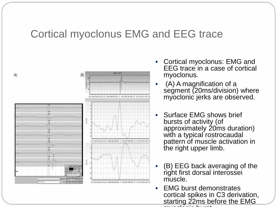
Cortical myoclonus EMG and EEG trace
Cortical myoclonus: EMG and EEG trace in a case of cortical myoclonus.
(A) A magnification of a segment (20ms/division) where myoclonic jerks are observed.
Surface EMG shows brief bursts of activity (of approximately 20ms duration) with a typical rostrocaudalpattern of muscle activation in the right upper limb.
(B) EEG back averaging of the right first dorsal interosseimuscle.
EMG burst demonstrates cortical spikes in C3 derivation, starting 22ms before the EMG myoclonic burst.

Subcortical myoclonus
In contrast with cortical myoclonus, in subcortical
myoclonus there are no signs of
hyperexcitability on the EEG and SSEP
recordings.
Brainstem myoclonus EMG
Simultaneous recording of surface EMG
(multichannel surface EMG) from different
muscles may give information on the distribution
and mode of spread of myoclonus in the case of
brainstem myoclonus.
The first activated muscle is
sternocleidomastoid or trapezius with
subsequent spread of activity to rostral and
caudal muscles.

Brainstem reticular myoclonus EMG
Brainstem reticular
myoclonus.
Multichannel EMG
recording:
Following acoustic
stimulation there was
an initial activation of
the right
sternocleidomastoid
muscle with a latency
of 68ms, followed by
the spread to rostral
and caudal muscles.
Propriospinal myoclonus
In propriospinal myoclonus, myoclonic bursts may
last from 50ms to 4s.
EMG jerks arise from abdominal or cervical
spinal segments and spread slowly rostrally and
caudally, sparing the cranial muscles.

Propriospinal myoclonus EMG
Propriospinal myoclonus. With the patient in a recumbent position, surface multichannel EMG from right-sided muscles shows a jerk of approximately 400ms duration.
This jerk is electrically evoked, starts with a latency of 200ms in the rectus abdominismuscle and is followed by activation of rostral and caudal muscles.

Spinal segmental myoclonus
In spinal segmental myoclonus, myoclonic
bursts are confined to one or two
contiguous myotomes.

Spinal segmental myoclonus EMG
Spinal segmental
myoclonus.
Multisurface EMG
shows myoclonic
bursts confined mainly
to the right triceps but
affecting also a few
adjacent myotomes.
Treatment
The treatment of myoclonus depends on the
underlying disorder.
Reversible causes of myoclonus include some
toxic–metabolic states, drug intoxications or
surgically treatable lesions.
Majority of cases, the underlying cause is not
correctable and symptomatic treatment is the
only possibility.
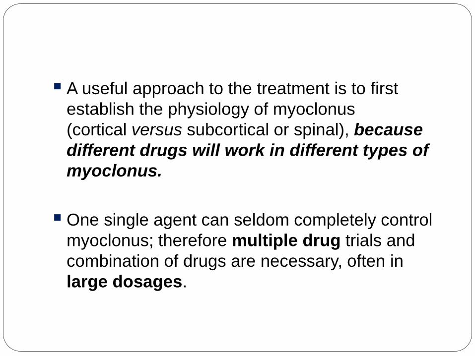
A useful approach to the treatment is to first
establish the physiology of myoclonus
(cortical versus subcortical or spinal), because
different drugs will work in different types of
myoclonus.
One single agent can seldom completely control
myoclonus; therefore multiple drug trials and
combination of drugs are necessary, often in
large dosages.
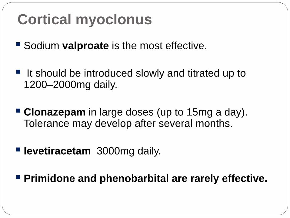
Cortical myoclonus
Sodium valproate is the most effective.
It should be introduced slowly and titrated up to 1200–2000mg daily.
Clonazepam in large doses (up to 15mg a day). Tolerance may develop after several months.
levetiracetam 3000mg daily.
Primidone and phenobarbital are rarely effective.
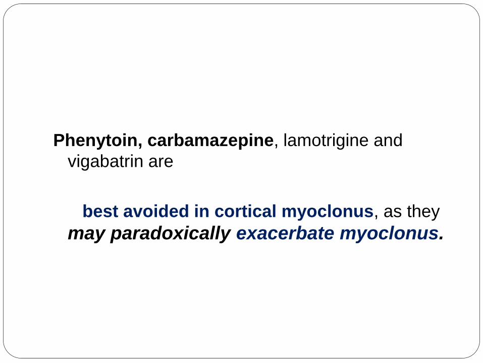
Phenytoin, carbamazepine, lamotrigine and
vigabatrin are
best avoided in cortical myoclonus, as they
may paradoxically exacerbate myoclonus.

Subcortical myoclonus
Antiepileptic drugs used in cortical myoclonus are
not effective in subcortical myoclonus.
Clonazepam is useful in hyperekplexia and
partially in reticular reflex myoclonus
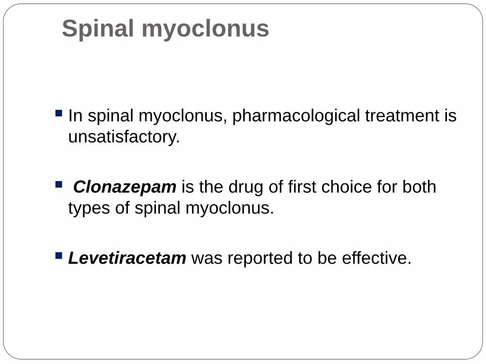
Spinal myoclonus
In spinal myoclonus, pharmacological treatment is
unsatisfactory.
Clonazepam is the drug of first choice for both
types of spinal myoclonus.
Levetiracetam was reported to be effective.
Peripheral myoclonus
Drugs are usually ineffective.
carbamazepine may have some effect.
Hemifacial spasm responds excellently to
botulinum toxin injections

Reference
http://www.ncbi.nlm.nih.gov/pmc/articles/PMC303
6960/
(Myoclonic disorders: a practical approach for
diagnosis and treatment)

Thanks