Myoclonus in Childhood - omsusa.org · Myoclonus in Childhood Michael R. Pranzatelli The term...
11
Myoclonus in Childhood Michael R . Pranzatelli The term "myoclonus" sounds esoteric, yet it is part of our normal physiology, occurring as a muscle jerk on drowsiness or falling asleep, during rapid eye movement (REM) sleep, and as hiccoughs . Myoclonus is also a developmental feature of the human nervous system, comprising some of the earliest fetal movements . In pathologic settings, myoclonus may be the only neurologic abnormality, as in essential myoclonus, but more often it is one symptom of a larger neurologic problem . The vast etiologic spectrum of symptomatic myoclonus can be bewildering, but defining the underlying problem may provide the opportunity to develop specific therapies. Otherwise, treatment is merely symptomatic . The approach to the patient should be to verify the nature of the movement disorder and establish a specific etiologic diagnosis . A battery of' neurophysiologic, neuroradiologic, and other laboratory studies is needed to localize the origin of the myoclonus and identify causative lesions . Drug treatment is largely empiric but must be systematic and aimed at restoring activities of everyday living . Unlike in epilepsies, in myoclonus multiple drugs usually must be combined to attain functional improvement. © 2003 Elsevier Inc . All rights reserved. M YOCLONUS IS a brief involuntary muscle jerk originating in the central nervous sys- tem .1 It is a paroxysmal event that may appear as an isolated finding or as a symptom of many diseases . Myoclonus affects all age groups and may be so severe as to be disabling or may be mild and require no treatment . Developmental and phys- iologic forms of myoclonus contribute to its uniqueness as a dyskinesia . 2 Physiologic myoclo- nus occurs episodically throughout life as hic- coughs (singultus) and hypnic (sleep) jerks . Myo- clonus is also distinguished from other movement disorders by its unusual association with epilepsy and ataxia and by the distinctive panel of drugs used in its treatment . This article focuses on recent advances in classification, etiology, diagnosis, and treatment of myoclonus. CLASSIFICATION Rather than one biologically "generic" myoclo- nus, there are several types . Different classification schema have been proposed, ranging from purely clinical classifications to those that use etiologic or neurophysiologic criteria (Table 1) .1 Differentiation between spontaneous, reflex, and movement-induced action myoclonus can be made clinically . The most common of these types is action myoclonus . Myoclonus may be activated by the intention of an action or the action itself . Reflex myoclonus is activated by sound, light, touch, or passive movement of a limb. Patients with myoclonus may exhibit postural lapses that correspond to a silent period on elec- tromyography (EMG) . This brief lack of muscle activity that sometimes follows a muscle discharge has been called "negative myoclonus," or asterixis, in contradistinction to the muscle discharges de- Seminars in Pediatric Neurology, Vol 10, No 1 (March), 2003: pp 41-51 noted as "positive myoclonus" . 3 Sudden loss of antigravitational muscle tone can be disabling and refractory to treatment . Many patients with severe myoclonus have a mixture of positive and negative myoclonus. Cortical, subcortical, and spinal myoclonus are defined neurophysiologically, but there are clinical clues as well (Table 2) . 4 Cortical myoclonus is focal and distal and typically found in the arm. Cortical reflex myoclonus may be activated by photic stimulation . Patients with subcortical myo- clonus have both proximal and distal generalized myoclonus, involving both agonist and antagonist muscle groups . Spinal myoclonus may be limited to muscles innervated by a few or multiple spinal segments and affects predominantly flexor mus- cles . l These myoclonic categories can also be classi- fied as epileptic or nonepileptic (Table 3) . 3 ' 4 Cor- tical reflex myoclonus, reticular reflex myoclonus, and the myoclonic jerks that herald a generalized seizure in patients with primary generalized epi- lepsy are examples of epileptic myoclonus . 4 Non- epileptic myoclonus encompasses normal physio- From the Departments of Neurology and Pediatrics, South- ern Illinois University School of Medicine, Springfield, IL. Supported by the American Medical Association Research and Education Foundation, Florence A . Carter Fellowship ; the Children's Miracle Network ; and the Southern Illinois Univer- sity School of Medicine. Address reprint requests to Michael R . Pranzatelli, MD, National Pediatric Myoclonus Center, Southern Illinois Univer- sity School of Medicine, P.O . Box 19658, Springfield, IL 62702. © 2003 Elsevier Inc . All rights reserved 1071-9091/03/1001-0007$30.00/0 do i:10.1053/spe n. 2 003.0000 41
-
Upload
doankhuong -
Category
Documents
-
view
218 -
download
0
Transcript of Myoclonus in Childhood - omsusa.org · Myoclonus in Childhood Michael R. Pranzatelli The term...
Myoclonus in Childhood
Michael R . Pranzatelli
The term "myoclonus" sounds esoteric, yet it is part of our normal physiology, occurring as a muscle jerk ondrowsiness or falling asleep, during rapid eye movement (REM) sleep, and as hiccoughs . Myoclonus is also adevelopmental feature of the human nervous system, comprising some of the earliest fetal movements . In
pathologic settings, myoclonus may be the only neurologic abnormality, as in essential myoclonus, but more oftenit is one symptom of a larger neurologic problem . The vast etiologic spectrum of symptomatic myoclonus can bebewildering, but defining the underlying problem may provide the opportunity to develop specific therapies.Otherwise, treatment is merely symptomatic . The approach to the patient should be to verify the nature of themovement disorder and establish a specific etiologic diagnosis. A battery of' neurophysiologic, neuroradiologic,and other laboratory studies is needed to localize the origin of the myoclonus and identify causative lesions . Drug
treatment is largely empiric but must be systematic and aimed at restoring activities of everyday living . Unlike in
epilepsies, in myoclonus multiple drugs usually must be combined to attain functional improvement.© 2003 Elsevier Inc. All rights reserved.
MYOCLONUS IS a brief involuntary musclejerk originating in the central nervous sys-
tem . 1 It is a paroxysmal event that may appear asan isolated finding or as a symptom of manydiseases . Myoclonus affects all age groups andmay be so severe as to be disabling or may be mildand require no treatment . Developmental and phys-iologic forms of myoclonus contribute to itsuniqueness as a dyskinesia . 2 Physiologic myoclo-nus occurs episodically throughout life as hic-coughs (singultus) and hypnic (sleep) jerks . Myo-clonus is also distinguished from other movementdisorders by its unusual association with epilepsyand ataxia and by the distinctive panel of drugsused in its treatment . This article focuses on recentadvances in classification, etiology, diagnosis, andtreatment of myoclonus.
CLASSIFICATION
Rather than one biologically "generic" myoclo-nus, there are several types . Different classificationschema have been proposed, ranging from purelyclinical classifications to those that use etiologic orneurophysiologic criteria (Table 1) . 1
Differentiation between spontaneous, reflex, andmovement-induced action myoclonus can be madeclinically . The most common of these types isaction myoclonus . Myoclonus may be activated bythe intention of an action or the action itself . Reflexmyoclonus is activated by sound, light, touch, orpassive movement of a limb.
Patients with myoclonus may exhibit posturallapses that correspond to a silent period on elec-tromyography (EMG) . This brief lack of muscleactivity that sometimes follows a muscle dischargehas been called "negative myoclonus," or asterixis,in contradistinction to the muscle discharges de-
Seminars in Pediatric Neurology, Vol 10, No 1 (March), 2003: pp 41-51
noted as "positive myoclonus" . 3 Sudden loss ofantigravitational muscle tone can be disabling andrefractory to treatment . Many patients with severemyoclonus have a mixture of positive and negativemyoclonus.
Cortical, subcortical, and spinal myoclonus aredefined neurophysiologically, but there are clinicalclues as well (Table 2) . 4 Cortical myoclonus isfocal and distal and typically found in the arm.Cortical reflex myoclonus may be activated byphotic stimulation . Patients with subcortical myo-clonus have both proximal and distal generalizedmyoclonus, involving both agonist and antagonistmuscle groups . Spinal myoclonus may be limitedto muscles innervated by a few or multiple spinalsegments and affects predominantly flexor mus-cles . l
These myoclonic categories can also be classi-fied as epileptic or nonepileptic (Table 3) . 3 ' 4 Cor-tical reflex myoclonus, reticular reflex myoclonus,and the myoclonic jerks that herald a generalizedseizure in patients with primary generalized epi-lepsy are examples of epileptic myoclonus . 4 Non-epileptic myoclonus encompasses normal physio-
From the Departments of Neurology and Pediatrics, South-ern Illinois University School of Medicine, Springfield, IL.
Supported by the American Medical Association Researchand Education Foundation, Florence A. Carter Fellowship ; theChildren's Miracle Network ; and the Southern Illinois Univer-sity School of Medicine.
Address reprint requests to Michael R . Pranzatelli, MD,National Pediatric Myoclonus Center, Southern Illinois Univer-sity School of Medicine, P.O . Box 19658, Springfield, IL 62702.
© 2003 Elsevier Inc. All rights reserved1071-9091/03/1001-0007$30.00/0do i:10.1053/spe n. 2 003.0000
41
42
MICHAEL R . PRANZATELLI
Table 1 . Classification of Myoclonus
Regularity
SynchronyRhythmic
SynchronousArrhythmic
AsynchronousOscillatory
Direction of joint displacementUpwardDownward
DevelopmentalPhysiologic
HiccupsHypnic jerksFragmentary nocturnal myoclonus
SubcorticalReticular reflexSpontaneous reticularBallistic overflowOscillatoryCortico-subcortical
Spinal
Non-epilepticExaggerated startlePeriodic movements of sleepMyoclonic ticsSegmental myoclonusPhysiologic myoclonus
ClinicalActivation
SpontaneousSensory-evokedAction-evoked
DistributionFocalMultifocalGeneralized
EtiologicEssentialSymptomatic
Genetic disordersAcquired disorders
NeurophysiologicLocalization
CorticalCortical reflexSpontaneous corticalEpilepsy partialis continua
Cortico-reticular
Relation to muscle tonePositiveNegative
Relation to epilepsyEpileptic
Cortical reflexReticular reflexPrimary generalized epileptic
logic phenomena, exaggerated startle, periodicmovements of sleep, some tics, essential myoclo-nus, and dystonic and segmental myoclonus.
DIFFERENTIAL DIAGNOSIS
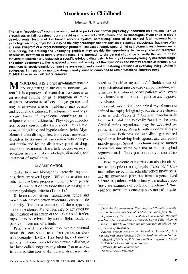
Myoclonus is differentiated from superficiallysimilar dyskinesias by its abruptness and brevity(Fig 1) . Although myoclonus is sometimes con-fused with tics, which are commonly myoclonic innature, tics are usually confined to the head and
shoulders and may be more complex in pattern.Choreiform movements in the fingers when thehands are held outstretched may also appear myo-clonic, but myoclonus is not typically limited to thefingers . When multiple dyskinesias are present,ancillary tests may be necessary to distinguishbetween them.
Although the term "palatal myoclonus" has beenused for many years to describe the rapid, rhythmicfluttering of the soft palate, newer studies indicate
Table 2 . Differentiation of Cortical and Subcortical Myoclonus
Cortical Myoclonus Subcortical Myoclonus
Pattern Focal GeneralizedLocation Distal Proximal and distalMuscle group One synergist group Agonist-antagonist cocontractionActivation Rostrocaudal activation order May propagate up brainstemEEG Time-locked Not time-lockedSSEPs "Giant" (enhanced) Normal amplitude
MYOCLONUS IN CHILDHOOD
43
Table 3 . Relation of Myoclonus to Epilepsy
EMG Burst Length (msec)
Type
Positive myoclonus
Negative myoclonus
Epileptic
<50
<400Non-epileptic
50-200
200-500
that "palatal tremor" is a more appropriate term.Periodic movements of sleep have sometimes beenconfused with "nocturnal myoclonus," which in-stead should refer to myoclonic jerks, such asfragmentary myoclonus, during different phases ofsleep . 5 The movements of restless legs are not trulymyoclonic and are distinguished by the associatedsensations . While startle is a component of brain-stem myoclonus, startle disorders such as hyperex-plexia are discrete syndromes . It also has beensuggested recently that essential myoclonus andmyoclonus dystonia, both of which are sensitive toethanol, are manifestations of the same disorder.Although the term "cortical tremor" implies a non-myoclonic disorder, this is actually a form ofcortical reflex myoclonus that looks similar toessential tremor but is found in patients with my-oclonus . 6
ETIOLOGIES
The treatable or reversible etiologies of myo-clonus need to be recognized (Table 4), especiallyiatrogenic causes . 2 '7-14 Too many diseases associ-ated with myoclonus are not yet reversible . Theseinclude degenerative disorders (eg, Rett's syn-drome, leukodystrophies and Hallervorden-Spatzdisease), metabolic disorders (eg, certain amino-acidurias, lysosomal storage diseases, Leigh's syn-drome, and lipidoses), neurocutaneous disorders(eg, tuberous sclerosis, Sturge-Weber syndrome,and linear nevus sebaceum), prion diseases (eg,slow virus infections), congenital brain anomalies(eg, dorsal and ventral induction defects, migra-tional and proliferation disorders, porencephaly,hydranencephaly, and agenesis of corpus callo-sum), and severe acquired disorders, such as heador spinal injury . Myoclonus is usually one of manyneurologic abnormalities seen in these disorders .
ciated with myoclonus (Fig 2) . It is fashionable todesignate "dyskinesia-plus" syndromes in the eti-ologic classification of other movement disorders,and myoclonus is amenable to the same approach.
Sleep Myoclonus
The term "benign neonatal sleep myoclonus"includes all of the component features of thiscontext-specific myoclonus . The diagnosis is con-firmed by waking the infant, which stops the myo-clonus, or through proprioceptive input by tappingon the limbs, which increases or elicits the jerks . 15
When the myoclonus is rhythmic, the onset is late,or the syndrome persists, the diagnosis may bemissed. Electroencephalography (EEG) findingsare normal . 16
Myoclonus is part of normal sleep physiology,as paradoxical excitation in rapid-eye-movementsleep . Beginning in fetal life, this is most abundantduring the first 6 to 8 months postnatal and persiststhrough life as fragmentary nocturnal myoclonus.Hypnic jerks, or myoclonus on sleep initiation, areassociated with the sensation of falling.
Isolated Myoclonus During Wakefulness inOtherwise Normal Children
Essential myoclonus occurs in otherwise normalindividuals and has an upper body distributionsimilar to that of myoclonic tics, with which it ismost often confused . "Benign myoclonus of earlyinfancy" is a term coined to designate nonepileptic"spasms" that otherwise resemble West's syn-drome . '7 Developmental and neurologic examina-
t
tNocturnalmyoclonus
Non-Myoclonic Disorders
Startledisease
Myoclonic
Myoclonic
palatalPeriodic
seizures
tics Essential tremormovements
tremor
Myoclonusepilepsy
r r rEssential
Segmentalmyoclonus myoclonus /
Myoclonic Disorders
CONTEXT-SPECIFIC MYOCLONUS
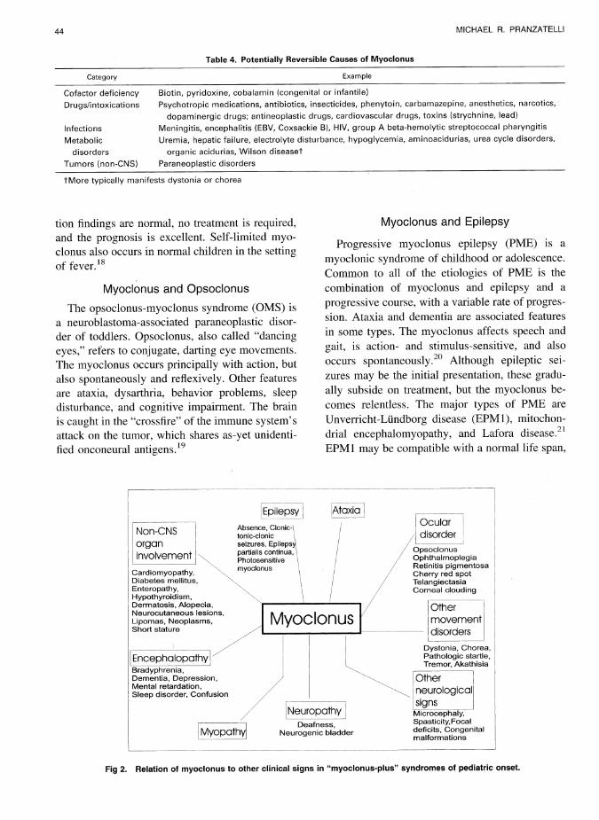
Identifying the specific context in which myo-clonus occurs aids diagnosis and helps organize theotherwise unruly heterogeneity of etiologies asso-
Fig 1 . The interface of myoclonus and related disordersnot properly designated as myoclonus . The pathophysiologicrelationship of some entities is poorly understood and con-troversial.
44
MICHAEL R . PRANZATELLI
Table 4 . Potentially Reversible Causes of Myoclonus
Category
Example
Cofactor deficiency
Biotin, pyridoxine, cobalamin (congenital or infantile)Drugs/intoxications
Psychotropic medications, antibiotics, insecticides, phenytoin, carbamazepine, anesthetics, narcotics,
dopaminergic drugs; antineoplastic drugs, cardiovascular drugs, toxins (strychnine, lead)
Infections
Meningitis, encephalitis (EBV, Coxsackie B), HIV, group A beta-hemolytic streptococcal pharyngitis
Metabolic
Uremia, hepatic failure, electrolyte disturbance, hypoglycemia, aminoacidurias, urea cycle disorders,
disorders
organic acidurias, Wilson diseasetTumors (non-CNS)
Paraneoplastic disorders
tMore typically manifests dystonia or chorea
tion findings are normal, no treatment is required,and the prognosis is excellent . Self-limited myo-clonus also occurs in normal children in the settingof fever.1 s
Myoclonus and Opsoclonus
The opsoclonus-myoclonus syndrome (OMS) isa neuroblastoma-associated paraneoplastic disor-der of toddlers . Opsoclonus, also called "dancingeyes," refers to conjugate, darting eye movements.The myoclonus occurs principally with action, butalso spontaneously and reflexively . Other featuresare ataxia, dysarthria, behavior problems, sleepdisturbance, and cognitive impairment . The brainis caught in the "crossfire" of the immune system'sattack on the tumor, which shares as-yet unidenti-fied onconeural antigens . 19
Myoclonus and Epilepsy
Progressive myoclonus epilepsy (PME) is amyoclonic syndrome of childhood or adolescence.Common to all of the etiologies of PME is thecombination of myoclonus and epilepsy and aprogressive course, with a variable rate of progres-sion. Ataxia and dementia are associated featuresin some types . The myoclonus affects speech andgait, is action- and stimulus-sensitive, and alsooccurs spontaneously . 20 Although epileptic sei-zures may be the initial presentation, these gradu-ally subside on treatment, but the myoclonus be-comes relentless . The major types of PME areUnverricht-Lundborg disease (EPM1), mitochon-drial encephalomyopathy, and Lafora disease . 21
EPM1 may be compatible with a normal life span,
Non-CNSorganinvolvement
Cardiomyopathy,
NDiabetes mellitus,Enteropathy,Hypothyroidism,Dermatosis, Alopecia,Neurocutaneous lesions,Lipomas, Neoplasms,Short stature
EncephalopathyBradyphrenia,Dementia, Depression,Mental retardation,Sleep disorder, Confusion
Oculardisorder
OpsoclonusOphthalmoplegiaRetinitis pigmentosaCherry red spotTelangiectasiaCorneal clouding
Othermovementdisorders
Dystonia, Chorea,Pathologic startle,Tremor, Akathisia
Otherneurologicalsigns
Microcephaly,Spasticity,Focaldeficits, Congenitalmalformations
Deafness,Neurogenic bladderMyopathy
Epilepsy
Ataxia
Absence, Clonic-Itonic-clonicseizures, Epilepsypartialis continua,Photosensitivemyoclonus
Myoclonus
Neuropathy
Fig 2 . Relation of myoclonus to other clinical signs in "myoclonus-plus" syndromes of pediatric onset .
MYOCLONUS IN CHILDHOOD
45
whereas the other two types are typically fatal inthe second decade of life . Both occur sporadicallyor due to autosomal-recessive inheritance . Patientswith EPM1 tend to have absence or tonic-clonicseizures, whereas those with Lafora disease mayhave clonic-tonic-clonic seizures or partial epi-lepsy . 22 Many clinical features differentiate othertypes of PME . 23-25
Juvenile myoclonus epilepsy (JME) representsprimary generalized epilepsy in which myoclonicjerks occur independently of seizures, particularlyon awakening . 26 JME is sometimes confused withPME. Reference to it as "myoclonic epilepsy" is amisnomer.
Because myoclonic seizures are a type of epi-lepsy, this author does not include them in discus-sions of myoclonus as a movement disorder. 27However, some authors do so . 25,28,29
Myoclonus and Ataxia
Myoclonus in the context of cerebellar ataxia isoften part of a progressive disorder, such as ataxia-telangiectasia, 30 in which case other dyskinesiasmay be present as well . Progressive myoclonusataxia (PMA), previously known as Ramsay-Huntsyndrome, may represent more than one disorder.
Myoclonus and Dystonia
The combination of myoclonus and dystoniawithout other abnormalities should suggest myo-clonus-dystonia, a genetic entity . 31 Ethanol respon-siveness and autosomal-dominant inheritance havebeen described.
Myoclonus and Toxic Encephalopathy
The so-called "serotonin syndrome" is an un-common but potentially lethal drug reaction occur-ring primarily in patients with psychiatric illnessduring treatment with serotonin reuptake inhibitorsand other serotonin-potentiating agents . 32 Seroto-nin syndrome is the prototype of drug-inducedmyoclonus . Patients also exhibit fever, confusion,restlessness, ataxia, hyperreflexia, and tremor.
PATHOPHYSIOLOGY
Anatomy and Circuitry
Because myoclonus is often one feature of amore diffuse neurologic disorder, the gamut ofneuropathologic lesions has obscured what mightbe an anatomic common denominator to myoclo-
nus . In cortical myoclonus, lack of inhibition fa-cilitates the transcallosal and cortical spread ofmyoclonus . 4 Abnormal activation of the sensori-motor cortex gives rise to cortical myoclonus, butwhether abnormalities of both cortices are requisiteremains controversial.
Brainstem myoclonus can be induced in exper-imental animals by injection of various drugs intothe brainstem reticular formation at the nucleusgigantocellularis reticularis or anatomically relatedstructures, such as the inferior olive . This medul-lary reticular region has also been implicated in theparadoxically excitatory manifestation of myoclo-nus during REM sleep. The most common forms ofbrainstem myoclonus appear to utilize the samecircuitry as the normal startle reflex . 33
The propriospinal system may be involved insome forms of spinal myoclonus . A spinal-step-ping generator has been proposed that may bereleased from supraspinal control.
Noninvasive functional neuroimaging shouldprovide some answers to the questions regardingthe circuitry involved in myoclonic disorders.Some of the subcortical structures involved, espe-cially in the brainstem, are difficult to resolve usingcurrent technology . In hereditary essential myo-clonus, cerebral blood flow studies have revealedreduced cortical cerebral blood flow contralateralto the myoclonus, suggesting a brainstem or basalganglia lesion. In epileptic negative myoclonus,EEG-single-photon emission computed tomogra-phy (SPECT) indicates involvement of the premo-tor cortex . 34
Neurotransmitters
Experiences with myoclonus-evoking drugs andintoxications, the therapeutic use of many pharma-cologically diverse categories of drugs, and cere-brospinal fluid (CSF) studies of neurotransmittersor their metabolites, have implicated more than oneneurotransmitter system in human myoclonic dis-orders . 35 Without yet being able to identify whichneurotransmitter is most proximal to the myoclo-nus induction mechanism, it is possible to say thaty-aminobutyric acid (GABA), glycine, serotonin,and glutamate seem to be primary.
Although the idea that various types of myoclo-nus involve different neurotransmitters locally andthrough projections because they involve differentanatomic pathways is plausible, very little data areavailable in humans . Distinctive pharmacologic
46
MICHAEL R . PRANZATELLI
responses in diverse types of myoclonus may bebased on unique circuitry of cortical, brainstem,and spinal myoclonus "generators," as well as thefunctional impact of specific human myoclonicdisorders on the anatomy and physiology of thatcircuitry . In spinal cords of experimental animals,local circuitry includes GABA- and glycine-medi-ated inhibition of Renshaw cells on spinal motorneurons . Neurotransmitter receptor subunit and ionchannel disorders may be involved in some formsof hereditary myoclonus.
Animal Models
Gene knockout animal models of myoclonus areallowing links to be made between gene defectsand brain neuropl~armacology . Of note are recentmodels of EPM1,36 neuronal ceroid lipofuscino-sis, 37 and mitochondrial disorders . 38 Profound lossof GABA-ergic interneurons and an autoantibodyinhibitory to glutamic acid decarboxylase (GAD)are new findings in neuronal ceroid lipofuscino-sis . 39,4o Mice lacking potassium channels Kv3 .1and Kv3.3 display myoclonus . 41
LABORATORY TESTS
Neurophysiologic Studies
Cortical, subcortical, and spinal myoclonus canbe differentiated by computer-facilitated back-av-eraging, which correlates EEG and electromyelog-raphy (EMG) activity . A cortical electrical poten-tial . occurs just before the myoclonic jerk incortical, but not subcortical, myoclonus . 42 Besidesthis time-locked EEG event, patients with corticalmyoclonus have enlarged somatosensory evokedpotentials (SSEPs) and enhanced long loop re-flexes . 6 Advanced magnetograms, which havethree-dimensional resolution, have aided localiza-tion of the premyoclonus spike in cortical myo-clonus, resolving the giant somatosensory evokedmagnetic field to the sensory or motor cortex. 33
Cortical myoclonus is typical of PME, Angelmansyndrome, and autosomal-dominant cortical myo-clonus and e ile s 23,43,44
P~ P Y•In subcortical myoclonus, SSEPs are normal,
and muscle group activation indicates that dis-charges may actually propagate up the brainstem.A common type of subcortical myoclonus is retic-ular reflex myoclonus . 4
Epileptic and nonepileptic myoclonus can bedifferentiated on EEG and EMG testing . The EMG
burst in epileptic myoclonus is very short, andthere is an EEG correlate . The innervation ofmuscles involved in jerking is synchronous, andthe muscle groups affected may be activated eitherin a rostrocaudal or ascending fashion .4 In contrast,nonepileptic myoclonus is associated with longerEMG discharges and asynchronous muscle groupjerking. Muscles are activated in a segmental fash-ion without an EEG correlate : 1
Evoked potentials differentiate two types of my-oclonus : one with and another without enhancedSSEPs . Physiologic myoclonus, such 'as duringsleep or startle, occurs without an enhanced evokedpotentials . Giant SSEPs occur in the epilepsies,including myoclonic generalized seizures . 33
Neuroimaging Studies
Magnetic resonance imaging or computed to-mography scans of the head or spinal cord .arenormal in essential myoclonus and in some typesof symptomatic myoclonus . SPECT may demon-strate hypoperfusion associated with myoclo-nus . 21,4s,46 MR spectroscopy may be positive whenother studies are negative.
Other Tests
Sophisticated biochemical and histochemicaltests enable a specific etiologic diagnosis . Theymay include biotinidase, biotin, organic and aminoacids, urinary oligosaccharides, as well as evokedresponses, skin biopsy, slit-lamp examination,muscle biopsy, and electroretinography . 47 An-tineuronal antibodies and antibodies to neurotrans-mitter pathway enzymes, such as GAD, can bemeasured . Determination of catecholamine pre-cursors and metabolites in CSF may be helpfulin diagnostically challenging cases .48 Advancesin the molecular genetics of movement disor-ders have produced new diagnostic tests (Ta-ble 5) .24,26,31,36,44,49-s9 p ediatric rating scales forthe evaluation of myoclonus are available . 6o,6 ~
TREATMENT OF MYOCLONUS
Treatment of the underlying pathophysiology ispreferable to symptomatic treatment . In drug-in-duced myoclonus, the offending agent should bestopped and symptomatic support given. The ap-proach to myoclonus due to infections is directedto the source of the infection . When myoclonusoccurs as the remote immunologic effect of can-
MYOCLONUS IN CHILDHOOD
47
Table 5 . Some Genetic Disorders Associated with Myoclonus
Disorder Chromosome Gene/Gene Product
ADCME 2p11 .1-g12 .2Angelman syndrome 15g11-q13 UBE3A, GABRB3(?)Ataxia-telangiectasia 11q ATMNCL 16p12 CLN1, CLN2, CLN3/lysosomal enzymes and transmembrane proteins
Biotinidase deficiency 3p25 BTD gene/biotinidaseDRPLA 12p DRPLA gene/atrophin-1JME 6p, 15qMitochondria)
MERRF mt RNA (Lys)MELAS mt RNA (Leu (UUR))PEO multiple point mutations and large deletions
Myoclonus-dystonia syndrome (DYT11) 7g21-q31 SGCE (gene for epsilon-sarcoglycan)11q23 D 2 dopamine receptor gene (?)
PMEU-L disease (EPM1) 21q22 .3 CSTB/cystatin BLafora (EPM2) 6q23-q25 EPM2A/laforinGM1-gangliosidosis 5p1 .13 Beta-galactosidase geneSialidosis, type 1 20 Sialidase geneJuvenile Gaucher (type III) 1 .g21 Glucocerebrosidase gene
SCA 2 12 SCA2 gene/ataxin-2
ADCME, autosomal dominant cortical myoclonus and epilepsyDRPLA, dentato-rubro-pallido-luysian atrophyJME, juvenile myoclonic epilepsyMERRF, mitochondria) encephalopathy with ragged red fibersMELAS, mitochondrial encephalopathy with lactic acidosis and stroke-like episodesNCL, neuronal ceroid lipofuscinosisPME, progressive myoclonus epilepsySCA, spinocerebellar ataxia
cers, the therapy is aimed at the cancer and alsoagainst the autoimmune process . 19
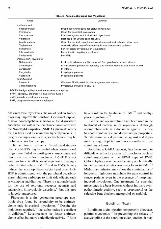
Anticonvulsants
Anticonvulsants (Table 6) are the mainstay oftreatment for myoclonus, whether it is epileptic ornonepileptic . 12,22,62-64 The 1,3-substituted benzo-diazepines clonazepam, nitrazepam, and loraz-epam are particularly useful due to the combina-tion of their anxiolytic, sedative, muscle relaxant,and anticonvulsant properties, but tolerance is lim-iting. 35 Anticonvulsants may have synergistic ef-fects in myoclonus . However, not all are antimyo-clonic, and some induce myoclonus . 65-71
Immunotherapy
Immunomodulation with intravenous immuno-globulins is an important treatment for myoclonusdue to autoimmune disorders . Immunosuppres-sants, such as azathioprine, cyclophosphamide, andmethotrexate, also have a role . Adrenocortico-tropic hormone is still the gold standard for OMS .
Case reports suggest that plasmapheresis also maybe effective . 72
Cofactors and Vitamins
Cofactors and vitamins are used to reverse un-derlying etiologic abnormalities . Coenzyme-Q andcarnitine may be administered in mitochondrialmyopathies . Although 'biotin's mechanism of ac-tion in myoclonus remains uncertain, replacementtherapy may be therapeutic in deficiencies of bi-otinidase, multiple carboxylases, or other biotin-dependent enzymes .47 A response to biotin mayoccur in the absence of biotin or biotinidase defi-ciency. Response to thiamine in opsoclonic cer-ebellopathy has been reported but is rare.
Other Drugs
Anticholinergic drugs (eg, trihexyphenidyl) are anoption in treating myoclonus dystonia but have alimited role in the therapy of other myoclonic disor-ders . Estrogen may be either a cause of or a treatmentfor movement disorders, 73 but when menstrual peri-
48
MICHAEL R . PRANZATELLI
Table 6 . Antiepileptic Drugs and Myoclonus
Effect
Comment
AntimyoclonicPhenobarbitalPrimidoneClonazepamValproateLevetiracetamTopiramateFelbamateEthosuximideZonisamide
Occasionally myoclonicGabapentinLamotrigineLorazepamPregabalinVigabatrin
Best Avoided ,PhenytoinCarbamazepine
Broad-spectrum ; good for status myoclonusGood for essential myoclonusEffective against opioid-induced myoclonusBest drug for EPM1 ; good for JMEGood for cortical myoclonus; avoid in mood and behavior disordersAnorexic effect may offset obesity in non-ambulatory patientsFor refractory myoclonus in youngstersFor epileptic negative myoclonusFor PME
In chronic refractory epilepsy ; good for opioid-induced myoclonusIn intractable generalized epilepsy and Lennox-Gastaut ; less often in JMEI n infantsIn epileptic patientsIn epileptic patients
Worsens EPM1 ; used for diaphragmatic myoclonusMyoclonus induced in BECTS
BECTS, benign epilepsy with centrotemporal spikesEPM1, epilepsy, progressive myoclonus, type 1JME, juvenile myoclonus epilepsyPME, progressive myoclonus epilepsy
ods exacerbate myoclonus, the use of oral contracep-tives may improve the situation . Dextromethorphan,a weak noncompetitive inhibitor at the dissociativeanesthetic site within the ion channel associated withthe N-methyl-D-aspartate (NMDA) glutamate recep-tor, has been used for nonketotic hyperglycinemia . Inprogressive myoclonus ataxia, acetazolamide may beuseful as adjunctive therapy.
The serotonin precursor 5-hydroxy-L-trypto-phan (L-5-HTP) may be useful when conventionaldrugs have failed in posthypoxic myoclonus andphotic cortical reflex myoclonus . L-5-HTP is notantimyoclonic in all types of myoclonus, having arather limited role in PME20 and in OMS. It mayinduce the eosinophilia-myalgia syndrome . L-5-HTP is administered with the peripheral decarbox-ylase inhibitor carbidopa to limit side effects, suchas cramping and diarrhea. There is a rationale basisfor the use of serotonin receptor agonists andantagonists in myoclonic disorders, 32 but this areais largely unexplored.
Piracetam is a first-generation prototype noo-tropic drug found by serendipity to be antimyo-clonic only in cortical myoclonus . 74 Despite thehigh doses required, 75 the safety index is excellentin children. 61 Levetiracetam has lesser antimyo-clonic effect but more antiepileptic activity . 76 Both
have a role in the treatment of PME" and posthy-poxic myoclonus . 78
Lisuride and apomorphine have been used in thetreatment of cortical reflex myoclonus . Althoughapomorphine acts as a dopamine agonist, lisuridehas both serotonergic and dopaminergic properties.Tetrabenazine is a dopamine antagonist and dopa-mine storage depleter used occasionally to treatspinal myoclonus.
Baclofen, a GABA agonist, has been used indifficult or refractory cases of myoclonus such asspinal myoclonus or the EPM1 type of PME.Chloral hydrate may be used acutely or chronicallyin the treatment of refractory myoclonus in PME . 79Midazolam infusion may allow the continuation oflong-term high-dose morphine for pain control incancer patients even in the presence of morphineinduced myoclonus . One treatment for essentialmyoclonus is a beta-blocker without intrinsic sym-pathomimetic activity, such as propranolol or theselective beta-blockers metoprolol or nadolol.
Botulinum Toxin
Botulinum toxin injection temporarily alleviatespainful myoclonus . 80 In preventing the release ofacetylcholine at the neuromuscular junction, it may
MYOCLONUS IN CHILDHOOD
49
block involuntary movement but will preservestrength . The effects last from weeks to months,but the injections can be repeated . Both botulinumtoxins A (Botox) and B are used clinically . Thecurrent trend is toward lower doses than thoserecommended initially.
Transcranial Magnetic Stimulation
Transcranial magnetic stimulation (TMS) is anoninvasive, safe, and painless way to stimulatethe human motor cortex in humans . 81 Types in-clude single-pulse, paired-pulse, and repetitive
TMS (rTMS) . 82 rTMS can be used to transientlyinactivate different cortical areas to study theirfunctions . Modulation of cortical excitability byrTMS has therapeutic potential in neurologic andpsychiatric disorders, because high-frequencyrTMS (5 Hz) increases cortical excitability,whereas low-frequency stimulation (1 Hz) reducescortical excitability . 83 Although only corticalstructures are currently accessible, TMS seemscapable of affecting activity in cortically linkeddeep brain structures . 84 The only serious side ef-fect is a possible induction of seizures . 85
REFERENCES
1. Marsden CD, Hallett M, Fahn S : The nosology andpathophysiology of myoclonus, in Marsden CD, Fahn S(eds) : Movement Disorders . London, Butterworths, 1982, pp196-248
2. Pranzatelli MR: Myoclonic disorders . Pediatr Ann 22 :33-37, 1993
3. Tassinari CA, Rubboli G, Parmeggiani L, et al : Epilepticnegative myoclonus. Adv Neurol 67 :181-197, 1995
4. Hallett M : Myoclonus : Relation to epilepsy . Epilepsia26 :567-577, 1985
5. Anstead M: Pediatric sleep disorders : New developmentsand evolving understanding . Curr Opin Pulm Med 6 :501-506,2000
6. Toro C, Pascual-Leone A, Deuschl G, et al : Corticaltremor: A common manifestation of cortical myoclonus . Neu-rology 43 :2346-2352, 1993
7. DiFazio MP, Morales J, Davis R: Acute myoclonus sec-ondary to group A beta-hemolytic streptococcus infection : APANDAS variant . J Child Neurol 13 :516-518, 1998
8. Maiteh M, Daoud AS : Myoclonic seizure following intra-venous verapamil injection : Case report and review of theliterature . Ann Trop Paediatr 21 :271-272, 2001
9. Grech V, Vella C, Mercieca V : Temporary myoclonuswith treatment of congenital transcobalamin 2 deficiency . Pe-diatr Neurol 24 :75-76, 2001
10. Ozer EA, Turker M, Bakiler AR, et al : Involuntarymovements in infantile cobalamin deficiency appearing aftertreatment . Pediatr Neurol 25 :81-83, 2001
11. McClain BC, Probst LA, Pinter E : Intravenous clonidineuse in a neonate experiencing opioid-induced myoclonus . An-esthesiology 95 :549-550, 2001
12. Mercadante S, Villari P, Fulfaro F: Gabapentin for opi-oid-related myoclonus in cancer patients . Support Care Cancer9 :205-206, 2001
13. Zaw W, Knoppert DC, da Silva 0 : Flumazenil's reversalof myoclonic-like movements associated with midazolam interm newborns . Pharmacotherapy 21 :642-646, 2001
14. Ghaziuddin N, Iqbal A, Khetarpal S : Myoclonus duringprolonged treatment with sertraline in an adolescent patient.J Child Adolesc Psychopharmacol 11 :199-202, 2001
15. Daoust-Roy J, Seshia SS : Benign neonatal sleep myo-clonus : A differential diagnosis of neonatal seizures . AJDC146 :1236-1241, 1992
16. Bye AM, Kok DJ, Ferenschild FT, et al : Paroxysmal
non-epileptic events in children: A retrospective study over aperiod of 10 years . J Paediatr Child Health 36 :244-248, 2000
17. Maydell BV, Berenson F, Rothern AD, et al : Benignmyoclonus of early infancy : An imitator of West's syndrome.J Child Neurol 16:109-112, 2001
18. Rajakumar K, Bodensteiner JB : Febrile myoclonus : Asurvey of pediatric neurologists . Clin Pediatr (Phila) 35 :331-332, 1996
19. Pranzatelli MR: Paraneoplastic syndromes : An unsolvedmurder . Semin Pediatr Neurol 7 :118-130, 2000
20. Pranzatelli MR, Tate E, Huang Y, et al : The neurophar-macology of progressive myoclonus epilepsy : Response to5-hydroxy-L-tryptophan . Epilepsia 36 :783-791, 1995
21. Berkovic SF, Carpenter S, Evans A, et al: Myoclonusepilepsy and ragged-red fibres (MERRF), 1 . A clinical, patho-logical, biochemical, magnetic resonance spectrographic andpositron emission tomographic study . Brain 112(pt 5) :1231-1260, 1989
22. Yoshimura I, Kaneko S, Yoshimura N, et al : Long-termobservations of two siblings with Lafora disease treated withzonisamide. Epilepsy Res 46 :283-287, 2001
23. Tobimatsu S, Fukui R, Shibasaki H, et al : Electrophys-iological studies of myoclonus in sialidosis type 2 . Clin Neu-rophysiol 60 :16-22, 1985
24. Tomoda A, Ikezawa M, Ohtani Y, et al : Progressivemyoclonus epilepsy : Dentato-rubro-pallido-luysian atrophy(DRPLA) in childhood. Brain Dev 13 :266-269, 1991
25. Minassian BA, Sainz J, Delgado-Escueta AV: Geneticsof myoclonus and myoclonus epilepsies . Clin Neurosci 3 :223-235, 1995
26. Delgado-Escueta AV, Medine MT, Serratosa JM, et al:Mapping and positional cloning of common idiopathic gener-alized epilepsies: Juvenile myoclonus epilepsy and childhoodabsence epilepsy . Adv Neurol 79 :351-374, 1999
27. Pranzatelli MR : Infantile spasms vs. myoclonus: Is therea connection?, in Schwartzkroin PA, Rho JM (eds) : Epilepsy,Infantile Spasms, and Developmental Encephalopathy. San Di-ego, Academic Press, 2002, pp 285-314
28. Fejerman N : Myoclonus and epilepsies . Indian J Pediatr64 :583-602, 1997
29. Oguni H, Fukuyama Y, Tanaka T, et al : Myoclonic-astatic epilepsy of early childhood : Clinical and EEG analysisof myoclonic-astatic seizures, and discussions on the nosologyof the syndrome. Brain Dev 23 :757-764, 2001
50
MICHAEL R . PRANZATELLI
30. Pagani F, Buratti E, Stuani C, et al : A new type ofmutation carries a splicing defect in ATM . Nat Genet 30:426-429, 2002
31. Klein C, Schilling K, Saunders-Pullman RJ, et al : Amajor focus for myoclonus-dystonia maps to chromosome 7q ineight families . Am J Hum Genet 67 :1314-1319, 2000
32. Pranzatelli MR: Serotonin and human myoclonus : Ra-tionale for the use of serotonin receptor agonists and antago-nists . Arch Neurol 51 :605-617, 1994
33. Shibasaki H: Electrophysiological studies of myoclonus.Muscle Nerve 23 :321-335, 2000
34. Baumgartner C, Podreka I, Olbrich A, et al : Epilepticnegative myoclonus : An EEG-single-photon emission CT studyindicating involvement of premotor cortex . Neurology 46:753-758, 1996
35. Pranzatelli MR, Nadi NS : Mechanism of action of anti-epileptic and antimyoclonic drugs . Adv Neurol 67 :329-360,1995
36. Pennacchio LA, Lehesjoki AE, Stone NE, et al: Muta-tions in the gene encoding cystatin B in progressive myoclonusepilepsy (EPM1) . Science 271 :1731-1734, 1996
37. Gupta P, Sajombo AA, Atashband A, et al : Disruption ofPPT1 or PPT2 causes neuronal ceroid lipofuscinosis in knock-out mice . Proc Natl Acad Sci U S A 98 :13566-13571, 2001
38. Wallace DC : Animal models for mitochondrial disease.Methods Mol Biol 197 :3-54, 2002
39. Cooper JD, Gupta P, Bihle E, et al : Profound loss ofGABAergic interneurons in the PPT1 knockout mouse model ofinfantile neuronal ceroid lipofuscinosis . Neuropathol Appl Neu-robiol 28 :158-159, 2002
40. Chattopadhyay S, Ito M, Cooper JD, et al : An autoanti-body inhibitory to glutamic acid decarboxylase in the neurode-generative disorder Batten disease . Hum Mol Genet 11 :1421-1431, 2002
41. Espinosa F, McMahon A, Chan E, et al : Alcohol hyper-sensitivity, increased locomotion, and spontaneous myoclonusin mice lacking the potassium channels Kv3 .1 and Kv3 .3.J Neurol Sci 21 :6657-6665, 2001
42. Shibasaki H, Ikeda A, Nagamino T, et al : Cortical reflexnegative myoclonus . Brain 117 :477-486, 1994
43. Guerrini R, De Lorey TM, Bonanni P, et al: Corticalmyoclonus in Angelman syndrome . Ann Neurol 40:39-48, 1996
44. Guerrini R, Bonanni P, Patrignani A, et al : Autosomaldominant cortical myoclonus and epilepsy (ADCME) withcomplex partial and generalized seizures : A newly recognizedepilepsy syndrome with linkage to chromosome 2p 11 .1-q 12 .2.Brain 124(pt 12) :2459-2475, 2001
45. Oguro K, Kobayashi J, Aiba H, et al : Opsoclonus-myoclonus syndrome with abnormal single photon emissioncomputed tomography imaging . Pediatr Neurol 16 :334-336,1997
46. Tanaka S, Osari S, Ozawa M, et al : Recurrent painattacks in a 3-year-old patient with myoclonus epilepsy associ-ated with ragged-red fibers (MERRF) : A single-photon emis-sion computed tomographic (SPECT) and electrophysiologicalstudy . Brain Dev 19 :205-208, 1997
47. Gascon GG, Ozand PT, Brismar J : Movement disordersin childhood organic acidurias : Clinical, neuroimaging, andbiochemical correlations. Brain Dev 16(suppl) :94-103, 1994
48. Hyland K, Arnold LA, Trugman JM : Defects of biop-terin metabolism and biogenic amine biosynthesis : Clinical,
diagnostic, and therapeutic aspects, in Fahn S, Marsden CD,DeLong M (eds) : Dystonia 3 : Advances in Neurology, Vol 78.Philadelphia, Lippincott-Raven, 1998, pp 301-308
49. Schols L, Gispert S, Vorgerd M, et al : Spinocerebellarataxia type 2 . Genotype and phenotype in German kindreds.Arch Neurol 54 :1073-1080, 1997
50. Bespalova IN, Adkins S, Pranzatelli, M, et al : Novelcystatin B mutation and diagnostic PCR assay in Unverricht-Lundborg (Baltic) progressive myoclonus epilepsy patient.Am J Med Genet (Neuropsych Gen) 74 :467-471, 1997
51. Serratosa JM, Gomez-Garre P, Gallaredo ME, et al : Anovel protein tyrosine phosphatase gene is mutated in progres-sive myoclonus epilepsy of the Lafora type (EPM2) . Hum MolGenet 8 :345-352, 1999
52. Larson GP, Ding S, Lafrenier RG, et al : Instability of theEPM1 minisatellite . Hum Mol Genet 8 :1985-1988, 1999
53. Borner GV, Zeviani M, Tiranti V, et al : Decreasedaminoacylation of mutant tRNAs in MELAS but not in MERRFpatients . Hum Mol Genet 9 :467-475, 2000
54. Wisniewski KE, Kida E, Connell F, et al : Neuronalceroid lipofuscinoses : Research update . Neurol Sci 21(suppl3) :549-556, 2000
55. Bonten EJ, Arts WF, Beck M, et al : Novel mutations inlysosomal neuraminidase identify functional domains and de-termine clinical severity in sialidosis . Hum Mol Genet 9 :2715-2725, 2000
56. Wood JD, Nucifara FC, Duan K, et al : Atrophin-1, thedentato-rubral and pallido-luysian atrophy gene product, inter-acts with ETO/MTG8 in the nuclear matrix and repressestranscription. J Cell Biol 150:939-948, 2000
57. Pshezhetsky AV, Ashmarina M : Lysosomal multien-zyme complex : Biochemistry, genetics and molecular patho-physiology. Prog Nucleic Acid Res Mol Biol 69:81-114, 2001
58. Minassian BA: Lafora's disease : Towards a clinical,pathologic, and molecular synthesis. Pediatr Neurol 25 :21-29,2001
59. Zimprich A, Grabowski M, Asmus F, et al : Mutations inthe gene encoding epsilon-sarcoglycan cause myoclonus-dys-tonia syndrome . Nat Genet 29 :66-69, 2001
60. Tate E, Pranzatelli MR, Ho H, et al : A sensitive andsemi-quantitative pediatric myoclonus evaluation scale . Neuro-sci Nurs 27 :287-291, 1995
61. Pranzatelli MR, Tate ED, Galvan I, et al : Controlled pilotstudy of piracetam for pediatric opsoclonus-myoclonus . ClinNeuropharmacol 24:352-357, 2001
62. Capovilla G, Beccaria F, Veggiotti P, et al : Ethosuxim-ide is effective in the treatment of epileptic negative myoclonusin childhood partial epilepsy . J Child Neurol 14 :395-400, 1999
63. Moretti R, Torre P, Antonello RM : Opsoclonus-myo-clonus syndrome : Gabapentin as a new therapeutic proposal.Eur J Neurol 7 :455-456, 2000
64. Wallace SJ: Newer antiepileptic drugs : Advantages anddisadvantages . Brain Dev 23 :277-283, 2001
65. Eldridge R, Iivanainen M, Stern R, et al : "Baltic" my-oclonus epilepsy: Hereditary disorder of childhood made worseby phenytoin. Lancet 9 :838-842, 1983
66. Marciani MG, Maschio M, Spanedda F, et al : Develop-ment of myoclonus in patients with partial epilepsy duringtreatment with vigabatrin : An electroencephalographic study.Acta Neurol Scand 91 :1-5, 1995
67. Guerrini R, Belmonte A, Parmeggiani L, et al : Myo-
MYOCLONUS IN CHILDHOOD
51
clonic status epilepticus following high-dosage lamotriginetherapy. Brain Dev 21 :420-424, 1999
68. Nanba Y, Maegaki Y : Epileptic negative myoclonusinduced by carbamazepine in a child with BECTS : Benignchildhood epilepsy with centrotemporal spikes . Pediatr Neurol21 :664-667, 1999
69. Asconape J, Diedrich A, DellaBadia J : Myoclonus asso-ciated with the use of gabapentin . Epilepsia 41 :479-481, 2000
70. Janszky J, Rasonyi G, Halasz P, et al : Disabling erraticmyoclonus during lamotrigine therapy with high serum level:Report of two cases . Clin Neuropharmacol 23 :86-89, 2000
71. Huppertz HJ, Feuerstein TJ, Schulze-Bonhage A : My-oclonus in epilepsy patients with anticonvulsive add-on therapywith pregabalin. Epilepsia 42 :790-792, 2001
72. Yiu VW, Kovithavongs T, McGonigle LF, et al : Plas-mapheresis as an effective treatment for opsoclonus-myoclonussyndrome. Pediatr Neurol 24 :72-74, 2001
73. Kompoliti K : Estrogen and movement disorders . ClinNeuropharmacol 22 :318-326, 1999
74. Gouliaev AH, Senning A : Piracetam and other structur-ally related nootropics . Brain Res Rev 19 :180-222, 1994
75. Ikeda A, Shibasaki H, Tashiro K, et al : Clinical trial ofpiracetam in patients with myoclonus : Nationwide multiinsti-tution study in Japan . Mov Disord 11 :691-700, 1996
76. Genton P, Van Vleymen B : Piracetam and levetiracetam:Close structural similarities but different pharmacological andclinical profiles. Epileptic Disord 2 :99-105, 2000
77. Feddi M, Reutens D, Dubeau F, et al : Long-term efficacyand safety of piracetam in the treatment of progressive myo-clonus epilepsy. Arch Neurol 58 :781-786, 2001
78. Krauss GL, Bergin A, Kramer RE, et al : Suppression ofpost-hypoxic and post-encephalitic myoclonus with levetirac-etam . Neurology 56 :411-412, 2001
79. Pranzatelli MR, Tate ED : Chloral hydrate for progres-sive myoclonus epilepsy : A new look at an old drug. PediatrNeurol 25 :385-389, 2001
80. Awaad Y, Tayem H, Elgamal A, et al : Treatment ofchildhood myoclonus with botulinum toxin type A. J ChildNeurol 14 :781-786, 1999
81. Chen, R: Studies of human motor physiology with trans-cranial magnetic stimulation . Muscle Nerve Suppl 9, S26-S32,2000
82. Amassian VE, Cracco RQ, Maccabee PS, et al : Somepositive effects of transcranial magnetic stimulation . Adv Neu-rol 67 :79-104, 1995
83. Triggs WJ, Kirshner HS : Improving brain function withtranscranial magnetic stimulation . Neurology 56 :429-430, 2001
84. McDonald WM, Greenberg BD : Electroconvulsive ther-apy in the treatment of neuropsychiatric conditions and trans-cranial magnetic stimulation as a pathophysiological probe inneuropsychiatry . Depress Anxiety 12 :135-143, 2000
85. Lisanby SH, Luber B, Sackeim HA, et al : Deliberateseizure induction with negative transcranial magnetic stimulationin nonhuman primates . Arch Gen Psychiatry 58 :199-200, 2001