Myocardial deformation after radiotherapy: a layer ...
10
RESEARCH Open Access Myocardial deformation after radiotherapy: a layer-specific and territorial longitudinal strain analysis in a cohort of left-sided breast cancer patients (BACCARAT study) Valentin Walker 1 , Olivier Lairez 2,3,4 , Olivier Fondard 5 , Gaëlle Jimenez 6 , Jérémy Camilleri 6 , Loïc Panh 7 , David Broggio 8 , Marie-Odile Bernier 1 , Dominique Laurier 9 , Jean Ferrières 2,10,11 and Sophie Jacob 1* Abstract Background: Radiotherapy for breast cancer (BC) and its resulting cardiac exposure are associated with subclinical left ventricular dysfunction characterized by early decrease of global longitudinal strain (LS) measurement based on 2D speckle-tracking echocardiography. Recent software allows multi-layer and segmental analysis of strain, which may be of interest to quantify and locate the impact of cardiac exposure on myocardial function and potentially increase the early detection of radiation-induced cardiotoxicity. The aim of the study was to evaluate whether decrease in LS 6 months after radiotherapy is layer-specific and if it varies according to the left ventricular regional level and the coronary arterial territories. Methods: LS was measured at baseline before radiotherapy and 6 months post-radiotherapy. The LS was obtained for each myocardial layer (endocardial, mid-myocardial, epicardial), left ventricular regional level (basal, mid, apical) and coronary artery territory (left anterior descending artery (LAD), circumflex artery, right coronary artery). Results: The study included 64 left-sided BC patients. Mean age was 58 years, mean doses to the heart, the left ventricle and the LAD were respectively 3.0, 6.7 and 16.4 Gy. The absolute decrease of LS was significant for the three layers (endocardial: - 20.0 ± 3.2% to - 18.8 ± 3.8%; mid-myocardial: - 16.0 ± 2.7% to - 15.0 ± 3.1%; epicardial: - 12.3 ± 2.5% to - 11.4 ± 2.8%, all p = 0.02), but only the relative decrease of LS in the endocardial layer was close to be significant ( - 4.7%, p = 0.05). More precisely, the LS of the endocardial layer was significantly decreased for the most exposed parts of the left ventricle corresponding to the apical level ( - 26.3 ± 6.0% vs. -24.2 ± 7.1%, p = 0.03) and LAD territory ( - 22.8 ± 4.0% vs. -21.4 ± 4.8%, p = 0.03). Conclusion: Six months post-radiotherapy, LS decreased predominantly in the endocardial layer of the most exposed part of the left ventricle. For precise evaluation of radiotherapy-induced cardiotoxicity and early left ventricular dysfunction, the endocardial layer-based LS might be the most sensitive parameter. Trial registration: ClinicalTrials.gov: NCT02605512, Registered 6 November 2015 - Retrospectively registered. Keywords: Radiation therapy, Cardiac toxicity, Echocardiography, Multilayer strain, Coronary arteries © The Author(s). 2020 Open Access This article is licensed under a Creative Commons Attribution 4.0 International License, which permits use, sharing, adaptation, distribution and reproduction in any medium or format, as long as you give appropriate credit to the original author(s) and the source, provide a link to the Creative Commons licence, and indicate if changes were made. The images or other third party material in this article are included in the article's Creative Commons licence, unless indicated otherwise in a credit line to the material. If material is not included in the article's Creative Commons licence and your intended use is not permitted by statutory regulation or exceeds the permitted use, you will need to obtain permission directly from the copyright holder. To view a copy of this licence, visit http://creativecommons.org/licenses/by/4.0/. The Creative Commons Public Domain Dedication waiver (http://creativecommons.org/publicdomain/zero/1.0/) applies to the data made available in this article, unless otherwise stated in a credit line to the data. * Correspondence: [email protected] 1 Pôle Santé-Environnement (PSE-SANTE), Service de recherche sur les effets biologiques et sanitaires des rayonnements ionisants (SESANE), Laboratoire d’épidémiologie des rayonnements ionisants (LEPID), Institute for Radiological Protection and Nuclear Safety (IRSN), BP17, 92262 Fontenay-aux-Roses cedex, France Full list of author information is available at the end of the article Walker et al. Radiation Oncology (2020) 15:201 https://doi.org/10.1186/s13014-020-01635-y
Transcript of Myocardial deformation after radiotherapy: a layer ...

RESEARCH Open Access
Myocardial deformation after radiotherapy:a layer-specific and territorial longitudinalstrain analysis in a cohort of left-sidedbreast cancer patients (BACCARAT study)Valentin Walker1, Olivier Lairez2,3,4, Olivier Fondard5, Gaëlle Jimenez6, Jérémy Camilleri6, Loïc Panh7, David Broggio8,Marie-Odile Bernier1, Dominique Laurier9, Jean Ferrières2,10,11 and Sophie Jacob1*
Abstract
Background: Radiotherapy for breast cancer (BC) and its resulting cardiac exposure are associated with subclinicalleft ventricular dysfunction characterized by early decrease of global longitudinal strain (LS) measurement based on2D speckle-tracking echocardiography. Recent software allows multi-layer and segmental analysis of strain, whichmay be of interest to quantify and locate the impact of cardiac exposure on myocardial function and potentiallyincrease the early detection of radiation-induced cardiotoxicity. The aim of the study was to evaluate whetherdecrease in LS 6 months after radiotherapy is layer-specific and if it varies according to the left ventricular regionallevel and the coronary arterial territories.
Methods: LS was measured at baseline before radiotherapy and 6 months post-radiotherapy. The LS was obtainedfor each myocardial layer (endocardial, mid-myocardial, epicardial), left ventricular regional level (basal, mid, apical)and coronary artery territory (left anterior descending artery (LAD), circumflex artery, right coronary artery).
Results: The study included 64 left-sided BC patients. Mean age was 58 years, mean doses to the heart, the left ventricle andthe LAD were respectively 3.0, 6.7 and 16.4 Gy. The absolute decrease of LS was significant for the three layers (endocardial:− 20.0 ± 3.2% to − 18.8 ± 3.8%; mid-myocardial: − 16.0 ± 2.7% to − 15.0 ± 3.1%; epicardial: − 12.3 ± 2.5% to − 11.4 ± 2.8%, all p =0.02), but only the relative decrease of LS in the endocardial layer was close to be significant (− 4.7%, p = 0.05). More precisely,the LS of the endocardial layer was significantly decreased for the most exposed parts of the left ventricle corresponding to theapical level (− 26.3 ± 6.0% vs. -24.2 ± 7.1%, p = 0.03) and LAD territory (− 22.8 ± 4.0% vs. -21.4 ± 4.8%, p = 0.03).
Conclusion: Six months post-radiotherapy, LS decreased predominantly in the endocardial layer of the most exposed part ofthe left ventricle. For precise evaluation of radiotherapy-induced cardiotoxicity and early left ventricular dysfunction, theendocardial layer-based LS might be the most sensitive parameter.
Trial registration: ClinicalTrials.gov: NCT02605512, Registered 6 November 2015 - Retrospectively registered.
Keywords: Radiation therapy, Cardiac toxicity, Echocardiography, Multilayer strain, Coronary arteries
© The Author(s). 2020 Open Access This article is licensed under a Creative Commons Attribution 4.0 International License,which permits use, sharing, adaptation, distribution and reproduction in any medium or format, as long as you giveappropriate credit to the original author(s) and the source, provide a link to the Creative Commons licence, and indicate ifchanges were made. The images or other third party material in this article are included in the article's Creative Commonslicence, unless indicated otherwise in a credit line to the material. If material is not included in the article's Creative Commonslicence and your intended use is not permitted by statutory regulation or exceeds the permitted use, you will need to obtainpermission directly from the copyright holder. To view a copy of this licence, visit http://creativecommons.org/licenses/by/4.0/.The Creative Commons Public Domain Dedication waiver (http://creativecommons.org/publicdomain/zero/1.0/) applies to thedata made available in this article, unless otherwise stated in a credit line to the data.
* Correspondence: [email protected]ôle Santé-Environnement (PSE-SANTE), Service de recherche sur les effetsbiologiques et sanitaires des rayonnements ionisants (SESANE), Laboratoired’épidémiologie des rayonnements ionisants (LEPID), Institute forRadiological Protection and Nuclear Safety (IRSN), BP17, 92262Fontenay-aux-Roses cedex, FranceFull list of author information is available at the end of the article
Walker et al. Radiation Oncology (2020) 15:201 https://doi.org/10.1186/s13014-020-01635-y
BackgroundRadiotherapy (RT) is a major component of breast cancertreatment. Despite its benefits, it is now commonly acceptedthat breast cancer RT can be associated with long-term car-diac complications, including coronary artery diseases, dueto the presence of cardiac tissues in the irradiation field [1–3]. Long before the onset of clinically detectable cardiacevents, sensitive parameters of left ventricular myocardialdysfunction based on echocardiography can be investigated.Two-dimensional speckle-tracking echocardiography is asemi-automated quantitative technique for assessment ofstrain, a measure of myocardial deformation to evaluate themyocardial systolic function. Global longitudinal strain (LS)is often considered as an optimal parameter of deformationfor the early detection of sub-clinical left ventricular dysfunc-tion [4, 5]. Many studies on early myocardial dysfunctionafter BC RT showed a significant decrease in global LSamong left-sided breast cancer patients at different timespost-RT, from few weeks to 3 years [6–14], whereas nomeasurable alteration of left ventricular ejection fraction(LVEF) was observed. Moreover, some previous worksshowed an association between the global LS decrease afterBC RT and the mean heart dose or the mean left ventriculardose [15].However, the left ventricular wall of the heart is
composed of three myocardial layers: endocardial, mid-myocardial, and epicardial. Of these 3 layers, the endo-cardial layer is the most susceptible of ischemic injury
[16] and potentially radiotherapy-induced subclinical is-chemic injury. Recent softwares allow multi-layer strainanalysis [17, 18], but separate evaluation of endocardial,mid-myocardial and epicardial myocardial deformationhas never been analyzed in breast cancer patients treatedwith RT. A careful evaluation of these layers, inparticular the endocardial layer, might increase the earlydetection of radiation-induced cardiotoxicity in thiscontext.Cardiac exposure due to breast cancer RT is not homo-
geneous [19]. Highest cardiac radiation doses are likely tobe delivered to the anterior part of the heart and the leftventricle, including the left anterior descending coronaryartery (LAD), and are observed in the apex and in theapical-anterior segment where some hot spots >50Gy canbe found [20]. One study based on strain imaging detecteda correlation between the reduction in regional myocardialfunction (basal level, mid-level and apical level) and thelocal radiation dose [10] as the decrease in global LS atthe apical level was the most important. Heterogeneity ofdoses among coronary arteries was also demonstrated [19,21], but it was never considered for LS analysis whereasleft ventricular segmentations provide segmental strainsthat can be assigned to coronary arterial territories [22].Thus, analysis of territorial myocardial function (LAD, cir-cumflex artery (Cx) and right coronary artery (RCA))might also be relevant in the context of early detection ofRT-induced cardiotoxicity.
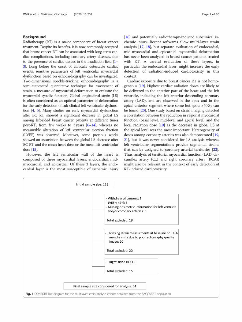
Fig. 1 CONSORT-like diagram for the multilayer strain analysis cohort obtained from the BACCARAT population
Walker et al. Radiation Oncology (2020) 15:201 Page 2 of 10

Based on the BACCARAT prospective cohort of leftand right BC patients treated with 3D-CRT, we had pre-sented a 6-month interim follow-up analysis on a second-ary outcome measure defined by a decrease of global LSfrom baseline to 6months after RT [15]. In the continu-ation of these previous results, the aim of this new paperwas to evaluate among left-sided BC patients whether thisdecrease in global LS at the scale of the left ventricle wasmyocardial layer-specific, depending on regional level andcoronary arterial territories, and whether coronary arteriesdoses were associated with territorial strain changes.
Patients and methodsStudy populationThe BACCARAT study initially included 118 female pa-tients of the Clinic Pasteur Toulouse from October 2015to December 2017, aged 40 to 75 years old, mainly withleft unilateral BC, and in a smaller proportion with right-sided unilateral BC. All patients were treated withadjuvant 3D-CRT after breast conserving surgery or mast-ectomy, without chemotherapy. Five patients withdrewconsent, 8 patients had abnormal LVEF before RT (LVEF< 45%) and 6 patients without available cardiac dosimetrywere excluded. We excluded patients with echocardiogra-phies for which the image quality was too low for a reli-able assessment of longitudinal strain (n = 20) remaining79 patients [15, 23]. For the analysis presented here, we fo-cused on left-sided BC patients, and finally, the patientstudy group consisted of 64 patients (Fig. 1). With afollow-up of 6-months after RT, none of the 64 patientsincluded had undergone chemotherapy.
Radiotherapy treatment and radiation doses evaluationAll patients were treated with 3D-CRT. The prescriptiondose was delivered over 5 weeks: either 50Gy in 25 daily
fractions of 2 Gy or 47Gy in 20 daily fractions of 2.35 Gy.The methods to evaluate radiation doses in BACCARATpatients were presented elsewhere [15, 19, 23]. Dose-Volume-Histogram (DVH) for the heart was generated bythe Clinic Pasteur radiotherapy department. Before RT, acoronary computed tomography angiography (CCTA) wasperformed for all patients as planned in the BACCARATprotocol. For dosimetric evaluation of coronary arteries,the simulation CT scan, the CCTA, the RT dose and RTstructure files in DICOM format were used. Merging ana-tomical information from the simulation CT scan and theCCTA was performed. Once inserted in ISOgray TPS (ver-sion 4.2, Dosisoft, Cachan, France; http://www.dosisoft.com/en/radiotherapy/planning-products.html), manual de-lineation was performed for the left ventricle (LV), the leftanterior descending artery (LAD), the left circumflex artery(Cx) and the right coronary artery (RCA). Using the 3Ddose matrix generated during treatment planning and thenew delineated substructures, DVH for LV and coronaryarteries were generated with ISOgray TPS by the dosimetricdepartment of IRSN in collaboration with the Clinic Pas-teur radiotherapy department. We thus obtained meandoses for the following cardiac structures: whole heart, leftventricle, left anterior descending artery, circumflex arteryand right coronary artery.
Transthoracic echocardiographyTransthoracic echocardiography was performed at base-line before RT and 6months after RT with ultrasoundAcuson S2000 device (Siemens Medical Solutions USA,Inc. Malvern, USA), using a 3MHz transducer. Imageanalysis was independently performed by a single blindedobserver unaware of clinical data. Longitudinal strain (LS)measurement was evaluated using two-dimensionalspeckle tracking [24] using a 16-segment model as
Fig. 2 “Bull-eye” presentation of the left ventricle: 16-segmental model and coronary artery territories. Legend: LAD - Left Anterior Descendingartery; Cx - Circumflex artery; RCA - Right Coronary Artery
Walker et al. Radiation Oncology (2020) 15:201 Page 3 of 10

recommended by the American Society of Echocardiog-raphy guidelines [25]. Global LS for the whole left ven-tricle was obtained for each myocardial layer (endocardial,mid-myocardial and epicardial). Mean LS for each re-gional level (basal, mid and apical) and for each coronaryartery territory (Territorial Longitudinal Strain (TLS)LAD, TLS Cx and TLS RCA) [22] was calculated as themean of segmental LS included in these levels (Fig. 2). Inparticular, the specific LS corresponding to the segmentsof the LAD (TLS LAD), was based on the average of seg-ments 1, 2, 7, 8, 13, 14 and 15 as indicated in Fig. 2.
Statistical analysisContinuous variables are presented with mean and stand-ard deviation or median and interquartile range values.Categorical values are presented with percentages. Stu-dent’s t-test or Wilcoxon non-parametric test was used tocompare continuous variables, adapted to paired samplesfor the comparison of echocardiographic variables beforeRT and 6months after RT. Comparison of layer-specificLS at baseline and RT + 6months were performed, meanrelative change was evaluated (Mean = V6 – V0 / V0).Specific analysis of segmental strains values according toregional level or coronary arteries territorial areas was per-formed, and the evolution of LS in these levels from base-line to RT + 6months was analyzed. We compared theseevolutions according to the group of exposure (“High” forpatients with cardiac doses >66th percentile of dose distri-bution, “Low” for others). Given the exploratory nature ofthis work, we presented unadjusted p-values for compari-sons, but in order to take into account multiple testing inthese comparisons we also applied the Holm–Bonferronimethod, a step-down procedure performed after conduct-ing the multiple comparison tests. Finally, p-value < 0.05was considered statistically significant. All statisticalanalysis was performed using SAS statistical software forWindows (Version 9.4 TS1M4 – SAS Institute, Cary, NC).
ResultsStudy populationSixty-four left-sided breast cancer patients were includedin the analysis. Baseline characteristics of the populationare shown in Table 1. The mean age at inclusion was 58 ±9 years. Concerning cardiac risk factors, 14 (22%), 20(31%) and 28 (44%) patients had hypertension, hyperchol-esterolemia and a BMI over 25 kg/m2, respectively. More-over, 8% of patients had diabetes, and 47% were currentsmokers. The mean dose received by the left ventricle wasmore than twice as high as the mean dose received by thewhole heart (6.68 ± 3.36 versus 3.05 ± 1.31, P = < 0.0001).For coronary arteries exposure, the highest mean dosewas found in the LAD (16.41 ± 7.41Gy), while the lowestmean dose was found in the RCA (0.71 ± 0.37 Gy).
Multilayer evolution of the global longitudinal strainEchocardiography parameters are displayed in Table 2. Leftventricular ejection fraction (LVEF) remained within nor-mal range after RT. A significant decrease in global longitu-dinal strain was observed for each myocardial layer, but thehighest mean relative change from baseline to RT + 6
Table 1 Baseline characteristics of the study population
Left-sided BCpatients n = 64
Age in years, mean ± SD 58 ± 9
Type of cancer, n (%)
In situ 11 (17%)
Invasive 53 (83%)
Surgery, n (%)
Breast conserving 61 (95%)
Mastectomy 3 (54%)
Regional lymph node irradiation 22 (34%)
Supraclavicular alone 1
Internal mammary alone 2
Both 19
Body mass index in kg/m2, mean ± SD 24.5 ± 4.2
Smoking, n (%)
Never-smokers 34 (53%)
Former smokers 20 (31%)
Current smokers 10 (16%)
Systolic blood pressure, in mmHg, mean ± SD 119 ± 12
Diastolic blood pressure in mmHg, mean ± SD 75 ± 10
Hypertension, n (%) 14 (22%)
Diabetes, n (%) 5 (8%)
Hypercholesterolemia, n (%) 20 (31%)
Cardiac doses in Gy
Whole Heart
Mean ± SD 3.05 ± 1.31
Min – Max 0.87–6.37
Left Ventricle
Mean ± SD 6.68 ± 3.36
Min – Max 1.16–13.42
Left Descending Artery
Mean ± SD 16.41 ± 7.41
Min – Max 1.68–34.63
Circumflex Artery
Mean ± SD 1.65 ± 0.82
Min – Max 0.53–4.34
Right Coronary Artery
Mean ± SD 0.71 ± 0.37
Min – Max 0.14–2.50
BC Breast Cancer, SD Standard Deviation
Walker et al. Radiation Oncology (2020) 15:201 Page 4 of 10

months was observed in the endocardial layer (− 4.7%, p =0.05) whereas for other layers, the mean relative changewas slightly lower.
Left ventricular regional evolution of the longitudinalstrain in the endocardial layerAmong the three regional left ventricular levels, a signifi-cant decrease of the LS was observed only in the apicallevel (− 26.3 ± 6.0% vs. -24.2 ± 7.1%, p = 0.03). AfterHolm-Bonferroni method for multiple testing for 3 tests,this decrease did not reach statistical significance. Whileseparating patients into two groups according to theirexposure to the left ventricle (Table 3), the regional ana-lysis showed that LS decreased significantly after RT atthe apical level in the highly exposed group correspond-ing to the 22 patients with LV Dose >66th percentile =8.6 Gy (− 25.5 ± 6.3 at V0 to − 22.7 ± 6.9 at V6; p = 0.04),which did not remain significant after multiple testingcorrection. More precisely, the segmental analysis ofstrain values (Fig. 3) showed a decrease in all segmentsof the apical level, with significant deteriorations in theapical inferior segment (segment 15) and the mid-anteroseptal segment (segment 8), but not significantafter Holm-Bonferroni method.
Coronary arteries territorial evolution of the endocardiallayer longitudinal strainThe coronary arteries territorial analysis showed no signifi-cant decrease for the Cx and the RCA. However, an
alteration of the LS was observed for the LAD territory (−22.8 ± 4.0% vs. -21.4 ± 4.8%, p = 0.03). While separating pa-tients into two groups according to their exposure to the leftventricle (Table 4), an alteration of the LS was observed forthe LAD territory in the highly exposed group correspondingto patients receiving > 8.6Gy to the LV (− 22.7 ± 3.4 at V0 to− 20.7 ± 4.5 at V6; p = 0.05).Moreover, longitudinal strain decrease in the LAD ter-
ritory could be associated with the dose level of the LADwith a significant decrease observed in the highly ex-posed group corresponding to the 22 patients with LADDose >66th percentile = 19.9 Gy (p = 0.02), and this re-sult remained significant after Holm-Bonferroni methodfor 2-tests comparisons. No significant difference couldbe observed in less exposed patients, corresponding to adecrease of LS from − 22.3% at V0 to − 19.5% at V6(Fig. 4). In comparison, no significant difference couldbe observed for the Cx and the RCA at the highest expo-sures, even by taking into account the precise dose tothese substructures (Figs. 5 and 6).
DiscussionAt the scale of the whole left ventricle, the previously re-ported deterioration of LS 6months after BC RT [15], isconfirmed on the three layers of the myocardial wall.However, our study suggests that the LS strain changemay be more relevant in the endocardial layer, in particu-lar in the most exposed areas of the left ventricle, corre-sponding to the apical region and the LAD territory.
Table 2 Comparison of baseline and follow-up measurements of echocardiographic data
Baseline n = 64 6months after RT n = 64 P-value Relative change (%)p-value
LVEF, % 61 ± 7 60 ± 9 0.073 Na.
GLS, %
Endocardial layer −20.0 ± 3.2 −18.8 ± 3.8 0.02 −4.7%; 0.05
Mid-myocardial layer −16.0 ± 2.7 −15.0 ± 3.1 0.02 − 4.4%; 0.11
Epicardial layer −12.3 ± 2.5 −11.4 ± 2.8 0.02 −4.2%; 0.25
LVEF Left Ventricular Ejection Fraction, GLS Global Longitudinal Strain, Na. Not assessed
Table 3 Regional analysis of longitudinal strains in the endocardial layer
Low dose to the LV n = 42 High dose to the LV n = 22
V0 V6 V0 V6
Basal level, % Mean ± SD −16.7 ± 5.6 −17.1 ± 5.0 −19.0 ± 3.5 −17.8 ± 3.2
p-value 0.52 0.10
Mid-level, % Mean ± SD − 18.6 ± 3.0 −17.0 ± 5.2 −17.6 ± 3.4 −17.5 ± 4.7
p-value 0.06 0.90
Apical level, % Mean ± SD −26.7 ± 5.8 −25.0 ± 7.2 −25.5 ± 6.3 −22.7 ± 6.9
p-value 0.18 0.04a
LV Left Ventricle. Low dose to the LV corresponds to patients receiving < 8.6 Gy to the LV (66th percentile of dose distribution among the 64 patients). High doseto the LV corresponds to patients receiving > 8.6 Gy. aNot significant after Holm-Bonferroni method for multiple testing
Walker et al. Radiation Oncology (2020) 15:201 Page 5 of 10

Difference of strain according to myocardial layersThe LS difference according to the myocardial layersthat we observed in either baseline or RT + 6monthsvalues, with higher values in the endocardial layerand lower values in the epicardial layer, has been pre-viously observed [26]. In normal heart, contraction isgreater in the endocardial layer than in the epicardiallayer [27] and the difference in amplitude of myocar-dial contraction between the endocardial and epicar-dial regions is related to the orientation pattern ofmyocardial fiber in the heart as the subendocardium
is predominantly composed of longitudinal myocardialfiber. Moreover, the longitudinal left ventricular me-chanics are predominantly governed by the subendo-cardial layer of the myocardium, which may explainthe significant decrease in global LS from baseline toRT + 6 months visits in the three layers. However,with greater contraction and higher energy require-ments, endocardial layer is more susceptible to injurywhich may explain that the relative decrease in LSwas slightly higher in the endocardial layer.
Location of LS deteriorationUnlike chemotherapy, which impact on myocardial func-tion can be considered global at the scale of the left ven-tricle, RT affects the heart in a more localized way as theapical level of the left ventricle is particularly exposed tothe tangential beams of the RT [19]. This may explain thestrongest decrease of the LS at the apical level as previ-ously observed [6]. Moreover, in a previous study of pa-tients with left-sided breast cancer [12], the segments witha significant strain reduction just after RT and 3 yearspost-RT were similar to those found in our study, particu-larly with regards to the mid-anterolateral segment andthe apical-inferior segment. However, the association be-tween LS decrease and cardiac dose is far to be clear andour correlations between LS decrease and doses to the dif-ferent cardiac structures were low (under 0.3), as previ-ously observed in several other studies [6, 10, 11].Concerning the coronary arteries territorial analysis of
LS, we found significant deterioration of the LS for theLAD. The strongest impact on the LAD territory could beexplained by the fact that the segments related to this cor-onary artery received highest radiation doses [19]. More-over, patients with the highest LAD doses (> 20Gy) werethose with a clear LS deterioration as illustrated in Fig. 3.
LS of the endocardial layer and LAD territoryIt is commonly accepted that the endocardium is themost susceptible target to ischemic injury [28, 29].Moreover, it has been shown that LS of the endocardial
Fig. 3 Segmental analysis of the endocardial layer according tolongitudinal strain based on bull’s eye representation (16 segmentmodel). Legend. Red for segments with significant decrease inlongitudinal strain from baseline to RT + 6 months (but notsignificant after Holm-Bonferroni method for multiple testing on 16tests); Orange for segments with non-significant decrease inlongitudinal strain; Blue for segments with non-significant increasein longitudinal strain
Table 4 Territorial analysis of longitudinal strains in the endocardial layer for the coronary arteries
Low dose to the LV N = 42 High dose to the LV N = 22
V0 V6 V0 V6
TLS - LAD, % Mean ± SD − 22.9 ± 4,3 −21.8 ± 5,0 − 22.7 ± 3.4 −20.7 ± 4.5
p-value 0.20 0.05a
TLS - Cx, % Mean ± SD − 19.0 ± 4.9 − 17.5 ± 5.1 −19.5 ± 3.8 −17.9 ± 5.5
p-value 0.10 0.25
TLS - RCA, % Mean ± SD − 16.2 ± 4.7 −16.2 ± 5.2 −16.9 ± 4.8 −15.6 ± 5.1
p-value 0.97 0.35
Left Ventricle; TLS Territorial Longitudinal Strain, LAD Left Anterior Descending artery, Cx Circumflex artery, RCA Right Coronary Artery. Low dose to the LVcorresponds to patients receiving < 8.6 Gy to the LV (66th percentile of dose distribution among the 64 patients). High dose to the LV corresponds to patientsreceiving > 8.6 Gy. aNot significant after Holm-Bonferroni method for multiple testing
Walker et al. Radiation Oncology (2020) 15:201 Page 6 of 10
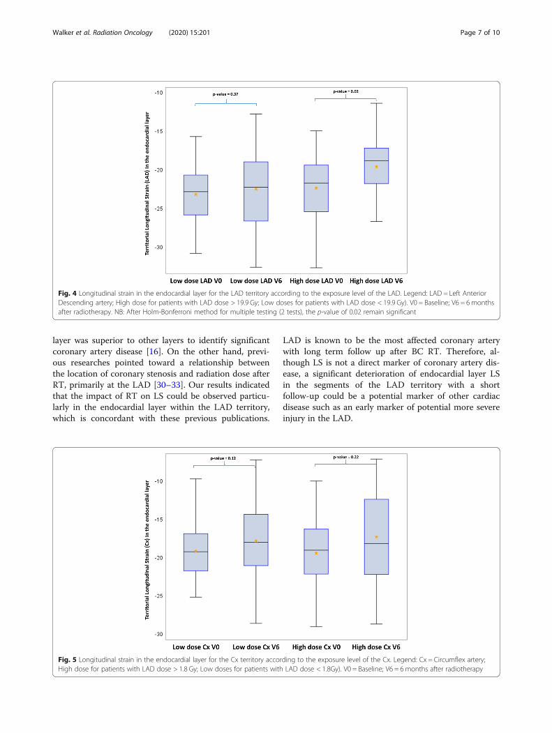
layer was superior to other layers to identify significantcoronary artery disease [16]. On the other hand, previ-ous researches pointed toward a relationship betweenthe location of coronary stenosis and radiation dose afterRT, primarily at the LAD [30–33]. Our results indicatedthat the impact of RT on LS could be observed particu-larly in the endocardial layer within the LAD territory,which is concordant with these previous publications.
LAD is known to be the most affected coronary arterywith long term follow up after BC RT. Therefore, al-though LS is not a direct marker of coronary artery dis-ease, a significant deterioration of endocardial layer LSin the segments of the LAD territory with a shortfollow-up could be a potential marker of other cardiacdisease such as an early marker of potential more severeinjury in the LAD.
Fig. 4 Longitudinal strain in the endocardial layer for the LAD territory according to the exposure level of the LAD. Legend: LAD = Left AnteriorDescending artery; High dose for patients with LAD dose > 19.9 Gy; Low doses for patients with LAD dose < 19.9 Gy). V0 = Baseline; V6 = 6 monthsafter radiotherapy. NB: After Holm-Bonferroni method for multiple testing (2 tests), the p-value of 0.02 remain significant
Fig. 5 Longitudinal strain in the endocardial layer for the Cx territory according to the exposure level of the Cx. Legend: Cx = Circumflex artery;High dose for patients with LAD dose > 1.8 Gy; Low doses for patients with LAD dose < 1.8Gy). V0 = Baseline; V6 = 6 months after radiotherapy
Walker et al. Radiation Oncology (2020) 15:201 Page 7 of 10

LimitationsSeveral limitations should be mentioned. The firstpoint is not specific of our study, but a general limi-tation of global LS to know how the changes in LSmight translate into clinical cardiac outcome parame-ters (morbidity/mortality). In the context of chemo-therapy, it has been showed that there is a correlationand a predictive value of GLS decrease on the laterpresence of the outcome CTRCD (CancerTherapeutics-Related Cardiac Dysfunction) defined bya decrease in LVEF of at least 10% to a value < 53%[4]. However, such CTRCD may be reversible in somecases and do not translate into clinical cardiac mor-bidity. Although the decrease in longitudinal strainand LVEF appears to at least partially persist through-out the treatment it is unknown what their evolutionwill be in subsequent years, and whether early de-formation measurements will predict persistent de-creases in LVEF or symptomatic heart failure. In thecontext of general population, a wide Danish studyestablished that lower GLS was associated with ahigher risk of a composite endpoint defined by inci-dent heart failure, acute myocardial infarction or car-diovascular death (HR 1.12 [1.08–1.17], p < 0.001 per1% decrease) [34]. In the context of radiotherapy, it isstill unknown whether changes in LS will translateinto clinical cardiac morbidity or mortality. In sum-mary, based on different studies in different contexts,use of GLS measurement in the specific context ofradiotherapy-treated patients is more and more fre-quent as an additional parameter (in particular to
LVEF) to potentially predict later cardiac morbi/mor-tality, but it has still to be investigated and validatedin observational studies with long follow-up. As a sec-ond limitation, our study is a prospective study butbased on short follow-up and a relatively small popu-lation of left-sided BC patients, which did not provideinformation on the value of specific myocardial de-formation parameters in the prognosis of cardiaccomplication, in particular injury to the LAD. Furtherstudies with clear clinical endpoints will be requiredto determine the clinical significance of our findings.In particular, the observation of decreased LS in theLAD territory as well as in the apical region of theleft ventricle should be compared with observationsfrom computed tomography coronary angiographyanalysis [19, 31]. Furthermore, multi-layer strain ana-lysis is controversial [35], limited by poor reproduci-bility and important variability [26]. Our results wereexploratory and should be confirmed by other studies.
ConclusionsWith a follow-up of 6 months after RT, LS decrease waspredominantly in the endocardial myocardial layer andappeared to be localized in the most exposed part of theleft ventricle. For precise evaluation of RT-induced car-diotoxicity and early left ventricular dysfunction, theendocardial layer-based LS might be the most sensitiveparameter, in particular to evaluate the impact of radi-ation exposure during BC RT to the LAD. However, thisexploratory analysis remains to be confirmed with largerstudies and longer follow-up.
Fig. 6 Longitudinal strain in the endocardial layer for the RCA territory according to the exposure level of the RCA. Legend: RCA = Right CoronaryArtery; High dose for patients with LAD dose > 0.8 Gy; Low doses for patients with LAD dose < 0.8Gy). V0 = Baseline; V6 = 6monthsafter radiotherapy
Walker et al. Radiation Oncology (2020) 15:201 Page 8 of 10

Abbreviations3D-CRT: Three-Dimensional Conformal Radiation Therapy; Cx: Circumflexartery; LAD: Left Anterior Descending coronary artery; LS: Longitudinal Strain;LVEF: Left Ventricular Ejection Fraction; RCA: Right Coronary Artery;RT: Radiotherapy (or Radiation Therapy)
AcknowledgementsNot applicable.
Authors’ contributionsConceived study: SJ, VW, OL, GJ, MOB, DL, JF. Data analysis: OL, OF, GJ, JC, LP,DB, JF, SJ. Statistical analysis: VW, MOB, JF, DL, SJ. All authors participated inthe writing, and manuscript edition. All authors read and approved the finalmanuscript.
Authors’ informationNot applicable.
FundingThe study received funding for collection of data from the FédérationFrançaise de Cardiologie (FFC), from Electricité de France (EDF) and from theH2020 Euratom research and training program 2014–2018 under grantagreement No 755523 in the frame of the MEDIRAD project.
Availability of data and materialsThe datasets used and/or analysed during the current study are availablefrom the corresponding author on reasonable request.
Ethics approval and consent to participateThis study received ethical approval from the French South West Committeefor Protection of Persons (ID: CPP2015/66/2015-A00990–69) and fromNational Agency for Medical and Health products Safety (Reference:150873B-12). All patients enrolled in the study provided their written in-formed consent.
Consent for publicationNot applicable.
Competing interestsDeclarations of interest: none.
Author details1Pôle Santé-Environnement (PSE-SANTE), Service de recherche sur les effetsbiologiques et sanitaires des rayonnements ionisants (SESANE), Laboratoired’épidémiologie des rayonnements ionisants (LEPID), Institute forRadiological Protection and Nuclear Safety (IRSN), BP17, 92262Fontenay-aux-Roses cedex, France. 2Department of Cardiology, RangueilUniversity Hospital, 31059 Toulouse, France. 3Cardiac Imaging Centre,Rangueil University Hospital, 31059 Toulouse, France. 4Medical School ofRangueil, University Paul Sabatier, 31400 Toulouse, France. 5Department ofCardiology, Clinique Pasteur, 31300 Toulouse, France. 6Department ofRadiation Oncology (Oncorad), Clinique Pasteur, 31300 Toulouse, France.7Department of Cardiac Arrhythmia, Clinique Pasteur, 31300 Toulouse,France. 8Department of dosimetry, Institute for Radiological Protection andNuclear Safety (IRSN), Fontenay-aux-Roses, France. 9Division of Health andEnvironment, Institute for Radiological Protection and Nuclear Safety (IRSN),Fontenay-aux-Roses, France. 10Medical School of Purpan, University PaulSabatier, 31000 Toulouse, France. 11INSERM, UMR1027, 31000 Toulouse,France.
Received: 23 June 2020 Accepted: 4 August 2020
References1. Gagliardi G, Lax I, Rutqvist LE. Partial irradiation of the heart. Semin Radiat
Oncol. 2001;11:224–33.2. Darby SC, Cutter DJ, Boerma M, Constine LS, Fajardo LF, Kodama K, et al.
Radiation-related heart disease: current knowledge and future prospects. IntJ Radiat Oncol. 2010;76:656–65.
3. Piroth MD, Baumann R, Budach W, Dunst J, Feyer P, Fietkau R, et al. Hearttoxicity from breast cancer radiotherapy: current findings, assessment, andprevention. Strahlenther Onkol. 2019;195:1–12.
4. Plana JC, Galderisi M, Barac A, Ewer MS, Ky B, Scherrer-Crosbie M, et al.Expert consensus for multimodality imaging evaluation of adult patientsduring and after cancer therapy: a report from the American Society ofEchocardiography and the European Association of Cardiovascular Imaging.Eur Heart J - Cardiovasc Imaging. 2014;15:1063–93.
5. Thavendiranathan P, Poulin F, Lim K-D, Plana JC, Woo A, Marwick TH. Use ofmyocardial strain imaging by echocardiography for the early detection ofCardiotoxicity in patients during and after Cancer chemotherapy. J Am CollCardiol. 2014;63:2751–68.
6. Erven K, Jurcut R, Weltens C, Giusca S, Ector J, Wildiers H, et al. Acuteradiation effects on cardiac function detected by strain rate imaging inbreast Cancer patients. Int J Radiat Oncol. 2011;79:1444–51.
7. Erven K, Florian A, Slagmolen P, Sweldens C, Jurcut R, Wildiers H, et al.Subclinical Cardiotoxicity detected by strain rate imaging up to 14 monthsafter breast radiation therapy. Int J Radiat Oncol. 2013;85:1172–8.
8. Heggemann F, Grotz H, Welzel G, Dösch C, Hansmann J, Kraus-TiefenbacherU, et al. Cardiac function after multimodal breast Cancer therapy assessedwith functional magnetic resonance imaging and echocardiographyimaging. Int J Radiat Oncol. 2015;93:836–44.
9. Sritharan HP, Delaney GP, Lo Q, Batumalai V, Xuan W, Thomas L.Evaluation of traditional and novel echocardiographic methods ofcardiac diastolic dysfunction post radiotherapy in breast cancer. Int JCardiol. 2017;243:204–8.
10. Lo Q, Hee L, Batumalai V, Allman C, MacDonald P, Lonergan D, et al. Strainimaging detects dose-dependent segmental cardiac dysfunction in theacute phase after breast irradiation. Int J Radiat Oncol. 2017;99:182–90.
11. Tuohinen SS, Skyttä T, Poutanen T, Huhtala H, Virtanen V, Kellokumpu-Lehtinen P-L, et al. Radiotherapy-induced global and regional differences inearly-stage left-sided versus right-sided breast cancer patients: speckletracking echocardiography study. Int J Cardiovasc Imaging. 2017;33:463–72.
12. Tuohinen SS, Skytta T, Huhtala H, Virtanen V, Kellokumpu-Lehtinen P-L,Raatikainen P. Left ventricular speckle tracking echocardiography changesamong early-stage breast Cancer patients three years after radiotherapy.Anticancer Res. 2019;39:4227–36.
13. Chen L, Ta S, Wu W, Wang C, Zhang Q. Prognostic and added value ofechocardiographic strain for prediction of adverse outcomes in patientswith locally advanced non-small cell lung Cancer after radiotherapy.Ultrasound Med Biol. 2019;45:98–107.
14. Trivedi SJ, Choudhary P, Lo Q, Sritharan HP, Iyer A, Batumalai V, et al.Persistent reduction in global longitudinal strain in the longer term afterradiation therapy in patients with breast cancer. Radiother Oncol. 2019;132:148–54.
15. Walker V, Lairez O, Fondard O, Pathak A, Pinel B, Chevelle C, et al. Earlydetection of subclinical left ventricular dysfunction after breast cancerradiation therapy using speckle-tracking echocardiography: associationbetween cardiac exposure and longitudinal strain reduction (BACCARATstudy). Radiat Oncol. 2019;14:204.
16. Sarvari SI, Haugaa KH, Zahid W, Bendz B, Aakhus S, Aaberge L, et al. Layer-specific quantification of myocardial deformation by strainechocardiography may reveal significant CAD in patients with non–ST-segment elevation acute coronary syndrome. JACC Cardiovasc Imaging.2013;6:535–44.
17. Adamu U, Schmitz F, Becker M, Kelm M, Hoffmann R. Advanced speckletracking echocardiography allowing a three-myocardial layer-specificanalysis of deformation parameters. Eur J Echocardiogr. 2008;10:303–8.
18. Leitman M, Lysiansky M, Lysyansky P, Friedman Z, Tyomkin V, Fuchs T, et al.Circumferential and longitudinal strain in 3 myocardial layers in Normalsubjects and in patients with regional left ventricular dysfunction. J Am SocEchocardiogr. 2010;23:64–70.
19. Jacob S, Camilleri J, Derreumaux S, Walker V, Lairez O, Lapeyre M, et al. Ismean heart dose a relevant surrogate parameter of left ventricle andcoronary arteries exposure during breast cancer radiotherapy: a dosimetricevaluation based on individually-determined radiation dose (BACCARATstudy). Radiat Oncol. 2019;14:29.
20. Moignier A, Broggio D, Derreumaux S, El Baf F, Mandin A-M, Girinsky T, et al.Dependence of coronary 3-dimensional dose maps on coronary topologiesand beam set in breast radiation therapy: a study based on CTangiographies. Int J Radiat Oncol. 2014;89:182–90.
Walker et al. Radiation Oncology (2020) 15:201 Page 9 of 10

21. Nilsson G, Witt Nyström P, Isacsson U, Garmo H, Duvernoy O, Sjögren I,et al. Radiation dose distribution in coronary arteries in breast cancerradiotherapy. Acta Oncol. 2016;55:959–63.
22. Lang RM, Badano LP, Mor-Avi V, Afilalo J, Armstrong A, Ernande L, et al.Recommendations for Cardiac Chamber Quantification byEchocardiography in Adults: An Update from the American Society ofEchocardiography and the European Association of Cardiovascular Imaging.J Am Soc Echocardiogr. 2015;28:1–39 e14.
23. Jacob S, Pathak A, Franck D, Latorzeff I, Jimenez G, Fondard O, et al. Earlydetection and prediction of cardiotoxicity after radiation therapy for breastcancer: the BACCARAT prospective cohort study. Radiat Oncol. 2016;11:54.
24. Voigt J-U, Pedrizzetti G, Lysyansky P, Marwick TH, Houle H, Baumann R, et al.Definitions for a common standard for 2D speckle trackingechocardiography: consensus document of the EACVI/ASE/industry taskforce to standardize deformation imaging. J Am Soc Echocardiogr Off PublAm Soc Echocardiogr. 2015;28:183–93.
25. Lang RM, Bierig M, Devereux RB, Flachskampf FA, Foster E, Pellikka PA, et al.Recommendations for chamber quantification: a report from the AmericanSociety of Echocardiography’s guidelines and standards committee and thechamber quantification writing group, developed in conjunction with theEuropean Association of Echocardiography, a branch of the EuropeanSociety of Cardiology. J Am Soc Echocardiogr. 2005;18:1440–63.
26. Ancedy Y, Ederhy S, Lang S, Hollebecque A, Dufour LS, Adavane-Scheuble S,et al. Multilayer global longitudinal strain in patients with cancer: acomparison of two vendors. Arch Cardiovasc Dis. 2018;111:285–96.
27. Tsutsui H, Uematsu M, Yamagishi M, Haruta S, Shimakura T, Miyatake K.Usefulness of the subendocardial myocardial velocity gradient in low-dosedobutamine stress echocardiography. Heart Vessel. 2000;15:11–7.
28. Duncker DJ, Traverse JH, Ishibashi Y, Bache RJ. Effect of NO on transmuraldistribution of blood flow in hypertrophied left ventricle during exercise.Am J Physiol-Heart Circ Physiol. 1999;276:H1305–12.
29. Geer JC, Crago CA, Little WC, Gardner LL, Bishop SP. Subendocardialischemic myocardial lesions associated with severe coronary atherosclerosis.Am J Pathol. 1980;98:18.
30. Correa CR, Litt HI, Hwang W-T, Ferrari VA, Solin LJ, Harris EE. Coronary arteryfindings after left-sided compared with right-sided radiation treatment forearly-stage breast Cancer. J Clin Oncol. 2007;25:3031–7.
31. Nilsson G, Holmberg L, Garmo H, Duvernoy O, Sjögren I, Lagerqvist B, et al.Distribution of coronary artery stenosis after radiation for breast Cancer. JClin Oncol. 2012;30:380–6.
32. Zagar TM, Marks LB. Breast Cancer radiotherapy and coronary arterystenosis: location, location, location. J Clin Oncol. 2012;30:350–2.
33. Wennstig A-K, Garmo H, Isacsson U, Gagliardi G, Rintelä N, Lagerqvist B,et al. The relationship between radiation doses to coronary arteries andlocation of coronary stenosis requiring intervention in breast cancersurvivors. Radiat Oncol. 2019;14:40 [cited 2020 Feb 28]. Available from:https://ro-journal.biomedcentral.com/articles/10.1186/s13014-019-1242-z.
34. Tor B-S, Reumert B-SS, Javier OF, Morten S, Godsk JP, Rasmus M, et al.Global Longitudinal Strain by Echocardiography Predicts Long-Term Risk ofCardiovascular Morbidity and Mortality in a Low-Risk General Population.Circ Cardiovasc Imaging. 2017;10:e005521.
35. Orloff E, Fournier P, Bouisset F, Moine T, Cournot M, Elbaz M, et al.Myocardial multilayer strain does not provide additional value for detectionof myocardial viability assessed by SPECT imaging over and beyondstandard strain. Echocardiography. 2018;35:1300–9.
Publisher’s NoteSpringer Nature remains neutral with regard to jurisdictional claims inpublished maps and institutional affiliations.
Walker et al. Radiation Oncology (2020) 15:201 Page 10 of 10