Myocardial cell death in fibrillating and dilated human ... · Paris and Le Plessis Robinson,...
10
Myocardial Cell Death in Fibrillating and Dilated Human Right Atria Christine Aime ´-Sempe ´, PHD,* Thierry Folliguet, MD,§ Catherine Ru ¨cker-Martin, PHD,‡ Maryla Krajewska, MD,\ Stanislaw Krajewski, MD,\ Miche `le Heimburger, PHD,² Michel Aubier, MD, PHD,* Jean-Jacques Mercadier, MD, PHD,² John C. Reed, MD, PHD,\ Stephane N. Hatem, MD, PHD² Paris and Le Plessis Robinson, France and La Jolla, California OBJECTIVES The aim of the present study was to determine if myocytes can die by apoptosis in fibrillating and dilated human atria. BACKGROUND The cellular remodeling that occurs during atrial fibrillation (AF) may reflect a degree of dedifferentiation of the atrial myocardium, a process that may be reversible. METHODS We examined human right atrial myocardium specimens (n 5 50) for the presence of apoptotic myocytes. We used immunohistochemical and Western blotting analysis to examine the expression of a final effector of programmed cell death, caspase-3 (CASP-3) and of regulatory proteins from the BCL-2 family. RESULTS Sections from atria in AF contained a high percentage of large myocytes with a disrupted sarcomeric apparatus replaced by glycogen granules (64.4 6 6.3% vs. 12.2 6 5.8%). These abnormal myocytes, which also predominated in atria from hearts with decreased left ventricular ejection fraction (42.3 6 10.1%), contained large nuclei, most of which were TUNEL positive, indicating a degree of DNA breakage. None of these abnormal myocytes expressed the proliferative antigen Ki-67. A small percentage of the enlarged nuclei (4.2 6 0.8%) contained condensed chromatin and were strongly TUNEL positive. Both the pro- and activated forms of CASP-3 were detected in diseased myocardial samples, which also showed stronger CASP-3 expression than controls. Expression of the antiapoptotic BCL-2 protein was decreased in diseased atria, whereas that of the proapoptotic BAX protein remained unchanged. CONCLUSIONS In fibrillating and dilated atria, apoptotic death of myocytes with myolysis contributes to cellular remodeling, which may not be entirely reversible. (J Am Coll Cardiol 1999;34: 1577– 86) © 1999 by the American College of Cardiology Atrial fibrillation (AF) is the most frequent sustained cardiac arrhythmia and causes serious deleterious effects such as impairment of cardiac function or thromboembolic events (1). This arrhythmia is due to circuits of micro- reentry of electrical impulses in the atrial wall, a process referred to as the multiple-wavelets mechanism (2). The incidence of wavelets that can coexist is determined by both the mass and the electrical vulnerability of the atrial myo- cardium, explaining why AF is often observed in clinical situations associated with atrial enlargement and shortening of the atrial refractory period (3,4). The other major characteristic of AF is its tendency to become sustained, suggesting that AF induces a vicious circle leading to the increase in its arrhythmogenic substrate (4). Distinct cellular alterations occur in AF that may also contribute to its perpetuation. In human atrial biopsies or in animal models of AF, a number of myocytes lose their sarcomeric apparatus, which is replaced by accumulation of glycogen granules (5–9). Because these abnormal myocytes reexpress some fetal proteins, it has been proposed that they are dedifferentiated, reflecting a certain degree of adaptation of the atrial myocardium to changes in its working condi- tions, a process that may be reversible (9). It is also conceivable that as the result of the persistence of patho- genic factors such as increased pressure load or high fre- quency of beating, some of the myocytes with structural alterations could activate a programmed cell death (PCD) pathway. Programmed cell death is an active cell suicide mecha- nism culminating in characteristic features commonly From *INSERM U408 and ²INSERM U460, Faculte ´ de Me ´decine, Xavier Bichat, Paris, France; ‡Universite ´ de Paris XI-CNRS ERS 566, Ho ˆ pital Marie Lannelongue, Le Plessis Robinson, France; §the Department de Chirurgie Cardiaque, Institut Mutualiste Montsouris, Paris, France; and \the Burnham Institute, La Jolla, Califor- nia. This work was supported by grants from the Institut National de la Sante ´ et de la Recherche Me ´dicale (INSERM), the Association Franc ¸aise contre les Myopathies (A.F.M.) and from the Assistance Public-Ho ˆ pitaux de Paris (AOB94038). Christine Aime ´-Sempe ´ was supported by a grant from the Fondation pour la Recherche Me ´dicale (FRM). Manuscript received March 12, 1999; revised manuscript received May 20, 1999, accepted June 29, 1999. Journal of the American College of Cardiology Vol. 34, No. 5, 1999 © 1999 by the American College of Cardiology ISSN 0735-1097/99/$20.00 Published by Elsevier Science Inc. PII S0735-1097(99)00382-4
Transcript of Myocardial cell death in fibrillating and dilated human ... · Paris and Le Plessis Robinson,...

Myocardial Cell Death inFibrillating and Dilated Human Right AtriaChristine Aime-Sempe, PHD,* Thierry Folliguet, MD,§ Catherine Rucker-Martin, PHD,‡Maryla Krajewska, MD,\ Stanislaw Krajewski, MD,\ Michele Heimburger, PHD,†Michel Aubier, MD, PHD,* Jean-Jacques Mercadier, MD, PHD,† John C. Reed, MD, PHD,\Stephane N. Hatem, MD, PHD†Paris and Le Plessis Robinson, France and La Jolla, California
OBJECTIVES The aim of the present study was to determine if myocytes can die by apoptosis in fibrillatingand dilated human atria.
BACKGROUND The cellular remodeling that occurs during atrial fibrillation (AF) may reflect a degree ofdedifferentiation of the atrial myocardium, a process that may be reversible.
METHODS We examined human right atrial myocardium specimens (n 5 50) for the presence ofapoptotic myocytes. We used immunohistochemical and Western blotting analysis toexamine the expression of a final effector of programmed cell death, caspase-3 (CASP-3) andof regulatory proteins from the BCL-2 family.
RESULTS Sections from atria in AF contained a high percentage of large myocytes with a disruptedsarcomeric apparatus replaced by glycogen granules (64.4 6 6.3% vs. 12.2 6 5.8%). Theseabnormal myocytes, which also predominated in atria from hearts with decreased leftventricular ejection fraction (42.3 6 10.1%), contained large nuclei, most of which wereTUNEL positive, indicating a degree of DNA breakage. None of these abnormal myocytesexpressed the proliferative antigen Ki-67. A small percentage of the enlarged nuclei (4.2 60.8%) contained condensed chromatin and were strongly TUNEL positive. Both the pro- andactivated forms of CASP-3 were detected in diseased myocardial samples, which also showedstronger CASP-3 expression than controls. Expression of the antiapoptotic BCL-2 proteinwas decreased in diseased atria, whereas that of the proapoptotic BAX protein remainedunchanged.
CONCLUSIONS In fibrillating and dilated atria, apoptotic death of myocytes with myolysis contributes tocellular remodeling, which may not be entirely reversible. (J Am Coll Cardiol 1999;34:1577–86) © 1999 by the American College of Cardiology
Atrial fibrillation (AF) is the most frequent sustainedcardiac arrhythmia and causes serious deleterious effectssuch as impairment of cardiac function or thromboembolicevents (1). This arrhythmia is due to circuits of micro-reentry of electrical impulses in the atrial wall, a processreferred to as the multiple-wavelets mechanism (2). Theincidence of wavelets that can coexist is determined by boththe mass and the electrical vulnerability of the atrial myo-cardium, explaining why AF is often observed in clinicalsituations associated with atrial enlargement and shortening
of the atrial refractory period (3,4). The other majorcharacteristic of AF is its tendency to become sustained,suggesting that AF induces a vicious circle leading to theincrease in its arrhythmogenic substrate (4).
Distinct cellular alterations occur in AF that may alsocontribute to its perpetuation. In human atrial biopsies or inanimal models of AF, a number of myocytes lose theirsarcomeric apparatus, which is replaced by accumulation ofglycogen granules (5–9). Because these abnormal myocytesreexpress some fetal proteins, it has been proposed that theyare dedifferentiated, reflecting a certain degree of adaptationof the atrial myocardium to changes in its working condi-tions, a process that may be reversible (9). It is alsoconceivable that as the result of the persistence of patho-genic factors such as increased pressure load or high fre-quency of beating, some of the myocytes with structuralalterations could activate a programmed cell death (PCD)pathway.
Programmed cell death is an active cell suicide mecha-nism culminating in characteristic features commonly
From *INSERM U408 and †INSERM U460, Faculte de Medecine, Xavier Bichat,Paris, France; ‡Universite de Paris XI-CNRS ERS 566, Hopital Marie Lannelongue,Le Plessis Robinson, France; §the Department de Chirurgie Cardiaque, InstitutMutualiste Montsouris, Paris, France; and \the Burnham Institute, La Jolla, Califor-nia. This work was supported by grants from the Institut National de la Sante et dela Recherche Medicale (INSERM), the Association Francaise contre les Myopathies(A.F.M.) and from the Assistance Public-Hopitaux de Paris (AOB94038). ChristineAime-Sempe was supported by a grant from the Fondation pour la RechercheMedicale (FRM).
Manuscript received March 12, 1999; revised manuscript received May 20, 1999,accepted June 29, 1999.
Journal of the American College of Cardiology Vol. 34, No. 5, 1999© 1999 by the American College of Cardiology ISSN 0735-1097/99/$20.00Published by Elsevier Science Inc. PII S0735-1097(99)00382-4
known as apoptosis. During this process, caspases, whichare highly specific cysteine proteases, function in both celldisassembly and in initiating this disassembly in response toproapoptotic signals by cleaving a discrete set of proteinssuch as ICAD/CAD, gelsolin or proteins involved in DNArepair such as Poly (ADP ribose) polymerase (10,11). Thecaspases are all expressed as proenzymes that contain threedomains: an NH2-terminal domain, a large and a smallsubunit. Activation involves proteolytic processing betweendomains, followed by association of the large and smallsubunits to form a functional heterodimer. The Caspase-3/CPP32 (CASP-3) is one of the major caspases involved inapoptosis (11). BCL-2 is the founding member of agrowing family of proteins that either protect against celldeath such as BCL-2 or that promote apoptosis such asBAX (12). The data available to date indicate that BCL-2family proteins may dictate whether effector caspases be-come active or remain quiescent after exposure of cells toapoptotic stimuli (13). Both caspases and BCL-2 familyproteins are involved in the apoptosis of cardiac myocytes,which occurs in various cardiopathies (14–17).
The aim of the present study was to examine if atrialmyocytes can undergo an apoptotic-like death process inchronic AF or in clinical settings associated with a highincidence of this arrhythmia.
METHODS
Cardiac specimens. With approval from our Ethics Com-mittee, specimens of the right atrial appendage were ob-tained from 50 patients (31 to 86 years old) undergoingheart surgery for coronary artery disease, valve disease orcongenital heart defects (Table 1). Patients with chronic AFhad left and right atrial dilation (.45 mm), most oftenassociated with increased systolic pulmonary pressure(.35 mm Hg) indicating longstanding valve disease (18).Specimens of the right atrial appendage were fixed imme-diately after excision and maintained in 10% bufferedformalin overnight or fixed 3 h in Bouin solution andembedded in paraffin or frozen in liquid nitrogen (L2N) orisopentane.
Histological analysis. Tissue sections were deparaffinized,transferred to xylene and rehydrated in decreasing concen-
trations of alcohol. Then slides were stained with hematein,eosin, orcein, Masson’s trichrome or periodic acid schiff.Myocyte diameter was determined by measuring the shortaxis of 100 randomly chosen cells per field. To quantify theamount and the percentage of myocytes with myolysis, twosections per atrial samples were analyzed, and at least 400cells per section were analyzed. Cells were scored bymorphometry as severely myolytic if .25% of the sarcomerewas absent.
Immunohistochemistry. Cryo-sections (5 mm) were incu-bated in phosphate-buffered saline (PBS) containing 5%BSA, followed by incubation with mouse anti-rabbit sarco-meric a-actinin antibody (1/400; Sigma Aldrich, St. Louis,Missouri), then incubated with goat biotinylated anti-mouse IgG secondary antibody (1/30; Vector Laboratories,Paris, France) and with streptavidin-Texas red or fluores-cein (1/30; Amersham). Some observations were carried outwith an MRC-1024 (Bio-Rad, UK) confocal scanning laserwith microscope (Nikon Optiphot Fluorescence) usingLasersharp (Bio-Rad).
For expression and localization of CASP-3, BCL-2 andBAX proteins, deparaffinized sections were either useddirectly or subjected to microwave oven antigen retrievaltreatment (19,20). Tissue sections were immunostainedwith a rabbit anti-CASP-3, anti-BCL-2 or anti-BAXantiserum. The immunoreactions were visualized with adiaminobenzidine (DAB)-based colorimetric method, aspreviously described (20). Slides were lightly counterstainedwith hematoxylin and permanently mounted with De-Pe-X(Fluka, Buchs, Germany) before being analyzed and photo-graphed with an Olympus BH-2 microscope connected to acamera. The specificity of the staining was confirmed byusing preimmune serum and/or by preabsorbing the anti-serum with specific peptide or recombinant protein beforeperforming immunostaining.
Western blot. Protein lysates were prepared, normalizedfor total protein content (200 or 100 mg per lane) andanalyzed by 12.5% or 10% SDS-PAGE, followed byelectro-transfer to polyvinylidene difluoride membranes(Bio-Rad). Immunodection was performed using anti-CASP-3 (19), anti-BCL-2, anti-BAX (20) antitubulin oranti-a-actinin sarcomeric monoclonal (Sigma, St. Louis,MO) antibodies followed by horseradish peroxydase(HRPase)-conjugated secondary antibody (Amersham LifeSciences). Detection was performed using enhanced chemi-luminescence detection method (ECL1; Amersham LifeScience). Densitometric analysis was performed using Gelanalyst system (ICONIX).
Staining of Ki-67 antigen. The staining of Ki-67 antigenwas performed on paraffin-embedded sections, using theMIB-1 monoclonal antibody (1/1,000 dilution; DakoA/S,Copenhagen, Denmark). Detection was accomplished usingstreptavidin biotinylated horseradish peroxydase method(Dako). DAB tetrahydrochloride 0.06% in PBS containing
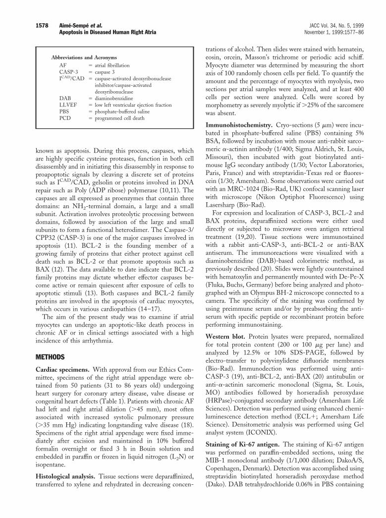
Abbreviations and AcronymsAF 5 atrial fibrillationCASP-3 5 caspase 3ICAD/CAD 5 caspase-activated deoxyribonuclease
inhibitor/caspase-activateddeoxyribonuclease
DAB 5 diaminobenzidineLLVEF 5 low left ventricular ejection fractionPBS 5 phosphate-buffered salinePCD 5 programmed cell death
1578 Aime-Sempe et al. JACC Vol. 34, No. 5, 1999Apoptosis in Diseased Human Right Atria November 1, 1999:1577–86

Table 1. Clinical Characteristics of Patients
Patient GenderAge
(years)
Rhythm(Time in
Year) DiagnosisEF(%)
LA(mm)
PAPSystolic
1 Male 78 AF (1 month) AS 32 55 , 252 Female 67 AF (10) MR 45 58 503 Female 74 AF (12) MS 46 65 , 254 Male 69 AF (13) MR 50 44 405 Male 54 AF (2) MS 52 63 356 Female 66 AF (20) MS 50 63 537 Male 69 AF (3) MS 52 56 , 258 Female 65 AF (6) AS 50 50 , 259 Male 51 AF (7) MS/AI 35 54 , 25
10 Female 73 AF (7) MS 54 65 4111 Male 86 AF (8) AS 50 50 3012 Male 34 AF (2) HypoRV 10 70 2413 Male 69 AF (5) MR 56 50 6014 Male 77 AF (10) CAD 50 59 , 2515 Male 71 AF (5) MR 58 48 4516 Male 64 AF (9) MR 35 70 4514 Male 77 AF (5) CAD 66 58 3515 Male 78 Flutter (2) AS 40 39 6015 Male 31 SR CAD 55 36 , 2516 Male 63 SR CAD 50 40 , 2517 Male 55 SR CAD 55 35 , 2518 Male 59 SR CAD 55 35 , 2519 Male 53 SR CAD 55 40 , 2520 Male 54 SR CAD , 20 . 50 . 5021 Male 59 SR CAD , 20 . 50 . 5022 Male 44 SR CAD , 20 . 50 . 5023 Female 74 SR CAD 29 49 , 2524 Female 38 SR AS/AI 60 29 , 2525 Female 44 SR CAD 50 30 , 2526 Male 65 SR CAD 47 30 , 2527 Male 47 SR CAD 70 30 , 2528 Male 60 SR CAD 20 30 , 2529 Female 48 SR CAD 60 31 , 2530 Male 70 SR CAD 47 35 , 2531 Male 57 SR CAD 47 37 , 2532 Male 81 SR AS 50 37 3033 Female 68 SR CAD 40 38 , 2534 Male 77 SR CAD 50 38 , 2535 Male 53 SR MS 50 39 5236 Female 62 SR CAD 60 39 , 2537 Female 45 SR MR 48 39 , 2538 Male 54 SR CAD 60 41 , 2539 Female 77 SR CAD 45 45 3040 Male 75 SR AS 45 46 , 2541 Male 78 SR CAD 60 46 , 2542 Female 82 SR CAD 60 47 , 2543 Female 75 SR CAD 30 47 4544 Male 66 SR AS 35 47 3545 Male 75 SR AS 58 48 , 2546 Male 38 SR MR 42 58 6547 Male 77 SR MR 50 45 4548 Female 74 SR AS/CAD 50 39 , 2549 Male 73 SR AS 11 54 3250 Male 70 SR AS 55 58 , 25
AI 5 aortic insufficiency; AS 5 aortic stenosis; CAD 5 coronary artery disease; EF 5 ejection fraction; HypoRV 5 hypo right ventricule; LA 5 left atrium; MR 5 mitralregurgitation; MS 5 mitral stenosis; PAP 5 pulmonary artery pressure; SR 5 sinus rhythm.
1579JACC Vol. 34, No. 5, 1999 Aime-Sempe et al.November 1, 1999:1577–86 Apoptosis in Diseased Human Right Atria

0.03% hydrogen peroxyde was used as a chromogen. Aspositive control, we used sections from squamous cellcarcinomas.
Nuclear staining with DAPI. Deparaffinized tissue sec-tions were incubated with the intercalating agent DAPI (49,6-diamidino-2-phenylindole, 0.3 mg/ml in PBS, 15 min) tovisualize nuclear morphology.
TUNEL assay. Sections were transferred to xylene andrehydrated in decreasing concentrations of alcohol. Slideswere then incubated (10 min, room temperature) with10 mg of proteinase K (Sigma-Aldrich, St. Louis, MO) permilliliter of PBS. Endogenous peroxydase was inactivatedby immunopure peroxydase suppressor for 30 min (Pierce).Tissue sections permeabilized with 1% Triton X-100 (4°C,2 min) were stained with an in situ cell detection (POD)system (Boehringer Mannheim, Mannheim, Germany).DNA-strand breaks were identified by labeling free 39-OHtermini with dUTP-FITC using the terminal deoxynucleo-tidyl transferase (Tdt; TUNEL). Incorporated fluoresceinwas detected by anti-fluorescein antibody Fab fragmentfrom sheep conjugated with horseradish peroxydase (POD).After reaction with the substrate metal-enhanced DAB(Boehringer Mannheim, Germany), sections were counter-stained with hematoxylin. Positive controls consisted ofincubating fixed and permeabilized sections with DNase I(1 mg/ml) (10 min, room temperature). For negative con-trols, sections were incubated in labeling solution withoutTdt. The percentage of TUNEL 2 positive nuclei wascalculated as followed: 50 randomly chosen fields per sectioncorresponding to approximately 700 cells were examined athigh magnification (3400).
DNA gel electrophoresis. Fragments of atrial myocar-dium were crushed in liquid nitrogen, homogenized, fixedwith 70% ethanol and incubated in 40 ml of phosphate-citrate buffer (pH 7.8). The pellet was resuspended in 0.5 mlof lysis buffer containing 75 mM NaCl, 0.5% SDS, 10 mMTris-HCl, 10 mM EDTA, pH 8.0 and 0.15 mg/ml pro-teinase K (Sigma-Aldrich) and incubated at 50°C for 3 h.The lysate was then incubated with 200 mg/ml RNase A(Sigma-Aldrich) at 37°C for 1 h (25). After extraction withphenol and chlorophorm, DNA was precipitated with 2 volof ethanol and centrifuged; the pellet was washed with 70%ethanol and resolubilized in an appropriate volume of TE(10 mM Tris [Ethylenediaminetetraacetate disodium]-HCl, 1 mM EDTA, pH 8.0). DNA was quantified bymeans of spectrophotometry. Equal amounts of each sample(10 mg) were loaded on 1.5% agarose gels containing0.5 mg/ml ethidium bromide, alongside 2 mg of molecularweight DNA marker (123-bp DNA ladder; Sigma-Aldrich). DNA laddering was visualized under ultravioletlight.
Statistical analysis. Data on structural cellular changeswere tested for statistical significance by a one-way analysisof variance for multiple comparison. A p value below 0.05
was considered significant after correction with Fischer posthoc statistical test. Values are expressed as mean 6 standarderror of the mean.
RESULTS
Marked structural cellular alterations in diseased atria.Figure 1 shows a typical tissue section of the right atrialspecimen of a patient in sinus rhythm without atrialdilation. Myocytes of relatively uniform size were regularlyarranged in parallel to their long axis. Muscle bundles weresurrounded by a thin connective tissue with absent-to-moderate interstitial fibrosis (Fig. 1, A and C). By contrast,tissue sections from a dilated and chronically fibrillatingatrium exhibited large areas of extensive structural alter-ations. Myocytes were organized in large and tortuousstrips, separated from each other by an important interstitialfibrosis made up of the accumulation of collagen and elasticfibers (Fig. 1, B and D). In these areas, myocytes were ofirregular shape and of large size (diameter: 21.2 6 0.3 vs.11.5 6 0.2 mm, in diseased and control samples respectively;p , 0.001). A number of these large myocytes showedmarked structural alterations, including 1) the depletion ofcontractile materials limited to the vicinity of the nucleus orfrequently involving the entire cytosol and 2) the cytosoldepleted of myofibrillar structure was filled with glycogengranules (Fig. 1, E and F). Figure 2 shows a confocalmicrophotograph of tissue sections labeled with anti-a-actinin antibodies. Whereas in control atria, myofibrils wereorganized in a well-aligned and striated network, diseasedatria showed vast areas of myofibrillar disruption. However,the remaining myofibrils maintained their striated organi-zation and showed no contraction bands, which indicatedpreservation of membrane integrity (Fig. 2).
In tissue section from fibrillating and dilated atria, 64.4 66.3% of total myocytes (n 5 11 patients) showed severemyolysis, whereas only 12.2 6 5.8% of total myocytes (n 59 patients) consisted of these cells in myocardium from atriain sinus rhythm. However, a high percentage of myocyteswith myolysis (42.3 6 10.1% of total myocytes, n 5 8patients) was also found in samples obtained from the heartsof patients with a low left ventricular ejection fraction(LLVEF) in sinus rhythm (Fig. 3). Most of these patientsshowed a dilated atria and/or increased pulmonary arterypressure, suggesting a degree of hemodynamic overload oftheir right atria (Table 1).
Nuclear alterations in myocytes with myolysis. In controlsections, the majority of the myocytes showed nuclei ofuniform size with an ovoid shape and an apparent regulardistribution of the heterochromatin (Fig. 4A). In sections ofdiseased myocardium, most of the myocytes with myolysishad markedly enlarged nuclei, with an evenly distributedheterochromatin (Fig. 4B). However, occasional nuclei wereshrunken, with condensed and reorganized heterochromatin(Fig. 4D), and more rarely, nuclei appeared fragmented(Fig. 4C). To determine if some of these large nuclei might
1580 Aime-Sempe et al. JACC Vol. 34, No. 5, 1999Apoptosis in Diseased Human Right Atria November 1, 1999:1577–86

represent attempted mitosis, sections (n 5 4) were stainedwith the antibody MIB-1 directed against the antigenKi-67, the expression of which is associated with the reentryof cells into the division cycle (21). In both control anddiseased specimens, we failed to detect the expression of thisantigen.
In situ detection of DNA cleavage using the TUNELmethod showed that a high percentage of myocytes withmyolysis had nuclei TUNEL positive (45.2 6 5%, n 5 8),whereas almost none of the myocytes without structuralalterations contained TUNEL-positive nuclei (Fig. 5A).The vast majority of nuclei with weak TUNEL stainingwere of large size with a uniform distribution of theirheterochromatin (counterstained with hematoxylin) (Fig.5B).
However, occasional nuclei (4.2 6 0.8%, n 5 8) wereshrunken with a strong TUNEL staining, indicating thatmore extensive DNA cleavage was associated with these
nuclear alterations (Fig. 5, C and D). The observation intissue sections of only rare cells with strong nuclear TUNELpositivity was also consistent with the lack of clear typicalladders of regular size DNA detected after gel electrophore-sis (Fig. 6).
Activation of PCD in diseased atrial myocardium. Thelack of detection of DNA nucleosome ladders may beinsufficient to eliminate the activation of PCD, which is atransient and fast process involving a limited number ofmyocytes. Indeed, immunohistochemistry analysis of theexpression of CASP-3 revealed that the expression of thisprotease was enhanced in diseased (n 5 5) compared withcontrol samples (n 5 6). This is illustrated in Figure 7,which shows that in a section from the diseased atrium (Fig.7, B to D), marked CASP-3 staining was observed predom-inantly in myocardial areas characterized by a high percent-age of myocytes with sarcomeric depletion. The CASP-3
Figure 1. Tissue sections from control (A, C, E) and dilated and fibrillating atria (B, D, F). Collagen and elastic fibers were stained withMasson’s trichrome (A, B), orcein (C, D) and glycogen granules with PAS (E, F), respectively (arrow), and nuclei were counterstainedwith hematoxylin. A to E, bar 5 40 mm; E and F, bar 5 20 mm.
1581JACC Vol. 34, No. 5, 1999 Aime-Sempe et al.November 1, 1999:1577–86 Apoptosis in Diseased Human Right Atria

immunostaining exhibited a coarse-grained appearance, andin some myocytes the deposits overlaid the nucleus. Themajority of the CASP-3-labeled myocytes showed hyper-chromatic nuclei with features of shrinkage. A fine perinu-clear weak staining was observed in myocytes from thecontrol atrium (Fig. 7A). In all protein samples studied byimmunoblotting, we detected the pro-form (p32) of theCASP-3 without significant difference in its level of expres-
sion among the different specimens. Only in patients withchronic AF or with LLVEF was the activated form (p17) ofthe CASP-3 detected (Fig. 8A). It is conceivable that thehigher expression of CASP-3 detected in diseased samplescompared with the control could result from the addition ofboth p32 and activated forms.
Further evidence that PCD is activated in diseased atrialmyocardium was provided by studying the expression of theproapoptotic BAX and the antiapoptotic BCL-2 proteins.The analysis of these two proteins by immunochemistry(data not shown) and immunoblotting showed that bothwere expressed in control and diseased myocardium (Fig.8B). Whereas BAX expression was not significantly differ-ent among the different samples studied, the expression ofBCL-2 was downregulated in patients with chronic AF orLLVEF. Densitometric analysis confirmed that the ratio ofp26-BCL-2/BAX was decreased in patients in AF or withLLVEF (1.42) compared with controls (2.15). Of note, inaddition to the classical p26-kDa band representing BCL-2,we consistently detected a smaller band of approximately23 kDa, which could indicate the presence of a cleaved formof BCL-2.
DISCUSSION
The results of this study indicate that fibrillating andhemodynamically overloaded atria contain a number ofmyocytes undergoing apoptosis. We found profound cyto-architectural and cellular alterations of the human rightatrial myocardium. These alterations predominated in bi-opsy specimens from chronically fibrillating and dilatedatria, but they were also seen in atrial samples from the heartin sinus rhythm with altered left ventricular function. Inboth groups of patients, a number of myocytes with myolysishad activated a cell suicide program resembling apoptosis.
Approximately 12% of myocytes in the control atriashowed marked sarcomeric depletion, compared with 60%in specimens of fibrillating atria and 40% in hearts withLLVEF. The cellular alterations observed here are moresevere that those found in previous studies. For instance,Mary-Rabine et al. (6) found that ,5% of myocytes hadstructural alterations in the majority of their adult right
Figure 2. Immunostaining of atrial sections with antibodiesagainst sarcomeric a-actinin observed by means of confocal mi-croscopy showing disruption of the myofibrillar apparatus inmyocytes from diseased (B) but not from control (A) atria. Insetsshow higher magnifications of the myofibrillar areas indicated byarrows in A and B, respectively. Bar 5 5 mm.
Figure 3. Percentage of myocytes with myolysis in tissue sectionsfrom fibrillating atria (n 5 11 patients), from heart in sinus rhythmwith a normal (EF . 45%, n 5 9 patients) or a low left ventricularfunction (EF , 45%, n 5 8 patients). **p , 0.001.
1582 Aime-Sempe et al. JACC Vol. 34, No. 5, 1999Apoptosis in Diseased Human Right Atria November 1, 1999:1577–86

atrial biopsies and that this value exceeded 10% only inmarkedly dilated and fibrillating atria. The discrepancybetween these two studies may reflect the fact that thisprocess predominates in the right atrial appendage, whereasthe anterior free wall was studied by Mary-Rabine et al. (6).Indeed, in a goat model of pacing-induced AF, cellularalterations similar to those observed in our study alsopredominated in the right appendage (9). Furthermore, themarked cellular remodeling observed in patients in sinusrhythm with LLVEF could contribute to the high incidenceof AF observed during heart failure (22).
The mechanisms underlying the cellular alterations seenin diseased atria are still poorly understood (8,9). Theresemblance between myolytic atrial cells and myocytes seenduring cardiac development or in less specialized myocardialtissue had led to suggestions that such myocytes may haveundergone a dedifferentiation process. This is also suggestedby the reexpression of some fetal phenotypic features such assmooth a-actin (8) and the transition from a- to b-myosinheavy chain observed in the present study (data not shown,23). The notion that myocytes with myolysis are in adedifferentiated state could explain why these cells contain a
Figure 4. Staining of nuclei with DAPI. (A) Section from controlatria. (B) Section from a fibrillating atria with number of myocyteswith myolysis showing large nuclei with a redistribution of theirheterochromatin. Examples of fragmented (C, arrow) and con-densated (D, arrow) nuclei from two sections of diseased atria.Bar 5 20 mm.
Figure 5. Staining of tissue sections with the TUNEL. (A) Noneof the nuclei of normally structured myocytes from control tissuesection was stained with TUNEL. (B) A high percentage of thenuclei of myocytes with myolysis showed a weak dUTP staining.(C and D) Examples of strong dUTP staining of nuclei showingmorphological alterations. Bar 5 20 mm.
1583JACC Vol. 34, No. 5, 1999 Aime-Sempe et al.November 1, 1999:1577–86 Apoptosis in Diseased Human Right Atria

large nucleus with a homogenous distribution of hetero-chromatin resembling that of fetal/embryonic cardiac myo-cytes. However, these myocytes did not express the nuclearantigen Ki-67, indicating that they had not reentered the
cell cycle. The high percentage of nuclei with dUTP(deoxyuridine triphosphate)-positive labeling but no nuclearabnormalities indicates a degree of DNA breakage, whichcould reflect some level of transcription or defective DNArepair. In chicken myoblasts and in human lymphocytes, ithas been shown that dedifferentiation is associated with theoccurrence of DNA-strand breaks, a process that maysignify some underlying mobility of the genome and berequired to relax tightly packed chromatin for the transcrip-tion of new genes (24,25).
Evidence of activation of PCD in atrial myocardium.Although myocytes with myolysis may exhibit some featuresof dedifferentiation, it remains to be determined whetherthe cells are in a long-term viable state that eventually can bereversible or they eventually die by activating PCD. Thepossibility that some myolytic myocytes undergo apoptoticdeath was raised by the observations of shrunken nuclei withclumps of chromatin and strong TUNEL staining. Becausemyocytes showing these nuclear and DNA alterations hadintact membranes (indicated by the persistence of striatedmyofibrils and the lack of contraction bands), cellular
Figure 6. Electrophoresis patterns of DNA extracted from twocontrol atria (lanes 1, 2) one dilated atria in sinus rhythm (lane 3)and two fibrillating atria (lanes 4, 5). No typical DNA ladderswere observed in all the samples studied.
Figure 7. Immunohistochemical detection of the protease CASP-3 in atrial myocardium. (A) Control adult atrium shows weak, diffuse,fine-granular CASP-3 immunoreactions in most myocytes. Note, that only singular fibers showed more intense CASP-3 immunoreactivity(arrow). (B to D) A number of myocytes from dilated and fibrillating atria heavily loaded with coarse-grained CASP-3 deposits (B,arrows). Note that nuclei of the most affected cells demonstrate hyperchromasia (C and D). Bar 5 20 mm.
1584 Aime-Sempe et al. JACC Vol. 34, No. 5, 1999Apoptosis in Diseased Human Right Atria November 1, 1999:1577–86

necrosis can be eliminated from consideration. Moreover,the activation and increased expression of CASP-3, amolecule involved in the final executional step of PCD, indiseased atria is also consistent with the notion that anumber of myocytes are undergoing an apoptotic death.This biological process is regulated by proteins such asmembers of the BCL-2 family, and whereas the expressionof the proapoptotic protein BAX was the same in allspecimens studied, we found that the expression level of theantiapoptotic protein BCL-2 was reduced in specimens ofdiseased atrial myocardium. BAX expression is also un-changed in human ventricular myocardium during terminalheart failure, while BCL-2 expression is increased (16). Thishas been interpreted as evidence for compensatory mecha-nisms during apoptosis associated with heart failure. Al-though there is no clear explanation for these differences inthe expression of BCL-2 proteins between atrial and ven-tricular myocardium, these results suggest that in the humanheart the ratio of BCL-2 and BAX may play a key role inthe balance between life and death (26,27). Recent studieshave shown that BCL-2 could be a substrate for proteases
such as CASP-3 (28), resulting in the cleavage of the26-kDa protein to produce a 23-kDa band and removal ofthe BH4 domain of the protein, a domain necessary for itsantiapoptotic function (26,29). The weak 23-kDa bandconsistently detected in atrial myocardium samples couldcorrespond to the cleaved form of BCL-2. However, wecannot rule out the possibility that this lower band repre-sents the BCL-2b, isoform or another unrecognized tissue-specific form of BCL-2 protein.
Cellular alterations characteristic of the final phase ofPCD, such as cytoplasm condensation or nuclear and DNAfragmentation, were rarely detected in this study. Onepossible explanation is that apoptosis involved a very limitednumber of atrial myocytes that may rapidly be removed fromthe myocardium. Indeed, the chronic course of atria diseasesis inconsistent with the notion that major cell loss plays animportant role in their pathogenesis. An alternative expla-nation is that myocytes expressing a high level of activateddeath effectors and reduced BCL-2 protein expression aremore susceptible to death signals. In this case, minoradditional noxious stimuli may be sufficient to trigger thedeath of occasional vulnerable myocytes in random fashion.This is somewhat reminiscent of neurodegenerative diseasessuch as Alzheimer’s disease, wherein neurons showingDNA alterations may not be undergoing apoptotic deathbut are rather in a living state in which they are morevulnerable to death stimuli (30).
Study limitations. In the goat model of chronic AF,Ausma et al. (8) found no evidence of nuclear alterationsresembling an apoptotic process. Beside the difference inspecies, the discrepancy between these two studies may beexplained by the relatively short period of AF in the goatmodel (a few weeks vs. a few years in the present study),which may be insufficient to activate an apoptotic process.Moreover, most of the patients with chronic AF in thepresent study had marked atrial dilation, suggesting chronichemodynamic overload of their atria, which may be animportant factor in triggering a PCD pathway. For instance,abnormal levels of resting tension have been shown toinduce apoptosis of ventricular myocytes (31). This mech-anism may be particularly important for myocytes of thinatrial walls, in which even moderate hemodynamic overloadcould induce substantial overstretching. This may alsoexplain why atrial samples from patients in sinus rhythmwith a LLVEF (which is often accompanied by a degree ofhemodynamic overload of the atria) also had marked histo-logical abnormalities and apoptotic cells. Our results raisequestions as to the nature of the pathogenic stimuli, i.e.,altered hemodynamic load or a high beating rate, thattrigger the cell suicide program in atrial myocardium.
Conclusion. Our observation that atrial myocytes withmyolysis can undergo apoptotic death implies that thecellular remodeling that occurs in diseased atrial myocar-dium may be partially irreversible. This could contribute to
Figure 8. Expression of CASP-3, BCL-2 and BAX protein. (A)Whereas p32 isoform of CASP-3 was detected in all patients, thep17 isoform was detected only in patients with AF (lanes 2 and 5)and in some patients with LLVEF (lanes 3 and 4). (B) A lowerexpression of BCL-2 was observed in patients with AF (lanes 2and 5) and with LLVEF (lanes 3, 4 and 7) compared with controlpatients (lanes 1 and 6) while BAX expression was unchanged. Inall samples analyzed, a 23-kDa band was detected with antibodydirected against BCL-2, the expression of which was enhanced inpatients with AF (lanes 2 and 5) or with LLVEF (lanes 3, 4 and7) compared with control patients (lanes 1 and 6). Tubulin wasused as a control of equal loading and, except for patient in lane 2,soluble tubulin was the predominant form.
1585JACC Vol. 34, No. 5, 1999 Aime-Sempe et al.November 1, 1999:1577–86 Apoptosis in Diseased Human Right Atria

the difficulty of restoring sinus rhythm and to the self-perpetuation of AF.
AcknowledgmentWe thank the Department of Pathology of Hopital MarieLannelongue for their technical assistance.
Reprint requests and correspondence: Dr. S. Hatem, INSERMUnite 460, Faculte de Medecine Xavier Bichat, 16 rue HenriHuchard, 75018 Paris, France. E-mail: [email protected].
REFERENCES
1. The National Heart, Lung, and Blood Institute Working Group onAtrial Fibrillation. Atrial fibrillation: current understandings andresearch imperatives. J Am Coll Cardiol 1993;22:1830–4.
2. Moe GK. On the multiple wavelet hypothesis of atrial fibrillation.Arch Int Pharmacodyn Ther 1962;140:183–8.
3. Le Grand B, Hatem S, Deroubaix E, Couetil JP, Coraboeuf E.Depressed transient outward and calcium currents in dilated humanatria. Cardiovasc Res 1994;28:548–56.
4. Allessie MA, Rensma PL, Brugada J, Smeets JLRM, Penn O, KirchofCJHJ. Pathophysiology of atrial fibrillation. In: Zipes DP, Jalife J,editors. Cardiac Electrophysiology. From Cell to Bedside. Philadel-phia: WB Saunders, 1990:59.
5. Fenoglio JJ, Duc Pham T, Hordof A, Edie RN, Wit AL. Right atrialultrastructure in congenital heart disease. Atrial septal defects: effectsof volume overload. Am J Cardiol 1979;43:820–7.
6. Mary-Rabine L, Albert A, Pham TD, Hordof A, et al. The relation-ship of human atrial cellular electrophysiology to clinical function andultrastructure. Circ Res 1983;52:188–99.
7. Frustaci A, Chimenti C, Belloci F, Morgante E, Russo M, Maseri A.Histological substrate of atrial biopsies in patients with lone atrialfibrillation. Circulation 1997;96:1180–4.
8. Ausma J, Wijffels M, van Eys G, et al. Dedifferentiation of atrialcardiomyocytes as a result of chronic atrial fibrillation. Am J Pathol1997;151:985–97.
9. Ausma J, Wijffels M, Thone F, Wouters L, Allessie M, Borgers M.Structural changes of atrial myocardium due to sustained atrialfibrillation in the goat. Circulation 1997;96:3157–63.
10. Thornberry NA, Lazebnik Y. Caspases: enemies within. Science1998;281:1312–6.
11. Tewari M, Quan LT, O’Rourke K, et al. Yama/CPP32b, a mamma-lian homolog of CED-3, is a CrmA-inhibitable protease that cleavesthe death substrate poly (ADP-Ribose) polymerase. Cell. 1995;81:801–9.
12. Adams JM, Cory S. The Bcl-2 protein family: arbitrers of cell survival.Science 1998;281:1322–6.
13. Reed JC, Zha H, Aime-Sempe C, Takayama S, Wang HV. Structure-function analysis of Bcl-2 family proteins. Regulators of programmedcell death. In: Gupta S, Cohen JJ, editors. Mechanisms of Lympho-cytes Activation and Immune Regulation, Vol. VI. New York: PlenumPress, 1996:99–112.
14. Narula J, Haider N, Virmani R, et al. Apoptosis in myocytes inend-stage heart failure. N Engl J Med 1996;335:1182–9.
15. Mallat Z, Tedgui A, Fontaliran F, Frank R, Durigon M, Fontaine G.Evidence of apoptosis in arrhythmogenic right ventricular dysplasia.N Engl J Med 1996;335:1190–6.
16. Olivetti G, Abbi R, Quaini F, et al. Apoptosis in the failing humanheart. N Engl J Med 1997;336:1131–41.
17. Misao J, Hayakawa Y, Ohno M, Kato S, Fujiwara T, Fujiwara H.Expression of Bcl-2 protein, an inhibitor of apoptosis, and Bax, anaccelerator of apoptosis in ventricular myocytes of human hearts withmyocardial infarction. Circulation 1996;94:1506–12.
18. Henry WL, Moranroth J, Pearlman AS, et al. Relation betweenechocardiographically determined left atrial size and atrial fibrillation.Circulation 1976;53:273–9.
19. Krajewska M, Wang HG, Krajewski S, et al. Immunohistochemicalanalysis of in vivo patterns of expression of CPP32 (Caspase-3), a celldeath protease. Cancer Res 1997;57:1605–13.
20. Krajewski S, Krajewska M, Shabaik A, Miyashita T, Wang HG, ReedJC. Immunohistochemical determination of in vivo distribution ofBax, a dominant inhibitor of Bcl-2. Am J Pathol 1994;145:1323–36.
21. Nakopoulou L, Vourlaki C, Zervas A, Tzonou A, Gakiopoulou H,Dimopoulos MA. The prevalence of bcl-2, p53, and Ki-67 intransitional cell bladder carcinomas and their clinicopathologic corre-lates. Hum Pathol 1998;129:146–54.
22. Benjamin EJ, Levy D, Vaziri S, Diagostino R, Belanger A, Wolf P.Independent risk factors for atrial fibrillation in a population-basedcohort. JAMA 1994;271:840–4.
23. Mercadier JJ, De La Bastie D, Menasche P, et al. Alpha-myosin heavychain isoform and atrial size in patients with various types of mitralvalve dysfunction: a quantitative study. J Am Coll Cardiol 1987;9:1024–31.
24. Farzaneh F, Zalin R, Brill D, Shall S. DNA strand breaks andADP-ribosyl transferase activation during cell differentiation. Nature1982;300:362–6.
25. Johnstone AP, Williams GT. Role of DNA breaks and ADP-ribosyltransferase activity in eukaryotic differentiation demonstrated in hu-man lymphocytes. Nature 1982;300:368–70.
26. Hanada M, Aime-Sempe C, Sato T, Reed JC. Structure-functionanalysis of Bcl-2 protein. Identification of conserved domains impor-tant for homodimerization with Bcl-2 and heterodimerization withBax. J Biol Chem 1995;270:11962–9.
27. Yin XM, Oltvai ZN, Korsmeyer SJ. BH1 and BH2 domains of bcl-2are required for inhibition of apoptosis and heterodimerization withBax. Nature 1994;369:321–33.
28. Cheng EHY, Kirsch DG, R.J. Clem RJ et al. Conversion of Bcl-2 toa Bax-like death effector by caspases. Science 1997;278:1966–8.
29. Hunter JJ, Bond BL, Parslow TG. Functional dissection of the humanBcl-2 protein: sequence requirements for inhibition of apoptosis. MolCell Biol 1996;16:877–83.
30. Stadelmann C, Bruck W, Bancher C, Jellinger K, Lassmann H.Alzheimer disease: DNA fragmentation indicates increased neuronalvulnerability, but not apoptosis. J Neuropath Exp Neurology 1988;57:456–64.
31. Cheng W, Li B, Kajstura P, Li P, Wolin MS, Sonnenblick EH,Hintze TH, Olivetti G, Anversa P. Stretch-induced programmedmyocyte cell death. J Clin Invest 1995;96:2247–59.
1586 Aime-Sempe et al. JACC Vol. 34, No. 5, 1999Apoptosis in Diseased Human Right Atria November 1, 1999:1577–86