Myeloperoxidase Inhibitors - rsc.org · IC. 50. values were calculated by determining the slope of...
27
Supporting Information: Triazolopyrimidines Identified as Reversible Myeloperoxidase Inhibitors Franck Duclos * , Lynn M. Abell * , David Harden , , Kristen Pike, Kimberly Nowak, Gregory A. Locke, Gerald J. Duke, Xiaoqin Liu, Gayani Fernando, Scott A. Shaw, Benjamin P. Vokits, Nicholas R. Wurtz, Andrew Viet, Meriah N. Valente, Sylwia Stachura, Paul Sleph, Javed Khan, Ji Gao, Ashok Dongre, Lei Zhao, Ruth R. Wexler, David Gordon, Ellen K. Kick Discovery Chemistry, Discovery Biology and Preclinical Candidate Optimization, Bristol-Myers Squibb, 311 Pennington-Rocky Hill Road Pennington, NJ 08534, U.S.A. Experimental Methods Abbreviations: acetate (Ac); acetonitrile (ACN); aminophenyl fluorescein (APF); amplex red (AR); apoA1 (apolipoprotein A1); argon (Ar); dimethoxyethane (DME); dimethylformamide (DMF); 4-dimethylaminopyridine (DMAP); dimethyl sulfoxide (DMSO); eosinophil peroxidase (EPX); equivalent (equiv.); ethyl (Et); excitation (ex); emission (em); high density lipoprotein (HDL); lactoperoxidase (LPO); methyl (Me); milligrams per kilogram (mpk); myeloperoxidase (MPO); room temperature (rt); tetrahydrofuran (THF); thyroid peroxidase (TPO); trifluoroacetic acid (TFA). Biological assay methods MPO Peroxidation Assay (Amplex Red Assay) MPO peroxidation activity was measured in 100 mM KPi (pH 7.4) by utilizing the non- fluorescent reagent Amplex Red (Invitrogen catalog # A12222) which can be oxidized to the highly fluorescent resorufin. Amplex Red is oxidized by the peroxidase action of MPO to resorufin. Reactions were carried out in 50 µL total volume by adding a 25 µL mixture of 200 pM myeloperoxidase (Calbiochem #475911) and 40 nM H 2 O 2 (Sigma #349887) to 100 nL inhibitor in 100% DMSO in a 384 well Perkin Elmer Optiplate. Enzyme and compound were preincubated for ten minutes at room temperature. After the ten minute preincubation, 25 µL of an Amplex Red mixture containing 200 µM Amplex Red and 10mM H 2 O 2 was added to the plate. Kinetic determinations were carried out immediately on a Perkin Elmer Envision (15 minute kinetic read, Ex: 535 nm, Em: 590 nm). Electronic Supplementary Material (ESI) for MedChemComm. This journal is © The Royal Society of Chemistry 2017
-
Upload
vuonghuong -
Category
Documents
-
view
213 -
download
0
Transcript of Myeloperoxidase Inhibitors - rsc.org · IC. 50. values were calculated by determining the slope of...

Supporting Information: Triazolopyrimidines Identified as Reversible
Myeloperoxidase Inhibitors
Franck Duclos*, Lynn M. Abell*, David Harden,, Kristen Pike, Kimberly Nowak, Gregory A. Locke, Gerald J. Duke, Xiaoqin Liu, Gayani Fernando, Scott A. Shaw, Benjamin P. Vokits, Nicholas R. Wurtz, Andrew Viet, Meriah N. Valente, Sylwia Stachura, Paul Sleph, Javed Khan, Ji Gao, Ashok Dongre, Lei Zhao, Ruth R. Wexler, David Gordon, Ellen K. Kick Discovery Chemistry, Discovery Biology and Preclinical Candidate Optimization, Bristol-Myers Squibb, 311 Pennington-Rocky Hill Road Pennington, NJ 08534, U.S.A.
Experimental Methods
Abbreviations: acetate (Ac); acetonitrile (ACN); aminophenyl fluorescein (APF); amplex red (AR); apoA1 (apolipoprotein A1); argon (Ar); dimethoxyethane (DME); dimethylformamide (DMF); 4-dimethylaminopyridine (DMAP); dimethyl sulfoxide (DMSO); eosinophil peroxidase (EPX); equivalent (equiv.); ethyl (Et); excitation (ex); emission (em); high density lipoprotein (HDL); lactoperoxidase (LPO); methyl (Me); milligrams per kilogram (mpk); myeloperoxidase (MPO); room temperature (rt); tetrahydrofuran (THF); thyroid peroxidase (TPO); trifluoroacetic acid (TFA).
Biological assay methods
MPO Peroxidation Assay (Amplex Red Assay)
MPO peroxidation activity was measured in 100 mM KPi (pH 7.4) by utilizing the non-
fluorescent reagent Amplex Red (Invitrogen catalog # A12222) which can be oxidized to the
highly fluorescent resorufin. Amplex Red is oxidized by the peroxidase action of MPO to
resorufin. Reactions were carried out in 50 µL total volume by adding a 25 µL mixture of 200 pM
myeloperoxidase (Calbiochem #475911) and 40 nM H2O2 (Sigma #349887) to 100 nL inhibitor
in 100% DMSO in a 384 well Perkin Elmer Optiplate. Enzyme and compound were preincubated
for ten minutes at room temperature.
After the ten minute preincubation, 25 µL of an Amplex Red mixture containing 200 µM
Amplex Red and 10mM H2O2 was added to the plate. Kinetic determinations were carried out
immediately on a Perkin Elmer Envision (15 minute kinetic read, Ex: 535 nm, Em: 590 nm).
Electronic Supplementary Material (ESI) for MedChemComm.This journal is © The Royal Society of Chemistry 2017

IC50 values were calculated by determining the slope of the linear portion of the kinetic
trace (180-540 secs), and using that calculated slope to determine % inhibition occurring at each
concentration of inhibitor using the following equation:
Y =
A +
B
A
1 +
(C/x)D
where A = minimal Y value (activity level of inhibited sample), B = maximal Y value (activity
level of uninhibited sample), C = LogIC50, D = Hill Slope, x = concentration of inhibitor.
MPO Chlorination Assay (APF Assay)
MPO chlorination activity was measured in 100 mM KPi (pH 7.4) by utilizing the non-
fluorescent reagent Aminophenyl fluorescein (APF-Invitrogen catalog # A36003). APF is cleaved
by (-OCl) to yield the fluorescent compound fluorescein. Reactions were carried out in 50 µL
total volume by adding a 25 µL mixture of 200 pM myeloperoxidase (Human MPO_Calbiochem
#475911; Mouse MPO_R&D Systems#3667-MP) and 40 nM H2O2 (Sigma #349887) to 100 nL
inhibitor in 100% DMSO in a 384 well Perkin Elmer Optiplate. Enzyme and compound were
preincubated for ten minutes at rt. After the ten minute preincubation, 25 µL of an APF mixture
containing 10 mM APF, 240 mM NaCl and 10 µM H2O2 was added to the plate. Kinetic
determinations were carried out immediately on a Perkin Elmer Envision (15 minute kinetic read,
ex: 485 nm, em: 535 nm). IC50 values for inhibitors were calculated by taking the slope of the
linear portion of the kinetic measurement (first ten minutes).
IC50 values were determined by calculating % inhibition occurring at each concentration of
inhibitor using the equation 100-(vi/v0*100) where vi = the slope of the linear portion of an
inhibited sample, v0 = slope of the linear portion of the non-inhibited control sample and fitting
data to:
where s is a slope factor, y is the percent activity remaining and x is the corresponding
concentration for the inhibitor.
s
50ICx1
100%y
+
=

Activity was also measured without the 10 minute pre-incubation step using a similar
procedure.
High throughput screening assays:
In order to enable the screening of BMS small molecule collection, the previously
described assays were modified to a miniaturized 1536 well format.
APF assay. Briefly, the APF assay was conducted in a 4 µL final volume in which 2µL of 1.75
nM MPO (Calbiochem #475911) with 20 µM H2O2 (final) were added to 20 nL of pre-plated
compound, 10µM (final) and pre-incubated for 10 mins. Following pre-incubation, 2 µL of the
APF reagent was added and change in fluorescence (485nmEx/535nm Em) was measured
kinetically at 30 second intervals for 5 minutes using a Viewlux™ (Perkin Elmer). The reaction
rate was fit to linear equation and the determined slope used for further data analysis. A series of
wells in columns 45 and 46 containing MPO and compound-equivalent concentration of DMSO
were used to set 0% inhibition, and columns 47-48 in which MPO was absent serve as 100%
inhibition. During the confirmation/retest phase of the experiment active compounds from the
primary screening were examined for assay interference by measuring compound effect on the
APF detection reaction, so called “NaOCl scavengers assay”. In these experiments, 50µM of
NaOCl was substituted for MPO. Following 15 minutes of incubation using the fluorescence
intensity measurement from the Viewlux™, compounds exhibiting 20% or greater inhibition of
the fluorescence were deemed interfering and excluded from further progression.
Amplex Red Assay. Similarly, the Amplex Red assay was modified to a miniaturized 1536
well format. MPO was added in a 2 µL volume (1.75 nM final) to 20 nL or pre-plated compounds.
Following 10 minutes of pre-incubation, the reaction was initiated with a 2µL addition of mixture
of 20 µM H2O2, 200 µM Amplex Red (final). The reaction was measured kinetically at 30 second
intervals for 5 minutes using a Viewlux, with the rate of fluorescence change fit to a linear equation
and the slope measurement further analyzed for percent inhibition determinations. To ensure
compounds did not interfere with the detection method, compounds were evaluated for an effect
on the conversion of the resazurin reagent to resorufin, so called “redox assay”. Test compounds
(10 µM final) were incubated with 5 µM (final) of the resazurin reagent for 30 minutes.
Fluorescence intensity was subsequently measured on an Envision™ (Perkin Elmer) plate reader.
Percent inhibition was determined by normalizing fluorescent values obtained from a known
reactive standard, (E)-1,6-dimethyl-3-styrylpyrimido[5,4-e][1,2,4]triazine-5,7(1H,6H)-dione (10
µM) final, which served as 100% control (Lor et al.)1.
MPO Spectral Scan Analysis
The formation and kinetics of the different MPO species in the presence of inhibitors was observed
using an Applied Photophysics stopped flow spectrophotometer. For these studies, syringe 1
typically contained 2 uM MPO (Calbiochem #475911) and 5 uM H2O2 in phosphate buffer.
Syringe 2 contained the desired concentration of inhibitor and 5 uM H2O2 in phosphate buffer.
Equal volumes (40 uL) of each syringe was rapidly mixed at 25oC and data was acquired using
PDA mode so that multiple MPO species could be monitored simultaneously. The amount of
peroxide used was purposefully controlled to limit the number of turnovers which were observed
in order to observe if any MPO II that was formed was converted back to resting MPO or if it
remained in the MPO II form. Single wavelength data was collected under similar conditions but
in separate experiments. Data shown is representative of numerous replicates.
Compound 6 Reversibility Studies
To determine whether the inhibitors were reversible or irreversible inhibitors of MPO, 20 nM MPO
(Calbiochem #475911) in phosphate buffer was incubated with 5x - 10x the AR IC50 for a given
compound when that IC50 is > 1 uM and 2 – 10x fold the enzyme concentration when the AR
IC50 is < 1 uM in the presence of 100 nM H2O2 for various times along with control samples which
contained peroxide 0.2% DMSO or the known irreversible inhibitor, 4-hydroxybenzoic acid
hydrazide.2 Typically after 2, 10, 20, and 30 mins pre-incubation, 80 uL of the reaction was
removed and passed through a G50 spin column to remove any unbound inhibitor. A 5 uL aliquot
of the flow-through was added to a 1x Amplex Red assay, and the rate of the reaction was measured
in the usual way. Reversibility experiments were also conducted in the presence of 100 mM NaCl
and 150 uM L-methionine. For the compound 6, the addition of these components did not affect
the results. The initial rate of the AR assay was used to calculate the percent activity remaining
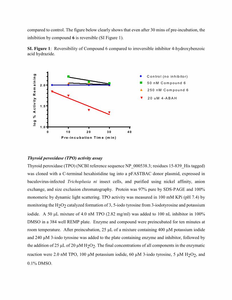
compared to control. The figure below clearly shows that even after 30 mins of pre-incubation, the
inhibition by compound 6 is reversible (SI Figure 1).
SI. Figure 1: Reversibility of Compound 6 compared to irreversible inhibitor 4-hydroxybenzoic acid hydrazide.
0 1 0 2 0 3 0 4 01 .0
1 .5
2 .05 0 n M C o m p o u n d 6
2 5 0 n M C o m p o u n d 6
2 0 u M 4 -A B A H
C o n tro l (n o in h ib ito r)
P re -in c u b a tio n T im e (m in )
log
% A
cti
vit
y R
em
ain
ing
Thyroid peroxidase (TPO) activity assay
Thyroid peroxidase (TPO) (NCBI reference sequence NP_000538.3; residues 15-839_His tagged)
was cloned with a C-terminal hexahistidine tag into a pFASTBAC donor plasmid, expressed in
baculovirus-infected Trichoplusia ni insect cells, and purified using nickel affinity, anion
exchange, and size exclusion chromatography. Protein was 97% pure by SDS-PAGE and 100%
monomeric by dynamic light scattering. TPO activity was measured in 100 mM KPi (pH 7.4) by
monitoring the H2O2 catalyzed formation of 3, 5-iodo tyrosine from 3-iodotyrosine and potassium
iodide. A 50 μL mixture of 4.0 nM TPO (2.82 mg/ml) was added to 100 nL inhibitor in 100%
DMSO in a 384 well REMP plate. Enzyme and compound were preincubated for ten minutes at
room temperature. After preincubation, 25 μL of a mixture containing 400 μM potassium iodide
and 240 μM 3-iodo tyrosine was added to the plate containing enzyme and inhibitor, followed by
the addition of 25 μL of 20 μM H2O2. The final concentrations of all components in the enzymatic
reaction were 2.0 nM TPO, 100 μM potassium iodide, 60 μM 3-iodo tyrosine, 5 μM H2O2, and
0.1% DMSO.
The reaction was allowed to proceed for 15 minutes, at which time it was quenched with 10 μL of
20% TCA bringing the TCA concentration to 2.0%. IC50 values were determined by calculating
the peak areas of 3-bromo-tyrosine present at the end of the 15 minute reaction and fitting the data
to:
𝑌𝑌 = 𝐴𝐴 +𝐵𝐵 − 𝐴𝐴
1 + (𝐶𝐶/𝑥𝑥)𝐷𝐷
where A = minimal Y value (activity level of inhibited sample), B = maximal Y value (activity
level of uninhibited sample), C = logIC50, D = Hill Slope, and x = concentration of inhibitor.
Lactoperoxidase (LPO) Assay
Lactoperoxidase (NCBI reference sequence NP_006142.1-isoform 1; residues 26-712) was cloned
into pFASTBAC donor plasmid, expressed in baculovirus-infected Trichoplusia ni insect cells and
purified using cation exchange, hydrophobic interaction and size exclusion chromatography
following procedures outlined in Shin et al.3 Biochemical and Biophysical Research
Communications 271,831-836 (2000). Protein was over 85% pure by SDS-PAGE and 99.9%
monomeric by dynamic light scattering. LPO activity was measured in 100 mM KPi (pH 7.4) by
monitoring the H2O2 catalyzed formation of 3,5-iodo tyrosine from 3-iodotyrosine and potassium
iodide. A 50 μL mixture of 2.0 nM LPO (1.75 mg/ml) was added to 100 nL inhibitor in 100%
DMSO in a 384 well REMP plate. Enzyme and compound were preincubated for ten minutes at
room temperature. After preincubation, 25 μL of a mixture containing 400 μM potassium iodide
and 240 μM 3-iodo tyrosine was added to the plate containing enzyme and inhibitor, followed by
the addition of 25 μL of 20 μM H2O2. The final concentrations of all components in the enzymatic
reaction were 1.0 nM LPO, 100 μM potassium iodide, 60 μM 3-iodo Tyrosine, 5 μM H2O2, and
0.1% DMSO.
The reaction was allowed to proceed for 15 minutes, at which time it was quenched with 10 μL of
20% TCA bringing the TCA concentration to 2.0%. IC50 values were determined by calculating
the peak areas of 3,5-diiodo-L-tyrosine present at the end of the 15 minute reaction and fitting the
data to:
𝑌𝑌 = 𝐴𝐴 +𝐵𝐵 − 𝐴𝐴
1 + (𝐶𝐶/𝑥𝑥)𝐷𝐷

where A = minimal Y value (activity level of inhibited sample), B = maximal Y value (activity
level of uninhibited sample), C = logIC50, D = Hill Slope, and x = concentration of inhibitor.
Eosinophil peroxidase (EPX) Bromination Activity
Eosinophil peroxidase (EPX) ) used in the assay was purchased from Lee Biosolutions (cat:342-
60), with purity ≥98% (SDS-PAGE). EPX bromination activity was measured in 100 mM KPi
(pH 7.4) by monitoring the H2O2 catalyzed formation of 3-bromo tyrosine from tyrosine and
potassium bromide. A 50 μL mixture of 0.6 μM EPX (Lee Biosolutions Cat# 342-60) was added
to 100 nL inhibitor in 100% DMSO in a 384 well REMP plate. Enzyme and compound were
preincubated for ten minutes at room temperature.
After the ten minute preincubation of enzyme and inhibitor, 25 μL of a mixture containing 400
μM tyrosine and 1200 μM potassium bromide was added to the plate containing enzyme and
inhibitor, followed by the addition of 25 μL of 20 μM H2O2. The reaction was allowed to proceed
for 15 minutes, at which time it was quenched with 10 μL of 20 % TCA. The final concentrations
of all components were 0.3 μM EPX, 100 μM Tyrosine, 400 μM potassium bromide, 5 μM H2O2,
0.1% DMSO, and 2.0% TCA.
IC50 values were determined by determining the peak areas of 3-bromo-tyrosine present at the
end of the 15 minute reaction and fitting the data to:
𝑌𝑌 = 𝐴𝐴 +𝐵𝐵 − 𝐴𝐴
1 + (𝐶𝐶/𝑥𝑥)𝐷𝐷
where A = minimal Y value (activity level of inhibited sample), B = maximal Y value (activity
level of uninhibited sample), C = LogIC50, D = Hill Slope, x = concentration of inhibitor.
Reversed-phase analysis was performed on a Waters Acquity Ultra Performance LC system using
an Acquity UPLC BEH C18 1.7 μM, 2.1 x 50 mm analytical column. The column was maintained
at 60 °C. Samples were eluted using a gradient of 0%-100% B over 2.5 minutes, followed by
equilibration with 100% A for 1 minute where A consisted of 0.1 % TFA and B consisted of 90%
MeOH/0.1 % TFA at a flow rate of 0.6 mL/min. The retention time of 3-bromo tyrosine was 1.22
min.

APOA1 modification protocol - cholesterol efflux and ApoA1 peptide oxidation LCMS
assay:
1. Human ApoA1 MPO modification:
Human ApoA1 (Biodesign A95120H) was first dialyzed against PBS pH7.4, and then 200
ug/mL treated with a mixture containing 100 µM diethylenetriamine penta-acetic acid (DTPA ,
Sigma D-6518), 250µM H2O2 (J.T Baker 2180-01) with or without 50nM MPO (Calbiochem
#475911), and small molecule inhibitors at 37 oC for 30 min. The reaction was stopped by adding
2 mM L-Methionine (Sigma M-9625).
2. THP1 cholesterol efflux (adapted from Kritharides et al, 1998)4
THP1 cells (ATCC TIB-202) were cultured in RPMI-1640 plus 10% fetal bovine serum,
L-glutamin (Gibco 25030-081), 2 mM, 25 mM hepes (Gibco 15630-080), 1% sodium pyruvate
(Gibco 11360-070), 0.05 mM 2-mercaptoethanol( Sigma M-3148), and 1% antibiotic-antimycotic
(Gibco 15240-096) in 5% CO2, differentiated with 100 nM PMA (Sigma P-8139) and plated in
96-well plates (1X 10⁵ cells/well) for 4 days. Then 5µCi/mL (1,2 3H)-cholesterol (Perkin Elmer
NET-139) was used for labelling serum lipoprotein particles (100µL/well) in the presence with
5% lipid-deficient serum (LDS Intracel RP-056) and 50 µg/mL ac-LDL (Biomedical Tech Inc,
BT-906) for 24 hours. For cholesterol efflux measurement, 20 µg/mL each apoA1 (treated with or
without MPO and inhibitors) with 0.2% fatty acid-free BSA (Sigma A-6003) in RPMI-1640 with
1% sodium pyruvate, 1% antibiotic-antimycotic and 0.05 mM 2-mercaptoethanol were incubated
with cells overnight. The apoA1 mediated cholesterol efflux in the THP1 cells media was
determined by counting 3H (Perkin Elmer Topcount-HTS, 12 DET, 96/384) present in cell media
and in cell extracts and % cholesterol efflux calculated based on the formula: 3H count in
media/total 3H (media+lysate) X 100.
3. Method for ApoA1 peptide LCMS assay
ApoA1 peptides measured:
Y192cl - LAEYclHAK
Y192 - LAEYHAK

W50- LLDNWDSVTSTFSK
Protein digestion: Protein samples were cleaned up with Zeba spin plate following vendor’s
protocol (Thermo Fisher Scientific). Trypsin was added to each well with 1 to 50 enzyme to protein
ratio. Samples were incubated in 37 °C overnight.
SPE (C18 solid phase extraction):
To each sample, add 80 uL working internal standard (IS) solution with W50 IS
(LLDNWDSVTSTFSK(13C, 15N)), Y192Cl IS (LAEYclHAK (13C, 15N)), and Y192 IS
(LAEYHAK(13C, 15N)). All heavy labeled internal standards were custom made by New England
Peptide Co. Samples then were desalted using 3M Empore C18 plate. Briefly, the plate was
conditioned with 300 uL 0.1% TFA in 90% acetonitrile followed by 300 uL 0.1% TFA in water.
Samples were loaded, and the plate was washed with 300 uL 0.1% TFA in water. Finally bound
peptides were eluted with 300 uL 0.1%TFA in 90% acetonitrile. Eluates were dried down by
SpeedVac.
LCMS: Samples were reconstituted in 0.1% acetic acid in water and injected onto the
column (Microm 0.3 x 50mm Halo C18 ) on Agilent 1200 CapLC system coupled with AB Sciex
5500 QTRAP.
Peak extraction was performed by AB Sciex Analysis software. Relative abundance of
Y192cl, Y192 and W50 was estimated according to the ratio of AUCs (area under the curve) of
the target peak and that of corresponding spiked in heavy labeled internal standard. Plots were
generated by GraphPad Prism 5.0.
SI Figure 2. Dose response of inhibitor 6 on MPO oxidation of ApoA1 Trp (W) 50 (blue) and
cholesterol efflux capacity (red). The EC50 values were ~10 µM for W50 protection and ApoA1
cholesterol efflux capacity in THP-1 macrophages.

Human Neutrophil PMA-induced MPO Activity Assay
Neutrophils from healthy donors were isolated by the Dextran Sedimentation Followed by
Ficoll-Paque method described by William M. Nauseef.5 Blood was drawn in BD Vacutainer (BD
Franklin Lakes 366480) and mixed with equal volumes of 3% dextran solution (Sigma D8906) by
repeated inversion (10 times), and tubes were set upright for 18–20 min. Then the straw-colored,
leukocyte-rich, erythrocyte-poor upper layer was aspirated with a sterile plastic pipet or a 10- to
15-mL sterile syringe and transfer to a sterile 50-mL conical tube. Leukocytes were pelleted by
centrifugation at 500 g for 10 min at 4 °C. The cell pellets was resuspended with 35 mL sterile
saline buffer. The leukocyte suspension was then carefully underlayed with 10 mL of Ficoll-Paque
solution, and gradients centrifuged at 400 g for 40 min at rt. After centrifugation, the supernatant
was discarded, and neutrophils were collected in sterile water supplemented with an equal volume
of 1.8 % saline to promptly restore tonicity. Cells were centrifuged at 500 g for 5 min at 4 °C, and
cells resuspended in HBSS (GIBCO # 14025) at about 30 million cells/mL.
The assay was adapted from protocol described by Gross et al.6 100 uL neutrophils at 1.106
Cells/mL were plated in Costar 96-well plate, and PMA ((Sigma # p8139) at 100 nM final
concentration were added. Inhibitor (1 µL) in vehicle (10% Ethanol, 50% Peg, and 40% H2O) from
a starting concentration of 22 µM down to 1 nM (1:3 dilution, total 10 doses) were incubated with
cells for 3 min at rt. Luminol (100 µM final concentration) was added, and plate was read on
LMaxII-384 luminometer (Molecular Devices) at 37 oC for 25 min for kinetics analysis. The rate
of formation of luminescence due to oxidation of luminol by MPO-produced HOCl was measured
C o m p o u n d 6E ff lu x & W 5 0 P ro te c t io n
-9 -8 -7 -6 -5 -4 -3-2 0
0
2 0
4 0
6 0
8 0
1 0 0
0
2 0
4 0
6 0
8 0
1 0 0A p o A 1 c h o l E fflu x
W 5 0 p ro te c t io n
L o g [C o m p o u n d 6 ] , M
% E
fflu
x R
ec
ov
ery %
W5
0 P
rote
ctio
n

in the initial linear region of the reaction. No signal was detected using neutrophils from MPO
knock-out mice used as negative control.
Acute in Vivo PMA Model
Mouse whole blood luminescence assay:
All animal studies were performed according to guidelines established by the American
Association for Accreditation of Laboratory Animal Care and protocols were approved by the
Bristol-Myers Squibb Hopewell Animal Care and Use Committee (ACUC). The protocol was
modified from a similar method described by Gross et al.6 C57Bl6 mice were orally dosed with
30 mpk 6 or vehicle 20 min prior to PMA (Sigma P-8139) retro-orbital injection at 0.62mg/kg.
Two minutes post PMA injection, blood was drawn and kept on ice before measurement of MPO-
produced HOCl quantification using luminol chemiluminescence as readout.
To measure mouse blood luminol activity, 100 uL of mouse blood was plated in a 96-well
plate (Costar 3925), combined with 2 µL of 100 mM luminol (Sigma A4685, dissolved in H2O),
and mixed. Luminol chemiluminescence was measured on LMaxII-384 luminometer (Molecular
Devices) at 37 oC for 25 min for kinetics analysis and the HOCl rate of production was calculated.
No luminol oxidation was detected in MPO knockout mice, which confirmed luminol
chemiluminescence was dependent on MPO formation of HOCl (SI Figure 3).7
SI. Figure 3. Luminol chemiluminescence measured in wild type mice and MPO KO mice
after PMA treatment.
0 .0
0 .5
1 .0
1 .5C 5 7 B L /6 (n = 4 )
M P O K O (n = 3 )
*
Init
ial
Ra
te (
RL
U)

Protein crystallization
Human MPO protein Isoform C was purchased from Lee Biosolutions Inc, St. Louis MO. The
purchased protein was further purified by gel–filtration chromatography (Superdex 75) in a buffer
containing 20 mM Na-Acetate pH 5.5, 100 mM NaCl. Crystals of MPO in complex with 6 were
obtained at 22 °C by the hanging drop vapor diffusion method. The protein (25 mg/ml
concentration) was incubated with 6 (protein:6 molar ratio of 1:5) at 4 °C for 4 hours prior to
crystallization setup. The reservoir solution contains 100 mM hepes (pH -7.5), 150 mM NaCl, and
20-25% (w/v) PEG3350. Crystals were cryoprotected by supplementing the mother liquor with
15% (v/v) ethylene glycol and harvested by flash-cooling in liquid nitrogen.
Data Collection and Structure determination. Data for the MPO/6 complex were collected at
beamline 08ID at the Canadian Light Source. The wavelength used was 1.0 Å and the detector was
a Rayonix MX225. The diffraction images were indexed, integrated, and scaled with HKL-2000.8
The structure was determined by molecular replacement using the program PHASER9 with a
previously reported structure of MPO (PDB entry 1CXP) used as the starting model in the
refinement. The structure was refined with the program autoBUSTER (Global Phasing Ltd,
Cambridge, England) and were examined and manually refitted with the program COOT.10 The
difference electron density clearly defined the position of the compound in the heme-binding
pocket. Additional cycles of refinement were performed to complete the refinement and add
solvent molecules. The crystallographic data-collection and reduction statistics are summarized in
Table A.
Table A. Summary of crystallographic information
MPO
+ Compound 6
PDB Code 5WDJ
Data collection
Space group P43212
Cell dimensions

a, b, c (Å) 103.9, 103.9, 242.3
α, β, γ (°) 90, 90, 90
Resolution (Å) 50-2.40
Rmerge (%) 11.3 (52.5)
I / σI 15.11 ( 3.1)
Completeness (%) 100 (100)
Redundancy 4.8 (4.8)
Refinement
Resolution (Å) 95.5 - 2.40
Number of reflections 47,290 (2676)
Rwork / Rfree (%) 19.4 / 24.8
No. atoms
Protein 9048
Ligand/ion 326
Water 522
B-factors (Å2)
Protein 24.3
Ligand/ion 30.1
Water 28.9
R.m.s. deviations
Bond lengths (Å) 0.006
Bond angles (°) 1.1
Ramachandran Plot Stats
Most allowed (%) 87.6
Additional allowed (%) 12.3
Outliers (%) 0.0 aAs defined by Laskowski et al., 1993.11

The numbers in parentheses are for the highest-resolution shell. One crystal was used for each data
collection.
Synthesis
All reactions were carried out under a static atmosphere of argon or nitrogen and stirred
magnetically unless otherwise stated. All reagents used were of commercial quality. 1HNMR
spectra were recorded on a Bruker 400 MHz system spectrometer unless otherwise noted.
Chemical shifts are given in parts per million (ppm), and coupling constants (J values) are given
in hertz (Hz). Selected data are reported in the following manner: chemical shift, multiplicity,
coupling constants, proton number. All solvents were removed by rotary evaporation under
vacuum using a standard rotary evaporator equipped with a dry ice condenser.
Final products were analyzed by reverse phase analytical HPLC: carried out on a Shimadzu
Analytical HPLC: system running Discovery VP software. Reverse phase preparative HPLC of
some final compounds was carried out using C18 columns with UV 220 nm or prep LCMS
detection eluting with gradients of Solvent A (90% water, 10% MeOH, 0.1% TFA) and Solvent B
(10% water, 90% MeOH, 0.1% TFA) or with gradients of Solvent A (90% water, 10% ACN, 0.1%
TFA) and Solvent B (10% water, 90% ACN, 0.1% TFA) or with gradients of Solvent A (95%
water, 2% ACN, 0.1% HCOOH) and Solvent B (98% ACN, 2% water, 0.1% HCOOH) or with
gradients of Solvent A (95% water, 5% ACN, 10 mM NH4OAc) and Solvent B (98% ACN, 5%
water, 10 mM NH4OAc) or with gradients of Solvent A (95% water, 2% ACN, 0.1% NH4OH)
and Solvent B (98% ACN, 2% water, 0.1% NH4OH).
1H NMR spectra were obtained with Bruker or JEOL® Fourier transform spectrometers operating
at frequencies as follows: 1H NMR: 400 MHz (Bruker or JEOL®) or 500 MHz (Bruker or
JEOL®). 13C NMR: 100 MHz (Bruker or JEOL®). Spectra data are reported in the format:
chemical shift (multiplicity, coupling constants, number of hydrogens). Chemical shifts are
specified in ppm downfield of a tetramethylsilane internal standard (δ units, tetramethylsilane = 0
ppm) and/or referenced to solvent peaks, which in 1H NMR spectra appear at 2.49 ppm for
CD2HSOCD3, 3.30 ppm for CD2HOD, 1.94 for CD3CN, and 7.24 ppm for CHCl3, and which in
13C NMR spectra appear at 39.7 ppm for CD3SOCD3, 49.0 ppm for CD3OD, and 77.0 ppm for
CDCl3.
The pKa values were determined in the pH range between 2 and 11, and the data were obtained
using a Sirius GLpKa instrument. The pKa values were measured using the spectrophotometric
titration method, and the compound was dissolved in varying ratios of methanol/water with
extrapolation to 0% cosolvent. The predicted pKa value was determined using the Jaguar pKa
GUI method in the Maestro version 8.5.207 from Schroedinger, Inc.
Compounds reported in the paper were prepared by methods similar to those reported by Chae, M-
Y. et al. 12 or by the methods below.
NN
O
N NH
NH2N
Ph
7-(benzyloxy)-3H-[1,2,3]triazolo[4,5-d]pyrimidin-5-amine (6).
A solution of 6N HCl (25 mL, 150 mmol) was added to 4-chloroaniline (5.0 g, 39 mmol) with
vigorous stirring at 0 °C. A solution of NaNO2 (2.7 g, 39 mmol) in water (4.0 mL) was added to
the reaction mixture, and the mixture was stirred at 0 °C for 30 min. Urea (0.47 g, 7.8 mmol) was
added to destroy residual NaNO2. This reaction mixture was added to a solution of 6-
chloropyrimidine-2,4-diamine (5.6 g, 39 mmol) in water (30 mL). After 30 min, sodium acetate
(14.5 g, 176 mmol) was added, and the reaction mixture was allowed to warm to rt overnight. The
mixture was filtered, and the solids were washed thoroughly with water, and dried under vacuum
overnight to obtain (E)-6-chloro-5-((4-chlorophenyl)diazenyl)pyrimidine-2,4-diamine (8.1 g, 28
mmol, 73 % yield). MS(ESI) m/z 284.2 (M+H).
To a mixture of (E)-6-chloro-5-((4-chlorophenyl)diazenyl)pyrimidine-2,4-diamine (3.4 g, 12
mmol), and benzyl alcohol (2.5 mL, 24 mmol) in DMSO (25 mL) was added Cs2CO3 (12 g, 36
mmol), and the resulting mixture was heated to 105 °C for 2h. The mixture was cooled to rt and
diluted with water. The resulting slurry was filtered. The solid was washed with water, and
triturated with ether to obtain a yellow solid (E)-6-(benzyloxy)-5-((4-

chlorophenyl)diazenyl)pyrimidine-2,4-diamine (4.2 g, 12 mmol), which was used without further
manipulation. MS(ESI) m/z 255.1 (M+H).
To a solution of (E)-6-(benzyloxy)-5-((4-chlorophenyl)diazenyl)pyrimidine-2,4-diamine (4.2 g,
12 mmol) in EtOH (10 mL) was added Zn powder (4.65 g, 71.0 mmol) followed by glacial AcOH
(2.0 mL, 36 mmol). The resulting reaction mixture was heated at 70 °C for 2h. The mixture was
filtered through a pad of celite, and the filtrate was concentrated to obtain an orange red product
that was purified by silica gel chromatography (eluted with 0-20% MeOH in EtOAc) to furnish 6-
(benzyloxy)pyrimidine-2,4,5-triamine (1.2 g, 5.2 mmol, 44 % yield). 1H NMR (500 MHz, MeOH-
d4) δ 7.49 - 7.42 (m, 2H), 7.40 - 7.34 (m, 2H), 7.34 - 7.29 (m, 1H), 5.37 (s, 2H). MS(ESI) m/z
232.2 (M+H).
To a solution of 6-(benzyloxy)pyrimidine-2,4,5-triamine (50 mg, 0.20 mmol) in DMSO (2 mL)
was added a solution of isoamyl nitrite (0.03 mL, 0.2 mmol). The resulting solution was stirred at
rt overnight. The reaction mixture was diluted with EtOAc, washed with saturated NaHCO3 and
brine, dried over MgSO4, and concentrated to obtain a reddish, sticky solid 7-(benzyloxy)-3H-
[1,2,3]triazolo[4,5-d]pyrimidin-5-amine (45 mg, 0.19 mmol, 86 % yield), which was purified by
silica gel chromatography (eluted with EtOAc/hex) and recrystallization from iPrOH. 1H NMR
(500 MHz, DMSO-d6) δ ppm 15.34 (br. s., 1 H), 7.55 (d, J=6.88 Hz, 2 H), 7.41 - 7.45 (m, 2 H),
7.35 - 7.40 (m, 1 H), 7.00 (br. s., 2 H), 5.57 (s, 2 H). 13C NMR (126 MHz, DMSO-d6) δ ppm
162.03, 160.43, 154.74, 135.82, 128.63, 128.38, 128.21, 119.65, 67.55. HRMS Observed:
243.09872; Calculated: 243.09899. HPLC Purity >96%.
5-(2,4,5-trichlorophenoxy)pyridin-2-amine (7).
N NO2
Br
N NH2
O
Cl
Cl
Cl
OH
Cl
Cl
Cl
1) Cs2CO3, DMF
2) H2, Pd/C
To a solution of 5-bromo-2-nitropyridine (0.47 g, 2.3 mmol) in DMF (7.7 mL) was added Cs2CO3
(1.13 g, 3.47 mmol) at 0 °C followed by 2,4,5-trichlorophenol (0.50 g, 2.6 mmol) in DMF (7.7
mL). The solution was stirred at rt overnight. Water and EtOAc were added to the mixture and
the organics were extracted from water with EtOAc. The organics were washed with water and

brine, then concentrated and purified by column chromatography (0-20% EtOAc/hexanes) to
furnish 2-nitro-5-(2,4,5-trichlorophenoxy)pyridine (0.51 g, 1.60 mmol, 6) which was taken on
directly. To a solution of 2-nitro-5-(2,4,5-trichlorophenoxy)pyridine (0.512 g, 1.60 mmol) in
MeOH (16 mL) was added Pd/C (0.17 g, 0.16 mmol), and the mixture was blanketed under H2
using a balloon. The reaction mixture was stirred overnight. The solution was filtered and
concentrated under vacuum. The residue was purified by HPLC (10-90% ACN/water/0.1% TFA,
Phenomonex AXIA 5u C18) to furnish 5-(2,4,5-trichlorophenoxy)pyridin-2-amine, TFA (0.047
g, 0.12 mmol, 5.2 % yield, two steps). 1H NMR (400 MHz, CHLOROFORM-d) δ 7.91 (d, J=2.7
Hz, 1H), 7.53 (s, 1H), 7.19 (dd, J=8.8, 2.7 Hz, 1H), 6.86 (s, 1H), 6.55 (d, J=8.8 Hz, 1H), 4.45 (br
s, 2H). MS(ESI) m/z 289.1 (M+H).
General Procedure to prepare triazolopyrimidine analogs.

N
NH2N
Cl
NH
CN
NH2 N
NH2N
Cl
N
CN
NNaNO2, AcOH
water, 0 °CN
N
NH2N
Cl
Cl
Et3N
BuOH, 100 °C
NH2
N
NH2N OH
OHNH2 MeEt3NCl, POCl3
NH
H2N NH2
ClHO
O O
O
HN
O
1)Na, EtOH2) HCl
1) carbonate base or NaH,ROH in DME2) KOtBu N
NH2N
O
N
NN
R
H
To an ethanol (230 mL) solution was added sodium (2.65 g, 115 mmol) under Ar with stirring.
After complete consumption of sodium, guanidine hydrochloride (11.0 g, 115 mmol) was added,
and the solution was stirred for 10 min. This solution was heated to reflux and treated with diethyl
acetamidomalonate (25.0 g, 115 mmol). After 2h, additional 460 mL of EtOH were added and
heating to reflux was continued overnight. The mixture was cooled to rt. The mixture was filtered,
and filtrate was washed with EtOH and CHCl3 to yield 21 g of intermediate acetamide. The
intermediate solid was taken up in 60 mL of 6 N HCl and heated to 70 °C. After 2h the reaction
mixture was cooled, and the solid was filtered followed by washing with 80 mL of 4N HCl. The
resulting solid was dried in a vacuum oven at 80 °C overnight to furnish 2,5-diaminopyrimidine-
4,6-diol, HCl (10 g, 56 mmol, 49 % yield). MS(ESI) m/z 143.0 (M+H).
Triethylmethylammonium chloride (9.6 g, 63 mmol) and 2,5-diaminopyrimidine-4,6-diol, HCl
(10 g, 56 mmol) were heated in POCl3 (13.6 mL, 146 mmol) to 105 °C overnight. The solution
was concentrated (caution, POCl3 was neutralized in the collection bulb with 10 N NaOH
solution), followed by addition of 200 mL water to the residue. The pH was adjusted to 4 (caution,
EXOTHERM), and the solution was stirred for 1 h. The solution was adjusted to pH = 7, diluted
with 1 L of EtOAc, and the solution was filtered through celite. The organic phase was separated,
passed through a silica plug and concentrated to furnish 4,6-dichloropyrimidine-2,5-diamine (2.55
g, 14.2 mmol, 25.4 % yield). 1H NMR (500 MHz, DMSO-d6) δ 6.49 (s, 2H), 4.71 (s, 2H).
To a solution of 4,6-dichloropyrimidine-2,5-diamine (2.0 g, 11 mmol) and 3-aminopropionitrile
(0.98 mL, 13 mmol) in butan-1-ol (45 mL) was added Et3N (1.6 mL, 11 mmol). The reaction
mixture was heated to 100 °C and stirred overnight. The mixture was concentrated, and the
volatiles were removed with MeOH azeotrope. The residue was carried forward as crude, 3-(2,5-
diamino-6-chloropyrimidin-4-ylamino)propanenitrile (2.4 g, 11 mmol). MS(ESI) m/z 213.1
(M+H).
To a solution of 3-(2,5-diamino-6-chloropyrimidin-4-ylamino)propanenitrile (2.4 g, 11 mmol) in
AcOH (9.3 mL)/water (28 mL) was added sodium nitrite (0.92 g, 13 mmol) in water (18 mL) at 0
°C. After 10 min, the reaction was complete by LCMS. The mixture was diluted with EtOAc and
adjusted to pH = 6 using sat aq. NaHCO3. The organic layer was washed with brine, dried over
Na2SO4, filtered and concentrated. The residue was purified by column chromatography (80-
100% EtOAc/hexanes) to furnish 3-(5-amino-7-chloro-3H-[1,2,3]triazolo[4,5-d]pyrimidin-3-
yl)propanenitrile (0.28 g, 1.2 mmol, 11 % yield). MS(ESI) m/z 239.1 (M+H).
To a solution of 3-(7-chloro-3H-[1,2,3]triazolo[4,5-d]pyrimidin-3-yl)propanenitrile (1 equiv.) and
the benzyl alcohol starting material (1.5 equiv.) in DME (0.2 M reaction concentration) was added
3 equiv. of K2CO3 or CsCO3 or 2 equiv. of NaH. The reaction mixture was heated to 80 °C
overnight. The reaction mixture was concentrated, and the residue purified by column
chromatography (0-100% EtOAc/hexanes gradient) to furnish the appropriately substituted 3-(7-
(benzyloxy)-3H-[1,2,3]triazolo[4,5-d]pyrimidin-3-yl)propanenitrile in 25-60 % yields. To a
solution of the propane nitrile protected intermediate (1 equiv) in THF (1 mL) at 0 °C was added
KOtBu (1 equiv). After 10 - 60 min, LCMS indicated formation of a single peak with product
molecular ion mass. The reaction was quenched with AcOH (2 equiv), and then the mixture was
concentrated. The residue was purified by prep HPLC to furnish the desired products in 5-70%
yields.

NN
O
N NH
N
7-(benzyloxy)-3H-[1,2,3]triazolo[4,5-d]pyrimidine (8).
Prepared from 3-(7-chloro-3H-[1,2,3]triazolo[4,5-d]pyrimidin-3-yl)propanenitrile and benzyl
alcohol by methods described in general procedure. 1H NMR (500 MHz, DMSO-d6) δ 8.77 (s,
1H), 7.63 - 7.53 (m, 2H), 7.49 - 7.43 (m, 3H), 5.73 (s, 2H). MS(ESI) m/z 227.1 (M+H).
7-(benzyloxy)-3H-[1,2,3]triazolo[4,5-d]pyrimidin-5-ol (9)
A suspension of 7-(benzyloxy)-3H-[1,2,3]triazolo[4,5-d]pyrimidin-5-amine, 6 (250 mg, 1.03
mmol) in acetone (4.5 mL) was poured into a solution of sodium nitrite (1.42 g, 20.6 mmol) in
water (4.5 mL). Acetic acid (2.4 mL, 1.0 mmol) was added to the suspension with
stirring. Minimum amounts of acetone were added as necessary to dissolve any suspended solid,
and the solution was stirred for 5 h at rt. The acetone was removed by rotovap, and an orange
precipitate formed in the aqueous solution. The precipitate was filtered from the solution, and
washed with water. The light orange solid was dried on the high vacuum overnight to provide
7-(benzyloxy)-3H-[1,2,3]triazolo[4,5-d]pyrimidin-5-ol (139 mg, 0.557 mmol, 54% yield).
1H NMR (500 MHz, DMSO-d6) δ ppm 12.10 (1 H, br. s.), 7.52 (2 H, d, J=7.1 Hz), 7.32 - 7.47 (3
H, m), 5.56 (2 H, s). MS(ESI) m/z + 244.1 (M+H).
NN
O
N NH
NNH
7-(benzyloxy)-N-ethyl-3H-[1,2,3]triazolo[4,5-d]pyrimidin-5-amine (10)
NN
O
N NH
NHO

Dissolved excess acetaldehyde (40 mg) and 7-(benzyloxy)-3H-[1,2,3]triazolo[4,5-d]pyrimidin-5-
amine (6, 0.05 g, 0.21 mmol) in MeOH (0.67 mL) with AcOH (0.067 mL), and a slightly opaque
solution resulted. The mixture was heated to reflux and molecular sieves were added (~100 mg)
followed by cooling to 40 °C. Sodium cyanoborohydride (0.052 g, 0.83 mmol) was added and the
mixture was stirred overnight. The sieves were removed and washed with MeOH. The product
was purified by reverse phase HPLC (10-90% MeOH in TFA buffered water, 30x75mm
Phenomenex Axia Luna 5u column) to afford 5 mgs (0.018 mmol) of product. 1H NMR (500 MHz,
DMSO-d6) δ 7.55 (br d, J=7.2 Hz, 2H), 7.46 - 7.41 (m, 2H), 7.39 (d, J=7.2 Hz, 1H), 5.57 (br s,
2H), 3.34 (m, 2H), 1.15 (br t, J=6.7 Hz, 3H). MS(ESI) m/z + 271.1 (M+H).
NN
O
N NN
H2N
NN
O
N NN
H2N
Mixture of 7-(benzyloxy)-1-methyl-3H-[1,2,3]triazolo[4,5-d]pyrimidin-5-amine and 7-(benzyloxy)-2-methyl-2H-[1,2,3]triazolo[4,5-d]pyrimidin-5-amine (11)
To a solution of 7-(benzyloxy)-3H-[1,2,3]triazolo[4,5-d]pyrimidin-5-amine (6, 0.050 g, 0.21
mmol) and potassium carbonate (0.031 g, 0.23 mmol) in DMF (2 mL) was added MeI (0.012 mL,
0.19 mmol) and the solution was allowed to stir overnight. The reaction was partitioned between
EtOAc and 0.1M pH 7 phosphate buffer. The organic phase was washed with buffer, brine, and
concentrated. The residue was dissolved in ACN and water, filtered and purified via preparative
chromatography, 30x250 5u Phenomanex Luna column, 10-90% ACN / water (TFA buffer). The
fraction were concentrated to furnish mixture of compounds 11 (20 mg, 0.027 mmol, 13% yield)
and compound 12 (31 mg, 0.083 mmol, 40% yield).
Compound 11 1H NMR (500 MHz, CDCl3) δ 7.56 - 7.37 (m, 10H), 5.67 (s, 2H), 5.63 (s, 2H), 4.38 (s, 3H), 4.32 (s, 3H). MS(ESI) m/z + 257.3 (M+H).
NN
O
N NN
H2N
Ph
7-(benzyloxy)-3-methyl-3H-[1,2,3]triazolo[4,5-d]pyrimidin-5-amine (12)

1H NMR (500 MHz, CDCl3) δ 7.51 (br d, J=7.2 Hz, 2H), 7.43 - 7.30 (m, 3H), 5.69 – 5.52 (m, 4H),
4.10 (s, 3H). MS(ESI) m/z + 257.4 (M+H).
NN
O
N NHH2N
Ph
6-(benzyloxy)-9H-purin-2-amine (13).
Purchased from commercial sources.
HNN
O
N NHH2N
Ph
O
2-amino-6-(benzyloxy)-7,9-dihydro-8H-purin-8-one (14).
Phosgene in toluene (20% solution, 0.057 mL, 0.11 mmol) was added to a solution of 6-
(benzyloxy)pyrimidine-2,4,5-triamine (25 mg, 0.11 mmol) in CH2Cl2 (2 mL). The resulting
mixture was stirred at rt for 2 h. The mixture was concentrated in vacuo and the residue was
purified by reverse phase HPLC to yield Example 13 (0.68 mg, 1.69 µmol, 1.6 % yield). MS (ESI)
m/z 258.4 (M+H)+. 1H NMR (500 MHz, CD3OD) δ 7.52 - 7.42 (m, 2H), 7.42 - 7.27 (m, 3H), 5.45
(s, 2H).
N
O
N NH
NH2N
Ph
4-(benzyloxy)-1H-pyrazolo[3,4-d]pyrimidin-6-amine (15)
Cesium carbonate (120 mg, 0.35 mmol) was added to a mixture of 4-chloro-1H-pyrazolo[3,4-
d]pyrimidin-6-amine (BIONET Supplier, 20 mg, 0.12 mmol) and phenylmethanol (0.061 mL, 0.59
mmol) in DMF (1 mL) and the mixture was heated in a microwave for 25 min at 200°C. The
mixture was purified by reverse phase HPLC to yield Example 14 (0.96 mg, 2.7 µmol, 2.3 %

yield). MS (ESI) m/z 242.2 (M+H)+. 1H NMR (500 MHz, DMSO-d6) δ 8.58 (s, 1H), 7.38 - 7.29
(m, 6H), 5.36 (s, 2H).
NN
O
N NH
N
Ph
H2N
7-phenoxy-3H-[1,2,3]triazolo[4,5-d]pyrimidin-5-amine (16)
Prepared from 3-(5-amino-7-chloro-3H-[1,2,3]triazolo[4,5-d]pyrimidin-3-yl)propanenitrile and
phenol as described in general procedure using NaH as base. The propanenitrile protecting group
was removed under the reaction conditions. 1H NMR (500 MHz, DMSO-d6) δ 7.55 - 7.44 (m, 2H),
7.38 - 7.27 (m, 3H), 6.94 (br d, J=2.2 Hz, 2H). MS(ESI) m/z 239.1 (M+H).
NN
O
N NH
NH2N
Ph
7-phenethoxy-3H-[1,2,3]triazolo[4,5-d]pyrimidin-5-amine (17)
Prepared from 3-(5-amino-7-chloro-3H-[1,2,3]triazolo[4,5-d]pyrimidin-3-yl)propanenitrile and 2-
phenylethanol as described in general procedure using NaH as base. The propanenitrile protecting
group was removed under the reaction conditions. 1H NMR (500 MHz, DMSO-d6) δ 7.39 - 7.31
(m, 4H), 7.28 - 7.22 (m, 1H), 7.06 - 6.84 (m, 2H), 4.69 (t, J=7.2 Hz, 2H), 3.15 (t, J=7.2 Hz, 2H).
MS(ESI) m/z 257.0 (M+H).
NN
O
N NH
NH2N
F
7-((2-fluorobenzyl)oxy)-3H-[1,2,3]triazolo[4,5-d]pyrimidin-5-amine (18)
Prepared from 3-(5-amino-7-chloro-3H-[1,2,3]triazolo[4,5-d]pyrimidin-3-yl)propanenitrile and
(2-fluorophenyl)methanol as described in general procedure using K2CO3, followed by reaction with
KOtBu in THF. 1H NMR (500 MHz, DMSO-d6) δ 7.69 (td, J=7.6, 1.7 Hz, 1H), 7.52 - 7.42 (m,
1H), 7.34 - 7.22 (m, 2H), 7.04 (br d, J=3.3 Hz, 2H), 5.62 (s, 2H). MS(ESI) m/z 260.9 (M+H).
NN
O
N NH
NH2N
Cl
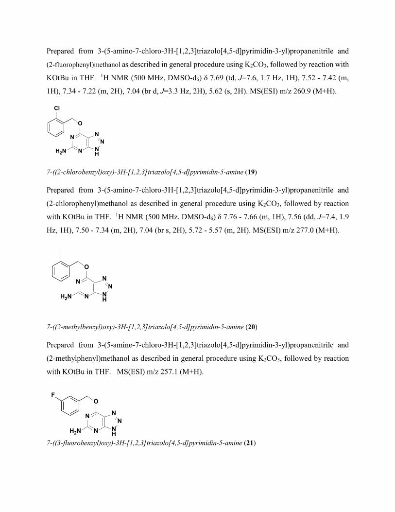
7-((2-chlorobenzyl)oxy)-3H-[1,2,3]triazolo[4,5-d]pyrimidin-5-amine (19)
Prepared from 3-(5-amino-7-chloro-3H-[1,2,3]triazolo[4,5-d]pyrimidin-3-yl)propanenitrile and
(2-chlorophenyl)methanol as described in general procedure using K2CO3, followed by reaction
with KOtBu in THF. 1H NMR (500 MHz, DMSO-d6) δ 7.76 - 7.66 (m, 1H), 7.56 (dd, J=7.4, 1.9
Hz, 1H), 7.50 - 7.34 (m, 2H), 7.04 (br s, 2H), 5.72 - 5.57 (m, 2H). MS(ESI) m/z 277.0 (M+H).
7-((2-methylbenzyl)oxy)-3H-[1,2,3]triazolo[4,5-d]pyrimidin-5-amine (20)
Prepared from 3-(5-amino-7-chloro-3H-[1,2,3]triazolo[4,5-d]pyrimidin-3-yl)propanenitrile and
(2-methylphenyl)methanol as described in general procedure using K2CO3, followed by reaction
with KOtBu in THF. MS(ESI) m/z 257.1 (M+H).
7-((3-fluorobenzyl)oxy)-3H-[1,2,3]triazolo[4,5-d]pyrimidin-5-amine (21)
NN
O
N NH
NH2N
F
NN
O
N NH
NH2N

Prepared from 3-(5-amino-7-chloro-3H-[1,2,3]triazolo[4,5-d]pyrimidin-3-yl)propanenitrile and
(3-fluorophenyl)methanol as described in general procedure using K2CO3, followed by reaction with
KOtBu in THF. 1H NMR (500 MHz, DMSO-d6) δ 7.51 - 7.44 (m, 1H), 7.43 - 7.37 (m, 2H), 7.26
- 7.17 (m, 1H), 7.05 - 6.98 (m, 1H), 7.08 - 6.96 (m, 1H), 7.11 - 6.93 (m, 1H), 5.58 (s, 2H). MS(ESI)
m/z 260.9 (M+H).
7-((3-chlorobenzyl)oxy)-3H-[1,2,3]triazolo[4,5-d]pyrimidin-5-amine (22)
Prepared from 3-(5-amino-7-chloro-3H-[1,2,3]triazolo[4,5-d]pyrimidin-3-yl)propanenitrile and
(3-chlorophenyl)methanol as described in general procedure using K2CO3, followed by reaction with
KOtBu in THF. 1H NMR (400 MHz, ACETONITRILE-d3) δ 7.54 (s, 1H), 7.48 - 7.30 (m, 3H),
6.95 - 6.33 (m, 2H), 5.56 (s, 2H). MS(ESI) m/z 275.3 (M+H).
7-((3-methoxybenzyl)oxy)-3H-[1,2,3]triazolo[4,5-d]pyrimidin-5-amine (23)
Prepared from 3-(5-amino-7-chloro-3H-[1,2,3]triazolo[4,5-d]pyrimidin-3-yl)propanenitrile and
(3-methoxyphenyl)methanol as described in general procedure using K2CO3 followed by reaction
with KOtBu in THF. MS(ESI) m/z 273.2 (M+H).
NN
O
N NH
NH2N
HF2CO
7-((3-(difluoromethoxy)benzyl)oxy)-3H-[1,2,3]triazolo[4,5-d]pyrimidin-5-amine (24)
NN
O
N NH
NH2N
Cl
NN
O
N NH
NH2N
MeO
Prepared from 3-(5-amino-7-chloro-3H-[1,2,3]triazolo[4,5-d]pyrimidin-3-yl)propanenitrile and
(3-(difluoromethoxy)phenyl)methanol as described in general procedure using K2CO3 followed
by reaction with KOtBu in THF. 1H NMR (500 MHz, ACETONITRILE-d3) δ 7.56 - 7.43 (m,
2H), 7.38 (s, 1H), 7.19 (br d, J=7.7 Hz, 1H), 7.02 - 6.64 (m, 1H), 5.70 - 5.60 (m, 2H). MS(ESI)
m/z 309.1 (M+H).
NN
O
N NH
NH2N
F
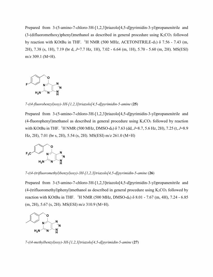
7-((4-fluorobenzyl)oxy)-3H-[1,2,3]triazolo[4,5-d]pyrimidin-5-amine (25)
Prepared from 3-(5-amino-7-chloro-3H-[1,2,3]triazolo[4,5-d]pyrimidin-3-yl)propanenitrile and
(4-fluorophenyl)methanol as described in general procedure using K2CO3 followed by reaction
with KOtBu in THF. 1H NMR (500 MHz, DMSO-d6) δ 7.63 (dd, J=8.7, 5.6 Hz, 2H), 7.25 (t, J=8.9
Hz, 2H), 7.01 (br s, 2H), 5.54 (s, 2H). MS(ESI) m/z 261.0 (M+H)
NN
O
N NH
NH2N
F3C
7-((4-(trifluoromethyl)benzyl)oxy)-3H-[1,2,3]triazolo[4,5-d]pyrimidin-5-amine (26)
Prepared from 3-(5-amino-7-chloro-3H-[1,2,3]triazolo[4,5-d]pyrimidin-3-yl)propanenitrile and
(4-(trifluoromethyl)phenyl)methanol as described in general procedure using K2CO3 followed by
reaction with KOtBu in THF. 1H NMR (500 MHz, DMSO-d6) δ 8.01 - 7.67 (m, 4H), 7.24 - 6.85
(m, 2H), 5.67 (s, 2H). MS(ESI) m/z 310.9 (M+H).
NN
O
N NH
NH2N
7-((4-methylbenzyl)oxy)-3H-[1,2,3]triazolo[4,5-d]pyrimidin-5-amine (27)
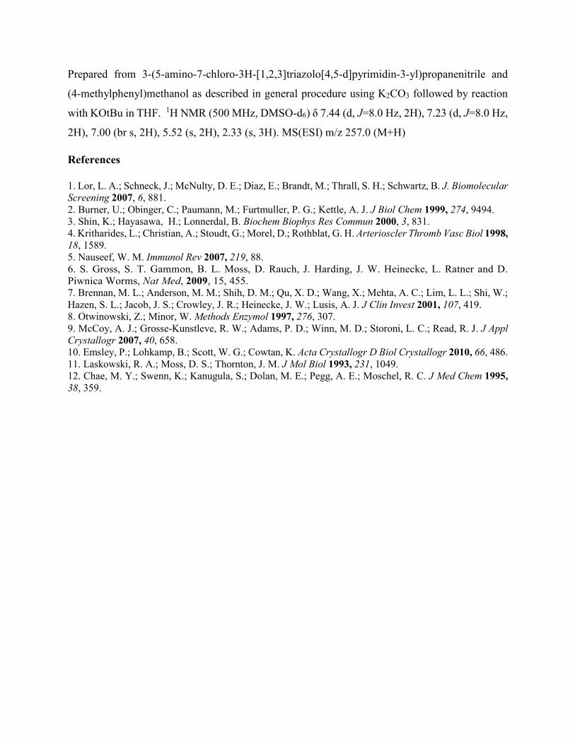
Prepared from 3-(5-amino-7-chloro-3H-[1,2,3]triazolo[4,5-d]pyrimidin-3-yl)propanenitrile and
(4-methylphenyl)methanol as described in general procedure using K2CO3 followed by reaction
with KOtBu in THF. 1H NMR (500 MHz, DMSO-d6) δ 7.44 (d, J=8.0 Hz, 2H), 7.23 (d, J=8.0 Hz,
2H), 7.00 (br s, 2H), 5.52 (s, 2H), 2.33 (s, 3H). MS(ESI) m/z 257.0 (M+H)
References
1. Lor, L. A.; Schneck, J.; McNulty, D. E.; Diaz, E.; Brandt, M.; Thrall, S. H.; Schwartz, B. J. Biomolecular Screening 2007, 6, 881. 2. Burner, U.; Obinger, C.; Paumann, M.; Furtmuller, P. G.; Kettle, A. J. J Biol Chem 1999, 274, 9494. 3. Shin, K.; Hayasawa, H.; Lonnerdal, B. Biochem Biophys Res Commun 2000, 3, 831. 4. Kritharides, L.; Christian, A.; Stoudt, G.; Morel, D.; Rothblat, G. H. Arterioscler Thromb Vasc Biol 1998, 18, 1589. 5. Nauseef, W. M. Immunol Rev 2007, 219, 88. 6. S. Gross, S. T. Gammon, B. L. Moss, D. Rauch, J. Harding, J. W. Heinecke, L. Ratner and D. Piwnica Worms, Nat Med, 2009, 15, 455. 7. Brennan, M. L.; Anderson, M. M.; Shih, D. M.; Qu, X. D.; Wang, X.; Mehta, A. C.; Lim, L. L.; Shi, W.; Hazen, S. L.; Jacob, J. S.; Crowley, J. R.; Heinecke, J. W.; Lusis, A. J. J Clin Invest 2001, 107, 419. 8. Otwinowski, Z.; Minor, W. Methods Enzymol 1997, 276, 307. 9. McCoy, A. J.; Grosse-Kunstleve, R. W.; Adams, P. D.; Winn, M. D.; Storoni, L. C.; Read, R. J. J Appl Crystallogr 2007, 40, 658. 10. Emsley, P.; Lohkamp, B.; Scott, W. G.; Cowtan, K. Acta Crystallogr D Biol Crystallogr 2010, 66, 486. 11. Laskowski, R. A.; Moss, D. S.; Thornton, J. M. J Mol Biol 1993, 231, 1049. 12. Chae, M. Y.; Swenn, K.; Kanugula, S.; Dolan, M. E.; Pegg, A. E.; Moschel, R. C. J Med Chem 1995, 38, 359.