Mutans Streptococci
80
Department of Pedodontics and Orthodontics, Institute of Dentistry, University of Helsinki and Department of Oral and Maxillofacial Diseases, Helsinki University Central Hospital, Helsinki, Finland Quantitative and Qualitative Characterization of Mutans Streptococci in Saliva and in the Dentition Lisa Grönroos Academic Dissertation To be presented with the assent of the Faculty of Medicine of the University of Helsinki for public examination in the main auditorium of the Institute of Dentistry, Mannerheimintie 172, Helsinki, on 18 August 2000, at 12 noon. Helsinki 2000
-
Upload
sarmad-salah -
Category
Documents
-
view
341 -
download
2
Transcript of Mutans Streptococci
Department of Pedodontics and Orthodontics,Institute of Dentistry, University of Helsinki
andDepartment of Oral and Maxillofacial Diseases,
Helsinki University Central Hospital, Helsinki, Finland
Quantitative and QualitativeCharacterization of Mutans Streptococci
in Saliva and in the Dentition
Lisa Grönroos
Academic Dissertation
To be presented with the assent of the Faculty of Medicine of the University of Helsinki for
public examination in the main auditorium of the Institute of Dentistry,
Mannerheimintie 172, Helsinki, on 18 August 2000, at 12 noon.
Helsinki 2000
Supervised by:
Professor Satu Alaluusua, DDS, PhD
Department of Pedodontics and Orthodontics
Institute of Dentistry
University of Helsinki, Finland
Reviewed by:
Docent Kaisu Pienihäkkinen, DDS, PhD
Department of Community Dentistry
Institute of Dentistry
University of Turku, Finland
and
Docent Jaana Vuopio-Varkila, MD, PhD
Department of Special Bacterial Pathogens
National Public Health Institute, Helsinki, Finland
Opponent:
Professor Markus Haapasalo, DDS, PhD
Division of Endodontics
Institute of Dentistry
University of Oslo, Norway
Cover photo: Mutans streptococci on mitis salivarius bacitracin agar (MSB). To the left,colony representing Streptococcus sobrinus. In the middle and to the right, coloniesrepresenting Streptococcus mutans.
ISBN 952-91-2381-7ISBN 952-91-2382-5 (PDF)ISBN 952-91-2383-3 (HTML)YliopistopainoHelsinki 2000
3
CONTENTS
LIST OF ORIGINAL PUBLICATIONS 5
ABBREVIATIONS 6
INTRODUCTION 7
REVIEW OF LITERATURE 9
General bacteriological aspects of mutans streptococci 9Historical background 9Taxonomy 9Epidemiology 14Culture methods and species identification 14Methods for direct detection:
monoclonal antibodies and DNA probes 16Typing of mutans streptococci 17
Phenotyping 17Genotyping 18
Primary acquisition and transmission of mutans streptococci 20
Occurrence of mutans streptococci in the oral cavity 22Dentition 22Saliva 23
Clinical illness connected with mutans streptococci 24Dental caries - a multifactorial disease 24
Nursing caries 25Mutans streptococci in caries prediction 25
Virulence factors of mutans streptococci 26Factors affecting adherence ability 27
Glucosyltransferases (GTFs) and fructosyltransferases (FTFs) 27Surface proteins 28
Acidogenicity and acid tolerance 28Mutacin production 29Production of intracellular polysaccharides (IPS) 29
Control of mutans streptococci by chlorhexidine (CHX) 30CHX resistance 30
AIMS OF THE STUDY 31
MATERIALS AND METHODS 32
Subjects and trial conditions 32Adolescents in follow-up on colonization of mutans streptococci (study I and unpublished results) 32
Mothers and their children in a preventive treatment programme(II, III, V, VI) 33
Nursing-caries children and their mothers (III, V, VI) 34Caries-active children referred for dental treatment under general
anaesthesia (IV) 34
4
Microbiological examination 35Sampling 35Culture and isolation 36
Typing of isolates 37Phenotyping of mutans streptococci 37
Serotyping 38Bacteriocin typing 38
Genotyping of mutans streptococci 38Ribotyping 38Arbitrarily primed polymerase chain reaction (AP-PCR) typing 39
Combining phenotyping and genotyping 40
Chlorhexidine susceptibility testing 40
Examination of virulence factors 41Glucosyltransferase (GTF) activity 41
Production of monoclonal antibodies 41Extraction of extracellular and cellbound GTFs 41Cross-dot assay 41
Mutacin production 42Stab culture technique 42
Statistics 43
RESULTS AND DISCUSSION
General culture and fermentation results 45Detection of mutans streptococci 45Proportion of mutans streptococci of total streptococcal count 48Detection of melibiose non-fermenting S. mutans 48
Effects of chlorhexidine sodium fluoride (CHX-NaF)-brushing onsalivary counts and transmission of mutans streptococci 49
Clonal diversity of mutans streptococci 50Total number of clonal types intra-individually 51Interindividual comparison 51Intra-individual site-specific evaluation:
implications for sampling technique 52
CHX susceptibility, GTFs and mutacin production of strains 53Intra-individual variation: strain specificity 55Interindividual variation 56Effects of CHX-NaF-brushing on mutacin production 56
Clonal diversity of mutans streptococci in nursing caries 57
Virulence factors studied and occurrence of nursing caries 57
Virulence factors studied and transmission of isolates from mother to child 58
SUMMARY AND CONCLUSIONS 62
ACKNOWLEDGEMENTS 64
REFERENCES 66
5
LIST OF ORIGINAL PUBLICATIONS
This thesis is based on the following original publications, which will be referred to in the
text by their Roman numerals, and on some unpublished results.
I. Alaluusua S, Grönroos L, Kleemola-Kujala E. Streptococcus mutans, not detected? Oral
Microbiol Immunol 1989: 4: 176-177.
II. Grönroos L, Mättö J, Saarela M, Luoma A-R, Luoma H, Jousimies-Somer H, Pyhälä L,
Asikainen S, Alaluusua S. Chlorhexidine susceptibilities of mutans streptococcal serotypes
and ribotypes. Antimicrob Agents Chemother 1995: 39: 894-898.
III. Alaluusua S, Mättö J, Grönroos L, Innilä S, Torkko H, Asikainen S, Jousimies-Somer H,
Saarela M. Oral colonization by more than one clonal type of mutans streptococcus in
children with nursing-bottle dental caries. Arch Oral Biol 1996: 41: 167-173.
IV. Grönroos L, Alaluusua S. Site-specific oral colonization of mutans streptococci detected
by arbitrarily primed PCR fingerprinting. In press.
V. Alaluusua S, Grönroos L, Zhu X, Saarela M, Mättö J, Asikainen S, Fukushima K.
Production of glucosyltransferases by clinical mutans streptococcal isolates as determined by
semiquantitative cross-dot assay. Arch Oral Biol 1997: 42: 417-422.
VI. Grönroos L, Saarela M, Mättö J, Tanner-Salo U, Vuorela A, Alaluusua S. Mutacin
production by Streptococcus mutans may promote transmission of bacteria from mother to
child. Infect Immun 1998: 66: 2595-2600.
6
ABBREVIATIONS
AP-PCR arbitrarily primed polymerase chain reaction
ATCC American Type Culture Collection
BHI broth brain heart infusion broth
bp base pair
CFU colony forming units
CHX chlorhexidine
DMF decayed, missing, filled
dmf for deciduous teeth decayed, missing, filled
DNA deoxyribonucleic acid
EDTA ethylenediaminetetraacetic acid
GTF glucosyltransferase
IDH Institute of Dentistry, Helsinki
IPS intracellular polysacharide
LB broth Luria-Bertani broth
MAb monoclonal antibody
MIC minimal inhibitory concentration
MS agar mitis salivarius agar
MSB agar mitis salivarius agar with sucrose and bacitracin
n number
NaF sodium fluoride
NCCLS National Committee for Clinical Laboratory Standards
PCR polymerase chain reaction
RAPD randomly amplified polymorphic DNA fingerprinting
REA restriction endonuclease analysis
RFLP restriction fragment length polymorphism
RNA ribonucleic acid
rRNA ribosomal RNA
SD standard deviation
SDS sodium dodecyl sulfate
0.5xTBE buffer 45 mM Tris, 45 mM boric acid, 1 mM EDTA
TE buffer 10 mM Tris-HCl, 1 mM EDTA
Tris tris(hydroxymethyl)aminomethane
TSA trypticase soy agar
TSB trypticase soy broth
UV ultraviolet
7
INTRODUCTION
The mutans streptococci comprise a group of seven species, of which Streptococcus mutans
and Streptococcus sobrinus are the predominant species isolated from human saliva and
dental plaque (Loesche, 1986). Experiments with gnotobiotic hamsters revealed these to be
the main initiator microorganisms in dental caries disease (Fitzgerald and Keyes, 1960).
Dental caries is a common infectious disease world-wide. The aetiology of the disease is
multifactorial, life habits and mutans streptococcus infection being the most important
factors (Johnson, 1991; Bratthall, 1997). In the disease process, the calcified tissues of the
tooth are demineralized and the organic substance is broken down.
In the western world, the prevalence of caries disease has declined, but 5 - 20% of the
population age groups remain at high risk (Paunio, 1993; Bolin, 1997; Watt and Sheiham,
1999). In developing countries, the rate of dental caries is rising, and because more than 80%
of the world's children live in these countries, dental caries disease is considered to be a
major public health problem (Cirino and Scantlebury, 1998). Dental caries disease causes
many people to experience a great deal of continuous discomfort through impaired function
and aesthetics as well as inconvenient treatment. Dental caries may even lead to life-
threatening infections, and the costs for operative dental treatment are significant both for
individuals and society. Therefore, a need exists to identify individuals at risk for the disease,
and to target preventive measures and active treatment for these individuals. Because mutans
streptococci are the main initiator microorganisms in dental caries disease, individuals
heavily colonized by the bacteria were earlier thought to automatically be at high risk for the
disease, but it became evident that on the individual level the caries risk rate could not be
accurately predicted on the basis of how heavily a child or adolescent was infected
(Alaluusua, 1993; van Houte, 1993; Tenovuo, 1997). Thus, research on the colonization
pattern and the virulence traits of mutans streptococci is essential.
The hypothesis inspiring this study was that individual oral isolates of mutans streptococci
can be qualitatively different, expressing varying degrees of virulence characteristics, and
that some strains are more likely to be associated with a high probability of transmission
between individuals and/or dental caries disease than are others. Previous reports have
shown that the oral cavity of an individual can be colonized by one or by multiple clonal
types of mutans streptococci (Bowden and Hamilton, 1998). Using a large study population
8
and numerous intra-individual mutans streptococcal isolates, we wanted to clarify this clonal
diversity. The goal of this study was to improve on the basic information needed for research
and for preventive dental treatment approaches.
9
REVIEW OF LITERATURE
General bacteriological aspects of mutans streptococci
Historical background
Streptococcus mutans was first described by J. Kilian Clarke in 1924. Kilian Clarke, a
microbiologist, had performed his study supported by a research grant from the Dental
Diseases Committee of the Medical Research Council. The money for the grant had been
raised by British dentists amongst themselves, to support dental research. His task was to
study the microbiology of dental caries disease. In deep dentin caries lesions, he found a
small, chained coccobacillus which was more oval than spherical in shape. He suggested that
these microorganisms were mutant streptococci and called them Streptococcus mutans
(Clarke, 1924). Clarke tried to prove the association of these streptococci with dental caries
disease, but since other researchers did not support his hypothesis, interest in S. mutans
waned. In the 1960s, the recently developed method of gnotobiotic animal research
stimulated studies on the microbiology of dental caries disease, and S. mutans was
convincingly connected to dental caries disease (Hamada, 1986a).
Taxonomy
According to the classification in Bergey's Manual of Determinative Bacteriology, 9th ed.
(Holt et al., 1994), the genus Streptococcus includes the pyogenic, oral and anaerobic groups
of streptococci, as well as a group of other streptococci. The cells are spherical or ovoid, 0.5-
2.0 µm in diameter, occurring in pairs or chains when grown in liquid media, and stain
Gram-positive. Streptococci require nutritionally rich media for growth. The metabolism is
fermentative, producing mainly lactate but no gas. The streptococci are catalase-negative,
and they commonly attack red blood cells, with either greenish discoloration (α-hemolysis)
or complete clearing (β-hemolysis). Optimum temperature for growth is 37°C, and growth is
usually restricted to 25-45°C. Streptococci constitute a major population in the oral cavity,
with several different species colonizing the various ecological niches of the mouth. Some of
the species exhibit Lancefield serological group antigens. Differentiation between the
10
pyogenic, oral and anaerobic groups may be laborious, and combined information is needed
for classification (Holt et al., 1994).
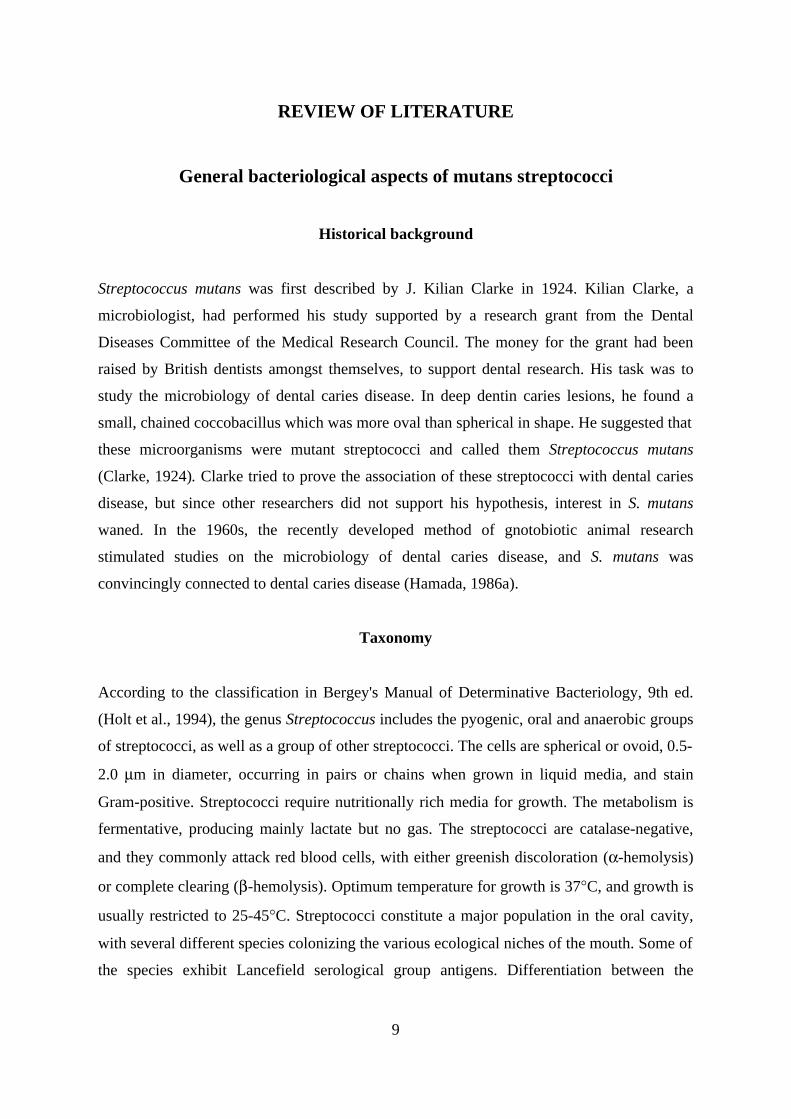
Fig. 1. Streptococci commonly found in the human mouth; phylogenetic relationships among
groups (information from Russell, 2000).
S. anginosus S. constellatus S. intermedius
anginosus group
mitis group
salivarius group
mutans group
S. oralis S. mitis S. gordonii S. sanguis S. parasanguis
S. vestibularis S. salivarius
S. mutans S. sobrinus
The oral group has sometimes been named the viridans streptococci, referring to the partial
clearing of the erythrocytes around the colony. However, the terms are not interchangeable,
as some species classified as viridans streptococci are not detected in the oral cavity. The
current classification of the oral streptococci places the bacteria into four species groups; the
anginosus, mitis, mutans and salivarius groups (Fig. 1). The classification is based on
chemotaxonomic and genotypic data, especially DNA-DNA base pairing and 16S rRNA
gene sequence analysis (Whiley and Beighton, 1998). The mutans group includes S. mutans,
S. sobrinus, Streptococcus cricetus, Streptococcus rattus, Streptococcus downeii and
Streptococcus macacae. Although the phylogenetic position of Streptococcus ferus has yet to
be determined by 16S rRNA sequencing, other data indicate that S. ferus also belongs to the
mutans group (Whiley and Beighton, 1998). The mutans streptococci represent eight
serotypes (Maiden et al., 1992; Whiley and Beighton, 1998).
11
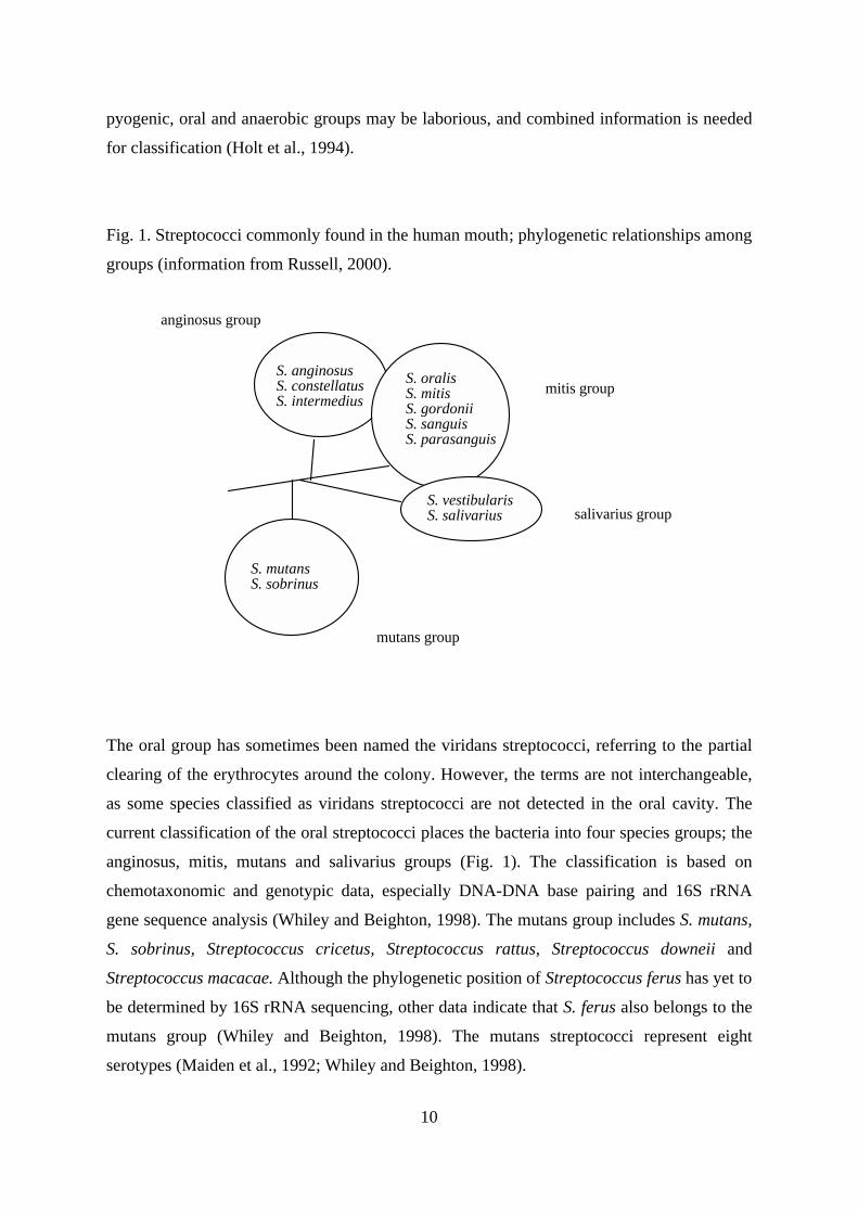
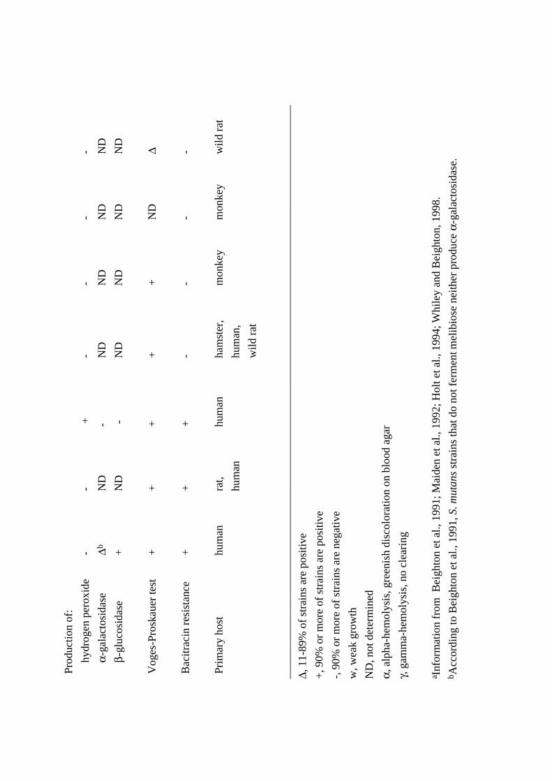
Table 1. Characteristics of the mutans streptococci groupa
__________________________________________________________________________________________________
Major cell wall
mol% G+C Serotype polysacharide constituentsb
__________________________________________________________________________________________________
S. mutans 36-38 c, e, f Rha, Glc
S. rattus 41-43 b Rha, Gal, Gro
S. sobrinus 44-46 d, g Rha, Glc, Gal
S. cricetus 42-44 a Rha, Glc, Gal
S. downei 41-42 h ND
S. macacae 35-36 c ND
S. ferus 43-45 c Rha, Glcc
__________________________________________________________________________________________________
aInformation from Maiden et al., 1992; Whiley and Beighton, 1998.
bAbbreviations: Rha, rhamnose; Glc, glucose; Gal, galactose; Gro, glycerol; ND, not
determined.
cS. ferus included in the mutans group by DNA-DNA hybridization, but not by multilocus
enzyme electrophoresis.
S. mutans cells are about 0.5-0.75 µm in diameter. S. mutans occurs in pairs or in short- or
medium-length chains, without capsules. Under acid conditions in broth and on some solid
media, these cocci may form short rods 1.5-3.0 µm in length. Rod-shaped morphology may
be evident on primary isolation from oral specimens. S. sobrinus are about 0.5 µm in
diameter. S. sobrinus occurs in pairs and in chains. The word sobrinus means male cousin on
mother's side and refers to the "distant relationship" between this species and S. mutans. S.
rattus, S. ferus and S. cricetus are about 0.5 µm in diameter, occurring in pairs or chains
(Hardie, 1986).
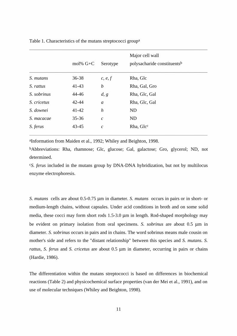
The differentiation within the mutans streptococci is based on differences in biochemical
reactions (Table 2) and physicochemical surface properties (van der Mei et al., 1991), and on
use of molecular techniques (Whiley and Beighton, 1998).
Tab
le 2
. Dif
fere
ntia
l cha
ract
eris
tics
of th
e m
utan
s st
rept
ococ
ci g
roup
a
Spe
cies
Cha
ract
eris
tic
S. m
utan
sS.
rat
tus
S. s
obri
nus
S. c
rice
tus
S. d
owne
iS.
mac
acae
S. fe
rus
Gro
wth
in a
ir∆
∆∆
∆+
w∆
Gro
wth
at:
1
0°C
--
--
ND
ND
-
4
5°C
∆∆
∆∆
--
-
Gro
wth
with
:
6
.5%
NaC
l-
-∆
∆-
--
Hem
olys
isγ
ND
γ or
αγ
ND
αN
D
Ferm
enta
tion
of:
m
anni
tol
++
++
++
+
s
orbi
tol
++
∆+
-+
+
r
affi
nose
++
∆+
-+
-
in
ulin
++
∆∆
+-
+
m
elib
iose
∆+
-N
DN
DN
DN
D
s
alic
in+
+-
++
ND
+
tr
ehal
ose
++
∆+
++
ND
Hyd
roly
sis
of:
a
rgin
ine
-+
--
--
-
e
scul
in+
+∆
∆-
++
Prod
uctio
n of
:
h
ydro
gen
pero
xide
--
+-
--
-
α
-gal
acto
sida
se∆b
ND
-N
DN
DN
DN
D
β
-glu
cosi
dase
+N
D-
ND
ND
ND
ND
Vog
es-P
rosk
auer
test
++
++
+N
D∆
Bac
itra
cin
resi
stan
ce+
++
--
--
Prim
ary
host
hum
anra
t,
hum
an
hum
anha
mst
er,
hum
an,
wild
rat
mon
key
mon
key
wild
rat
____
____
____
____
____
____
____
____
____
____
____
____
____
____
____
____
____
____
____
____
____
____
____
____
____
___
∆, 1
1-89
% o
f st
rain
s ar
e po
sitiv
e
+, 9
0% o
r m
ore
of s
trai
ns a
re p
ositi
ve
-, 9
0% o
r m
ore
of s
trai
ns a
re n
egat
ive
w, w
eak
grow
th
ND
, not
det
erm
ined
α, a
lpha
-hem
olys
is, g
reen
ish
disc
olor
atio
n on
blo
od a
gar
γ, g
amm
a-he
mol
ysis
, no
clea
ring
a Inf
orm
atio
n fr
om B
eigh
ton
et a
l., 1
991;
Mai
den
et a
l., 1
992;
Hol
t et a
l., 1
994;
Whi
ley
and
Bei
ghto
n, 1
998.
b Acc
ordi
ng to
Bei
ghto
n et
al.,
199
1, S
. mut
ans
stra
ins
that
do
not f
erm
ent m
elib
iose
nei
ther
pro
duce
α-g
alac
tosi
dase
.
14
Epidemiology
Epidemiological studies show that human populations world-wide carry mutans streptococci
(Loesche, 1986). Mutans streptococci have been demonstrated in nearly all subjects in
populations with high, low and very low prevalence of caries (Carlsson, 1988). These
microorganisms are harboured by 33-75% of 4-year-old children (Carlsson et al., 1975;
Alaluusua and Renkonen, 1984; Köhler et al., 1988; Caufield et al., 1993), 80-90% of
adolescents, and virtually all adults (Loesche, 1986; Carlson, 1988; Alaluusua et al., 1990).
Of the serotypes, c / e / f (representing S. mutans) and d / g (representing S. sobrinus) have
been detected in humans with high frequency. Some reports of finding serotypes a (S.
cricetus) and b (S. rattus) have also been published (Loesche, 1986; van der Mei et al.,
1991). Serotype c is predominant in plaque and saliva samples from humans (Loesche,
1986). In samples from Finnish children, 75-90% of S. mutans isolates are serotype c, 10-
20% serotype e, and only a few percent represent serotype f (Alaluusua et al., 1989, 1994).
S. sobrinus is not as prevalent as S. mutans and is usually detected together with S. mutans
(Loesche, 1986). The prevalence of S. sobrinus has been reported to range between a very
low frequency and 30% in different populations. In selected subject groups, a higher
frequency of S. sobrinus has been reported (Lindquist, 1991). Only a few studies report
finding S. sobrinus as the sole mutans species, and subjects who harbour both S. mutans and
S. sobrinus tend to have higher salivary mutans streptococcal counts than subjects
harbouring S. mutans alone (Köhler et al., 1988).
Culture methods and species identification
Mutans streptococci are facultative anaerobes and their optimal growth is at 37°C (Ma and
Marquis, 1997).
On MS agar, S. mutans colonies are small, raised, irregularly margined and adherent, while
S. sobrinus colonies are surrounded by a zooglea with a gelatinous consistency (Hamada &
Slade, 1980b). On sucrose-containing agar, most strains of S. mutans produce colonies of
about 1 mm in diameter, with beads, droplets or puddles containing soluble extracellular
polysaccharide (Hardie, 1986). On blood agar incubated anaerobically for two days, S.
mutans colonies are white or gray, circular or irregular, 0.5-1.0 mm in diameter, sometimes
tending to adhere to the surface of the agar (Hardie, 1986).
15
For primary isolation of mutans streptococci, the most frequently used medium is mitis
salivarius bacitracin (MSB) agar (Gold et al., 1973), which is composed of mitis salivarius
agar with sucrose, bacitracin and potassium tellurite. MSB agar is selective for S. mutans, S.
sobrinus and S. rattus. TYCSB agar contains TYC agar (trypticase, yeast extract and cystine)
with sucrose and bacitracin (van Palenstein-Helderman et al., 1983). GSTB agar contains a
basal GS agar (trypticase and yeast extract) with glucose, sucrose, bacitracin and potassium
tellurite (Tanzer et al., 1984). TSY20B agar contains trypticase soy agar, yeast extract,
sucrose and bacitracin (Schaeken et al., 1986). MSKB medium is composed of mitis
salivarius agar, sorbitol, kanamycin sulfate, bacitracin and potassium tellurite (Kimmel and
Tinanoff, 1991). Differences in culture result when different media are used (Little et al.,
1977; Jordan, 1986; Schaeken et al., 1986; Dasanayake et al., 1995). Little et al. (1977)
found that the use of blood-sucrose media produced the highest recoveries of mutans
steptococci as compared with the selective media MSB and the Carlsson medium. Schaeken
et al. (1986) also reported that MSB agar is inhibitory to mutans streptococci, especially S.
sobrinus, finding higher recoveries on TYCSB agar. Dasanayake et al. (1995) found higher
counts on MSB agar than on GSTB agar.
In addition, chair-side methods for detection and enumeration of mutans streptococci have
been developed. In the Dentocult SM® dip slide (Orion Diagnostica, Espoo, Finland), a
special slide was coated with mitis salivarius agar containing 20% sucrose (Alaluusua et al.,
1984). After inoculation of the slide with saliva, two discs containing bacitracin (5 µg) were
placed on the agar surface, and the growth density of mutans streptococci was scored after
incubation at 37°C for 48 h in an atmosphere created by CO2-generating tablets that were
placed into the cover tubes of the slides. This type of chair-side slide had a short shelf-life
because of the mitis salivarius-sucrose agar. The Dentocult-SM® Strip Mutans test was
introduced in 1989 (Jensen and Bratthall, 1989). When performing this test, a bacitracin disc
is added to the broth at least 15 min before use. After the test subject has chewed a piece of
paraffin for at least 1 min, a plastic strip is turned around in the mouth. The strip is
withdrawn through closed lips such that a thin layer of saliva remains on the strip. The strip
is closed in the cover tube with selective broth and incubated at 37°C for 48 h. After
incubation, the strip with attached colonies is compared with the model chart for counts 0 to
3. These strips can be dried and stored for long periods. For validation of method, these
chair-side techniques have been thoroughly compared with conventional selective agar plate
16
culture. The comparison yields a good correlation between the methods with regard to
detection of mutans streptococci, as well as counts on agar and score values (Alaluusua et al.,
1984; Jensen and Bratthall, 1989).
A number of other chair-side culture methods for mutans streptococci exist, including
methods using the adherence ability of isolates and using tooth picks or wooden spatulas for
sampling (Matsukubo et al., 1981; Jordan et al., 1987; Bratthall et al., 1996).
The identification of mutans streptococci is based on distinctive colonial morphology on
selective and nonselective agar, Gram staining, distinctive cell shape on light microscopy,
specific growth characteristics, and sugar fermentation and enzymatic patterns. The
identification scheme used in the present study is presented in Table 2. The scheme is based
on information in Bergey's Manual of Determinative Bacteriology (9th ed., 1994) and on
additional information on phenotypic properties. S. mutans isolates can also be identified by
the commercial biochemical test system API 20 Strep (Bio Mérieux, Marcy-l'Étoile, France).
Methods for direct detection: monoclonal antibodies and DNA probes
Monoclonal antibodies (MAbs) directed against specific species of mutans streptococci, and
also against cell markers and enzymes have been developed. The MAbs have been used in a
number of studies on mutans streptococci, for detection in epidemiological studies and in
studies on microbial mechanisms (de Soet et al., 1987, 1990a, 1990b; Takei et al., 1992;
Fukushima et al., 1993; Shi et al., 1998).
A DNA probe for detection of S. mutans was developed by using glucosyltransferase B gene
(gtfB) and fructosyltransferase gene (ftf) fragments, enabling detection without culture
(Smorawinska and Kuramitsu, 1992). The dextranase gene (dexA) has also been used to
construct a DNA probe for detection of S. mutans (Ida et al., 1998). For detection of S.
sobrinus, a DNA fragment in the dextranase gene (SSB-3) has been suggested to be useful
(Ida et al., 1999).
17
Typing of mutans streptococci
Typing of isolates is applied in epidemiological studies to determine bacterial occurrence
and modes of transmission. Typing of isolates is also performed for evaluation of whether
certain strains are associated with specific clinical disease conditions and to characterize the
heterogeneity of infection, i.e., whether subjects are colonized by one or multiple types of
the microorganism. Evaluation criteria for typing methods include typeability (ability to give
an outcome for every isolate included), reproducibility (ability to give the same result when
repeating the analysis) and discriminatory power (ability to differentiate between unrelated
strains) (Arbeit, 1999). Two main types of epidemiological typing systems for
microorganisms are available, the phenotypic and the genotypic methods.
Phenotyping
Traditional methods of characterizing bacterial isolates have relied on measurement of
characteristics expressed by the microorganisms, such as bacteriocin production and
sensitivity to bacteriocins, serotype, biochemical properties, antibiotic resistance and
bacteriophage type (Maslow and Mulligan, 1996).
Bacteriocin typing
One of the first epidemiological typing systems for oral streptococci was bacteriocin typing
(Kelstrup et al., 1970). Bacteriocins are proteinaceous substances produced by the bacteria
that inhibit the growth of other, mostly closely related, bacteria. The typing is performed by
measuring the inhibiting effect on bacterial growth of certain indicator strains, and by
measuring the sensitivity of the bacteria to be typed to bacteriocins from other strains (Jack
et al., 1995). Heterogeneity among strains of mutans streptococci within one individual was
first shown by bacteriocin typing (Kelstrup et al., 1970).
Serotyping
Using Ouchterlony immunodiffusion, Bratthall demonstrated five serological groups of
mutans streptococci (Bratthall, 1970). A total of eight serotypes were subsequently
recognized (Perch et al., 1974; Beighton et al., 1981). The classification is based on cell-wall
carbohydrate antigen (Table 1). Serotyping by immunodiffusion, immunofluorescence or
immunoelectrophoresis has been widely applied for typing of mutans streptococci.
18
Biotyping
In 1974, Shklair and Keene divided mutans streptococci into five biotypes (a-e) on the basis
of fermentation characteristics, arginine hydrolysis and bacteriocin sensitivity, and they
reported that these biotypes corresponded with the serotypes reported in 1970. Their later
scheme (Shklair and Keene, 1976) also includes serotypes f and g.
Among other phenotypic methods are cellular fatty acid analysis, whole-cell protein analysis
and multilocus enzyme electrophoresis (MEE). MEE is based on the relative electrophoretic
mobility of metabolite cellular enzymes. MEE has been successfully applied in studies with
many organisms, but only one report using MEE in strain identification of mutans
streptococci has been published (Gilmour et al., 1987). Phenotypic typing is, in most cases,
relatively inexpensive to perform and typeability of mutans streptococcal isolates is high,
however, reproducibility and discriminatory power are usually somewhat poorer (Arbeit,
1999).
Genotyping
For isolate fingerprinting, molecular typing methods have a higher discriminatory ability and
reproducibility since these methods do not examine the gene expression but rather the DNA
of the microorganisms to be studied (Arbeit, 1999; Olive and Bean, 1999). Among these
typing methods are plasmid analysis, restriction endonuclease analysis (REA), restriction
fragment length polymorphism (RFLP) (including ribotyping), pulsed field gel
electrophoresis (PFGE) and arbitrarily primed polymerase chain reaction (AP-PCR).
Plasmid analysis
Plasmids are extrachromosomal circles of DNA that encode many properties, including
antimicrobial resistance, many virulence traits and hydrocarbon metabolism (Madigan et al.,
1997a). Plasmid analysis was the first DNA-based technique applied in epidemiological
studies on mutans streptococci (Caufield et al., 1982). Because plasmids are infrequently
detected in mutans streptococci, in only 5% of strains (Hamada and Slade, 1980b), plasmid
analysis is not applicable to typing of these bacteria.
19
Restriction endonuclease analysis (REA)
In restriction endonuclease analysis (REA), bacterial chromosomal DNA is cut with a
restriction endonuclease and separated by gel electrophoresis. The restriction endonucleases
are enzymes that cut the DNA chain at specific recognition sequences. The restriction
enzymes are nowadays synthetically fabricated, but were originally isolated from bacteria,
with their original function being defence against other bacteria. After separation by gel
electrophoresis, gels are stained with ethidium bromide and detected under UV light,
whereby the banding patterns obtained for different strains are compared. Often the process
results in fingerprints with many bands, thus the interpretation of the REA profiles can be
complicated. REA has been applied for evaluation of relatedness of mutans streptococcal
isolates (Caufield and Walker, 1989; Kulkarni et al., 1989).
Restriction fragment length polymorphism (RFLP), including ribotyping
After cleaving the chromosomal DNA of the microorganisms to be studied, the separation
products can be labelled with either DNA or RNA probes in the Southern blot technique
(Southern, 1975). The use of a probe derived from the Escherichia coli ribosomal operon
was introduced by Grimont and Grimont in 1986. They had discovered that variations of the
genes encoding ribosomal ribonucleic acid (rRNA), and variations in sites flanking those
loci, could serve as a means of typing strains since ribosomal sequences are highly
conserved. In ribotyping an isolate, after the gel electrophoresis of the cleaved DNA, the
fragments are hybridized with the rRNA probe. When detecting the hybrids, every fragment
containing a ribosomal gene will be highlighted. The banding patterns obtained in ribotyping
include only a small number of bands, thus rendering comparison of fingerprints among
isolates easier than comparing REA patterns. The term ribotyping was introduced in 1988
(Stull et al., 1988), and ribotyping of mutans streptococci has been applied since 1993
(Saarela et al., 1993) mainly in studies on transmission of mutans streptococci and stability
of infection (Alaluusua et al., 1994).
Pulsed field gel electrophoresis (PFGE)
In pulsed field gel electrophoresis, a variation of agarose gel electrophoresis, the orientation
of the electric field across the gel is changed periodically ("pulsed"), thus larger bacterial
DNA fragments can be analysed than by REA (Arbeit, 1999). PFGE is considered the "gold
standard" of molecular typing methods, with excellent discriminatory power and
20
reproducibility (Arbeit, 1999; Olive and Bean, 1999). This method has not been applied for
typing of mutans streptococci.
Arbitrarily primed polymerase chain reaction (AP-PCR)
Of the genotyping methods thus far applied to mutans streptococci, AP-PCR is perhaps the
least laborious (Olive and Bean, 1999). AP-PCR can be performed with a very small sample
volume. For the polymerase chain reaction, the template is annealed to one or more short
primers (typically 9-10 bp) at low stringency. Amplification results in an array of DNA
fragments, often termed random amplified polymorphic DNA (RAPD), that can be resolved
by gel electrophoresis. AP-PCR requires no previous knowledge of the DNA to be analysed
(Welsh and McClelland, 1990; Williams et al., 1990). A limitation of the method is that it is
very sensitive to even minor variations in technical factors such as temperature, Mg2+
concentration and polymerase source. Interlaboratory comparison of typing results is
impeded by only a fair reproducibility (Arbeit, 1999), and only isolates processed
simultaneously and fingerprints obtained concomitantly can be compared. This fair
reproducibility is, however, complemented by a good discriminatory power (Arbeit, 1999).
AP-PCR typing has been shown to be well applicable to typing of mutans streptococci
(Saarela et al., 1996; Li and Caufield, 1998).
Primary acquisition and transmission of mutans streptococci
Normally, before birth the foetus is sterile. The inoculation of the human oral cavity starts
with the first tactile contacts with the mother and other persons present at the parturition, and
contact with air and equipment. The oral cavity of the toothless child contains only epithelial
surfaces and the first colonizers are species not requiring a nonshedding surface. Early
colonizers include some streptococci, Veillonella, Actinomyces, Fusobacterium and a few
Gram-negative rods (Könönen et al., 1994, 1999). Streptococcus salivarius is among the first
permanent colonizers, colonizing the dorsum of the tongue in the edentulous infant
(Socransky and Manganiello, 1971). In the first months, most of the detected strains are
transient colonizers. Streptococcus sanguis and the mutans streptococci are stably colonized
only after the first tooth has erupted (Berkowitz et al., 1975b).
In general, the acquisition of microorganisms by the human body is by transmission directly
from one host to another, or indirectly by means of another living agent (vector). Pathogens
21
can also be transmitted by inanimate objects and disease vehicles such as food and water
(Madigan et al., 1997b). Saliva is regarded as the most important vehicle of transmission of
mutans streptococci via physical contact (Duchin and van Houte, 1978; Köhler and Bratthall,
1978) or use of shared objects, e.g. spoons and forks (Bratthall, 1997); mutans streptococci
can be recovered from a metal plate 24-48 h after inoculation (Köhler and Bratthall, 1978).
The mother is considered to be the most important source of infection for the child, the first
indication of this obtained by using a phenotypic typing technique of S. mutans, bacteriocin
typing (Berkowitz and Jordan 1975a; Masuda et al., 1985). Li and Caufield (1995) detected
by molecular typing of mutans streptococcal isolates a homology of strains in 71% of
mothers and their children, and Kozai et al. (1999) found that 51.4% of mutans streptococcal
genotypes found in children were also identified in their mothers. Fathers and infants have a
far lower strain match (Rogers, 1981; Davey and Rogers, 1984; Kulkarni et al., 1989; Li and
Caufield, 1995, 1998). Only Kozai et al. (1999) found an abundant similarity of strains in
fathers and children, 31.4% of strains detected in children were in agreement with those in
their fathers. The time period when most children gain mutans streptococci in their oral flora
is when the primary teeth are erupting, i.e., between 8 months and 3 years of age (Caufield et
al., 1993). The probability of colonization with mutans streptococci is high when inoculation
with mutans streptococci is frequent and microbial cell count is at least 105 per ml saliva
(Berkowitz et al., 1981). Another suggested prerequisite for early colonization is that the
baby's diet includes frequent intake of refined carbohydrates (Alaluusua, 1991a). Recently, it
has been shown that mothers can diminish the probability of transmitting mutans
streptococci to their children by using xylitol chewing gum (Söderling et al., 2000). The
exact mechanism of action regarding xylitol is unknown. The sugar substitute xylitol is not
fermented by mutans streptococci into cariogenic acid end-products, and in the oral
environment, xylitol presumably selects for mutans streptococci with a weakened virulence
(Trahan, 1995). It has previously been shown that in mothers harbouring high numbers of
mutans streptococci in their saliva, preventive measures aimed at decreasing mutans
streptococcal counts also result in decreased caries counts in their children, even after 15
years (Köhler et al., 1982, 1983, 1994; Bratthall, 1997).
22
Occurrence of mutans streptococci in the oral cavity
The primary habitat of S. mutans and S. sobrinus is the human dentition. In addition, after
mutans streptococci have colonized the dentition, they can be detected in saliva, on the
tongue, on oral mucous membranes, on denture surfaces and on surfaces of orthodontic
appliances. When a mutans streptococcal strain has achieved stable colonization of the oral
cavity, the strain usually persists for a long time period (Alaluusua et al., 1994). Mutans
streptococci can also be detected in faeces (Hamada et al., 1980a), but otherwise, very few
reports on finding mutans streptococci outside the oral cavity are available. The organism
has been detected in endocarditis (Parker and Ball, 1976) and in purulent eyes of a neonate
(Reeder et al., 1985).
Dentition
The first tooth erupts at the age of about 8 months (± 2 months) (Kreiborg 1991), and only
after that can there be stable colonization by mutans streptococci. Mutans streptococci can
occur in the dentition on the tooth surfaces in dental plaque and in carious enamel or dentin.
In enamel caries, the subsurface tissue is invaded by microorganisms. The identification of
the bacteria invading dental enamel is as yet incomplete, but in experiments with gnotobiotic
rats, in combination with scanning electron microscopy, S. mutans and S. sobrinus have been
shown to be able to invade the enamel (Luoma et al., 1987; Seppä et al., 1989).
In carious dentin, the number of bacteria recovered per mg of dentin has been reported to be
higher in superficial layers of the lesion as compared with deeper layers, with regard to both
mutans streptococci and total bacterial count (Edwardsson, 1974; Hoshino, 1985). Many
bacterial species are present, but no evidence suggests that a specific combination of
organisms acting in symbiosis are involved in the decomposition of dentin (Edwardsson,
1974).
The dental plaque consists of cells (about 70% of the overall volume) and plaque matrix
(Jenkins, 1978). The water content ranges between 80% and 85%. Of the dry weight, 40-
50% is protein, 13-17% carbohydrate, 10-14% lipid and 10% ash. The concentrations of
calcium and phosphate are quite variable, but typical figures are calcium 8 µg per mg and
23
inorganic phosphate (as P) 16 µg per mg (Jenkins, 1978). Plaque maximum thickness on
smooth surfaces is 300 µm, on approximal surfaces 5 mm and in fissures 2 mm (Sissons,
1997). The microbial composition of dental plaque differs both qualitatively and
quantitatively from the bacterial communities of other oral surfaces, and the microflora also
varies over time. Plaque microorganisms exhibit a high genetic heterogeneity, with 30 to 300
species represented (Sissons, 1997). This conglomerate of bacteria and interbacterial
substances can be regarded as a prototype for a biofilm, a bacterial community. The biofilm
forms a dynamic link between the microbial flora, the tooth surfaces, and the different
constituents of saliva, with the microorganisms living in symbiosis and antibiosis with each
other (Costerton and Lewandowski, 1997). Survival of oral bacteria is enhanced by dental
plaque formation (Bowden and Hamilton, 1998).
The frequency of isolation of mutans streptococci in dental plaque has been reported to be
site-dependent, such that the isolation frequency steadily increases from approximal surfaces
of mandibular incisors, approximal surfaces of maxillary incisors, approximal surfaces of
molars, to fissures of molars (Loesche, 1986). S. mutans and S. sobrinus show a somewhat
similar colonization pattern in the dentition, but S. sobrinus is more frequently isolated from
posterior than anterior teeth. In subjects harbouring both S. mutans and S. sobrinus, the
species colonize buccal surfaces in comparable numbers, but on all other surfaces, S. mutans
is predominant (Lindquist, 1991).
Saliva
Saliva is a complex mixture of several components (Whelton, 1996). Whole saliva (oral
fluid) is formed primarily from salivary gland secretions, but also contains gingival fluid,
desquamated epithelial cells, bacteria, leucocytes, and possibly food residues, blood and
viruses (Dawes, 1996; Whelton, 1996). Saliva is essential for maintenance of healthy oral
tissues; it coats the oral mucosa and protects against irritation, forms an ion reservoir for
tooth remineralization, functions as a buffer, aids in swallowing, exerts antimicrobial action,
participates in pellicle formation and enzymic digestion of starch with amylase, and also
participates in taste sensation by acting as a solvent (Whelton, 1996). Antimicrobial
components in saliva include immunoglobulins, lysozymes, lactoferrins, salivary
peroxidases, myeloperoxidases, histatins, amylases and anionic proteins (Bowen, 1996;
Tenovuo, 1998). Moreover, organic components in saliva, especially mucous glycoproteins,
24
function as nutrients for many oral bacteria (Bowen, 1996). Salivary proteins can be
degraded by proteases produced by, for example, S. mutans and S. sanguis (Bowen, 1996).
The bacterial content of saliva is estimated to approach 109 bacteria per ml (Bowen, 1996).
Saliva helps to control invasion of the mouth by microorganisms, and lack of saliva results in
increased numbers of bacteria in the mouth. Saliva can act as a selective medium for
bacterial growth, but continuously repeated swallowing results in clearing of bacteria
(Bowen, 1996).
Salivary mutans streptococcal counts rarely exceed 107 CFU per ml. A highly significant
correlation has been demonstrated between the salivary numbers of mutans streptococci and
their prevalence in the dentition, both in terms of the number of tooth surfaces colonized and
the level of infection of tooth surfaces (Duchin and van Houte, 1978; Lindquist, 1991).
Clinical illness connected with mutans streptococci
Dental caries - a multifactorial disease
Dental caries disease includes a breakdown of enamel, the hardest material in the human
body, and a subsequent breakdown of the underlying dentin. The disease is the most
prevalent of the chronic diseases affecting the human race. The aetiology of dental caries
disease is multifactorial in that simultaneous participation of multiple factors is required for
caries to occur (Tanzer, 1992). The original still-prevailing theory explaining the disease
process implicates carbohydrates, oral microorganisms, and acids as the main factors in the
caries process. This acidogenic theory (or Miller's chemoparasitic theory, 1902) states that
"Dental decay is a chemico-parasitic process consisting of two stages, the decalcification of
enamel, which results in its total destruction, and the decalcification of dentin, as a
preliminary stage, followed by dissolution of the softened residue. The acid which affects
this primary decalcification is derived from the fermentation of starches and sugar lodged in
the retaining centers of the teeth." Miller isolated many microorganisms from the human oral
cavity, some acidogenic and some proteolytic. Miller believed that caries is caused by a
variety of microorganisms. Two other theories explaining the disease process are the
proteolytic theory and the proteolysis-chelation theory (Shafer et al., 1983).
25
Today, mutans streptococci are considered to be the main aetiological microorganisms in
caries disease, with lactobacilli and other microorganisms participating in the disease
progression (Tanzer, 1992). Occasionally, some other microorganisms have been traced as
initiator microorganisms. Severe dental caries has been induced in hyposalivated rats
infected with Lactobacillus fermentum (Ooshima et al., 1994). Acidogenesis at a low pH has
also been reported for a group of non-mutans streptococci (van Houte et al., 1991).
Nursing caries
When the caries disease has a very fast progression it is called rampant caries. A specific
form of rampant caries involves the dentition of very young children. The condition includes
a demineralization and cavitation of the labial surfaces of the maxillary primary incisors,
followed by involvement of the first primary molars (Ripa 1988). The disease onset is
between 1 and 2 years of age. A variety of names have been given to the condition; nursing
caries, nursing bottle caries, early childhood caries, baby bottle tooth decay and baby bottle
caries (Curzon and Pollard, 1994; Tinanoff, 1997). The prevalence of nursing caries in
different populations varies between 1% and 80% of preschool children, the proportion of
affected children being very low in many western societies (Curzon and Pollard, 1994;
Milnes, 1996). Some reports have been made on increasing incidence of this condition in
western countries (Duperon, 1995). In Finland, the prevalence can be estimated at about 1%,
exact figures being unavailable (Paunio, 1993; Alaluusua, 1999). The aetiology of this
condition is also considered multifactorial. In most cases, the affected child has frequently
received fermentable carbohydrates as a drink when going to sleep, combined with a lack of
toothbrushing with fluoride toothpaste (Curzon and Pollard, 1994). Moreover, the
combination of frequent breast-feeding and low additional fluoride use are considered to be
contributing factors in the process of nursing caries (Hallonsten et al., 1995; Weerheijm et
al., 1998). In children with nursing caries, mutans streptococci can be so dominant that the
plaque above the caries lesions consists almost entirely of these organisms (van Houte et al.,
1982).
Mutans streptococci in caries prediction
The fact that dental caries is a world-wide disease requiring vast economic resources and
causing a great deal of discomfort has called upon attempts aimed at developing an accurate
26
screening method for detection of the 5-20% of subjects comprising the high-caries-risk
group (Pienihäkkinen, 1987; Roeters, 1994; Hausen, 1997). Because mutans streptococci are
considered to be the predominant pathogens of dental caries disease, individuals heavily
colonized by mutans streptococci were thought to automatically be at high risk for caries.
Indeed, in young children, early mutans streptococcal colonization on tooth surfaces has
been recognized as an indicator of later high scores of decayed, missing and filled surfaces in
deciduous teeth (dmfs index) (Alaluusua and Renkonen, 1983; Köhler et al., 1984, 1988;
Jokela, 1997). However, it has become evident that, although prevalence of the infection is
indicative of the disease status on a population level, when it comes to an older child or
adult, on the individual level, the caries risk rate cannot be accurately predicted on the basis
of how heavily the subject is colonized by mutans streptococci (Stecksen-Blicks, 1985;
Alaluusua et al., 1987, 1990; Alaluusua, 1993; Alanen et al., 1994; Hausen, 1997).
Virulence factors of mutans streptococci
The term virulence describes the capacity of a parasite (a microorganism) to cause disease to
its host, the organism it lives on or in. The property is quantitative and expresses the degree
of pathogenicity, the ability to inflict damage to the host. The relationship between host and
parasite is dynamic and depends on their individual characteristics and their interrelationship
as well as on external factors. Virulence consists of bacterial properties required in the
interaction between host and parasite, factors that promote the entry, colonization and
growth of the pathogen within the host, including those required for opposing host defences
and for nutrient acquisition (Madigan, 1997b). The virulence factors of microorganisms can
be studied by the classical approach, by isolating bacteria from healthy and diseased subjects
and comparing the phenotypic properties of these microorganisms. During the last two
decades, approaches utilized in virulence studies have also included molecular biology
methods, techniques for the manipulation of DNA in vitro (Hensel and Holden, 1996).
Studies on the expression of the modified bacterial chromosome can subsequently be
extended to cell culture and animal experiments.
As regards the mutans streptococci, properties that affect their ability to cause dental caries
disease are virulence factors promoting their colonization and survival in the biofilm, the
dental plaque, that covers the tooth surfaces. Recognized virulence factors of mutans
streptococci are adhesin-like cell surface proteins, acid tolerance, acid production, and
27
production of glucosyltransferases, mutacin and intracellular polysaccharides (Kuramitsu,
1993). In addition to the recognized virulence factors, other properties of the microorganism
may influence virulence. One of the suggested virulence factors is the proteolytic activity of
mutans streptococci (Homer et al., 1990; Harrington and Russell, 1994; Jackson et al., 1997).
S. mutans has been shown to produce two extracellular proteases, possibly metalloproteases,
capable of degrading both gelatin and collagen-like substrates (Harrington and Russell,
1994). Currently under debate is whether metalloproteases detected in connection with
mutans streptococci are produced by the microorganism or whether they are host-derived
(Tjäderhane et al., 1998). Regarding the sIgA protease activity of mutans streptococci, the
presiding view is that the organism itself does not produce this protease (Marcotte and
Lavoie, 1998). Many oral streptococci do produce sIgA protease, which impairs the host
defence by cleaving the secretory IgA present, and apparently mutans streptococci benefit
from protease produced by the primary colonizers (Marcotte and Lavoie, 1998). Another trait
enabling survival is the ability of mutans streptococci to rapidly adapt to the environment by
microbial genetics phenomena; this property has been suggested to be an essential element in
the dominance of S. mutans in cariogenic dental plaque (Burne et al., 1997; Burne, 1998). As
a rule, the ability of cells to take up exogenous DNA, the regulation of natural genetic
competence in bacteria, is dictated by nutritional conditions and cell-to-cell signalling
(Solomon and Grossman, 1996).
Factors affecting adherence ability
The mutans streptococci synthesize extracellular polysaccharides from sucrose to increase
their stickiness. Surface proteins of S. mutans also participate in adherence. In S. sobrinus,
the adherence is probably primarily mediated by extracellular polysaccharides, with a minor
influence by surface proteins (Gibbons et al., 1986). In addition to the microbial properties,
host factors may affect adherence, and salivary components can function as receptors in oral
pellicles for microbial adhesion to host surfaces (Scannapieco, 1994).
Glucosyltransferases and fructosyltransferases
Glucosyltransferases (GTFs) and fructosyltransferases (FTFs) catalyse the synthesis of
water-soluble and water-insoluble glucan and fructan polymers from sucrose (Loesche,
1986). The nucleotide sequences of gtf genes from different oral streptococci comply with
28
the same basic pattern, and the GTFs are approximately 1500 amino acids long (Russell,
1994). Streptococcal GTFs have two common functional domains. The amino-terminal
portion, the catalytic domain, is responsible for the cleavage of sucrose, and the carboxyl-
terminal portion, the glucan binding domain, is responsible for glucan binding (Colby and
Russell, 1997). S. mutans produces at least one FTF and three GTFs (Sato and Kuramitsu,
1986; Russell, 1994). GTF-I and GTF-SI enzymes, products of gtfB and gtfC genes,
primarily catalyse the synthesis of water-insoluble glucans, whereas GTF-S, the product of
gtfD, mainly catalyses the synthesis of water-soluble glucans. S. sobrinus has four gtf genes
(Russell, 1994). Of the four GTFs of S. sobrinus, GTF-I produces water-insoluble glucans,
while the other three produce water-soluble glucans (Hanada et al., 1993). The three gtf
genes from S. mutans have been thoroughly assessed by comparative sequence analysis,
revealing that interstrain differences of gtfB and gtfD are limited, but gtfC exhibits
significant interstrain variability (Fujiwara et al., 1998).
The in vitro effects of the GTFs have been extensively studied; however, few studies with
clinical strains have been performed to date.
Surface proteins
S. mutans cells express a predominant surface protein called P1 (or I/II, B, IF, SR or PAc),
which functions in the binding of mutans streptococci to human salivary pellicle-coated
surfaces (Crowley et al., 1999). The apparent functional equivalent to this protein is the spaA
protein of S. sobrinus (Kuramitsu, 1993).
Acidogenicity and acid tolerance
The mutans streptococci ferment many different sugars, and they appear to metabolize
sucrose to lactic acid more rapidly than other oral bacteria. This is thought to be related to
the multitude of enzyme systems catalysing the reactions of transport and metabolism of
sucrose expressed by these organisms (Kuramitsu, 1993). These metabolic reactions render
the dental plaque acidic in the presence of a fermentable carbon source, and the acid
tolerance of the mutans streptococci enables them to continue metabolisms even at low pH.
It has been demonstrated that strains of mutans streptococci are more acid tolerant than all
other bacteria examined, with the exception of lactobacilli (Loesche, 1986). An inducible
29
property exists in mutans streptococci which permits adaptation to acidic environments
(Hamilton and Buckley, 1991; Birkhed et al., 1993). This property of acid tolerance (or
acidurance) appears to be connected with the membrane-associated H+(proton)-translocating
ATPase of these organisms (Bender at al., 1986).
Mutacin production
Many bacteria produce bacteriocins, i.e. antibacterial peptides, to interfere with the growth of
other closely related microorganisms (Jack et al., 1995). Bacteriocins are ribosomally
synthesized and usually require extensive posttranslational modification for activity. The
genes involved in the synthesis and modification of bacteriocins are often carried by a
plasmid or a transposon (Madigan et al., 1997a). Bacteriocins are frequently named
according to the bacterial species producing them; bacteriocin produced by mutans
streptococci is called mutacin. Mutacin production is usually not plasmid encoded (Caufield
et al., 1990). When bacteriocin activity is plasmid encoded, the plasmid generally also
confers bacteriocin immunity to the microorganism (Jack et al., 1995). Some bacteriocins
also have a commercial value. Nisin, which is produced by Lactococcus lactis strains, has
been used for more than 30 years as a preservative in the food industry (Jack et al., 1995).
Several mutacins have been purified and biochemically characterized (Fukushima et al.,
1982; Ikeda et al., 1982; Hamada et al., 1986b; Loyola-Rodriguez et al.,1992; Novak et al.
1994; Chikindas et al., 1995). In replacement experiments, strains producing increased
amounts of mutacin have been shown to colonize more easily (van der Hoeven and Rogers,
1979; Hillman et al., 1987).
Production of intracellular polysaccharides
Most strains of S. mutans and S. sobrinus produce intracellular iodine-staining
polysaccharides (IPS) from sucrose, which, according to results from experiments with rats,
may contribute to their virulence (Kuramitsu, 1993). Because of this intracellular
polysaccharide storage, these cariogenic bacteria have the ability to continue fermentation in
the absence of exogenous food supplies (Loesche, 1986).
30
Control of mutans streptococci by chlorhexidine (CHX)
The traditional preventive treatment approaches for dental caries disease include dietary
counselling on reduced intake of refined sugars, oral hygiene instruction and topical
application of fluoride. To aid these preventive measures, antimicrobial preparations, such as
chlorhexidine (CHX), can be used to reduce the numbers of mutans streptococci colonizing
an individual (Emilson and Fornell, 1976; Twetman and Petersson, 1999). The outcome of
all these measures, including CHX use, is largely dependent on patient compliance.
Chlorhexidine, developed in the 1950's, is a bisbiguanide that typically exerts its action on
Gram-positive bacteria, particularly on mutans streptococci (Hennessey, 1973; Emilson,
1994; Järvinen et al., 1995). Clinically, the reduction in mutans streptococcal counts not only
reduces the caries increment, but may also reduce the probability of transmission of mutans
streptcocci from mothers to their young children (Emilson, 1994; Köhler and Andréen,
1994). The antibacterial action of CHX is based on its adsorption on bacterial surfaces. CHX
can be administered in solutions, dental gels, varnish or even chewing tablets (Luoma, 1992;
Nuuja, 1992). The combination of CHX with sodium fluoride is a more potent inhibitor of
acidogenic streptococci than either CHX or fluoride alone (Luoma, 1972; McDermid et al.,
1985). The inhibiting effect of CHX on plaque formation has been reported to persist in
human subjects even upon 2 years of continuous use (Gjermo and Eriksen, 1974). Surfaces
which are heavily infected by mutans streptococci are more rapidly recolonized after
antimicrobial treatment. In practice, posterior teeth are recolonized more readily than other
tooth surfaces (Lindquist, 1991). Adverse effects of CHX are rare, but both IgE-mediated
local responses and anaphylactic shock reactions may occur (Ebo et al., 1998).
CHX resistance
Bacterial resistance to antiseptics and disinfectants may be an inherent or acquired property
of an organism. Antimicrobial resistance can evolve through acquisition of genetic material,
such as plasmids or transposons, or by mutation. Thus far, laboratory tests have failed to
conclusively demonstrate the possibility of "training" organisms to become chlorhexidine-
resistant (Russell and Day, 1993). Many bacterial species, e.g. Proteus and Providencia,
have an intrinsic chlorhexidine resistance (Russell and Day, 1993).
31
AIMS OF THE STUDY
For improvement of high-caries-risk subject selection and for planning of preventive dental
treatment approaches, more basic information is required on the virulence characteristics and
colonization patterns of mutans streptococci. To this end, two longitudinal and two cross-
sectional studies were planned to assess the quantitative and qualitative characteristics of
oral mutans streptococci. The classical approach chosen consisted of isolating and
characterizing strains from healthy and diseased subjects, and comparing the culture and
characterizing results with clinical findings. The microbial typing methods included the
molecular biology methods of ribotyping and AP-PCR typing combined with phenotypic
methods.
The specific aims of this study were:
1. to follow the detection of oral mutans streptococci in subjects in whom these
microorganisms were initially not detected,
2. to study the number of clonal types of mutans streptococci detected within one individual,
to compare clonal types between individuals, and in young children, to compare the intra-
individual clonal diversity of mutans streptococci with clinical caries status,
3. to study a possible site-specificity of mutans streptococcal infection within the oral cavity
of an individual, and to draw inference from this for sampling techniques,
4. to examine the in vitro differences in chlorhexidine (CHX) susceptibility of mutans
streptococcal isolates of different serotypes and genotypes, to elucidate whether mutans
streptococci have evolved CHX resistance as compared with earlier reports, to examine the
in vivo changes in CHX susceptibility of isolates and in levels of salivary mutans
streptococci after 1 year of periodic use of low-concentration CHX-NaF gel in an instructed
trial, and to examine the stability of the infection during the treatment, and
5. to study the glucosyltransferase production and mutacin activity of individual
fingerprinted strains isolated from mothers and their young children, and to compare these
results on phenotypic properties with the event of transmission of strains from mother to
child and to clinical caries status of the child.
32
MATERIALS AND METHODS
Subjects and trial conditions
The study subjects comprised a total of 402 participants in two longitudinal studies and two
cross-sectional studies. A more detailed analysis was performed using mutans streptococcal
isolates from 24 adolescents, 40 mothers with their 1.5- to 3-year-old children (one twin
pair) and seven 3- to 7-year-old children.
Adolescents in follow-up on colonization of mutans streptococci(study I and unpublished results)
A group of 182 adolescents aged 12 to 17 years were invited to participate in a longitudinalstudy on salivary levels of mutans streptococci. Since their birth the subjects had beenannually attending a longitudinal study on facial development and oral health at theDepartment of Pedodontics and Orthodontics, Institute of Dentistry, University of Helsinki(Haataja et al., 1976; Nyström, 1982; Nyström et al., 1990; Könönen and Nyström, 1993)(Table 3).
Table 3. Adolescents over an 8-year follow-up on colonization of mutans streptococci
__________________________________________________________________________
12- to 17-year-old adolescents invited to participate (n=182)
Sampling I: Chair-side test for mutans streptococci (n=146);
mutans streptococci not detected in 24 subjects
Sampling II: Annual chair-side test for mutans streptococci (n=23)
Sampling III: Annual chair-side test for mutans streptococci (n=24)
Sampling IV: Annual chair-side test for mutans streptococci (n=24),
complemented by conventional agar plate culture of
pooled plaque samples (n=10)
Sampling V: Five years later, chair-side test and conventional agar plate
culture for detection of mutans streptococci (n=22)
__________________________________________________________________________
After excluding subjects who had been on antibiotics or chlorhexidine during the last twomonths, and those who had fixed orthodontic appliances, a salivary sample was obtainedfrom 146 subjects. The subjects were annually sampled and examined clinically for decayed,missing and filled teeth during the next three years. At the first sampling, 24 subjects (8girls, 16 boys) had undetectable levels of mutans streptococci as assessed by a chair-side dip-slide method (Dentocult-SM®). In 10 of these 24 adolescents, on the fourth samplingoccasion, the salivary examination was complemented by an examination of mutansstreptococci in plaque. The results from this 3-year follow-up with four sampling occasions
33
are presented in study I. Eight years after the first microbiological examination, salivarysamples from 22 of these 24 subjects were assessed for mutans streptococci using a chair-side method complemented by conventional agar plate culture, and clinical examinationswere performed. The results from the 8-year follow-up examination are presented in thisthesis.
Mothers and their children in a preventive treatment programme(II, III, V, VI and unpublished results)
The study population for study II and a portion of the subjects in studies III, V and VI wereparticipants in a preventive programme that targeted pregnant women at a community healthcentre in Espoo, Finland. A total of 153 mothers were screened before childbirth for mutansstreptococci in saliva by a commercial strip method (Dentocult-SM STRIP MUTANS test),and subjects with Dentocult-SM STRIP MUTANS test scores of 2 and 3 (corresponding tocounts of •100 000 CFU/ml, according to the manufacturer) (n=105) were randomly selectedfor the test group (n=75) or control group (n=30). The mothers in the test group brushed theirteeth periodically with a gel that contained 0.3% CHX digluconate and 0.2% NaF, pH 5.8.Mothers' gel use occurred when the child was between 6 months and 3 years of age, with thegel being used twice a day for the first 10 days of each month. The control mothers weretreated according to the regular prophylaxis protocol provided for all pregnant women at thehealth centres in Espoo, without periodic CHX-NaF gel use. The children were screened atthe ages of approximately 18 and 36 months for the colonization of mutans streptococci(Table 4). At the samplings, the subjects were examined clinically for decayed, missing andfilled teeth.
Table 4. Mothers and their children in a preventive treatment programme using
CHX-NaF gel__________________________________________________________________________
Sampling I: Mothers screened for mutans streptococci in saliva before childbirth
by Dentocult-SM STRIP MUTANS test (n=153)____________________________________________________________________
Mothers with test scores of 2 and 3 (n=105)
test group (n=75) control group (n=30)____________________________________________________________________
Mothers included for 3 additional samplings,
when child about
Sampling II: - 6 months
Sampling III: - 18 months
Sampling IV: - 36 months____________________________________________________________________
Samples from children
at about
(n=53) - 18 months (n=27)(n=47) - 36 months (n=15)
__________________________________________________________________________
34
In study II, 24 mothers from the test group and 10 mothers from the control group wereincluded for examination of the effect of CHX on salivary mutans streptococcal levels, andfor testing of CHX susceptibility of their mutans streptococcal isolates. The CHX study wasstarted when the preventive programme was still ongoing, and those subjects who hadalready attended three sampling occasions were also included in the CHX study.
In study III, on occurrence of multiple clonal types of mutans streptococci in children, andstudy V, on GTF production by mutans streptococci, isolates from six mother-child pairswere included; three from the test group and three from the control group. The mother-childpairs included were those in the test and control groups who were first found culture-positiveregarding mutans streptococci in the child.
In study VI, on mutacin production by S. mutans, isolates from 13 mother-child pairs wereincluded; eight from the test group and five from the control group. Seven of the childrenharboured mutans streptococci, and six children did not. The mother-child pairs with mutansstreptococci not detected in the child were chosen at random from among the mothersincluded for ribotyping of isolates in the CHX susceptibility study.
Nursing-caries children and their mothers (III, V, VI)
A group of six 1.5- to 3-year-old children with nursing caries and their mothers participatedin studies III, V and VI.
In study III, the number of clonal types of mutans streptococci detected in plaque from thesix children with nursing caries were compared with the number of clonal types detected insix age-matched caries-free children of the preventive treatment programme.
Mutans streptococcal isolates from these children and their mothers were also studied forproduction of glucosyltransferases (V).
In study VI, mutacin production by mutans streptococcal isolates from the nursing-carieschildren and their mothers was studied. We also included mutans streptococcal isolates fromthe twin brother of one of the children.
Caries-active children referred for dental treatment under general anaesthesia (IV)
Seven children referred to the Helsinki University Dental Clinic for treatment under generalanaesthesia were enrolled in a study on the intra-individual clonal diversity of mutansstreptococci in caries-active children. The children were 3 to 7 years of age and had a meannumber of 7.6 decayed teeth (range, 3 - 15). Their general health was good and they were noton medication.
The study protocols were approved by the Ethics Committee of the Institute of Dentistry,
University of Helsinki, or by the Ethics Committee of the Espoo Health Centre. Informed
consent was obtained from the subjects or their parents.
35
In study VI, we included 39 mutans streptococcal isolates from our IDH (Institute ofDentistry, Helsinki) collection. Of these, 38 isolates had been obtained from five initially 5-year-old children who had attended a longitudinal study on dental health up to the age of 12years (Alaluusua et al., 1989). These five children were caries-free or had a moderate cariesactivity (dmfs range 0-8, mean 1.1). For mutacin activity testing, a clinical strain from theIDH collection exhibiting very distinct antibacterial activity was used as a control. Thisstrain had originally been isolated from the mouth of a Japanese child (Alaluusua et al.,1991b).
Microbiological examination
Sampling
Paraffin-stimulated salivary samples were obtained from the adolescents (I), the mothers (II,
III, V, VI) and the children referred for dental treatment under general anaesthesia (IV) by
collecting saliva into receptacles over a 5-min period while the subject was chewing a piece
of paraffin. From the mothers participating in the preventive programme, saliva was
collected on four occasions: before childbirth, when each child was 6 months old, one year
later and finally when the child was about 3 years old. In the test group, salivary samples
were taken 7 to 11 days after the CHX-NaF gel usage period.
From 10 of the adolescents in follow-up (I), on the fourth sampling occasion, two pooled
plaque samples were collected. One sample was collected with a sharp sterile dental explorer
from the occlusal surfaces of the premolars and molars of an upper and the opposite lower
quadrant teeth. Another sample was collected from the corresponding approximal contacts
by using dental floss (Butler, unwaxed floss), which was drawn through the approximal
contacts and moved buccolingually 4 to 5 times to and fro in the contact area. Samples from
the seven nursing-caries children and the children in the preventive programme were
obtained by taking a pooled plaque sample with a sterile explorer. Follow-up samples also
included, when possible, those taken with dental floss from one or two approximal contact
sites between a first and second primary molar.
The children in the preventive programme were sampled on two occasions, at about 18
months and 36 months of age. First, plaque samples were obtained from 80 children (53 in
test group and 27 in control group), and on the second occasion, from 62 children (47 test
and 15 control). At least one sample was obtained from 83 children (56 test and 27 control),
and both samples were obtained from 59 children (44 test and 15 control).
36
From the children referred for dental treatment under general anaesthesia, separate plaque
samples were gathered from a total of 13 caries-free sites, from a caries-free buccal surface
or from a caries-free occlusal surface, with a sterile excavator. In addition, the superficial and
deep layers of dentin caries lesions were separately sampled, with two or three lesions for
each child. After the superficial layer was sampled with a sterile excavator, the cavity walls
were cleaned using sterile absorbent cotton pellets, first for 1 s by a pellet dipped in 0.5%
sodium hypochlorite, and then for another 1 s by a pellet dipped in 3% hydrogen peroxide.
Finally, the cavity was rinsed with water. After this cleaning, more carious dentin was
removed by a high-speed bur with water cooling, and the deepest layers of carious dentin
were removed with a sterile excavator into the transport medium. A total of 19 superficial-
layer and 18 deep-layer samples were obtained from the seven children.
Samples from the mothers and their children, and saliva samples from adolescents were
transported to the laboratory in VMGA III transport medium (Möller, 1966). Samples from
children treated under general anaesthesia and plaque samples from adolescents, which were
cultured within two hours, were transported in small vials containing 20% skim milk and
glass beads (Sutter et al., 1984), or thioglycollate, respectively.
Culture and isolation
A chair-side method for detection of mutans streptococci was used for salivary samples from
the adolescents in longitudinal follow-up. On the four first sampling occasions, the culture
was by Dentocult SM® dip slide (Orion Diagnostica, Espoo, Finland) (Alaluusua et al.,
1984). The fifth salivary sample was cultured by a strip method, Dentocult-SM STRIP
MUTANS® test (Orion Diagnostica, Espoo, Finland) (Jensen and Bratthall, 1989).
Of the samples transported to the laboratory in VMGA III transport medium, skim milk or
thioglycollate, tenfold dilutions were made in peptone water. Appropriate 100 to 105
dilutions were plated on agar. In the longitudinal study on adolescents, on the fourth
sampling occasion, samples from 10 subjects were cultured on TSY20B agar (Schaeken et
al., 1986) for detection of mutans streptococci. The agar plates were incubated at 37°C for 4
days in Gas Pack. Samples from the mothers and their children, the adolescents on the fifth
sampling occasion and the children treated under general anaesthesia were cultured on mitis
37
salivarius bacitracin (MSB) agar (Gold et al., 1973) for mutans streptococci, and incubated at
37°C for 48 h in candle jars.
For total streptococcal count, appropriate dilutions of samples were cultured on mitis
salivarius (MS) agar. Plates were incubated at 37°C for 48 h in candle jars. In addition,
dentin samples from the children treated under general anaesthesia were cultured on Brucella
base blood agar for total anaerobe count. Plates were incubated at 37°C in an anaerobe
chamber for 7 days.
Colonies of mutans streptococci on agar were examined with a dissecting microscope and
identified by distinctive colonial morphology, complemented by Gram staining, biochemical
tests and tests for growth requirements (Table 2). Selected isolates were tested with the API
20 Strep system (Bio Mérieux, Marcy-l'Étoile, France). All isolates from the caries-active
children treated under general anaesthesia were tested for fermentation of melibiose.
From the mothers and children in the preventive programme, at each sampling time, at least
four S. mutans isolates and four S. sobrinus isolates, if available, were picked and tested.
From the nursing-caries children and their mothers, 3 - 13 isolates from each sample were
included. From the children treated under general anaesthesia, 9 - 29 isolates from each
sample, if available, were included. Among the isolates obtained, a total of 505 isolates from
the mothers, 115 isolates from their children and 598 isolates from the children treated under
general anaesthesia were included for genotyping and/or assessment of phenotypic
properties.
In addition to the bacterial isolates obtained from the study subjects, as previously stated, we
included 39 mutans streptococcal isolates from our IDH collection (VI).
Isolates were stored at -70°C in skim milk until tested.
Typing of isolates
Phenotyping of mutans streptococci
For typing of isolates, the phenotypic expression of selected isolates was studied regarding
serotype and mutacin activity.
38
Serotyping
Rabbit antisera raised against mutans streptococcal reference strains MT8148 (S. mutansserotype c), MT703R (S. mutans serotype e), OMZ175 (S. mutans serotype f), B13 (S.sobrinus serotype d) and 6715 (S. sobrinus serotype g), kindly provided by S. Hamada,Osaka University, were used to characterize the selected isolates by their carbohydrateantigens, by Ouchterlony immunodiffusion (Hamada et al., 1980a). Antisera againstserotypes d and g showed cross-reactions, and serotype-specific antisera were prepared byabsorption with cross-reactive strains (Hamada et al., 1978). Antigen extracts were preparedby autoclaving the cells (Rantz and Randall, 1955).
Bacteriocin typing
In study VI, 107 isolates from the mothers and their children were bacteriocin (mutacin)typed by assessment of their mutacin activity. The procedure is described in more detail inthe section on testing for virulence attributes.
Genotyping of mutans streptococci
Of the genotyping methods available, ribotyping and AP-PCR typing were chosen for
determination of clonality.
Ribotyping
A total of 110 isolates from the mothers and 81 isolates from their children were ribotyped aspart of this study.
DNA extractionChromosomal DNA of isolates was isolated by the method of Ushiro et al. (1991).
Restriction endonuclease digestion and gel electrophoresisChromosomal DNA of isolates (2-3 µg) was digested to completion with the restrictionendonuclease HindIII (Promega, Madison, WI) according to the manufacturer's instructions.In two cases, where two unrelated mothers were found to harbour identical ribotypes byanalysis with HindIII, the isolates were also ribotyped with EcoRI. The restriction fragmentswere separated by electrophoresis through 0.9% (wt/vol) agarose gels, and lambda DNA(marker III; Boehringer Mannheim GmbH, Germany) was used as the molecular size markerfor gel electrophoresis.
Preparation of probe for ribotypingThe recombinant plasmid pKK3535, consisting of the rrnB rRNA operon of the E. colichromosome and the cloning vector pBR322 (Brosius et al., 1981), was used as a probe forribotyping. It was possible to use the whole plasmid as a probe because the cloning vectorregion did not hybridize with the chromosomal DNA of mutans streptococci. The E. colistrain DH5α with the plasmid pKK3535 (kindly provided by P. A. Lawson, London HospitalMedical College, London) was cultured in LB broth supplemented with 50 µg/ml ampicillin,

39
and plasmid DNA was isolated by standard techniques. The plasmids were labelled by therandom primed labelling method with digoxigenin.
Southern blotting, hybridization and detection of DNA-DNA hybridsThe DNA restriction fragments were transferred to a positively charged nylon membrane(Boehringer Mannheim GmbH) and fixed by UV irradiation at 302 nm for 4 min. Fragmentswere hybridized to the recombinant plasmid pKK3535. Hybridization, as well as labellingand detection, was performed using the non-radioactive DIG DNA labelling and detectionkit (Boehringer Mannheim GmbH).
Ribotypes were considered identical when they exhibited the same numbers and sizes ofhybridizing fragments. Strains with closely resembling hybridization patterns were alwayscompared after running the digests in the same gel. Any one band difference was considereddiscriminatory.
Arbitrarily primed polymerase chain reaction (AP-PCR) typing
A total of 598 mutans streptococcal isolates from the children referred for dental treatmentunder general anaesthesia were included for typing by the arbitrarily primed polymerasechain reaction method.
Template preparationTemplates for PCR reaction were crude cell lysates, prepared by the DNA isolation methodof Bollet et al. (1991), but without treatment with phenol and chloroform. In brief, bacterialcells were harvested from a 5-ml BHI culture inoculated with one colony and incubated at37°C for 48 h. Cells were washed with TE buffer (10 mM Tris-HCl, 1 mM EDTA, pH 8.0)and incubated with sodium dodecyl sulphate (SDS) in TE buffer, 3% final concentration, at65°C for 30 min. After being heated with SDS, bacterial cells were harvested and treated in amicrowave oven at 490 W for 2 min 30 s. The pellets were dissolved in 250 µl TE buffer,and the supernatants diluted to 1:50 in sterile water served as templates for the PCR reaction,immediately after cell lysis or after overnight freezing.
PCR amplification and resolving the ampliconsPCR amplification was performed with single-stranded 10-mer oligonucleotide primer OPA-05 5'-AGGGGTCTTG-3'. Reaction mixture and PCR cycle were as described by Saarela etal. (1996). Amplification was performed in 50 µl reaction volumes containing 2 µl of the1:50 water dilution of the lysis supernatant as the template; 10 mM Tris-HCl (pH 8.3); 50mM KCl; 4 mM MgCl2; 0.2 mM of each deoxynucleoside triphosphate (Pharmacia Biotech.,Piscataway, NJ); 0.4 µM primer OPA-05 and 2.5 U Ampli-Taq® DNA polymerase (Perkin-Elmer Cetus, Norwalk, CT). PCR performed with sterile water only as the template servedfor the negative control and was included in every amplification. As a method reference, aclinical control strain was repeatedly included in the whole process from lysis to gelelectrophoresis. The amplification was performed in a thermocycler (Perkin-Elmer Cetus)with an initial denaturation at 94°C for 5 min, followed by 35 cycles of denaturation (94°C,1 min), annealing (36°C, 2 min) and extension (72°C, 2 min). The final extension step was at72°C for 5 min. The AP-PCR products were resolved by electrophoresis through 1% (wt/vol)TBE agarose gels (Boehringer GmbH, Mannheim, Germany) with 0.5 µg/ml ethidiumbromide. Gels were photographed under UV light. A 1 kbp DNA ladder (Gibco BRL LifeTechnologies, Gaithersburg, MD) served as a molecular size marker in the gel.
40
Analysis of AP-PCR banding patternsThe AP-PCR fingerprints were analysed by side-by-side visual comparison, as suggested in arecent study (Burr and Pepper, 1997). Only isolates processed simultaneously, starting withlysing of cultures up to electrophoresis in the same gel, were used for the final judgement ofsimilarity or dissimilarity. Fingerprints were considered similar when all major bands wereidentical, and minor bands had no more than two differences. Any repeatable differenceregarding the strong bands was considered discriminatory.
Combining phenotyping and genotyping
In study VI, in two cases, two mutans streptococcal isolates from unrelated mothers had had
similar ribotype profiles. However, the mutacin activity profiles of these isolates were not
identical, and by combining the results of ribotyping and bacteriocin typing, these isolates
could be differentiated.
Chlorhexidine susceptibility testing
In study II, the minimum inhibitory concentrations (MICs) of chlorhexidine (CHX) for 379
clinical mutans streptococcal isolates derived from the mothers participating in the
preventive programme were determined by the agar dilution method, according to the
standards of the National Committee for Clinical Laboratory Standards (NCCLS, 1991).
For CHX susceptibility testing, strains were cultured from frozen stocks on Brucella baseblood agar in 5% CO2 for 48 h. Approximately 20 colonies were transferred to 5 ml of Todd-Hewitt broth and grown for 24 h. Suspensions were adjusted to 0.5 on the McFarlandturbidity standard and further diluted to 1:10 in sterile broth. With a multipoint inoculator, afinal inoculum of approximately 104 CFU per spot was delivered onto Mueller-Hinton bloodagar supplemented with 5% defibrinated sheep blood (NCCLS, 1991). The CHXconcentration in agar varied from 0.03 to 128 µg/ml, and plates were prepared with 20%CHX digluconate stock solution. The plates were read after incubation at 35°C in 5% CO2 for48 h. The following eight mutans streptococcal laboratory strains, kindly provided by S.Hamada, Osaka University, were inoculated on each plate: S. mutans MT 8148 (serotype c),S. mutans LM 7 (e), S. mutans OMZ 175 (f), S. sobrinus B13 (d), S. sobrinus 6715 (g ), S.sobrinus MT 6223 (g), S. cricetus E49 (a) and S. rattus FA-1 (b). Staphylococcus aureusATCC 25923 and ATCC 29213, Enterococcus faecalis ATCC 29212 and Streptococcuspyogenes ATCC 19615 from the ATCC collection were also included as controls. In orderto facilitate the intra-individual comparison, the strains from one subject were always testedat the same time on the same series of Mueller-Hinton agar. A difference in MIC values inone plating series was interpreted when the results differed more than one twofold dilutionstep. Generally, the acceptable reproducibility of the test is within one twofold dilution of theactual endpoint (NCCLS, 1991).
41
Examination of virulence factors
Glucosyltransferase activity and mutacin production by part of the isolates were studied in
detail. Preliminary tests on other suggested virulence factors were also performed.
Glucosyltransferase (GTF) activity
The mutans streptococcal isolates included for glucosyltransferase activity testing were
derived from six nursing-caries children and their mothers, and six caries-free children of the
preventive programme and their mothers, before the commencement of CHX-NaF gel
brushing. The isolates represented all distinct ribotypes detected in the subjects. The total
number of isolates included for GTF activity testing was 44. Among these isolates were five
derived from test-group mothers of the preventive programme.
Production of monoclonal antibodies
The monoclonal antibodies P72, P32, P4 and B17 were prepared as described by Oschiai etal. (1990) and Fukushima et al. (1993). These antibodies react monospecifically with GTF-I,GTF-SI and GTF-S obtained from S. mutans, and GTF-I from S. sobrinus, respectively. Thespecificities and potencies of these monoclonals were confirmed by enzyme immunoassay(Fukushima et al., 1993, 1994).
Extraction of extracellular and cell-bound GTFs
The isolates were cultured in Todd-Hewitt broth, supplemented with 56 mM glucose, at37°C for 18 h in candle jars. Extracellular GTF was precipitated using a saturatedammonium sulphate solution. Cell-associated GTF was extracted by the method of Hamadaet al. (1989), with several modifications, using urea. The extraction supernatants were pooledand subjected to testing as the sample containing total GTFs.
Cross-dot assay
The assay was as described by Tomita et al. (1996), with slight modifications. For the assay,a Clear Blot Membrane-P (Atto, Tokyo) was assembled into the cross-dot apparatus. In theapparatus, diluted enzyme preparations were allowed to react with monoclonal antibodiesP72, P32, P4 or B17. The staining was by using horseradish peroxidase-conjugated goat anti-mouse immunoglobulin and 0.05% 4-chloro-1-naphtol. The results were developed by aDensitograph (AE-6920-MLR, Atto, Tokyo). Results were marked with 1-7 plus signs,corresponding to positive reactions with twofold dilutions of the enzyme preparation from 20
to 26.
42
Mutacin production
In study VI, the isolates included in evaluation of mutacin activity were selected from the
mother-child material such that every distinct ribotype was represented in duplicate. When
ribotypes were shared by mother and child, four isolates exhibiting the same ribotype, if
available, were selected, two from the mother and two from the child. The material also
included isolates obtained at follow-up. From the IDH collection, we included 38 isolates
that originated from a follow-up study of 5-year-old children. In total, 145 clinical mutans
streptococcal isolates were included for mutacin activity testing. In addition, a clinical strain
exhibiting very distinct antibacterial activity was used as a control in the stab culture assay
on each plate.
Stab culture technique
The mutacin activity of the mutans streptococcal clinical isolates was tested by amodification of the deferred antagonism method of Fredericq, the stab culture method(Hamada and Ooshima, 1975).
For mutacin activity testing, the following 14 stock culture strains were utilized as indicatorstrains: S. mutans MT 8148 (serotype c), S. mutans LM 7 (e), S. mutans OMZ 175 (f), S.sobrinus 6715 (g ), S. sobrinus B13 (d), S. rattus FA-1 (b), S. sanguis ATCC 10556, ST3,ST202 and B220, S. oralis ATCC 10557, S. gordonii ATCC 10558, S. salivarius HHT andS. pyogenes SV. Serving as a control in the stab culture assay, one clinical strain exhibitingvery clear antibacterial activity was used on every plate.
The strains were grown from the frozen stocks on Mitis Salivarius agar, and about 20colonies were transferred to 4 ml of Brain Heart Infusion broth and cultured overnight incandle jars. The strains were inoculated into Trypticase soy agar (TSA) (BBL, 2% agar) witha 0.6-mm-thick needle. A clinical mutans streptococcal control strain was included on eachplate. After a 48-h culture at 37°C in candle jars, the stab cultures were overlaid with 0.5 ml(about 107 CFU) of an overnight Trypticase soy broth (TSB) culture of the indicator strainsuspended in soft TSA (4 ml, 0.8% agar). After overnight incubation at 37°C, the diameterof the inhibition zone was measured. The isolate was recorded as mutacin-active if thediameter was 4 mm or greater. A difference in mutacin activity values was interpreted whenthe size of the inhibition zone diameter differed by more than 4 mm. In order to facilitateintra-individual comparison, the strains from one subject were always tested in duplicate atthe same time on the same series of TSA. For interindividual comparisons, the experimentwas performed at least twice, and the mean size of inhibition zones was calculated. Thesurface pH of the TSA inside the inhibition zone was measured with a Ross CombinationFlat Surface Electrode, model 8135 (Orion Research Inc, Boston, MA) before covering thesurface with the soft indicator agar. The surface pH on the agar after a 48-h culture in acandle jar did not fall below 6.0 around any of the stab-inoculated strains.
43
Statistics
Because the salivary mutans streptococcal counts, typing results, and GTF and mutacin
production results did not follow a normal distribution, either non-parametric statistical
methods were used to evaluate differences between groups, or comparisons were made after
logarithmic transformation. Throughout the studies, the differences between salivary mutans
streptococcal counts of the subject groups were evaluated with the Mann-Whitney U test. In
the study on the effects of chlorhexidine on mutans streptococci (II), analysis of variance
was used for testing differences in bacterial counts between group means at baseline and
post-treatment after logarithmic transformation. Bacterial counts within groups on different
sampling occasions were compared by Scheffé's test. When results followed a normal
distribution, the Mann-Whitney U test and Student's t-test were used to compare the serotype
distribution and MICs of CHX for isolates from CHX-treated subjects and control subjects at
three samplings.
In the study on clonal diversity of mutans streptococci in children with nursing caries (III),
the differences in frequencies of ribotypes between nursing-caries children and caries-free
children was evaluated using Fisher's exact probability test. The association between the
number of ribotypes and the proportion of mutans streptococci in plaque was studied by the
Kruskal-Wallis test. In the study on intra-individual clonal diversity of mutans streptococci
(IV), the frequency distribution of AP-PCR types of mutans streptococci in different sites
within the same mouth was compared with the chi-square test.
Ribotyping and mutacin typing were used to study the transmission of mutans streptococci
between mothers and their children. A strain was considered transmitted if representative
isolates from a mother and her child had similar ribotype profiles. Furthermore, in study VI,
to be considered transmitted, strains must also have been identical in mutacin typing. The
odds ratio was used to estimate the likelihood of ribotype matching between family
members.
In the study on glycosyltransferase production by clinical mutans streptococcal isolates (V),
relative amounts of GTFs of isolates obtained from participant groups were compared by the
Mann-Whitney U test. In the study on mutacin production by S. mutans (VI), the Mann-
Whitney U test was used to analyse differences between the sizes of inhibition zone
44
produced against single indicator strains by isolates derived from different subject groups.
When the numbers of indicator strains inhibited by transmitted strains and nontransmitted
strains were compared, the equality of medians was tested by Fisher's exact test. The total
inhibitory activity of isolates among subject groups was compared by the Wilcoxon signed-
rank test for mean inhibition zone sizes.
P < 0.05 was considered statistically significant.
45
RESULTS AND DISCUSSION
General culture and fermentation results
Detection of mutans streptococci
In study I, 24 adolescents initially had undetectable levels of salivary mutans streptococci as
assessed by a chair-side dip-slide method. During the three-year follow-up, mutans
streptococci were readily detected using the dip-slide in 15 of these subjects in at least one of
the annual samples. On the fourth sampling occasion, saliva samples were supplemented
with plaque samples, but this did not increase the isolation frequency. Five years after the
fourth sampling occasion (8-year follow-up), saliva samples from 22 of the subjects were
tested by using the chair-side strip method Dentocult-SM STRIP MUTANS® test and
conventional culturing. Mutans streptococci were detected in 11 subjects. In two samples,
the number of mutans streptococci was low, in eight samples moderate and in only one
sample high. MSB agar plate culture of saliva samples revealed mutans streptococci in
samples from two subjects in whom the strip method did not detect mutans streptococci. In
the 8-year follow-up, mutans streptococci were not detected in four subjects on any of the
five sampling occasions, neither in saliva, nor in plaque. A fifth subject in whom mutans
streptococci could not be detected also existed, but this subject had been on antibiotics
recently before sampling, and thus, his results were excluded. At the final sampling, the ages
of the four subjects were 22, 23, 23, and 24 years, and their DMF indices were 0, 0, 5 and 7,
respectively. In these four subjects with five negative sampling results over an eight-year
period, no new manifest caries lesions had developed. The 23-year-old subject with DMF 0
had one white spot lesion at the eight-year check-up. Because the DMF indices of all
subjects were not zero, it is possible that mutans streptococci had at some time been present
and then disappeared. It is also possible that the fillings that had been made earlier had not
been made because of actual caries lesions but were prophylactic fillings. The possibility still
remains as well that mutans streptococci actually were present in these subjects, but in such
low numbers that they went undetected. Molecular biology methods are more sensitive than
conventional selective agar plating of samples, and it is possible, even probable, that mutans
streptococci would have been detected in more subjects using DNA probes or PCR detection
(Smorawinska and Kuramitsu, 1992; Rupf et al., 1999). Yet another possibility is that the
initiator microorganism for dental caries disease in these subjects was a microorganism other
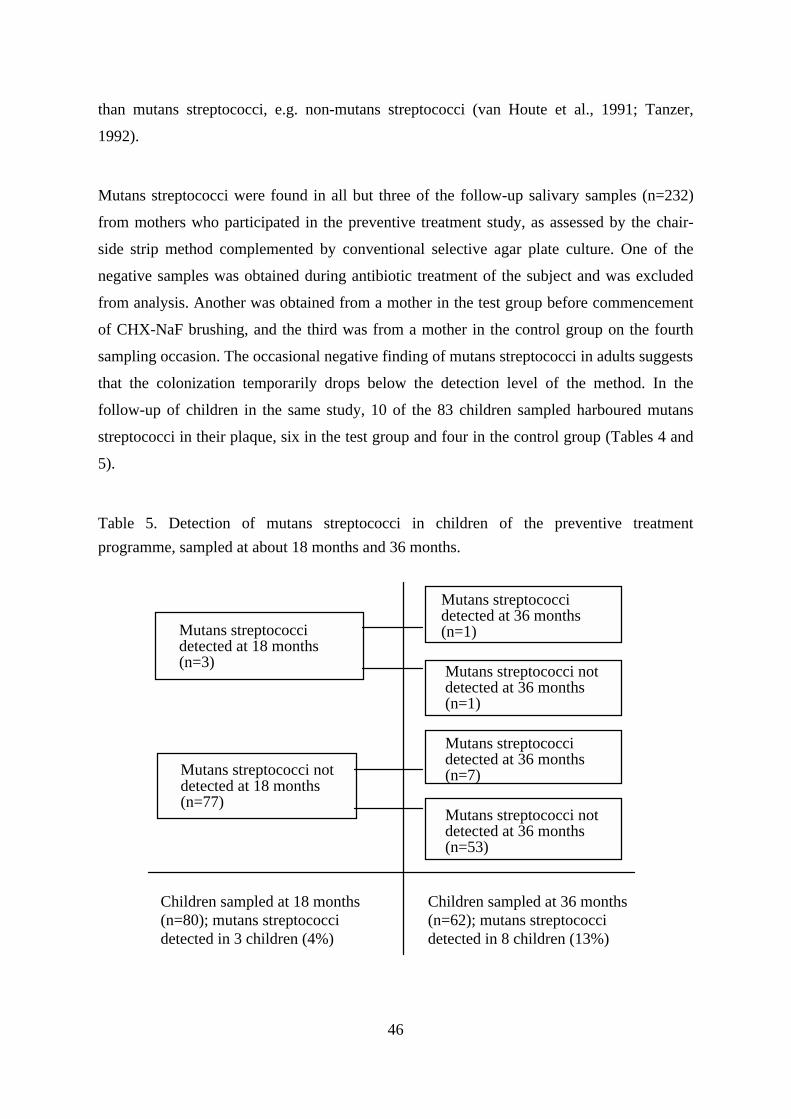
46
than mutans streptococci, e.g. non-mutans streptococci (van Houte et al., 1991; Tanzer,
1992).
Mutans streptococci were found in all but three of the follow-up salivary samples (n=232)
from mothers who participated in the preventive treatment study, as assessed by the chair-
side strip method complemented by conventional selective agar plate culture. One of the
negative samples was obtained during antibiotic treatment of the subject and was excluded
from analysis. Another was obtained from a mother in the test group before commencement
of CHX-NaF brushing, and the third was from a mother in the control group on the fourth
sampling occasion. The occasional negative finding of mutans streptococci in adults suggests
that the colonization temporarily drops below the detection level of the method. In the
follow-up of children in the same study, 10 of the 83 children sampled harboured mutans
streptococci in their plaque, six in the test group and four in the control group (Tables 4 and
5).
Table 5. Detection of mutans streptococci in children of the preventive treatment
programme, sampled at about 18 months and 36 months.
Children sampled at 18 months (n=80); mutans streptococci detected in 3 children (4%)
Children sampled at 36 months (n=62); mutans streptococci detected in 8 children (13%)
Mutans streptococci not detected at 18 months (n=77)
Mutans streptococci detected at 18 months (n=3)
Mutans streptococci detected at 36 months (n=1)
Mutans streptococci not detected at 36 months (n=1)
Mutans streptococci detected at 36 months (n=7)
Mutans streptococci not detected at 36 months (n=53)
47
Seven children were culture-negative for mutans streptococci at the first sampling, but at the
second sampling tested positive. One culture-positive child contributed a sample only at the
first sampling, and one child harboured mutans streptococci at both samplings. Another child
had a positive sample at first sampling at 16 months, but a negative sample at 36 months. In
this child, the colonization may have been transient, or at the second sampling, mutans
streptococci may have been present but in numbers under the detection level (II, III, V and
VI).
In the preventive study, only 12% of the 1.5- to 3-year old-children were colonized by
mutans streptococci. This is a low percentage as compared with earlier results from other
populations. Caufield et al. (1993) reported finding 75% of 31-month-old children to be
culture-positive. Alaluusua and Renkonen (1983) reported mutans streptococci in plaque
samples in 31% of 3-year-old children, and Pienihäkkinen and Jokela (1995) detected
mutans streptococci in plaque by the strip method in 25% of 2-to 3-year-old children. The
differences between the studies may be partly due to differences in sampling methods. As the
levels of mutans streptococci are low during the first years of the child's life, the amount of
plaque sample obtained and the dilutions used may cause differences in prevalence figures.
When comparing our test and control groups, however, the results are comparable because
samples from both groups were taken and handled in the same way. It is also noteworthy that
the preventive treatment programme at the Health Centre in Espoo, at the time of the
experiment, was strongly orientated to prevention of early mutans streptococcal colonization
by providing initial information to all parents well before childbirth and supporting
information for the first years of the child's life.
The seven 1.5- to 3-year-old nursing-caries children all harboured mutans streptococci.
Similarly, all seven 3- to 7-year-old children treated under general anaesthesia (IV)
harboured mutans streptococci. Among the 55 samples obtained from children treated under
general anaesthesia, mutans streptococci were detected by culture on MSB agar in 40
samples. No mutans streptococci were detected in 5 out of 13 samples from caries-free sites,
in 1 out of 19 samples from the superficial layer of caries lesions, or in 9 out of 18 samples
from deep layers of lesions. All five salivary samples obtained exhibited mutans streptococci
upon culture. These findings support earlier reports of a higher isolation frequency in
superficial layers of caries lesions as compared with deep layers or caries-free sites
(Edwardsson, 1974). In deep layers, other species are more prevalent (Tanzer, 1992). Counts
48
of mutans streptococci obtained by culture in the candle jar and in the anaerobe chamber
were at the same level, indicating that the strains present were aerotolerant. It has previously
been suggested that strains may be obligate anaerobes on first isolation, and only later
become facultative (Pulliam et al., 1980).
Proportion of mutans streptococci of total streptococcal count
The proportion of mutans streptococci of the total streptococcal count in the plaque samples
from caries-free 1.5- to 3-year-old children varied between <1% and 3.3% (median <1%),
whereas in nursing-caries children, the proportion varied between 8.3% and 98% (median
30%). In the 3- to 7-year-old caries-active children, the corresponding proportion in salivary
samples varied between 0.09% and 8.2% (median 0.5%). The mean proportion of mutans
streptococci of total streptococcal count in the culture-positive samples from caries-free sites,
from superficial layers of caries lesions and from deep layers of caries lesions was 1.1%
(range, < 0.01% to 4%), 14.7% (range, 0.16% to 93%) and 10% (range, 0.05% to 27%),
respectively. This finding of significantly higher proportions of mutans streptococci in dental
plaque in nursing-caries children concurs with the earlier studies of van Houte et al. (1982)
and Ripa (1988) who reported higher percentages of mutans streptococci in dental plaque
from nursing-caries children as compared with plaque from caries-free children or children
with dental caries not conforming to the criteria stipulated for nursing caries.
Detection of melibiose non-fermenting S.mutans
Among the 40 culture-positive samples from the 3- to 7-year-old caries-active children, a
total of 598 mutans streptococcal isolates were included for further analysis. Melibiose
fermentation is one of the biochemical tests used for distinguishing the various mutans
streptococci. All isolates were included for testing; 557 were identified as S. mutans and 41
as S. sobrinus. Among the S. mutans isolates, 215 (39%) did not ferment melibiose. This
percentage is higher than that (12%) reported earlier by Beighton et al. (1991). We also
found that in the two subjects who harboured both melibiose-fermenting and melibiose-non-
fermenting S. mutans isolates, the non-fermenters made up the majority of isolates. Thus, it
can be questioned whether our finding of melibiose-negative isolates was random or
suggestive of some property common to melibiose non-fermenters giving them a higher
virulence and potential to survive. Earlier suggestions have been made that S. mutans isolates
49
unable to ferment melibiose are genetically related to each other in having a large
chromosomal deletion including the multiple sugar metabolism (msm) operon which encodes
many genes involved in the uptake and metabolism of a number of sugars including
melibiose (Ushiro et al., 1991; Russell et al., 1992). Recently, however, it has been shown
that the melibiose-negative phenotype can arise through more than one mechanism; the
genotype and the phenotypic properties of these isolates can vary (Colby et al., 1995). So, as
regards any possible significance of our present findings on increased amounts of melibiose
non-fermenters, no final conclusions can be drawn before a comprehensive analysis of the
genetic elements of the strains isolated has been performed.
Effects of chlorhexidine - sodium fluoride (CHX-NaF) gel brushing on
salivary counts and transmission of mutans streptococci
The clinical effects of chlorhexidine-sodium fluoride (CHX-NaF) gel brushing on salivary
counts of mutans streptococci was evaluated using follow-up samples from 24 mothers in the
test group and 10 mothers in the control group (study II). For the comparison, samples were
obtained on three occasions: before childbirth, when the child was about 6 months old and
the CHX-NaF gel brushing was to commence in the test group, and when the child was about
18 months old and the periodic gel brushing had been ongoing for 1 year. The statistical
analysis of the differences between bacterial counts in treatment and control groups indicated
no significant effect of CHX gel brushing in the instructed trial on the salivary counts of
mutans streptococci. These negligible effects of periodic low-concentration CHX brushing
are comparable with earlier results (Emilson, 1994). When the gel is applied in trays, or
when the brushing is done professionally, and the concentration of CHX in gel is 1%, a
reducing effect is to be expected on salivary mutans streptococcal count (Emilson, 1994).
The poor effect of CHX brushing in an instructed trial might be related to the bioavailability
of the formulation when administered by the patient him/herself and also to the compliance
of the patient.
The effect of CHX-NaF gel brushing on transmission of isolates from mother to child was
also evaluated. Culture of dental plaque samples from the children revealed that mutans
streptococci had been colonized in 6 out of 56 children in the test group and in 4 out of 27
children in the control group. The colonization frequencies in the two groups did not differ
significantly from each other (Fisher's exact test, p > 0.05, ns) (unpublished result).
50
As a conclusion from the preventive programme, the periodic low-level CHX-NaF gel use
did not significantly affect the salivary counts of mutans streptococci in the mothers or the
colonization of mutans streptococci in their children. In earlier studies, when the salivary
mutans streptococcal counts in mothers of young children have been significantly reduced by
using 1% CHX gel in individual trays, controlled by culture of salivary samples, the
probability of colonization of mutans streptococci in the child has been reduced as well
(Köhler and Andréen, 1994). Use of CHX-NaF gel in standard trays by the mothers, without
control of salivary mutans streptococcal counts, did not significantly reduce the colonization
of mutans streptococci in their young children (Tenovuo et al., 1992). The difference in
results can thus be attributed to differences in study set-up.
In the preventive programme study, the number of drop-out subjects was rather high. A 3-
year sample was obtained from 47 children of the 75 mothers initially included in the test
group, and from 15 children of the 30 mothers initially included in the control group. As the
proportion of young inhabitants in the city of Espoo is quite high, and they change residence
rather frequently, these low numbers could be anticipated.
The mutans streptococcal isolates derived from the preventive treatment programme were
included for further studies on qualitative characteristics of mutans streptococci only after
the effects of the periodic CHX-NaF gel use had been evaluated to be negligible.
Clonal diversity of mutans streptococci
The clonality of isolates was studied by ribotyping and AP-PCR typing. Before proceeding
to genotyping by these molecular biology methods, mutans streptococcal isolates from the
1.5- to 3-year-old children and 40 mothers were phenotyped by serotyping. Serotyping
revealed one, two, or three different types of mutans streptococci within the subjects. Among
the 1.5- to 3-year-old children, 13 out of 14 children studied had serotype c isolates, 2
children had serotype e isolates and 3 had serotype g isolates. Two concomitant serotypes
were detected in 3 children, the other children had only one serotype present on the actual
sampling occasion. One caries-free child harboured serotype c isolates at first sampling and
serotype g isolates at second sampling. In the 40 mothers included for further studies, 37
51
harboured serotype c, 13 serotype g, 11 serotype e, 1 serotype f and 1 serotype d strains. One
serotype was detected in 20 mothers, with two and three concomitant serotypes found in 17
and 3 mothers, respectively (II, III, V, VI).
Total number of clonal types intra-individually
By ribotyping, one, two, three or four different clonal types of mutans streptococci were
detected within each subject. Among the 19 mothers studied for clonal diversity, 6 mothers
harboured one, 7 harboured two, 5 harboured three and 1 mother harboured four ribotypes.
In the 1.5- to 3-year-old caries-free children, one ribotype was detected in 6 out of 7 children
studied and two ribotypes in 1 child. In nursing-caries children, one ribotype was detected in
4 children, two in 2 children and three in 1 child.
In the seven 3- to 7-year-old caries-active children referred for dental treatment under
general anaesthesia, by AP-PCR typing of 90, 74, 81, 53, 83, 103 and 114 isolates from the
children, respectively, 2 children were found to harbour one, 3 children two, 1 child three
and 1 child four different AP-PCR types of mutans streptococci. The culture method of
samples did not alter the number of clonal types detected; culture in candle jar and in
anaerobe chamber yielded the same AP-PCR types of mutans streptococci.
In our material, molecular biology methods revealed more types intra-individually than did
phenotyping by serotyping, as has generally proven to be the case (Caufield and Walker,
1989; Alonso et al., 1993; Russell, 1994). In earlier studies, the number of clonal types
detected within an individual has varied between one and four (Bowden and Hamilton,
1998). In this study, the increased number of isolates included for genotyping failed in
detecting more clonal types; no more than four genotypes of mutans streptococci could be
found within any individual.
Interindividual comparison
The mutans streptococcal isolates in studies III, V and VI, and isolates from seven mothers
in study II, represented 52 different ribotypes. The isolates originated from 19 mothers and
their 18-month- to 3-year-old children (one twin pair), and five originally 5-year-old children
who had been studied in follow-up (Alaluusua et al., 1989). When comparing isolates from
52
unrelated subjects, identical ribotypes were detected in two cases. The isolates from these
two pairs of mothers could not be distinguished by ribotyping with HindIII or with EcoRI. In
comparison of ribotypes between mothers and their children, out of 14 mother-child pairs,
identical ribotypes were detected in 9 pairs (64%).
In study IV, AP-PCR typing was performed for 598 isolates originating from seven children.
Fourteen different genotypes were identified. By AP-PCR typing, isolates from one of the
children, all representing the same AP-PCR type, could not be distinguished from isolates
representing one out of two AP-PCR types detected in another child.
Our typing results confirmed earlier results of a high degree of genetic heterogeneity within
mutans streptococci (Saarela et al. 1993; Bowden and Hamilton, 1998). Isolates from
unrelated subjects that could not be distinguished by genotyping were detected in only two
pairs of adults, where ribotyping failed to show a difference, and in one pair of children,
where AP-PCR typing failed in discerning between isolates. The genotyping methods thus
proved to be useful in transmission studies, especially in combination with phenotypic
typing (VI).
Intra-individual site-specific evaluation: implications for sampling technique
In study IV, 598 mutans streptococcal isolates derived from 50 tooth-site samples and 5
salivary samples from the seven 3- to 7-year-old caries-active children were genotyped by
AP-PCR typing. The tooth-site samples included 7, 5, 7, 6, 9, 9 and 7 samples from the
subjects, respectively.
Two children harboured both S. mutans and S. sobrinus isolates. In one of the children, the
difference in frequency distribution of S. mutans and S. sobrinus isolates at different sites
was statistically significant; S. sobrinus constituted the majority of isolates in two out of
three samples from superficial layers of caries lesions, but remained undetected in one caries
lesion and in the one caries-free site sampled. By comparing the frequency distribution of
AP-PCR types of S. mutans in different sites within the subjects, it was revealed that mutans
streptococcal clones may selectively colonize specific hard-tissue sites; in three of the seven
children, the distribution of AP-PCR types of S. mutans differed depending on tooth site.
53
Five children in study IV had contributed saliva samples. In two, one of the AP-PCR types
detected in the tooth-site samples was not detected in the saliva sample. In these five
children, 8 out of 10, or 80%, of all genotypes of mutans streptococci detected appeared in
the saliva samples. The representativeness of tooth-site sampling was evaluated for all seven
children. In the present sampling setting and with the present number of isolates included,
when the first caries-free site sampled per child was considered, 6 out of 15, or 40%, of all
mutans streptococcal genotypes were detectable. Including the first caries-free site sampled
and both the superficial and the deep layer of the first caries lesion sampled revealed 11 out
of 15, or 73%, of the genotypes. Including the second caries lesion sampled, as well,
revealed all genotypes detected in the whole material. In conclusion, sampling of saliva is
fairly efficient for mutans streptococcal strain isolation, but does not necessarily reveal all
genotypes. When sampling for mutans streptococci for qualitative analysis, it is necessary to
sample multiple sites of the dentition.
CHX susceptibility, GTFs and mutacin production of strains
When testing the clinical isolates for CHX susceptibility (II), S. mutans isolates were found
to be more susceptible than S. sobrinus isolates, and analyses of laboratory strains also
suggested that S. mutans is more susceptible than S. sobrinus, S. cricetus and S. rattus. Of
the 379 clinical isolates studied, 50% were inhibited at 1 µg of CHX per ml, 90% were
inhibited at 2 µg/ml, and all were inhibited at 4 µg/ml. Development of resistant strains
during CHX-NaF gel use was not detected, and the serotype distribution of isolates remained
unchanged by CHX-NaF gel use. Similarly, Kozai et al. (1991) reported that strains isolated
after CHX varnish treatment were no more resistant to CHX than those isolated prior to
treatment. When evaluating long-term CHX susceptibility, earlier studies have reported that
mutans streptococci are inhibited at a CHX concentration of 1 to 4 µg/ml in agar (Meurman
et al., 1989; Järvinen et al., 1993). The MIC values obtained in the present study were at the
same level, and thus it can be concluded that mutans streptococci have not developed
resistance to CHX over the years of treatment. When using antibiotics and biocides such as
CHX, it is possible that resistant strains of commensal microorganisms and potential
pathogens such as Proteus mirabilis are selected, but the clinical relevance of this possibility
is still unclear (Russell et al., 1998).
54
In the analysis of GTF production by the strains in an 18-hr candle-jar culture in 56 mM
glucose-supplemented Todd-Hewitt broth (V), all S. mutans isolates were shown to produce
GTF-I and GTF-S, and all but two produced GTF-SI of S. mutans. All S. sobrinus isolates
produced GTF-I of S. sobrinus. The relative amount of GTFs produced by serotype c and e
isolates were often variable. No significant difference in production activity between
serotypes c and e was found. Earlier studies on the environmental regulation of GTF
production by S. mutans have shown that an addition of sucrose can increase the production
up to twofold in batch culture (Hudson and Curtiss, 1990). Moreover, many other dietary
components, such as various carbohydrates, milk and kappa casein, may also affect
glucosyltransferase activity (Vacca-Smith and Bowen, 1995). In our study, the altering of the
composition of the culture broth might have influenced detected levels of GTF production.
A result of the study on mutacin production of mutans streptococci (VI) was that the
production of mutacins also varies among isolates. Of the 145 strains tested, 88% produced
mutacin against more than 1 of the 14 indicator strains. The inhibition zone sizes for
producer strains varied from 4 to 26 mm in diameter. On average, the isolates produced
mutacins against 7 of the 14 indicator strains. S. mutans isolates showed more inhibitory
activity than did S. sobrinus isolates. Four isolates, representing two ribotypes of S. mutans
serotype e, were active against all indicator strains. S. sobrinus serotype g strains produced
mutacins against only one or no indicator strain. On average, serotype c isolates produced
mutacins against 8 indicator strains (range, 1 to 13), serotype e against 10 (range, 3 to 14)
and serotype f against 6 (range, 3 to 6). The mutacin activity of isolates found in this material
was at the same level as in earlier reports (Alaluusua et al., 1991b).
The stability of mutacin activity of clinical isolates was evaluated using samples in follow-
up. The spectrum of mutacin activity of baseline and follow-up isolates of identical ribotypes
within a subject was compared. The isolates were obtained at 1- to 7-year intervals (mean,
3.1; SD, 2.3) from 10 mothers and 5 initially 5-year-old children, producing a total of 19
comparable isolate pairs. The mutacin activity of the isolates initially and in follow-up was
similar in 14 subjects, whereas only the isolates obtained from one child 5 years apart
showed a difference in mutacin activity against two indicator strains. Thus, the results
indicated that the bacteriocinogenecity of S. mutans strains is reasonably stable.
55
Intra-individual variation: strain specificity
Among the isolates included for CHX susceptibility testing were serotype c isolates from
seven mothers that exhibited intra-individual variability in CHX susceptibility. By
ribotyping these isolates, it was shown that if the CHX MICs for isolates from one subject
were not the same (they differed by more than one dilution step), then the subject harboured
two or three ribotypes. These findings suggest that each intra-individual strain presents
unique susceptibility to CHX.
The analysis of GTF production of S. mutans strains revealed variability in the GTF-I, GTF-
SI and GTF-S production of isolates exhibiting a different ribotype. Eight participants (three
children and five mothers) harboured more than one ribotype of S. mutans: four had two
ribotypes and four had three ribotypes. When comparing the production activity of GTFs of
the intra-individual strains, eight out of the 16 comparable pairs had a difference in the
relative amounts of one or two enzymes. As concerns mutans streptococcal ribotypes shared
by a mother and her child, the production of the three GTFs of S. mutans was at the same
level in five out of six mother-child pairs. In the exceptional pair, the difference may have
been due to a technical problem, such as a failing culture of a strain. In most cases, the GTF
production was ribotype-specific.
Identical ribotypes had similar mutacin activity profiles within a subject initially and in
follow-up in all but two cases. To reach this conclusion, strains of an identical ribotype
within a subject, isolated on the same occasion, were used. The data included 43 cases of two
or more isolates of identical ribotype within a subject, obtained from the same sample. In 40
of these comparable cases, the sizes of the inhibition zones were identical, i.e., differed by
less than 4 mm. With two isolate pairs, the sizes of the inhibition zones differed for one
indicator strain. This difference for 1 indicator out of 14 fell within the range of accuracy for
the method. One isolate pair differed in mutacin activity profile for four indicator strains.
Thus, the mutacin activity of mutans streptococal isolates, defined by the stab culture
method, was ribotype-specific in 42 out of 43 cases. Mothers and children shared 10
ribotypes, and the isolates representing these ribotypes exhibited a similar mutacin activity
profile. Furthermore, all four isolates of the same ribotype, obtained from the twin brothers
with nursing caries, showed an identical mutacin activity profile.

56
In conclusion, isolates that were similar as evaluated by genotypic methods had similar
expression of phenotypic properties in most cases, and isolates that were genetically different
differed also, in most cases, in their phenotypic properties. Differences between isolates
derived from different sources were earlier suggested with regard to acid production (Köhler
et al., 1995). The present finding of strain specificity of phenotypic properties of bacterial
isolates supports the view that certain strains may more likely be associated with disease
than others, by the inherited properties of the microorganism, not only by environmental
modification of these properties.
Interindividual variation
As a rule, mutans streptococcal isolates from unrelated subjects could be differentiated by
genotyping. The phenotypic properties showed insufficient variability for comprehensive
differentiation between isolates. Identical ribotypes from related subjects, i.e. mother-child
pairs, were in most cases similar in their phenotypic properties.
Effects of CHX-NaF-brushing on mutacin production
In the preventive study, the effect of CHX-NaF brushing on mutacin activity of clinical
isolates was evaluated using samples in follow-up. Follow-up samples were obtained after 1
year of periodic CHX use for five subjects, and after 2.5 years of periodic CHX use for one
subject. A total of seven identical ribotype pairs were compared, and for all pairs, the
mutacin activity was identical in the initial and follow-up samples. In addition, in two cases,
a shared ribotype of mutans streptococci obtained from a mother during CHX treatment and
her child had identical mutacin activity profiles. These results indicate that periodic CHX
administration by tooth brushing does not affect the phenotypic property of mutacin activity.
By contrast, earlier in vitro studies have shown that CHX can affect phenotypic properties
of oral streptococci, e.g. acid production and sugar transport (Marsh et al., 1983). However,
the outcome of the present study, with periodic low-concentration CHX-NaF-gel brushing in
an instructed trial is not comparable with in vitro results, as apparently variability exists in
the bioavailability of different CHX preparations, administered in different modes (Emilson,
1994).
57
Clonal diversity of mutans streptococci in nursing caries
The results of study III on increased proportions of mutans streptococci of total streptococcal
count in dental plaque of children with nursing caries was expected based on earlier reports
(van Houte et al., 1982; Ripa, 1988). Further, in study III, providing that only clonal types
detected simultaneously in a subject were considered, the number of clonal types detected
was significantly higher in nursing-caries children as compared with caries-free children.
This is contradictory to the finding of Kreulen et al. (1997), who, in family studies, found
less clonal types of mutans streptococci in nursing-caries children than in siblings without
nursing caries. The difference in findings may be the result of a difference in sampling
technique; Kreulen et al. sampled only one tooth site for analysis, whereas we gathered
pooled plaque samples. Because mutans streptococci were found to selectively colonize
tooth sites, multiple sites should always be sampled for qualitative analysis of these bacteria.
Virulence factors studied and occurrence of nursing caries
In the study on glucosyltransferase activity of mutans streptococci, among the twelve 1.5- to
3-year-old children included, the six children with nursing caries harboured nine different S.
mutans ribotypes and the six caries-free children harboured seven. Analysis using
monoclonal antibodies revealed that isolates of the caries-free children produced more GTF-
SI than isolates of the children with nursing caries. Regarding enzymes GTF-I and GTF-S,
no significant difference between the groups was found. This finding of less GTF-SI
produced by isolates from nursing-caries children was unexpected because inactivation of
the gtfC gene, responsible for GTF-SI production, has been reported to drastically reduce
adherence to smooth surfaces (Tsumori and Kuramitsu, 1997). Possibly, in the mouths of
children with nursing caries, whose diets are rich in sucrose, special properties are not as
important for a colonizing strain as in less favourable environments.
Mutacin activity was studied for isolates obtained from fourteen 1.5- to 3-year-old children,
comprising 7 nursing-caries children and 7 caries-free children. The sample included a total
of 17 distinct S. mutans ribotypes, with nine of these detected in the nursing-caries children.
Overall, the ribotypes from nursing-caries children tended to inhibit more indicator strains
than those detected in caries-free children, the former inhibiting a mean number of 10.2
indicator strains (SD 2.9), and the latter 9.2 indicator strains (SD 2.2). When comparing the
58
inhibition zone sizes against single indicators, the zones for all indicator strains were bigger
for ribotypes originating in the oral flora of nursing-caries children than for ribotypes
isolated from caries-free children, the difference being statistically significant for indicator
strain S. sobrinus B13 (b) (Mann-Whitney U-test, p < 0.04) (unpublished results). Findings
on mutacin activity of mutans streptococcal isolates from nursing-caries children have not
been reported earlier. In a study by Alaluusua et al. (1991b) on older children aged 4 to 6
years, no association between caries experience and mutacin activity of mutans streptococci
isolated was found.
Virulence factors studied and transmission of
isolates from mother to child
Transmission of isolates between subjects can be supposed to have happened if isolates
derived from the subjects are similar upon typing (Berkowitz and Jordan, 1975a; Li and
Caufield, 1995). The methods with the highest discriminatory power are the genotypic
typing methods (Arbeit, 1999). As concerns transmission studies between mothers and their
young children, detection of microbial isolates with identical genotypes in mother and child
has been considered to mean that the particular strain has been transmitted from the mother
to her child, since at birth the child does not harbour any microorganisms (Könönen et al.,
1992; Caufield et al., 1993). With older children and adults, a cross-sectional study does not
allow for the determination of the direction of transmission, i.e., who is the source and who
the recipient of the infecting organism.
In study VI, a statistical analysis was included for strain matching between mothers and
children, and for evaluating the probability of transmission. Ribotyping and mutacin typing
were used to study transmission. In the children who harboured mutans streptococci and in
their mothers, a total of 36 ribotypes were detected. A ribotype match for mothers and
children was found for 10 of the 36 ribotypes detected. The study also included isolates from
5 initially 5-year-old children, unrelated to each other and to the other subjects. When
comparing ribotypes of these 5 children and the 19 unrelated adult women in the study (a
total of 42 ribotypes), identical ribotypes were detected in two cases, where two mothers had
the same ribotype. The odds ratio of the occurrence of shared identical strains, determined by
ribotyping with one enzyme, in mothers and children living in the same household (10 out of
36) as compared with that in unrelated subjects not living in the same household (2 out of
59
42), was 7.69. When the results of ribotyping were complemented with the results of mutacin
typing, no case of identical strains detected in epidemiologically unrelated persons remained.
The presence of identical ribotypes in 9 of the 14 mother-child pairs indicated maternal
transmission of mutans streptococci in 64% of mutans streptococcus culture-positive
children. This result agrees with previous studies; Li and Caufield (1995) reported finding
identical genotypes in 71% of mother-child pairs.
Transmitted mutans streptococcal strains produced the same levels of GTFs as strains that
were not shared by mothers and their children, as assessed by the semiquantitative cross-dot
assay using monoclonal antibodies P72, P32, P4 and B17 against GTF-I, GTF-SI and GTF-S
from S. mutans and GTF-I from S. sobrinus (V). The impact of the mutans streptococcal
glucosyltransferases in the early transmission of isolates from mother to child has not been
extensively studied to date, and should undergo comprehensive examination to add vital
information on the transmission process.
As regards the mutacin production of S. mutans isolates, mutacins may affect the
transmission from mother to child (study VI). S. mutans isolates harboured by the mothers
represented 35 distinct types, determined by ribotyping and mutacin typing. Eight of these
had been transmitted to the child. The transmitted strains inhibited a mean of 10.6 indicator
strains (SD 1.9; median, 10; range, 9 to 14), and nontransmitted strains inhibited a mean of
7.0 indicator strains (SD, 3.4; median, 6; range, 1 to 14); the difference was statistically
significant (equality of medians tested by Fisher's exact test, p < 0.002). When the sizes of
inhibition zone against single indicators were compared, for all indicator strains the zones
were bigger for transmitted strains than for nontransmitted strains, the difference being
statistically significant for 8 of the 14 indicator strains. The 8 S. mutans types that had been
transmitted, compared with the 27 types that had been available from the mothers but had
not been transmitted, showed significantly more mutacin activity (the mean zone sizes
against the indicator strains were compared by the Wilcoxon signed-rank test, p < 0.003).
Five ribotypes representing S. sobrinus isolates harboured by the mothers inhibited only one
or none of the indicator strains. The S. sobrinus mutacin production activity could not be
linked to transmission.
60
Earlier, bacteriocins produced by mutans streptococci have been shown to enhance
colonization in rats and adult humans (van der Hoeven and Rogers, 1979; Hillman et al.,
1987). In this regard, our present finding that mutacins may promote transmission of S.
mutans from mother to child clearly confirms results obtained two decades ago on enhanced
colonization by bacteriocin production. Hillman et al. (1990; 1994) have tried to construct an
effector strain for replacement therapy of mutans streptococci by using a strain with
increased bacteriocin activity to render high-level colonization potential. Unfortunately, the
deletion that rendered the low-level acid production proved to be lethal for the strain.
However, the bacteriocin produced by the strain JH1000 clearly enhanced colonization in
adults who already had a stable oral flora. As stated previously, bacteriocins are potent
inhibitors of bacterial growth. Nisin, commercially prepared bacteriocin from Lactococcus
lactis, has been used in dairy products as a preservative for decades (Jack et al., 1995). In
dentistry, commercially prepared bacteriocins could perhaps aid in maintaining a
nonpathogenic commensal oral microbial population. Experiments in beagle dogs by Howell
et al. (1993) indicated that nisin in a mouthrinse has a clear antibacterial potential. Thus far,
no microbial resistance or anaphylaxis problems have been reported for the bacteriocins.
As regards the clinical significance of the present findings on differences in the virulence
characteristics and colonization pattern of oral mutans streptococci, the results are not as yet
applicable to the selection of individuals at risk. The virulence of bacterial isolates, on the
whole, is dependent on the environment. Molecular biology methods, such as PCR detection,
can be developed and applied for selection of virulent strains once the genetic elements
coding for a defined property have been recognized, but when evaluating the clinical
significance, many factors must be considered, depending on the multifactorial aetiology of
dental caries disease. The virulence attributes remain to be clarified; it is probable that in the
future the adherence process during colonization (Kolenbrander and London, 1993), and the
proteolytic activity of mutans streptococci (Homer et al., 1990; Harrington and Russell,
1994; Jackson et al., 1997) will attract an increasing interest. In any case, as the present
study could identify differences between individual strains affecting the transmission of
strains from mother to child, it seems realistic to anticipate that selection for virulent strains
by molecular biology methods will be part of upcoming preventive treatment approaches.
With regard to the possible intra-individual site-specific colonization pattern of mutans
streptococci, our findings support the suggested site-directed antimicrobial varnish treatment
of initial caries lesion sites (Twetman, 1998; Twetman and Petersson, 1999). In the
61
suggested treatment protocol the antimicrobial agent chlorhexidine is locally administered to
eradicate the pathogenic mutans streptococci above the lesion, enabling colonization of a less
acidogenic microflora at the site.
62
SUMMARY AND CONCLUSIONS
The present studies were performed for quantitative and qualitative characterization of oral
mutans streptococci. For detection and isolation of mutans streptococci, saliva and/or plaque
samples from 97 children, 146 adolescents and 159 adults were included. The subjects,
participating in two cross-sectional studies and in two longitudinal studies, were also
examined clinically for decayed, missing and filled teeth. Further, for qualitative analysis,
bacterial isolates obtained from 64 individuals were subjected to genotyping and/or to
phenotyping, in order to study the clonal diversity and site-specific colonization of intra-
individual strains, and certain virulence factors of the organism. Phenotypical
characterization included serotyping and determination of chlorhexidine (CHX)
susceptibility, glucosyltransferase activity and bacteriocin (mutacin) activity of the isolates.
In addition, the effects of periodic low-concentration CHX-NaF gel brushing in an instructed
trial on salivary counts and transmission of mutans streptococci from mother to child was
studied. Genotyping was performed by ribotyping 191 of the obtained isolates, and 598
isolates were fingerprinted by the AP-PCR method. Serotyping was performed for 661
isolates by Ouchterlony immunodiffusion using rabbit antisera. CHX susceptibility of 379
isolates was tested by the agar dilution method, and for testing of glucosyltransferase
activity, using monoclonal antibodies in a semiquantitative cross-dot assay, 44 isolates were
included. Mutacin production of 145 isolates was tested by the stab culture method,
including 38 clinical mutans streptococcal isolates from our IDH (Institute of Dentistry,
Helsinki) collection .
The main findings were as follows:
In cross-sectional studies of adolescents, when mutans streptococci are not detected by chair-
side methods or by conventional agar plate culture methods of plaque and saliva, mutans
streptococci may either be absent or present in numbers under the detection level. In this
study, a longitudinal evaluation of adolescents with no mutans streptococci initially detected,
revealed that only a few subjects remained culture-negative for the organism during follow-
up.
The number of clonal types detected within a subject was from one to four, with saliva
revealing 80% of the genotypes present. The isolates from related subjects were often
63
similar, but from unrelated subjects they were, in almost all cases, distinguished by
genotyping. The children with nursing caries harboured more types of mutans streptococci
than caries-free children. The colonization of genotypes tended to be site-specific, implying
that sampling for intra-individual mutans streptococcal genotypes should include sampling
of multiple sites.
The CHX susceptibility of mutans streptococcal isolates was ribotype-specific, and the
susceptibility exhibited stability at follow-up. S. sobrinus isolates were somewhat more
resistant to CHX than S. mutans isolates. In the past, no CHX resistance in mutans
streptococci has appeared. In this study, periodic low-concentration CHX-NaF gel brushing
did not significantly affect salivary counts of mutans streptococci, CHX susceptibility of
isolates or transmission of isolates from mother to child.
Glucosyltransferase activity and mutacin production by mutans streptococcal isolates were
also ribotype-specific. The mutacin activity of isolates was fairly stable, and mutacin
production of S. mutans isolates was shown to have an impact on the probability of strain
transmission from mothers to their children.
These results support the original study hypothesis that mutans streptococcal strains may
differ in their phenotypic properties; CHX susceptibility, glucosyltransferase activity and
mutacin production were strain-specific. Moreover, the results imply that S. mutans strains
that produce increased amounts of mutacin may be more easily transmitted from mothers to
their young children. Therefore, the role of mutans streptococci in human dental caries may
be characterized by not only the quantity of the bacteria, but also the quality of strain(s)
colonizing.
64
ACKNOWLEDGEMENTS
This study was carried out at the Institute of Dentistry, University of Helsinki, and the
Department of Oral and Maxillofacial Diseases, Helsinki University Central Hospital,
Helsinki. I wish to thank Professor Jukka Ainamo, DDS, PhD, and Professor Heikki
Murtomaa, DDS, PhD, the former Deans of the Institute of Dentistry, and Professor Jukka H.
Meurman, DDS, PhD, the present Dean of the Institute of Dentistry for placing facilities at
my disposal.
I am indebted to my supervisor, Professor Satu Alaluusua, DDS, PhD, for persuading me
into scientific writing, and thereby introducing me to the pluralistic, fascinating world of
science.
I wish to express heartfelt gratitude towards my official referees, Docent Kaisu
Pienihäkkinen, DDS, PhD, and Docent Jaana Vuopio-Varkila, MD, PhD, for their valuable
comments and constructive criticism of this thesis. Their sound scientific advice and positive
attitude have been reviving.
I owe my warm thanks to all my collaborators at the Espoo City Health Centre, Espoo, the
Anaerobe Reference Laboratory and the Laboratory Animal Unit of the National Public
Health Institute, Helsinki, the School of Dentistry at Matsudo, Nihon University, Japan, the
former Department of Preventive Dentistry and Cariology, University of Kuopio, and the
Institute of Dentistry, University of Helsinki; Professor emeritus Heikki Luoma, DDS, PhD,
Professor Kazuo Fukushima, DDS, PhD, Docent Hannele Jousimies-Somer, PhD, Docent
Sirkka Asikainen, DDS, PhD, Docent Maria Saarela, PhD, Dr Eija Kleemola-Kujala, DDS,
PhD, Dr Anja-Riitta Luoma, DDS, PhD, Jaana Mättö, PhD, Liisa Pyhälä, MSc, Heini
Torkko, MSc, Dr Xia Zhu, Dr Ulla Tanner-Salo, Dr Sini Innilä and Dr Aura Vuorela. It has
been an honour and a privilege to share a piece of your vast knowledge. All researchers and
assisting personnel participating in the studies are warmly acknowledged for their generous
contribution. Especially the researchers engaged in the longitudinal study, initiated at the
Department of Pedodontics and Orthodontics by the late Professor Johannes Haataja, deserve
recognition for their positive research interest.
65
I am grateful for having had the opportunity to attend the basic courses in microbiology and
genetics at the Faculty of Science as part of my basic training towards a doctoral thesis in the
Faculty of Medicine.
I also wish to thank Carol Pelli, HonBSc, for kindly revising my English in this thesis, Riitta
Auvinen, IT-professional, for helping me with computers, and all the mothers, children and
adolescents attending the studies who made this thesis possible.
Finally, I thank my family, relatives, colleagues and friends for patience, support, friendship
and love.
The study was supported by grants from the Academy of Finland, the Finnish Dental
Society, the Finnish Woman Dentists´ Association, the University of Helsinki, and
Odontologiska Samfundet i Finland.
Helsinki - Helsingfors, June 2000
Lisa Grönroos
Munksgaard International Publishers Ltd (Oral Microbiology and Immunology), Elsevier
Science (Archives of Oral Biology), American Society of Microbiology (Antimicrobial
Agents and Chemotherapy; Infection and Immunity) and S. Karger AG, Basel (Caries
Research) are acknowledged for their kind permission to reprint articles upon which this
thesis is based.
66
REFERENCES
Alaluusua S. Transmission of mutans streptococci. Proc Finn Dent Soc 1991a; 87: 443-447.
Alaluusua S. Salivary counts of mutans streptococci and lactobacilli and past cariesexperience in caries prediction. Caries Res 1993; 27(suppl 1): 68-71.
Alaluusua S. Yleisanestesia lasten hammashoidossa. Duodecim 1999; 115: 1368-1369.
Alaluusua S, Alaluusua SJ, Karjalainen J, Saarela M, Holttinen T, Kallio M, Hölttä P,Torkko H, Relander P, Asikainen S. The demonstration by ribotyping of the stability of oralStreptococcus mutans infection over 5 to 7 years in children. Arch Oral Biol 1994; 39: 467-471.
Alaluusua S, Kleemola-Kujala E, Grönroos L, Evälahti M. Salivary caries-related tests aspredictors of future caries increment in teenagers. Oral Microbiol Immunol 1990; 5: 77-81.
Alaluusua S, Kleemola-Kujala E, Nyström M, Evälahti M, Grönroos L. Caries in the primaryteeth and salivary Streptococcus mutans and lactobacillus levels as indicators of caries inpermanent teeth. Pediatric Dentistry 1987; 9: 126-130.
Alaluusua S, Myllärniemi S, Kallio M. Streptococcus mutans infection level and caries in agroup of 5-year-old children. Caries Res 1989; 23: 190-194.
Alaluusua S, Renkonen O-V. Streptococcus mutans establishment and dental cariesexperience in children from 2 to 4 years old. Scand J Dent Res 1983; 91: 453-457.
Alaluusua S, Savolainen J, Tuompo H, Grönroos L. Slide-scoring method for estimation ofStreptococcus mutans levels in saliva. Scand J Dent Res 1984; 92: 127-133.
Alaluusua S, Takei T, Ooshima T, Hamada S. Mutacin activity of strains isolated fromchildren with varying caries prevalence. Arch Oral Biol 1991b; 36: 251-255
Alanen P, Hurskainen K, Isokangas P, Pietilä I, Levänen J, Saarni U-M, Tiekso J. Clinician'sability to identify caries risk subjects. Community Dent Oral Epidemiol 1994; 22: 86-89.
Alonso R, Aucken HM, Perez-Diaz JC, Cookson BD, Baquero F, Pitt TL. Comparison ofserotype, biotype and bacteriocin type with rDNA RFLP patterns for the type identificationof Serratia marcescens. Epidemiol Infect 1993; 11: 99-107.
Arbeit RD. Laboratory procedures for the epidemiologic analysis of microorganisms. In:Manual of clinical microbiology. Murray PR, Baron EJ, Pfaller MA, Tenover FC, YolkenRH, eds. 7th ed. ASM Press: Washington, D. C. 1999; pp. 116-137.
Beighton D, Russell RR, Hayday H. The isolation and characterization of Streptococcusmutans serotype h from dental plaque of monkeys (Macaca fascicularis). J Gen Microbiol1981; 124: 271-279.
Beighton D, Russell RRB, Whiley RA. A simple biochemical scheme for the differentiationof Streptococcus mutans and Streptococcus sobrinus. Caries Res 1991; 25: 174-178.
67
Bender GR, Sutton SV, Marquis RE. Acid tolerance, proton permeabilities, and membraneATPases of oral streptococci. Infect Immun 1986; 53: 331-338.
Berkowitz RJ, Jordan HV. Similarity of bacteriocins of Streptococcus mutans from motherand infant. Arch Oral Biol 1975a; 20: 1-6.
Berkowitz RJ, Jordan HV, White G. The early establishment of Streptococcus mutans in themouths of infants. Arch Oral Biol 1975b; 20: 171-174.
Berkowitz RJ, Turner J, Green P. Maternal salivary level of Streptococcus mutans andprimary oral infection of infants. Arch Oral Biol 1981; 26: 147-149.
Birkhed D, Imfeld T, Edwardsson S. pH changes in human dental plaque from lactose andmilk before and after adaptation. Caries Res 1993; 27: 43-50.
Bolin A-K. Children’s dental health in Europe. An epidemiological investigation of 5- and12-year-old children from eight EU countries. Thesis. Stockholm; Karolinska Institutet;Swed Dent J Suppl 122, 1997.
Bollet C, Gevaudan MJ, de Lamballerie X, Zandotti C, de Micco P. A simple method for theisolation of chromosomal DNA from Gram positive or acid-fast bacteria. Nucleic Acids Res1991; 19: 1955.
Bowden GHW, Hamilton IR. Survival of oral bacteria. Crit Rev Oral Biol Med 1998; 9: 54-85.
Bowen WH. Salivary influences on the oral microflora. In: Saliva and oral health. EdgarWM, O’Mullane DM, eds. 2nd ed. British Dental Journal: London 1996; pp. 95-103.
Bratthall D. Demonstration of five serological groups of streptococcal strains resemblingStreptococcus mutans. Odontol Revy 1970; 21: 143-152.
Bratthall D. Discovery! A Streptococcus mutans safari! J Dent Res 1997; 76: 1332-1336.
Bratthall D, Hoszek A, Zhao X. Evaluation of a simplified method for site-specificdetermination of mutans streptococci levels. Swed Dent J 1996; 20: 215-220.
Brosius J, Ullrich A, Raker MA, Gray A, Dull TJ, Gutell RR, Noller HF. Construction andfine mapping of recombinant plasmids containing the rrnB ribosomal RNA operon of E.coli. Plasmid 1981; 6: 112-118.
Burne RA. Concise review. Oral streptococci...products of their environment. J Dent Res1998; 77: 445-452.
Burne RA, Chen Y-YM, Penders JEC. Analysis of gene expression in Streptococcus mutansin biofilms in vitro. Adv Dent Res 1997; 11: 100-109.
Burr MD, Pepper IL. Variability in presence-absence scoring of AP PCR fingerprints affectscomputer matching of bacterial isolates. J Microbiol Methods 1997; 29: 63-68.
68
Carlsson J, Grahnén H, Jonsson G. Lactobacilli and streptococci in the mouth of children.Caries Res 1975; 9: 333-339.
Carlsson P. On the epidemiology of mutans streptococci. Thesis, Malmö: University ofLund, 1988.
Caufield PW, Cutter GR, Dasanayake AP. Initial acquisition of mutans streptococciinfections in infants: evidence for a discrete window of infectivity. J Dent Res 1993; 72: 37-45.
Caufield PW, Shah G, Hollingshead SK, Parrot M, Lavoie MC. Evidence that mutacin IIproduction is not mediated by a 5.6-kb plasmid in Streptococcus mutans. Plasmid 1990; 24:110-118.
Caufield PW, Walker TM. Genetic diversity within Streptococcus mutans evident fromchromosomal DNA restriction fragment polymorphism. J Clin Microbiol 1989; 27: 274-278.
Caufield PW, Wannemuehler Y, Hansen JB. Familial clustering of the Streptococcus mutanscryptic plasmid strain in a dental clinic population. Infect Immun 1982; 38: 785-787.
Chikindas ML, Novak J, Driessen AJ, Konings WN, Schilling KM, Caufield PW. MutacinII, a bactericidal lantibiotic from Streptococcus mutans. Antimicrob Agents Chemother1995; 39: 2656-2660.
Cirino SM, Scantlebury S. Dental caries in developing countries. Preventive and restorativeapproaches to treatment. New York State Dental Journal 1998; 64: 32-39.
Clarke JK. On the bacterial factor in the aetiology of dental caries. British J Exp Pathol1924; 5: 141-147.
Colby SM, Harrington DJ, Russell RRB. Identification and genetic characterisation ofmelibiose-negative isolates of Streptococcus mutans. Caries Res 1995; 29: 407-412.
Colby SM, Russell RRB. Sugar metabolism by mutans streptococci. J Appl Microbiol 1997;83: 80S-88S.
Costerton JW, Lewandowski Z. The biofilm lifestyle. Adv Dent Res 1997; 11: 192-195.
Crowley PJ, Brady LJ, Michalek SM, Bleiweis AS. Virulence of a spaP mutant ofStreptococcus mutans in a gnotobiotic rat model. Infect Immun 1999; 67: 1201-1206.
Curzon MEJ, Pollard MA. Nursing caries: the extent and nature of the problem. In:Proceedings of International Workshop, Maritim Rhein-Main-Hotel, Darmstadt, November17-18, 1994. Curzon ME, Diehl JM, Ghraf R, Lentze MJ, eds. Urban & Vogel: München1994; pp. 19-30.
Dasanayake AP, Caufield PW, Cutter GR, Roseman JM, Köhler B. Differences in thedetection and enumeration of mutans streptococci due to differences in methods. Arch OralBiol 1995; 40: 345-351.

69
Davey AL, Rogers AH. Multiple types of the bacterium Streptococcus mutans in the humanmouth and their intrafamily transmission. Arch Oral Biol 1984; 29: 453-460.
Dawes C. Factors influencing salivary flow rate and composition. In: Saliva and oral health.Edgar WM, O’Mullane DM, eds. 2nd ed. British Dental Journal: London 1996; pp. 27-41.
Duchin S, van Houte J. Colonization of teeth in humans by Streptococcus mutans as relatedto its concentration in saliva and host age. Infect Immun 1978; 20: 120-125.
Duperon DF. Early childhood caries: a continuing dilemma. Can Assoc Dent J 1995; 23: 15-24.
Ebo DG, Stevens WJ, Bridts CH, Matthieu L. Contact allergic dermatitis and life-threateninganaphylaxis to chlorhexidine. J Allergy Clin Immunol 1998; 101: 128-9.
Edwardsson S. Bacteriological studies on deep areas of carious dentine. Odontol Rev 1974;25(suppl 32): 1-143.
Emilson CG. Potential efficacy of chlorhexidine against mutans streptococci and humandental caries. J Dent Res 1994; 85: 255-265.
Emilson CG, Fornell J. Effect of toothbrushing with chlorhexidine gel on salivarymicroflora, oral hygiene, and caries. Scand J Dent Res 1976; 84: 308-319.
Fitzgerald RJ, Keyes PH. Demonstration of the etiologic role of streptococci in experimentalcaries in the hamster. J Am Dent Assoc 1960; 61: 9-19.
Fujiwara T, Terao Y, Hoshino T, Kawabata S, Ooshima T, Sobue S, Kimura S, Hamada S.Molecular analyses of glucosyltransferase genes among strains of Streptococcus mutans.FEMS Microbiol Lett 1998; 161: 331-336.
Fukushima H, Fukushima S, Umemoto T, Fukuhara H, Sagawa H. Purification and chemicalanalysis of a bacteriocin from the oral bacterium Streptococcus mutans RM-10. Arch OralBiol 1982; 27: 721-727.
Fukushima K, Namiki Y, Ikemi T. Semi-quantitative cross-dot assay for threeglucosyltransferases of Streptococcus mutans. Dentistry in Japan 1994; 31: 27-29.
Fukushima K, Okada T, Ochiai K. Production, characterization, and application ofmonoclonal antibodies which distinguish three glucosyltransferases from Streptococcusmutans. Infect Immun 1993; 61: 323-328.
Gibbons RJ, Cohen L, Hay DI. Strains of Streptococcus mutans and Streptococcus sobrinusattach to different pellicle receptors. Infect Immun 1986; 52: 555-561.
Gilmour MN, Whittam TS, Kilian M, Selander RK. Genetic relationships among the oralstreptococci. J Bacteriol 1987; 169: 5247-5257.
Gjermo P, Eriksen HM. Unchanged plaque inhibiting effect of chlorhexidine in humansubjects after two years of continuous use. Arch Oral Biol 1974; 19: 317-319.
70
Gold OG, Jordan HV, van Houte J. A selective medium for Streptococcus mutans. Arch OralBiol 1973; 18: 1357-1364.
Grimont F, Grimont PAD. Ribosomal ribonucleic acid gene restriction patterns as potentialtaxonomic tools. Ann Inst Pasteur/Microbiol 1986; 137B: 165-175.
Haataja J, Kaartinen A, Kleemola-Kujala E. Hampaistokehityksen ja karieksenseurantatutkimus, preliminääri raportti. Neuvolatyö 1976; 1.
Hallonsten A-L, Wendt LK, Mejare I, Birkhed D, Håkansson C, Lindvall AM, EdwardssonS. Dental caries and prolonged breast-feeding in 18-month-old Swedish children. Int J PaedDent 1995; 5: 149-155.
Hamada S. Overview of the biology of Streptococcus mutans. In: Molecular microbiologyand immunobiology of Streptococcus mutans. Hamada S, Michalek S, Kiyono H, MenakerL, McGhee J, eds. Elsevier Science Publishing Inc.: New York 1986a; pp.7-20.
Hamada S, Horikoshi T, Minami T, Okahashi N, Koga T. Purification and characterizationof cell-associated glucosyltransferase synthesizing water-insoluble glucan from serotype cStreptococcus mutans. J Gen Microbiol 1989; 135: 335-344.
Hamada S, Imanishi H, Ooshima T. Isolation and mode of action of a cell-free bacteriocin(mutacin) from serotype g Streptococcus mutans MT3791. Zentralbl Bakteriol MikrobiolHyg Ser A 1986b; 261: 287-298.
Hamada S, Masuda N, Kotani S. Demonstration of serotype d and g specificities ofStreptococcus mutans by immunodiffusion. Arch Oral Biol 1978; 23: 495-499.
Hamada S, Masuda N, Kotani S. Isolation and serotyping of Streptococcus mutans fromteeth and faeces of children. J Clin Microbiol 1980a; 11: 314-318.
Hamada S, Ooshima T. Inhibitory spectrum of bacteriocinlike substance (mutacin) producedby some strains of Streptococcus mutans. J Dent Res 1975; 54: 140-145.
Hamada S, Slade HD. Biology, immunology, and cariogenicity of Streptococcus mutans.Microbiol Rev 1980b; 44: 331-384.
Hamilton IR, Buckley ND. Adaptation by Streptococcus mutans to acid tolerance. OralMicrobiol Immunol 1991; 6: 65-71.
Hanada N, Katayama T, Kunimori A, Yamashita Y, Takehara T. Four different types ofglucans synthesised by glucosyltransferases from Streptococcus sobrinus. Microbios 1993;73: 23-35.
Hardie JM. Oral streptococci. In: Bergey’s manual of systematic bacteriology. Volume 2.Sneath PHA, Mair NS, Sharpe ME, Holt JG, eds. Williams & Wilkins: Baltimore, Maryland1986; pp. 1059-1063.
Harrington DJ, Russell RR. Identification and characterisation of two extracellular proteasesof Streptococcus mutans. FEMS Microbiol Lett 1994; 121: 237-241.
71
Hausen H. Caries prediction - state of the art. Community Dent Oral Epidemiol 1997; 25:87-96.
Hennessey TD. Some antibacterial properties of chlorhexidine. J Periodontal Res 1973; 8:61-67.
Hensel M, Holden DW. Molecular genetic approaches for the study of virulence in bothpathogenic bacteria and fungi. Microbiology 1996; 142: 1049-1058.
Hillman JD, Chen A, Duncan M, Lee SW. Evidence that L-(+)-lactate dehydrogenasedeficiency is lethal in Streptococcus mutans. Infect Immun 1994; 62: 60-64.
Hillman JD, Duncan MJ, Stashenko KP. Cloning and expression of the gene encoding thefructose-1,6-diphosphate-dependent L-(+)-lactate dehydrogenase of Streptococcus mutans.Infect Immun 1990; 58: 1290-1295.
Hillman JD, Dzuback AL, Andrews SW. Colonization of the human oral cavity by aStreptococcus mutans mutant producing increased bacteriocin. J Dent Res 1987; 66: 1092-1094.
van der Hoeven JS, Rogers AH. Stability of the resident microflora and bacteriocinogeny ofStreptococcus mutans as factors affecting its establishment in specific pathogen-free rats.Infect Immun 1979; 23: 206-212.
Holt JG, Krieg NR, Sneath PHA, Staley JT, Williams ST, eds. Bergey's manual ofdeterminative bacteriology. 9th ed. Williams & Wilkins: Baltimore, Maryland 1994; pp. 20& 527-558.
Homer KA, Whiley RA, Beighton D. Proteolytic activity of oral streptococci. FEMSMicrobiol Lett 1990; 55: 257-60.
Hoshino E. Predominant obligate anaerobes in human carious dentin. J Dent Res 1985; 64:1195-1198.
van Houte J. Microbiological predictors of caries risk. Adv Dent Res 1993; 7: 87-96.
van Houte J, Gibbs G, Butera C. Oral flora of children with "nursing bottle caries". J DentRes 1982; 61: 382-385.
van Houte J, Sansone C, Joshipura K, Kent R. Mutans streptococci, non-mutans streptococciacidogenic at low pH and in vitro acidogenic potential of dental plaque in two different areasof the human dentition. J Dent Res 1991; 70: 1503-1507.
Howell TH, Fiorellini JP, Blackburn P, Projan SJ, de la Harpe J, Williams RC. The effect ofa mouthrinse based on nisin, a bacteriocin, on developing plaque and gingivitis in beagledogs. J Clin Periodontol 1993; 20: 335-339.
Hudson MC, Curtiss R. III. Regulation of expression of Streptococcus mutans genesimportant to virulence. Infect Immun 1990; 58: 464-470.
72
Ida H, Igarashi T, Goto N, Sasa R. Identification of Streptococcus mutans by dexA DNAprobe. Pediatr Dent J 1998; 8: 1-5.
Ida H, Igarashi T, Yamamoto A, Goto N, Sasa R. A DNA probe specific to Streptococcussobrinus. Oral Microbiol Immunol 1999; 14: 233-237.
Ikeda T, Iwanami T, Hirasawa M, Watanabe C, McGhee JR, Shiota T. Purification andcertain properties of a bacteriocin from Streptococcus mutans. Infect Immun 1982; 35: 861-868.
Jack RW, Tagg JR, Ray B. Bacteriocins of gram-positive bacteria. Microbiol Rev 1995; 59:171-200.
Jackson RJ, Lim DV, Dao ML. Identification and analysis of a collagenolytic activity inStreptococcus mutans. Curr Microbiol 1997; 34: 49-54.
Järvinen H, Pienihäkkinen K, Tenovuo J, Huovinen P. Susceptibility of Streptococcusmutans and Streptococcus sobrinus to antimicrobial agents after short-term oralchlorhexidine treatments. Eur J Oral Sci 1995; 103: 32-35.
Järvinen H, Tenovuo J, Huovinen P. In vitro susceptibility of Streptococcus mutans tochlorhexidine and six other antimicrobial agents. Antimicrob Agents Chemother 1993; 37:1158-1159.
Jenkins GN. Pellicle, plaque and calculus. In: The physiology and biochemistry of themouth. Jenkins GN, ed. 4th ed. Blackwell Scientific Publications: Oxford London EdinburghMelbourne 1978; pp. 371-373.
Jensen B, Bratthall D. A new method for the estimation of mutans streptococci in humansaliva. J Dent Res 1989; 68: 468-471.
Johnson NW. Introduction: the nature of the caries process and the need for markers of risk.In: Risk markers for oral diseases. vol. I. Dental caries. Markers of high and low risk groupsand individuals. Johnson NW, ed. Cambridge University Press: Cambridge 1991; pp.1-12.
Jokela J. Kariesriskin arviointi ja prevention kohdentaminen alle kouluikäisillä. Thesis,Turku: University of Turku, 1997.
Jordan HV. Cultural methods for the identification and quantitation of Streptococcus mutansand lactobacilli in oral samples. Oral Microbiol Immunol 1986; 1: 23-27.
Jordan HV, Laraway R, Snirch R, Marmel M. A simplified diagnostic system for culturaldetection and enumeration of Streptococcus mutans. J Dent Res 1987; 66: 57-61.
Kelstrup J, Richmond S, West C, Gibbons RJ. Fingerprinting human oral streptococci bybacteriocin production and sensitivity. Arch Oral Bio1 1970; 15: 1109-1116.
Kimmel L, Tinanoff N. A modified mitis salivarius medium for a caries diagnostic test. OralMicrobiol Immunol 1991; 6: 275-279.
73
Köhler B, Andréen I. Influence of caries-preventive measures in mothers on cariogenicbacteria and caries experience in their children. Arch Oral Biol 1994; 39: 907-911.
Köhler B, Andréen I, Jonsson B. The effect of caries-preventive measures in mothers ondental caries and the oral presence of the bacteria Streptococcus mutans and lactobacilli intheir children. Arch Oral Biol 1984; 29: 879-883.
Köhler B, Andréen I, Jonsson B. The earlier the colonization by mutans streptococci, thehigher the caries prevalence at 4 years of age. Oral Microbiol Immunol 1988; 3: 14-17.
Köhler B, Andréen I, Jonsson B, Hultqvist E. Effect of caries preventive measures onStreptococcus mutans and lactobacilli in selected mothers. Scand J Dent Res 1982; 90: 102-108.
Köhler B, Birkhed D, Olsson S. Acid production by human strains of Streptococcus mutansand Streptococcus sobrinus. Caries Res 1995; 29: 402-406.
Köhler B, Bratthall D. Intrafamilial levels of Streptococcus mutans and some aspects of thebacterial transmission. Scand J Dent Res 1978; 86: 35-42.
Köhler B, Bratthall D, Krasse B. Preventive measures in mothers influence the establishmentof the bacterium Streptococcus mutans in their infants. Arch Oral Biol 1983; 28: 225-231.
Kolenbrander PE, London J. Adhere today, here tomorrow: oral bacterial adherence. JBacteriol 1993; 175: 3247-3252.
Könönen E, Asikainen S, Saarela M, Karjalainen J, Jousimies-Somer H. The oral Gram-negative anaerobic microflora in young children: Longitudinal changes from edentulous todentate mouth. Oral Microbiol Immunol 1994; 9: 136-141.
Könönen E, Jousimies-Somer H, Asikainen S. Relationship between oral gram-negativeanaerobic bacteria in saliva of the mother and the colonization of her edentulous infant. OralMicrobiol Immunol 1992; 7: 273-276.
Könönen E, Kanervo A, Takala A, Asikainen S, Jousimies-Somer H. Establishment of oralanaerobes during the first year of life. J Dent Res 1999; 78: 1634-1639.
Könönen M, Nyström M. A longitudinal study of craniomandibular disorders in Finnishadolescents. J Orofacial Pain 1993; 7: 329-336.
Kozai K, Wang DS, Sandham HJ, Phillips HI. Changes in strains of mutans streptococciinduced by treatment with chlorhexidine varnish. J Dent Res 1991; 70: 1252-1257.
Kozai K, Nakayama R, Tedjosasongko U, Kuwahara S, Suzuki J, Okada M, Nagasaka N.Intrafamilial distribution of mutans streptococci in Japanese families and possibility offather-to-child transmission. Microbiol Immunol 1999; 43: 99-106.
Kreiborg S, Rasmussen P, Thesleff I. Normal dental and occlusal development. In:Pedodontics - A clinical approach. Koch G, Modéer T, Poulsen S, Rasmussen P, eds.Munksgaard: Copenhagen 1991; p. 48.
74
Kreulen CM, de Soet HJ, Hogeveen R, Veerkamp JS. Streptococcus mutans in childrenusing nursing bottles. J Dent Child 1997; 64: 107-111.
Kulkarni GV, Chan KH, Sandham HJ. An investigation into the use of restrictionendonuclease analysis for the study of transmission of mutans streptococci. J Dent Res 1989;68: 1155-1161.
Kuramitsu HK. Virulence factors of mutans streptococci: role of molecular genetics. CritRev Oral Biol and Med 1993; 4: 159-176.
Li Y, Caufield PW. The fidelity of initial acquisition of mutans streptococci by infants fromtheir mothers. J Dent Res 1995; 74: 681-685.
Li Y, Caufield PW. Arbitrarily primed polymerase chain reaction fingerprinting for thegenotypic identification of mutans streptococci from humans. Oral Microbiol Immunol1998; 13: 17-22.
Lindquist B. Mutans streptococci in human dentition. Thesis, Göteborg: University ofGöteborg, 1991.
Little WA, Korts DC, Thomson LA, Bowen WH. Comparative recovery of Streptococcusmutans on ten isolation media. J Clin Microbiol 1977; 5: 578-583.
Loesche WJ. Role of Streptococcus mutans in human dental decay. Microbiol Rev 1986; 50:353-380.
Loyola-Rodriguez JP, Morisaki I, Kitamura K, Hamada S. Purification and properties ofextracellular mutacin, a bacteriocin from Streptococcus sobrinus. J Gen Microbiol 1992;138: 269-274.
Luoma H. The effects of chlorhexidine and fluoride combinations on the potassium, sodiumand phosphorus content and acid production of cariogenic streptococci. Arch Oral Biol 1972;17: 1431-1437.
Luoma H. Chlorhexidine solutions, gels and varnishes in caries prevention. Proc Finn DentSoc 1992; 88: 147-151.
Luoma H, Seppä L, Spets-Happonen S, Tulla M, Pelkonen K. Early penetration ofStreptococcus mutans into fissure enamel of gnotobiotic rats. Oral Microbiol Immunol 1987;2: 30-34.
Ma Y, Marquis RE. Thermophysiology of Streptococcus mutans and related lactic-acidbacteria. Antonie van Leeuwenhoek 1997; 72: 91-100.
Madigan MT, Martinko JM, Parker J. Plasmids. In: Brock biology of microorganisms.Madigan MT, Martinko JM, Parker J, eds. Prentice Hall: Englewood Cliffs, NJ 1997a; pp.328-333.
Madigan MT, Martinko JM, Parker J. Host-parasite relationships. In: Brock biology ofmicroorganisms. Madigan MT, Martinko JM, Parker J, eds. Prentice Hall: Englewood Cliffs,NJ 1997b; pp. 785-812.
75
Maiden MFJ, Lai C-H, Tanner A. Characteristics of oral gram-positive bacteria. In:Contemporary Oral Microbiology and Immunology. Slots J, Taubman MA, eds. Mosby: St.Louis, MI 1992; pp. 342-372.
Marcotte H, Lavoie MC. Oral microbial ecology and the role of salivary immunoglobulin A.Microbiol Mol Biol Rev 1998; 62: 71-109.
Marsh PD, Keevil CW, McDermid AS, Williamson MI, Ellwood DC. Inhibition by theantimicrobial agent chlorhexidine of acid production and sugar transport in oral streptococcalbacteria. Arch Oral Biol 1983; 28: 233-240.
Maslow J, Mulligan ME. Epidemiologic typing systems. Infect Control Hosp Epid 1996; 17:595-604.
Masuda N, Shimamoto T, Kitamura K, Sobue S, Hamada S. Transmission of Streptococcusmutans in some selected families. Microbios 1985; 44: 223-232.
Matsukubo T, Ohta K, Maki Y, Takeuchi M, Takazoe I. A semiquantitative determination ofStreptococcus mutans using its adherent ability in a selective medium. Caries Res 1981; 15:40-45.
McDermid AS, Marsh PD, Keevil CW, Ellwood DC. Additive inhibitory effects ofcombinations of fluoride and chlorhexidine on acid production by Streptococcus mutans andStreptococcus sanguis. Caries Res 1985; 19: 64-71.
van der Mei HC, de Soet JJ, de Graaff J, Rouxhet PG, Busscher HJ. Comparison of thephysicochemical surface properties of Streptococcus rattus with those of other mutansstreptococcal species. Caries Res 1991; 25: 415-423.
Meurman JH, Jousimies-Somer H, Suomala P, Alaluusua S, Torkko H, Asikainen S.Activity of amine-stannous fluoride combination and chlorhexidine against some aerobic andanaerobic oral bacteria. Oral Microbiol Immunol 1989; 4: 117-119.
Milnes AR. Description and epidemiology of nursing caries. J Publ Health Dent 1996; 56:38-50.
Möller ÅJR. Examination of root canals and periapical tissues of human teeth. OdontolTidskr 1966; 74: 1-380.
National Committee for Clinical Laboratory Standards. Approved standard M7-A2. Methodsfor dilution antimicrobial susceptibility tests for bacteria that grow aerobically. NationalCommittee for Clinical Laboratory Standards, Villanova, PA. 1991.
Novak J, Caufield PW, Miller EJ. Isolation and biochemical characterization of a novellantibiotic mutacin from Streptococcus mutans. J Bacteriol 1994; 176: 4316-4320.
Nuuja TT, Murtomaa HT, Meurman JH, Pesonen TJ. The effect of an experimental chewableantiplaque preparation containing chlorhexidine on plaque and gingival index scores. J DentRes 1992; 71: 1156-1158.
76
Nyström M. Development of the deciduous dentition in a series of Finnish children. Thesis,Helsinki: University of Helsinki, 1982.
Nyström M, Könönen M, Alaluusua S, Evälahti M, Vartiovaara J. Development ofhorizontal tooth wear in maxillary anterior teeth from five to 18 years of age. J Dent Res1990; 69: 1765-1770.
Ochiai K, Fukushima K, Shiota T, Ikeda T. Characterization of monoclonal antibodiesagainst glucosyltransferase synthesizing water-insoluble glucan from Streptococcus sobrinusB13. J Dent Res 1990; 69: 477-482.
Olive DM, Bean P. Principles and applications of methods for DNA-based typing ofmicrobial organisms. J Clin Microbiol 1999; 37: 1661-1669.
Ooshima T, Yoshida T, Hamada S. Detection of caries-inducing microorganisms inhyposalivated rats without infection of mutans streptococci. Microbiol Immunol 1994; 38:39-45.
van Palenstein Helderman WH, Ijsseldijk M, Huis in ‘t Veld JHJ. A selective medium for thetwo major subgroups of the bacterium Streptococcus mutans isolated from human dentalplaque and saliva. Arch Oral Biol 1983; 28: 599-603.
Parker MT, Ball LC. Streptocooci and aerococci associated with systemic infection in man. JMed Microbiol 1976; 9: 275-302.
Paunio P. Dental health habits in early childhood. Thesis, Turku: University of Turku, 1993.
Perch B, Kjems E, Ravn T. Biochemical and serological properties of Streptococcus mutansfrom various human and animal sources. Acta Path Microbiol Scand Section B 1974; 82:357-370.
Pienihäkkinen K. Screening for high caries increment in children. Thesis, Turku: Universityof Turku, 1987.
Pienihäkkinen K, Jokela J. A simple method for monitoring mutans streptococci in youngchildren. Eur J Oral Sci 1995; 103: 61-62.
Pulliam L, Porschen RK, Hadley WK. Biochemical properties of CO2-dependentstreptococci. J Clin Microbiol 1980; 12: 27-31.
Rantz LA, Randall E. Use of autoclaved extracts of hemolytic streptococci for serologicalgrouping. Stanford Med Bull 1955; 13: 290-291.
Reeder JC, Westwell AJ, Hutchinson DN. Indifferent streptococci in normal and purulenteyes of neonates. J Clin Pathol 1985; 38: 942-945.
Ripa LW. Nursing bottle caries: a comprehensive review. Pediatr Dent 1988; 10: 268-280.
Roeters J. Prediction of future caries prevalence in pre-school children. Thesis, Nijmegen:University of Nijmegen, 1994.
77
Rogers AH. The source of infection in the intrafamilial transfer of Streptococcus mutans.Caries Res 1981; 15: 26-31.
Rupf S, Kneist S, Merte K, Eschrich K. Quantitative determination of Streptococcus mutansby using competitive polymerase chain reaction. Eur J Oral Sci 1999; 107: 75-81.
Russell AD, Day MJ. Antibacterial activity of chlorhexidine. J Hosp Infect 1993; 25: 229-238.
Russell AD, Tattawasart U, Maillard J-Y, Furr JR. Possible link between bacterial resistanceand use of antibiotics and biocides. Antimicrob Agents Chemother 1998; 42: 2151.
Russell RRB. The application of molecular genetics to the microbiology of dental caries.Caries Res 1994; 28: 69-82.
Russell RRB. Pathogenesis of oral streptococci. In: Gram-positive pathogens. Fischetti VA,Novick RP, Ferretti JJ, Portnoy DA, Rood JI, eds. ASM Press: Washington, DC 2000; pp.272-279.
Russell RRB, Aduse-Opoku J, Sutcliffe IC, Tao L, Ferretti JJ. A binding protein-dependenttransport system in Streptococcus mutans responsible for multiple sugar metabolism. J BiolChem 1992; 267: 4631-4637.
Saarela M, Alaluusua S, Takei T, Asikainen S. Genetic diversity within isolates of mutansstreptococci recognized by an rRNA gene probe. J Clin Microbiol 1993; 31: 584-587.
Saarela M, Hannula J, Mättö J, Asikainen S, Alaluusua S. Typing of mutans streptococci byarbitrarily primed polymerase chain reaction. Arch Oral Biol 1996; 41: 821-826.
Sato S, Kuramitsu HK. Isolation and characterization of a fructosyltransferase gene fromStreptococcus mutans GS-5. Infect Immun 1986; 52: 166-170.
Scannapieco FA. Saliva-bacterium interactions in oral microbial ecology. Crit Rev Oral BiolMed 1994; 5: 203-248.
Schaeken MJM, van der Hoeven JS, Franken HCM. Comparative recovery of Streptococcusmutans on five isolation media, including a new simple selective medium. J Dent Res 1986;65: 906-908.
Seppä L, Luoma H, Forss H, Spets-Happonen S, Markkanen S, Pelkonen K. Invasion ofStreptococcus mutans and Lactobacillus salivarius in early caries lesions of gnotobiotic rats.Caries Res 1989; 23: 371-374.
Shafer WG, Hine MK, Levy BM. Dental caries. In: A textbook of oral pathology. WBSaunders Company: Philadelphia 1983; pp. 409-418.
Shi W, Jewett A, Hume WR. Rapid and quantitative detection of Streptococcus mutans withspecies-specific monoclonal antibodies. Hybridoma 1998; 17; 365-371.
Shklair IL, Keene HJ. A biochemical scheme for the separation of the five varieties ofStreptococcus mutans. Arch Oral Biol 1974; 19: 1079-1081.
78
Shklair IL, Keene HJ. Biochemical characterization and distribution of Streptococcus mutansin three diverse populations. In: Microbial aspects of dental caries. Stiles HM, Loesche WJ,O’Brien TC, eds. Information Retrieval Inc: Washington, DC 1976; pp. 201-210.
Sissons CH. Artificial dental plaque biofilm model systems. Adv Dent Res 1997; 11: 110-126.
Smorawinska M, Kuramitsu HK. DNA probes for detection of cariogenic Streptococcusmutans. Oral Microbiol Immunol 1992; 7: 177-181.
Socransky SS, Manganiello SD. The oral microbiota of man from birth to senility. JPeriodontol 1971; 42: 485-496.
Söderling E, Isokangas P, Pienihäkkinen K, Tenovuo J. Influence of maternal xylitolconsumption on acquisition of mutans streptococci by infants. J Dent Res 2000; 79: 882-887.
de Soet JJ, van Dalen PJ, Appelmelk BJ, de Graaff J. Identification of Streptococcussobrinus with monoclonal antibodies. J Clin Microbiol 1987; 25: 2285-2288.
de Soet JJ, van Dalen PJ, Paviciic MJAMP, de Graaff J. Enumeration of mutans streptococciin clinical samples using monoclonal antibodies. J Clin Microbiol 1990a; 28: 2467-2472.
de Soet JJ, van Dalen PJ, Russell RR, de Graaff J. Identification of mutans streptococci withmonoclonal antibodies. Antonie van Leeuwenhoek 1990b; 58: 219-225.
Solomon JM, Grossman AD. Who's competent and when: regulation of natural geneticcompetence in bacteria. Trends Genet 1996; 12: 150-154.
Southern EM. Detection of specific sequences among DNA fragments separated by gelelectrophoresis. J Mol Biol 1975; 98: 503-517.
Stecksen-Blicks C. Salivary counts of lactobacilli and Streptococcus mutans in cariesprediction. Scand J Dent Res 1985; 93: 104-212.
Stull TL, LiPuma JJ, Edlind TD. A broad-spectrum probe for molecular epidemiology ofbacteria: ribosomal RNA. J Infect Dis 1988; 157: 280-286.
Sutter VL, Citron DM, Edelstein MAC, Finegold SM. Wadsworth anaerobic bacteriologymanual. 4th ed. Star Publishing: Belmont, CA 1985.
Takei T, Ogawa T, Alaluusua S, Fujiwara T, Morisaki I, Ooshima T, Sobue S, Hamada S.Latex agglutination test for detection of mutans streptococci in relation to dental caries inchildren. Arch Oral Biol 1992; 37: 99-104.
Tanzer JM, Börjesson A-C, Laskowski L, Kurasz AB, Testa M. Glucose-sucrose-potassiumtellurite-bacitracin agar, an alternative to mitis salivarius-bacitracin agar for enumeration ofStreptococcus mutans. J Clin Microbiol 1984; 20: 653-659.
Tanzer JM. Microbiology of dental caries. In: Contemporary Oral Microbiology andImmunology. Slots J, Taubman MA, eds. Mosby: St. Louis, MI 1992; pp. 377-424.
79
Tenovuo J. Salivary parameters of relevance for assessing caries activity in individuals andpopulations. Community Dent Oral Epidemiol 1997; 25: 82-86.
Tenovuo J. Antimicrobial function of human saliva - how important is it for oral health?Acta Odontol Scand 1998; 56: 250-256.
Tenovuo J, Häkkinen P, Paunio P, Emilson CG. Effects of chlorhexidine-fluoride geltreatments in mothers on the establishment of mutans streptococci in primary teeth and thedevelopment of dental caries in children. Caries Res 1992; 26: 275-280.
Tinanoff N. The early childhood caries conference - October 18-19, 1997. Pediatr Dent1997; 19: 453-454.
Tjäderhane L, Larjava H, Sorsa T, Uitto V-J, Larmas M, Salo T. The activation and functionof host matrix metalloproteinases in dentin matrix breakdown in caries lesions. J Dent Res1998; 77: 1622-1629.
Tomita Y, Zhu X, Ochiai K, Namiki Y, Okada T, Ikemi T, Fukushima K. Evaluation of threeindividual glucosyltransferases produced by Streptococcus mutans using monoclonalantibodies. FEMS Microbiol Lett 1996; 145: 427-432.
Trahan L. Xylitol: a review of its action on mutans streptococci and dental plaque -- itsclinical significance. International Dental Journal 1995; 45: 77-92.
Tsumori H, Kuramitsu H. The role of the Streptococcus mutans glucosyltransferases in thesucrose-dependent attachment to smooth surfaces: the essential role of the GtfC enzyme.Oral Microbiol Immunol 1997; 12: 274-280.
Twetman S. Use of antibacterial varnishes for caries prevention in children. Ital J PediatricDent 1998; 1: 117-118.
Twetman S, Petersson LG. Interdental caries incidence and progression in relation to mutansstreptococci suppression after chlorhexidine-thymol varnish treatments in schoolchildren.Acta Odontol Scand 1999; 57: 144-148.
Ushiro I, Lumb SM, Aduse-Opoku J, Ferretti JJ, Russell RRB. Chromosomal deletions inmelibiose-negative isolates of Streptococcus mutans. J Dent Res 1991; 70: 1422-1426.
Vacca-Smith AM, Bowen WH. The effect of milk and kappa casein on streptococcalglucosyltransferase. Caries Res 1995; 29: 498-506.
Watt R, Sheiham A. Inequalities in oral health: a review of the evidence andrecommendations for action. British Dent J 1999; 187: 6-12.
Weerheijm KL, Uyttendaele-Speybrouck BF, Euwe HC, Groen HJ. Prolonged demandbreast-feeding and nursing caries. Caries Res 1998; 32: 46-50.
Welsh J, McClelland M. Fingerprinting genomes using PCR with arbitrary primers. NucleicAcids Res 1990; 18: 7213-7218.
80
Whelton H. Introduction: the anatomy and physiology of the salivary glands. In: Saliva andoral health. Edgar WM, O’Mullane DM, eds. 2nd ed. British Dental Journal: London 1996;pp. 1-8.
Whiley RA, Beighton D. Current classification of the oral streptococci. Oral MicrobiolImmunol 1998; 13: 195-216.
Williams JGK, Kubelik AR, Livak KJ, Rafalski JA, Tingey SV. DNA polymorphismsamplified by arbitrary primers are useful as genetic markers. Nucleic Acids Res 1990; 18:6531-6535.