MUSCULOSKELETAL MANIFESTATIONS OF DIABETES MELLITUS
19
See discussions, stats, and author profiles for this publication at: https://www.researchgate.net/publication/277406163 Musculoskeletal manifestations of diabetes mellitus Article in QJM: monthly journal of the Association of Physicians · May 2015 DOI: 10.1093/qjmed/hcv106 · Source: PubMed CITATIONS 39 READS 548 3 authors: Some of the authors of this publication are also working on these related projects: Th17 cells in autoimmune disease View project Global health View project Mira Merashli American University of Beirut 73 PUBLICATIONS 440 CITATIONS SEE PROFILE Tahseen A Chowdhury Barts Health NHS Trust 214 PUBLICATIONS 4,684 CITATIONS SEE PROFILE Ali S M Jawad Barts Health NHS Trust 210 PUBLICATIONS 1,577 CITATIONS SEE PROFILE All content following this page was uploaded by Mira Merashli on 02 June 2015. The user has requested enhancement of the downloaded file.
Transcript of MUSCULOSKELETAL MANIFESTATIONS OF DIABETES MELLITUS

See discussions, stats, and author profiles for this publication at: https://www.researchgate.net/publication/277406163
Musculoskeletal manifestations of diabetes mellitus
Article in QJM: monthly journal of the Association of Physicians · May 2015
DOI: 10.1093/qjmed/hcv106 · Source: PubMed
CITATIONS
39READS
548
3 authors:
Some of the authors of this publication are also working on these related projects:
Th17 cells in autoimmune disease View project
Global health View project
Mira Merashli
American University of Beirut
73 PUBLICATIONS 440 CITATIONS
SEE PROFILE
Tahseen A Chowdhury
Barts Health NHS Trust
214 PUBLICATIONS 4,684 CITATIONS
SEE PROFILE
Ali S M Jawad
Barts Health NHS Trust
210 PUBLICATIONS 1,577 CITATIONS
SEE PROFILE
All content following this page was uploaded by Mira Merashli on 02 June 2015.
The user has requested enhancement of the downloaded file.
1
MUSCULOSKELETAL MANIFESTATIONS OF DIABETES MELLITUS
Mira Merashli1 MD
Specialist trainee in Rheumatology
Tahseen A Chowdhury2 MD, FRCP
Consultant in Diabetes and Metabolism
Ali S M Jawad1 FRCP
Clinical Professor of Rheumatology
Departments of 1Rheumatology and
2Diabetes and Metabolism,
The Royal London Hospital, London, E1 1BB
Address for correspondence:
Dr Tahseen A Chowdhury
Department of Diabetes and Metabolism
The Royal London Hospital
Whitechapel
London
E1 1BB
© The Author 2015. Published by Oxford University Press on behalf of the Association of Physicians. All rights reserved. For Permissions, please email: [email protected]
QJM Advance Access published May 28, 2015by guest on June 2, 2015
Dow
nloaded from

2
Summary
The prevalence of type 1 and type 2 diabetes are increasing significantly worldwide. Whilst vascular
complications of diabetes are well recognised, and account for principle mortality and morbidity
from the condition, musculoskeletal manifestations of diabetes are common and whilst not life
threatening, are an important cause of morbidity, pain and disability. Joints affected by diabetes
include peripheral joints, and the axial skeleton. Charcot neuroarthropathy is an important cause of
deformity and amputation associated with peripheral neuropathy. A number of fibrosing conditions
of the hands and shoulder are recognised, including carpal tunnel syndrome, adhesive capsulitis,
tenosynovitis and limited joint mobility. People with diabetes are more prone to gout and
osteoporosis. Management of these conditions requires early recognition and close liaison between
diabetes and rheumatology specialists.
Key words
Endocrinology
Musculoskeletal disease
by guest on June 2, 2015D
ownloaded from

3
Introduction
Diabetes mellitus affects nearly 387 million people worldwide.1
The number of adults with diabetes
in Europe is estimated to be over 50 million with a prevalence of 7.9% and it is estimated around
17.2 million people remain undiagnosed. Around 90% of patients with diabetes have type 2 diabetes
(T2D), characterised by variable degrees of insulin resistance and deficiency. Type 1 diabetes (T1D) is
an autoimmune condition, characterised by the destruction of the insulin producing beta cells,
resulting in lifelong insulin deficiency. In developed countries, T1D is increasing at a rate of around
4% a year.2
Whilst diabetes is widely recognised as causing significant morbidity and premature mortality due to
myocardial infarction, stroke, renal failure, visual impairment and foot ulceration, it is less widely
known that diabetes is associated with a number of musculoskeletal conditions. Patients with
diabetes may develop several musculoskeletal syndromes or symptoms, many of which are
associated with the severity and duration of the disease. These conditions may affect joints, soft
tissues, nerves, muscles or tendons. Some conditions are unique to people with diabetes, while
others are seen in the general population but have a higher prevalence in the diabetic population.
While some of these conditions stem from other complications of diabetes, such as peripheral
neuropathy, others seem to be directly caused by the metabolic abnormality, with direct
glycosylation damaging tissues. Though musculoskeletal complications of diabetes are rarely life
threatening, they usually occur in patients who have other complications such as cardiovascular,
neuropathic, nephropathic or retinal conditions, and can cause significant disability. This article aims
to review some of the musculoskeletal manifestations of diabetes that may be seen in clinical
practice.
Diabetic neuropathic arthropathy (Charcot neuroarthropathy)
It is estimated that around a half of all patients with diabetes develop some degree of peripheral
neuropathy.3
Peripheral neuropathy may lead to loss of protective sensation, which increases risk of
foot ulceration. Distal motor neuropathy also causes atrophy of the intrinsic muscles of the feet
leading to claw toe deformity, and as a result callus may form over the now weight bearing
metatarsal heads, with further collapse of the mid foot arch adding to the deformity and disability.
An uncommon sequel of severe diabetic peripheral neuropathy is the development of Charcot
neuroarthropathy. Jean-Martin Charcot was the first to give a detailed description of arthropathy
by guest on June 2, 2015D
ownloaded from

4
associated with neuropathy in 1868, in a patient suffering with tabes dorsalis. 4
In diabetic
neuropathy associated neuroarthropathy, the joints involved in order of frequency are ankle,
metatarsophalangeal and tarsometatarsal joints. This distribution differentiates diabetic
neuroarthropathy from tabes dorsalis, in which the knee is more commonly involved.
The pathogenesis of this condition is thought to be primarily caused by neuropathy of the
sympathetic fibres leading to an increase in the blood flow to subchondral bone and a consequent
increase in osteoclastic activity, bone resorption, fragmentation, disorganisation and destruction
(Figures 1). 5
A proposed mechanism is that advanced glycation end-products (AGE) bind to receptors
(RAGE) on chondrocytes up-regulating matrix metalloproteinase leading to more damage and
degeneration. It is thought that increased numbers of RAGE receptors in diabetes may be
responsible for inflammation and accelerated atherosclerosis. 6
Generally, patients with diabetic Charcot neuroarthropathy are relatively pain free, and even when
they suffer pain, the severity is much less than what would be expected from the clinical and
radiologic appearance of the affected areas. The condition should be considered in a diabetic patient
with a unilateral, red and warm ankle or foot especially if the patient is known to have a peripheral
sensory neuropathy. Early recognition is crucial in preventing deformity and reducing progression of
the condition.
The main aim of treatment is to maintain a stable plantigrade foot that is free of ulceration and
infection. Even with good podiatry and the use of specialised footwear and orthoses, this can be
difficult to achieve. Avoiding weight bearing on the affected joint is important, and must be
continued until the erythema and swelling settles with improvement in radiologic appearance. A
difference in temperature between the legs may be useful in monitoring activity of the condition.
Immobilisation with total contact casting or air cast boots may be used. Some clinical trials have
suggested using intravenous bisphosphonates, but systematic reviews suggest there is insufficient
evidence for their routine use. 7
Involvement of the ankle and hind foot has a worse outcome than
disease of the mid foot, and in patients presenting with late in the disease, the deformity can be
difficult to treat, and lead to high risk of skin ulceration. Surgical intervention has little to offer in
management of the Charcot joint, apart from debridement or amputation in severe infection.
by guest on June 2, 2015D
ownloaded from

5
Gout, hyperuricaemia and metabolic syndrome
Obesity and metabolic syndrome are risk factors for the development of T2D. After adjusting for age,
body mass index, smoking, family history of T2D, alcohol intake, dietary factors and presence of
individual components of the metabolic syndrome, the multivariate risk for T2D among men with
gout at baseline compared to men without gout was 1.34 [95% CI 1.09–1.64]. 8
These findings from
men with a high cardiovascular risk suggest that men with gout are at a higher future risk of
developing T2D, independent of other known risk factors.
Co-occurrence of rheumatoid arthritis (RA) and T1D
It has been long recognised that people with RA have a higher risk of T1D, and vice versa, but a
recent study from Sweden has concluded that the association of RA and T1D appears to be limited
and specific to those RA patients with positive anti-citrullinated peptides antibodies. 9
The risk in
patients with T1D of developing RA in later life was attributed partly to the presence of a specific
allele 620W PTPN22, possibly representing a common pathway for both autoimmune diseases.
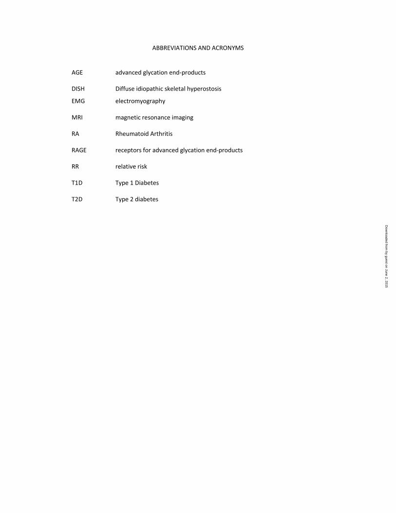
Diffuse idiopathic skeletal hyperostosis (DISH)
DISH or Forestier’s disease, was originally described as confined to the axial skeleton, but is now
recognised as a condition that involves ossification of tendon and ligament attachments in both
spinal and extraspinal locations as well as hyperostosis at bony prominences (figure 2). Nearly half of
patients with DISH have diabetes, and it is more common in elderly diabetic subjects. 10
Glycaemic
control or presence of other diabetic complications does not appear to be related to the onset or
extent of DISH. Whilst many patients may be asymptomatic and DISH noted on routine radiology,
DISH may present with back stiffness and pain, or even dysphagia due to exostoses. Atlanto-axial
subluxation in DISH has been described. Physiotherapy and anti-inflammatories drugs may help with
symptomatic relief and function, although prolonged courses of anti-inflammatories is not desirable
due to the renal and cardiovascular risks with these drugs in diabetic subjects.
Fibrosing syndromes
Diabetes is associated with a number of fibrosing syndromes including adhesive capsulitis of the
shoulder, tenosynovitis of the long flexor of the fingers and thumbs (trigger finger/thumb) (figure 3),
by guest on June 2, 2015D
ownloaded from

6
or the long abductor and the short extensor of the thumbs (De Quervain’s tenosynovitis), carpal
tunnel syndrome, Dupuytren’s contracture (figure 4) and diabetic stiff hand syndrome
(cheiroarthropathy). 11-15
The prevalence of hand or shoulder disorders is higher in people with
diabetes compared to non-diabetic subjects, and correlates with the duration, but not the type, of
diabetes. 16
In patients with diabetes, adhesive capsulitis of the shoulder can occur at a much earlier
age than a non-diabetic frozen shoulder, which rarely occurs before the age of 40 years.
The aetiology of these syndromes is at least in part due to abnormal collagen deposition in the
periarticular connective tissues. In addition, glycosylation, increased hydration and abnormal cross
linking of collagen may be important. 17
Microangiopathy and neuropathy aggravate the problem
(figure 5). These conditions are therefore not strictly inflammatory in nature, and their natural
history is that they generally tend to improve with time. The joints involved are particularly those of
the upper limbs, although the feet may also be involved.
Limitation of joint movement is most marked in the small joints of the hands with thickening and
waxiness of the skin, particularly on the dorsal surface of the fingers, but these skin changes may
occur in the absence of limited joint mobility. It can occur early in the course of the disease. The
prevalence also increases with age and cigarette smoking. Whilst previous studies suggested a
prevalence of anywhere between 20 and 50% of people with diabetes, more recent studies suggest
a reduction in incidence by around 50%, although the cause of this reduction is unclear. 18
The
patient shows a positive “prayer sign” (figure 6), and may be unable to place his hand and fingers flat
on a table (table top sign). The condition is usually painless, and may be asymptomatic.
Management strategies for fibrosing syndromes related to diabetes are guided towards
symptomatic relief and improvement in function. There is no evidence that tightening of glycaemic
control or cessation of smoking will help limited joint mobility, although as the pathogenesis may be
related to glycosylation, poor glycaemic control is undesirable. Physiotherapy with passive palmar
stretching and occupational therapy may improve function. In patients with carpal tunnel syndrome,
electromyography (EMG) is important diagnostically and prognostically, and management involves
resting wrist splints, physiotherapy, anti-inflammatories, and injection of local corticosteroid. Cases
not responding to these measures or with evidence of progressive neurological changes should be
considered for surgical decompression, which can be performed endoscopically.
by guest on June 2, 2015D
ownloaded from

7
A resting wrist, hand and finger splint for patients with limited joint mobility or a thumb splint for
patients with De Quervain’s tenosynovitis may be helpful. Corticosteroid injections may also used,
but they tend to be less effective in people with diabetes compared to people without diabetes.
Adhesive capsulitis of the shoulder may be treated with mobilisation and corticosteroid injection if
there is pain. Recurrence is common.
Complex regional pain syndrome type 1 (reflex sympathetic dystrophy)
Though it is thought to be common in patients with diabetes, the evidence for this association is not
well documented. 19
Sudek's atrophy (reflex sympathetic dystrophy or ‘shoulder-hand syndrome’)
can occur in association with shoulder pathologies. As in other cases, the complaint is of pain with
evidence of skin changes, hyperasthesiae and vasomotor instability.
Osteoporosis
In patients with diabetes, fracture risk appears to be increased in patients with vascular,
nephropathic and neuropathic complications of the disease. 20-22
Hyperglycaemia itself may cause
increased urinary calcium loss, leading to reduced bone mineral density. In T2D, there is a paradox of
increased fractures despite normal BMD because of increased porosity of cortical and trabecular
bone. A meta-analysis of 12 studies reported a relative risk (RR) of 1.7 (95% CI: 1.3–2.2) for hip
fracture in both men and women with T2D. 22
The risk of all clinical fractures was also increased with
a summary RR of 1.2 (95% CI: 1.0–1.5). There appears to be a direct association between the
duration of diabetes and increased fracture risk, although poorer glycaemic control has not
consistently been found to be a risk factor. Pioglitazone, an oral hypoglycaemic agent, may increase
risk of fractures in postmenopausal women. 23
Diabetic muscle infarction (aseptic myonecrosis, infarcted myonecrosis)
Diabetic muscle infarction is a rare complication that mainly occurs in patients with long standing
and poorly controlled disease with microvascular complications, particularly diabetic nephropathy
related end stage renal failure. It presents with acute muscle pain and swelling, most commonly in
the thigh with mild constitutional symptoms. Diagnosis is made with MRI without the need for a
biopsy. 24,25
Creatine kinase may be elevated in around 50% of patients, and the elevation may be
by guest on June 2, 2015D
ownloaded from

8
modest. It generally improves with conservative treatment, and surgical treatment may only be
needed if compartment syndrome develops.
Conclusions
The musculoskeletal complications of diabetes are common and though not life threatening, can
lead to significant pain and disability. They usually occur in patients with poorly controlled diabetes
of long duration and in those who have other more serious complications such as vascular,
neuropathic, renal and retinal problems. Early recognition of these complications, and
multidisciplinary management between diabetes and rheumatology specialists is necessary to
reduce morbidity from these complications.
Word count: 1987
Competing interests: None
by guest on June 2, 2015D
ownloaded from

9
References:
1. http://www.idf.org/sites/default/files/DA-regional-factsheets-2014_FINAL.pdf Accessed 07.04.15
2. Lehuen A. A double-edged sword against type 1 diabetes. N Engl J Med 2015;372:778-780. Doi:
10.1056/NEJMcibr1414708.
3. Edwards JL, Vincent AM, Cheng HT, Feldman EL. Diabetic neuropathy: mechanisms to management.
Pharmacol Ther 2008;120:1-34.doi: 10.1016/j.pharmthera.2008.05.005.
4. Charcot JM. Sur quelaques arthropathies qui paraissent depender d'une lesion du cerveauou de la
moeleepiniere. Arch Des Physiol Norm et Path 1868;1:161-71.
5. Jeffcoate WJ, Game F, Cavanagh PR: The role of proinflammatory cytokines in the cause of
neuropathic osteoarthropathy (acute Charcot foot) in diabetes. Lancet 2005;366:2058-61.
6. Barlovic DP, Soro-Paavonen A, Jandeleit-Dahm KA. RAGE biology, atherosclerosis and diabetes. Clin Sci
(Lond) 2011;121:43-55. doi: 10.1042/CS20100501.
7. Richard JL, Almasri M, Schuldiner S. Treatment of acute Charcot foot with bisphosphonates: a
systematic review of the literature. Diabetologia 2012;55:1258-64. doi: 10.1007/s00125-012-2507-3
8. Choi HK, De Vera MA, Krishnan E. Gout and the risk of type 2 diabetes among men with a high
cardiovascular risk profile. Rheumatology 2008;47:1567–70.doi: 10.1093/rheumatology/ken305.
9. Liao KP, Gunnarsson M, Kallberg H, Ding B, Plenge RM, Padyukov L, et al. Specific association of type
diabetes mellitus (DM) with anticyclic citrullinated peptide-positive rheumatoid arthritis. Arthritis
Rheum 2009;60:653–60.doi: 10.1002/art.24362.
10. Coaccioli S, Fatati G, Di Cato L, Marioli D, Patucchi E, Pizzuti C, et al. Diffuse idiopathic skeletal
hyperostosis in diabetes mellitus, impaired glucose tolerance and obesity. Panminerva Med. 2000;
42:247-51.
11. Cagliero E, Apruzzese W, Perlmutter GS, Nathan DM. Musculoskeletal disorders of the hand and
shoulder in patients with diabetes mellitus. Am J Med 2002; 112: 487-490.
12. Craig ME, Duffin AC, Gallego PH, et al. Plantar fascia thickness, a measure of tissue glycation, predicts
the development of complications in adolescents with type 1 diabetes. Diabetes Care 2008;31:1201-
1206.
13. Fitzgibbon PG: Hand manifestations of diabetes mellitus. J Hand Surg [Am] 2008;33:771-775.
14. Loos B, Puschkin V, and Horch RE. 50 years’ experience with Dupuytren’s contracture in the Erlangen
University Hospital: a retrospective analysis of 2919 operated hands from 1956 to 2006. BMC
Musculoskelet Disord 2007;8:60.
15. Blyth MJ, Ross DJ: Diabetes and trigger finger. J Hand Surg [Br] 1996;21:244-245.
16. Thomas SJ, McDougall C, Brown ID, et al. Prevalence of symptoms and signs of shoulder problems in
people with diabetes mellitus. J Shoulder Elbow Surg 2007;16:748-751.
17. Brownlee M, Cerami A, Vlassara H. Advanced glycosylation end products in tissue and the biochemical
basis of diabetic complications. N Engl J Med 1988;318:1315–1321.
18. Lindsay JR, Kennedy L, Atkinson AB, Bell PM, Carson DJ, McCance DR, Hunter SJ. Reduced prevalence
of limited joint mobility in type 1 diabetes in a UK clinic population over a 20-year period. Diabetes
Care 2005;28:658-61.
19. Marshall AT, Crisp AJ. Reflex sympathetic dystrophy. Rheumatology 2000;39:692-5.
20. Ivers RQ, Cumming RG, Mitchell P, Peduto AJ. Diabetes and the risk of fracture: The Blue Mountain
Eye Study. Diabetes Care 2001; 24: 1198.
21. Janghorbani M, van Dam RM, Willett WC, Hu FB. Systematic review of type 1 and type 2 diabetes
mellitus and risk of fracture. Am J Epidemiol 2007;166:495-505.
22. de L II, van der Klift M, de Laet CE, van Daele CE, Hofman A, Pols HA. Bone mineral density and
fracture risk in type 2 diabetes: the Rotterdam study. Osteoporosis Int 2005;16:1713-20.
23. Zhu ZN, Jiang YF, Ding T. Risk of fracture with thiazolidinediones: an updated meta-analysis of
randomized clinical trials. Bone 2014 ;68:115-23. doi: 10.1016/j.bone.2014.08.010.
by guest on June 2, 2015D
ownloaded from

10
24. Silberstein L, Britton KE, Marsh FP, et al. An unexpected cause of muscle pain in diabetes. Ann Rheum
Dis.2001;60:310-312.
25. Kapur S, Brunet JA, McKendry RJ. Diabetic muscle infarction: case report and review. J Rheumatol
2004;31:190-4.
by guest on June 2, 2015D
ownloaded from

11
Legends to Figures
Figure 1. Plain radiographs of a 67-year-old patient with diabetic neuroarthropathy of the
right ankle and foot showing soft tissue swelling, bone resorption, fragmentation, disorganisation
and destruction. There is also some sclerosis.
Figure 2. Plain radiograph of a 60-year-old man with T2 and DISH. Bridging osteophytes are
seen on four consecutive vertebrae on the right side of the mid dorsal spine.
Figure 3. Tendinopathy of the long flexor tendon of the ring finger. The finger has to be
pushed back to be straightened. Note the the thickened tendon inside the black oval. Also note the
scar of carpal tunnel release.
Figure 4. Dupuytren's contracture
Figure 5. Pathogenesis of limited joint mobility
Figure 6. Positive 'prayer's sign' in a patient with diabetic cheiroarthropathy
by guest on June 2, 2015D
ownloaded from
ABBREVIATIONS AND ACRONYMS
AGE advanced glycation end-products
DISH Diffuse idiopathic skeletal hyperostosis
EMG electromyography
MRI magnetic resonance imaging
RA Rheumatoid Arthritis
RAGE receptors for advanced glycation end-products
RR relative risk
T1D Type 1 Diabetes
T2D Type 2 diabetes
by guest on June 2, 2015D
ownloaded from

Figure 1
by guest on June 2, 2015D
ownloaded from
Figure 2.
by guest on June 2, 2015D
ownloaded from

Figure 3.
by guest on June 2, 2015D
ownloaded from

Figure 4.
by guest on June 2, 2015D
ownloaded from
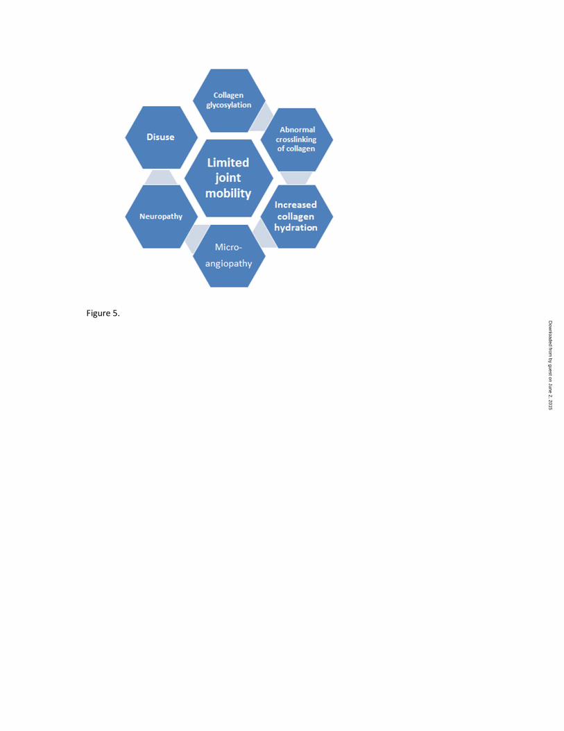
Figure 5.
by guest on June 2, 2015D
ownloaded from

Figure 6.
by guest on June 2, 2015D
ownloaded from
View publication statsView publication stats