Muscular System · the musculoskeletal, digestive, and cardio-respiratory systems, specifically as...
41
Muscular System
Transcript of Muscular System · the musculoskeletal, digestive, and cardio-respiratory systems, specifically as...

Muscular System

Bell Work
How does the muscular system relate to
the following organ systems
Digestive

Health Science Standards
8) Outline basic concepts of normal
structure and function of all body systems,
and explain how homeostasis is
maintained. (muscular)

Exercise Science Standards
8) Review the gross and cellular anatomy and physiology of the
musculoskeletal, nervous, and cardiovascular systems. Define the terms
neuromuscular integration and central command. Summarize how
neuromuscular integration, central command, and training and/or
rehabilitation plans are based on the integration of the muscle nerve with
the muscles of these systems.
9) Identify the two types of muscle fibers and their subtypes, slow twitch and
fast twitch. Relate the concepts of histochemistry, immunocytochemistry,
and physiologic contraction times to the performance of athletes in various
sports. Evaluate the role genetics and training play in muscle fiber
adaptations. From this research, generate an informational artifact to share
with athletes or clients as part of an exercise/training program.

Anatomy & Physiology
Standards 14) Classify the three categories of muscle fibers, differentiating between
cells and tissue. Draw evidence from informational texts to explain the
locations, behavioral properties, and functional roles unique to each
category. Draw on knowledge of biological processes, such as the body’s
conversion of ATP into energy, to illustrate phenomena such as muscle
fatigue.
15) Explain the guidelines used in naming skeletal muscles, such as
location, size, direction, etc. Develop a graphic that identifies the name of
the muscle, the directional motion, location, and function of the following
muscle groups:
a. Muscles of facial expressions
b. Muscles of mastication
c. Muscles of the neck
d. Muscles of the trunk and upper extremities
e. Muscles of lower extremities

Diagnostic Imaging Standards
12) Outline the in-depth normal structure and function of
the musculoskeletal, digestive, and cardio-respiratory
systems, specifically as they relate to diagnostic medical
imaging. Review directions, planes, and sections of the
body in order to perform diagnostic imaging procedures.
Summarize appropriate medical text(s) in order to list
signs and symptoms of common diseases and disorders
associated with each system.

OBJECTIVES
Review the muscular system’s types,
functions, characteristics, points of
attachment, and categories of naming by
creating a crossword puzzle.
Develop a graphic of 15 common muscles
in the human body.

Introduction
You have over 600 skeletal muscles (656-
850 depending on who you talk to)
Muscles account for 40% of our body
weight
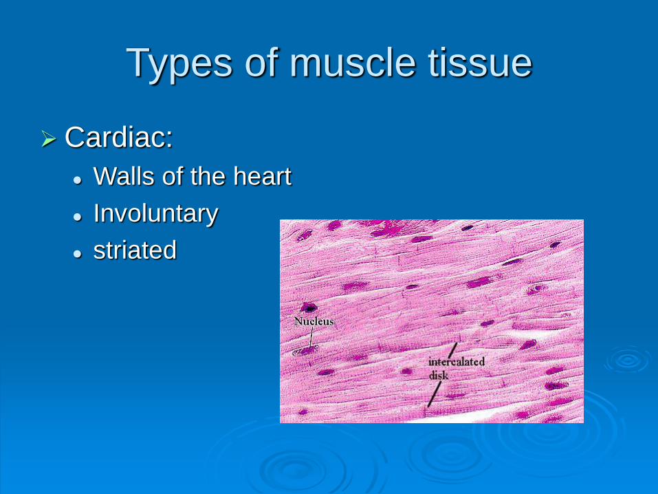
Types of muscle tissue
Cardiac:
Walls of the heart
Involuntary
striated

Smooth or visceral
Found in internal organs: respiratory tract,
digestive tract, blood vessels and eyes
Long circular fibers – peristalsis
Smooth (no striations)
INVOLUNTARY

Skeletal or striated
Attached to the bones to cause skeletal
movement
Striated
VOLUNTARY

Functions of Skeletal Muscle
Produce skeletal movement
Maintain posture and body movement
(muscle tone)
Protection of soft tissue: abdominal wall
and pelvic floor
Maintain body temperature: ex. shivering

Characteristics of Muscle Tissue
Irritability/excitability: ability of a muscle to
respond to a stimulus—nerve impulse
Contractibility: when stimulated muscles
respond by contracting
Extensibility: ability to be stretched
Elasticity: ability to return to its normal
shape after being stretched or contracted

Types of Muscle Contraction
Isometric: same length – ex. Wall sits
Concentric: muscle shortens while
contracting: ex. Bicep curl
Eccentric: muscle lengthens while
contracting: bicep as you are lowering the
weight

Points of Attachment
Origin: less moveable attachment point,
usually more proximal
Insertion: more moveable attachment
point, usually distal
Skeletal muscles are attached to bones by
tendons
(Ligaments connect bone to bone)
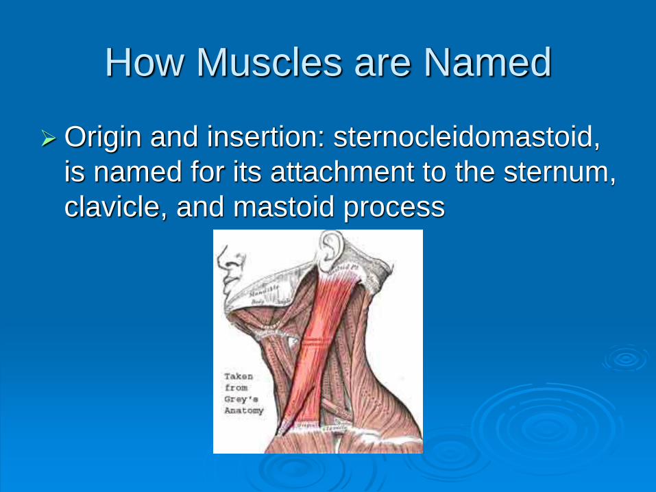
How Muscles are Named
Origin and insertion: sternocleidomastoid,
is named for its attachment to the sternum,
clavicle, and mastoid process

Location or region:
Pectoralis – chest
Gluteus – buttocks
Brachii – arm
Abdominus – abdomen
Femoris -- femur

Action: flexor, extensor, adductor, abductor
Fibers: the direction that the fibers run
Rectus – straight, rectus abdominis
Tranverse – across, transversus abdominus
Oblique – diagonal, external oblique
Obiqularis – circular, obiqularis oris
Divisions
Bi – two
Tri – three
Quad -- four

Size:
Vastus: Huge, vastus lateralis
Maximus: large, gluteus maximus
Minimus: small, gluteus minumus
Longus: long, abductor pollicus longus
Brevis: short, abductor pollicus brevis
Shape
Deltiod: triangular
Rhomboid: rhombus
Trapezius: trapezoid
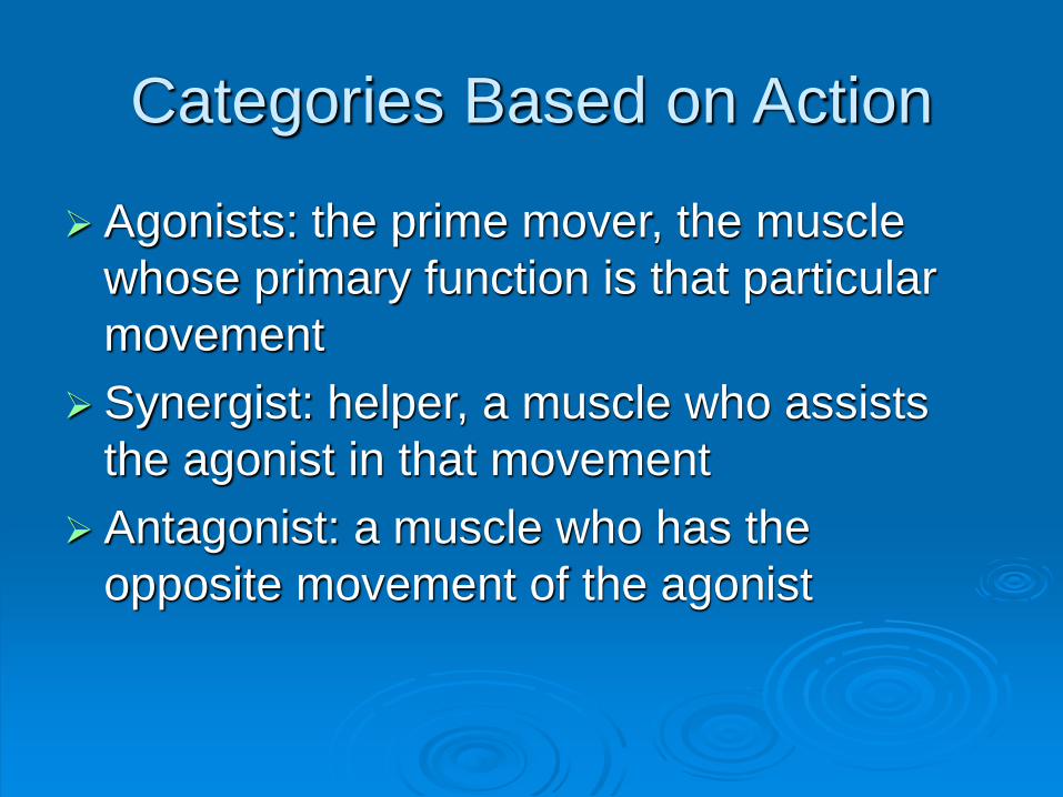
Categories Based on Action
Agonists: the prime mover, the muscle
whose primary function is that particular
movement
Synergist: helper, a muscle who assists
the agonist in that movement
Antagonist: a muscle who has the
opposite movement of the agonist

Example: for the movement of elbow
flexion
Agonist: biceps brachii
Synergist: brachioradialis
Antagonist: triceps brachii

Muscles to Know!!!
(Page 167 old or 321 new book)1. Sternocleidomastoid (SCM)
2. Pectoralis Major (chest)
3. Deltoid (upper/side of arm)(injection site)
4. Biceps Brachii (muscle with 2 heads at the front of upper arm)
5. Triceps Brachii (muscle with 3 heads at the back of upper
arm)
6. Intercostals (inter=between; costae= ribs) (this is not in book)
7. Rectus Abdominus (Abs)(Abd; rectus= straight)
8. Trapezius (Upper Back/Neck)
9. Lattisimus Dorsi (Dorsal = back)
10. Gluteus Maximus (Maximus= large muscle) (injection site)
11. Gastrocnemius (Calf)
12. Quadriceps Femoris (muscle with 4 heads on top of femur)
(injection site) (this name is not in book)
13. Sartorius (crosses over thigh like a seat belt)
14. Tibialis Anterior (front of tibia)
15. Orbicularis Oris (surrounds lips)

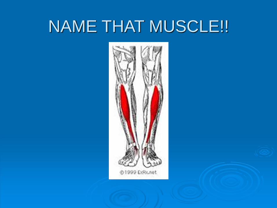
NAME THAT MUSCLE!!

NAME THAT MUSCLE!!
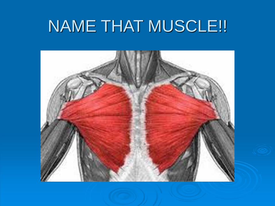
NAME THAT MUSCLE!!
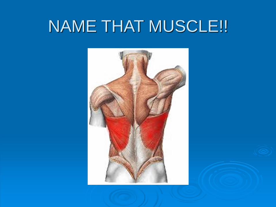
NAME THAT MUSCLE!!

NAME THAT MUSCLE!!

NAME THAT MUSCLE!!

NAME THAT MUSCLE!!

NAME THAT MUSCLE!!

NAME THAT MUSCLE!!
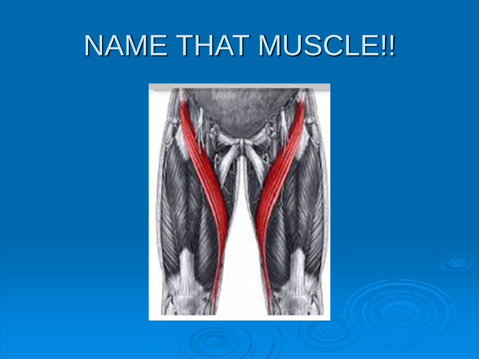
NAME THAT MUSCLE!!

NAME THAT MUSCLE!!

NAME THAT MUSCLE!!

NAME THAT MUSCLE!!
n.
1670s, from
Latinized form of
Greek
gastroknemia "calf
of the leg”, from
gaster "belly" +
kneme "leg“. So
called for its form –
belly of the leg.
NAME THAT MUSCLE!!

NAME THAT MUSCLE!!

Activity
Create your own
crossword puzzle
using key terms
from the following
headings:
Characteristics
Functions
Types
Attachments
Make sure to make
an “empty”
crossword as well
as a “key” to the
crossword puzzle.
Use at least 20
terms…
(E.g. movement,
cardiac, elasticity.)

Group Activity
Case Scenario Discussion groups
Find your match games
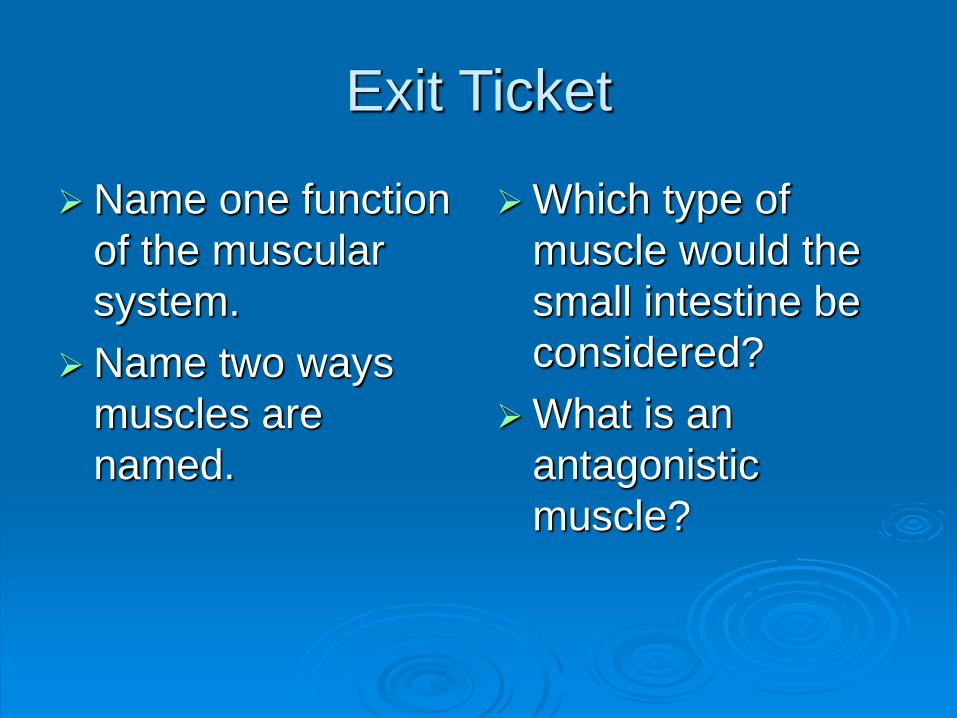
Exit Ticket
Name one function
of the muscular
system.
Name two ways
muscles are
named.
Which type of
muscle would the
small intestine be
considered?
What is an
antagonistic
muscle?

Sternocleidomastoid
Pectoralis Major
Deltoid
Biceps Brachii
Triceps Brachii
Intercostals
Rectus Abdominus
Trapezius
Lattisimus Dorsi
Gluteus Maximus
Gastrocnemius
Quadriceps Femoris
Sartorius
Tibialis Anterior
Orbicularis Oris
Skull
Mandible
Ulna
Tibia
Radius
Fibula
Carpals
Metacarpals
Femur
Tarsals
Phalanges
Clavicle
Sternum
Metatarsals
Ribs
Lumbar spine
Pelvis