Muscular System
44
Muscular System Chapter 6
-
Upload
jeanette-lara -
Category
Documents
-
view
20 -
download
0
description
Muscular System. Chapter 6. The 3 Muscle Types. The job of all muscles is to contract They are all fibrous because cells are elongated The 3 Muscle Types Are: Skeletal Muscle Cardiac Muscle Smooth Muscle. Skeletal Muscle. Cigar Shaped Multinucleated Striated Voluntary - PowerPoint PPT Presentation
Transcript of Muscular System

Muscular System
Chapter 6


The 3 Muscle Types
• The job of all muscles is to contract
• They are all fibrous because cells are elongated
• The 3 Muscle Types Are:
–Skeletal Muscle
–Cardiac Muscle
–Smooth Muscle

Skeletal Muscle
• Cigar Shaped• Multinucleated• Striated• Voluntary• Can Be Involuntary When Reflexes Are
Involved• Very Strong and Fast But Need Rest• Most Attached to Bone
Skeletal Muscle OverviewOf
Skeletal Muscle

Skeletal Muscle Cell

Smooth Muscle
Long thin nuclei and no striations

How are Muscles Structured?
• Muscle Cells have a plasma membrane called a Sarcolemma.
• The muscle fiber is enclosed in Endomysieum. (Endo= , Mys= )
• Many Muscle fibers bound together make a Fascicle.
• The Fascicle is wrapped in a membrane called the Perimysieum (Peri= ).

Muscle Structure Continued
• Many fascicles are wrapped together by an Epimysium. (Epi= )
• Epimysia attach to tendons or Aponeuroses. (Pg. 86)
• Tendons: Strong, Thin, and made up of collagen (dense connective tissue). Aponeuroses are sheet-like tendons.

Skeletal Muscle (pg. 174)
QuizYourself

What is the major Organelle of the Muscle Cell?
• Myofibril(s)=Working unit of the muscle cell. Made of Subunits called sarcomeres.–Give muscles the striped or striated
appearance–The light band is the I-band–The dark band is the A-band–Between the I-bands is the Z-line–Between the A-bands is the H-zone
Match theTerms
Mechanism of muscle contraction
• The above micrographs show that the sarcomere gets shorter when the muscle contracts
• The light (I) bands become shorter• The dark bands (A) bands stay the same length
Relaxedm uscle
Contractedm uscle
relaxed sarcom ere
contracted sarcom ere

Take a long deep breath, its not that bad.
• And remember a bicycle cannot stand alone, because it is two tired.
• Now lets go on.
• But lets first watch this short video clip.
Overview of the Job of the Bands

So how do these bands work?
• The myofibrils are surrounded by the sarcoplasmic reticulum, a specialized form of smooth endoplasmic reticulum that releases calcium.
• They are made of bands of– Actin (the thin filaments) that make up the I-
bands – Myosin (the thick filaments) that make up the
A-bands

So what is the Molecular Basis of Muscle Contraction? (pg. 176)
• 1) Nerve sends out Acetylcholine or Ach
• 2) Motor Unit= All muscles triggered by nerve. (1 nerveTriggers 100’s of cells)
• 3) The Sarcolema becomes permeable to Na+
• 4) Na+ causes an action potential because it disturbs the electrical conditions of the sarcolema (pg.178)

How does ACh stimulate the muscle?
• ACh causes the sarcolema to release Calcium (Ca+)
• Ca+ binds to the actin causing it to change shape.
• Myosine finds actin’s new shape attractive and grabs hold.

What happens after the Myosin grabs hold? (pg. 179)
• Myosin’s head snap towards the H-band of the sarcomere.
• ATP releases and re-cocks the myosin
• Only some myosin heads move at one time.
• Over all: Pg. 181
Description of Muscle movement

How does the muscle relax?
• When the action potential ends:
–Sarcomere absorb Ca+
–ATP releases myosin heads
–Actin takes on its former and less attractive shape.
–Muscle Cells can relax Best Movie on Muscle Contraction
Revisited

Write one paragraph explaining how a muscle works.
Do this from memory.
This will help you learn.

So, What is this Action Potential?
• Action Potential– Electrical Current or Charge– In order to return the cell to its original
condition, K+ is pumped into the cell by the sodium potassium pump. Fig 3.10
Sodium/Potassium
Pump

How do muscles work together?
• Prime Mover: Major muscle doing the bulk of the work contracting.
• Synergist: Group of muscles working together to contract.
• Antagonist: Muscle that works against the prime mover and or synergists.
How do Muscles work Together?

• http://www.bbc.co.uk/science/humanbody/body/interactives/3djigsaw_02/index.shtml?muscles
• http://www.healthchecksystems.com/exercise1.htm#hyper
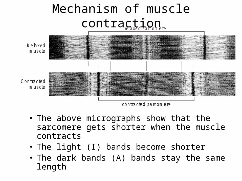
Types of Muscle Movements


To exercise the:
Quadriceps

To exercise the:
Gastronemius

To exercise the:
Hamstring GroupIncludes the bicepts femoris

Exercise the:Trapezius

Exercise for:Latissimus Dorsi

Exercise for:Deltoid

Exercise For: Gluteus Maximus

Exercises for:Oblique

Exercises for: Rectus Abdominus

Exercise for:
Pectoralis Major
Push up: Works Pecs on The Up-Motion

Exercises for:Bicep

Exercises for: Triceps

Now Quiz Yourself!
See if you know your muscles…
Another goodQuiz site

Interesting Aspects:
• Some of us may have a spare muscle: the Plantaris muscle 1 in 10 don’t.
• Want to see it? Click the link.
• Groin pull? Here is the problem:
Groin Pull
PlantarisIn
Action