Muscle Contraction - Oak Park Independent · 2013-01-18 · Muscle Contraction Skeletal Muscle...
3
Muscle Contraction Skeletal Muscle Physiology Part 1 - Muscle Contraction What is needed for a muscle to contract? **2 and 3 called excitation-contraction coupling** 1. 2. 3. a change in membrane potential (nervous stimulation) action potential across sarcolemma quick increase in intracellular calcium ion levels Neuromuscular Junction · region where a motor neuron comes into close contact with a skeletal muscle fiber 3 parts: axonal endings, synaptic cleft, junctional folds · motor neurons - nerve cells that activate skeletal muscle fibers · axonal endings - ends of motor neuron · synaptic cleft - space between axonal endings and muscle fiber · junctional folds - house ACh receptors on sarcolemma acetylcholine (ACh) - neurotransmitter responsible for muscle contraction · when a nerve impulse reaches axonal endings, ACh is released into the synaptic cleft · ACh diffuses across and binds to ACh receptor on sarcolemma (muscle fiber) · changes membrane potential and generates an action potential NeuroMuscular Junction Action Potential · change in membrane potential · resting muscle cell is polarized polarized - negative inside, positive outside · steps of action potential depolarization - inside become pos. (sodium rushes in) generation and propagation - once threshold is reached AP can travel along muscle fiber repolarization - sarcolemma restored to its original polarized state · trigged when ACh binds to receptors on sarcolemma
Transcript of Muscle Contraction - Oak Park Independent · 2013-01-18 · Muscle Contraction Skeletal Muscle...
Muscle Contraction
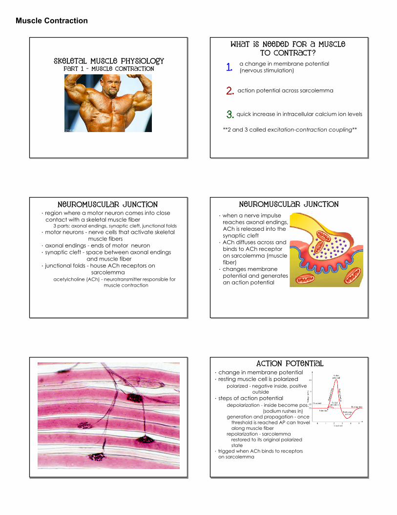
Skeletal Muscle Physiology Part 1 - Muscle Contraction
What is needed for a muscle to contract?
**2 and 3 called excitation-contraction coupling**
1.
2.
3.
a change in membrane potential (nervous stimulation)
action potential across sarcolemma
quick increase in intracellular calcium ion levels
Neuromuscular Junction· region where a motor neuron comes into close contact with a skeletal muscle fiber
3 parts: axonal endings, synaptic cleft, junctional folds· motor neurons - nerve cells that activate skeletal muscle fibers· axonal endings - ends of motor neuron· synaptic cleft - space between axonal endings and muscle fiber· junctional folds - house ACh receptors on sarcolemma
acetylcholine (ACh) - neurotransmitter responsible for muscle contraction
· when a nerve impulse reaches axonal endings, ACh is released into the synaptic cleft· ACh diffuses across and binds to ACh receptor on sarcolemma (muscle fiber)· changes membrane potential and generates an action potential
NeuroMuscular Junction
Action Potential· change in membrane potential· resting muscle cell is polarized
polarized - negative inside, positive outside· steps of action potential
depolarization - inside become pos. (sodium rushes in)
generation and propagation - once threshold is reached AP can travel along muscle fiber
repolarization - sarcolemma restored to its original polarized state· trigged when ACh binds to receptors on sarcolemma

Muscle Contraction
Excitation-Contraction Coupling· occurs during latent period
time between AP and beginning of contraction· AP reaches T tubules· T tubules change shape and stimulate sarcoplasmic reticulum to release calcium into muscle fiber
Role of Calcium· cross bridge formation dependent on calcium
low levels of calcium = relaxationhigh levels of calcium = contraction
· during relaxation, tropomyosin blocks the active sites on actin· when calcium levels rise, calcium binds to troponin· troponin changes shape and moves tropomyosin exposing the active sites on actin· cross bridges can now form
Cross Bridge Cycle1. cross bridge formation
energized myosin attaches to actin2. power stroke
ADP + P are releasedmyosin head pivots pulling actin toward the M line
3. cross bridge detachmentnew ATP binds to myosin head and actin is released
4. cocking of myosin headATP converted to ADP + Pmyosin cocked into "ready" position
***only about 1/2 of myosin heads form cross bridges at a time; other 1/2 searching for new attachment site*** Click for animations -
Action Potential & Myofilament Contraction
Muscle Twitch· contraction of a muscle fiber· myogram - graph of muscle contraction
latent period - time between AP and muscle contraction
contraction phase - cross bridges are activerelaxation phase - calcium reenters SR and
sarcomere returns to original length· muscles contract faster than they relax

Muscle Contraction
Muscular Responses1. threshold stimulus - minimal stimulus required to produce a contraction2. all-or-none response - muscle fibers contract completely or not at all
increases in intensity, rate or duration of impulses does NOT strengthen the contraction3. summation - contractions add together
incomplete tetanus - sustained by quivering contractioncomplete tetanus - sustained contraction, NO relaxation
4. tonus (muscle tone) - muscles during normal posture (always partially contracted)5. graded response - variations in contraction strength
changes in frequency of stimulationchanges in strength of stimulation
Muscular responses