Murine Vaginal Colonization Model for Investigating ... · Murine Vaginal Colonization Model for...
12
Murine Vaginal Colonization Model for Investigating Asymptomatic Mucosal Carriage of Streptococcus pyogenes Michael E. Watson, Jr., a Hailyn V. Nielsen, b Scott J. Hultgren, b,c Michael G. Caparon b,c Divison of Pediatric Infectious Diseases, Department of Pediatrics, Washington University School of Medicine, St. Louis, Missouri, USA a ; Department of Molecular Microbiology, Washington University Medical School, St. Louis, Missouri, USA b ; Center for Women’s Infectious Disease Research, Washington University School of Medicine, St. Louis, Missouri, USA c While many virulence factors promoting Streptococcus pyogenes invasive disease have been described, specific streptococcal fac- tors and host properties influencing asymptomatic mucosal carriage remain uncertain. To address the need for a refined model of prolonged S. pyogenes asymptomatic mucosal colonization, we have adapted a preestrogenized murine vaginal colonization model for S. pyogenes. In this model, derivatives of strains HSC5, SF370, JRS4, NZ131, and MEW123 established a reproducible, asymptomatic colonization of the vaginal mucosa over a period of typically 3 to 4 weeks’ duration at a relatively high coloniza- tion efficiency. Prior treatment with estradiol prolonged streptococcal colonization and was associated with reduced inflamma- tion in the colonized vaginal epithelium as well as a decreased leukocyte presence in vaginal fluid compared to the levels of in- flammation and leukocyte presence in non-estradiol-treated control mice. The utility of our model for investigating S. pyogenes factors contributing to mucosal carriage was verified, as a mutant with a mutation in the transcriptional regulator catabolite control protein A (CcpA) demonstrated significant impairment in vaginal colonization. An assessment of in vivo transcriptional activity in the CcpA strain for several known CcpA-regulated genes identified significantly elevated transcription of lactate oxi- dase (lctO) correlating with excessive generation of hydrogen peroxide to self-lethal levels. Deletion of lctO did not impair colo- nization, but deletion of lctO in a CcpA strain prolonged carriage, exceeding even that of the wild-type strain. Thus, while LctO is not essential for vaginal colonization, its dysregulation is deleterious, highlighting the critical role of CcpA in promoting mu- cosal colonization. The vaginal colonization model should prove effective for future analyses of S. pyogenes mucosal colonization. T he Gram-positive pathogen Streptococcus pyogenes, or group A streptococcus, is responsible for a wide variety of clinical man- ifestations ranging from the relatively benign and superficial otitis media, impetigo, and pharyngitis to less common and more inva- sive conditions, including necrotizing fasciitis and toxic shock syndrome, in addition to the postinfectious complications rheu- matic fever and glomerulonephritis (1). The ability to produce such a diversity of infections in so many different tissue compart- ments is testament to the extensive plasticity of the S. pyogenes transcriptome and an abundance of secreted virulence factors (2). While not considered normal human flora, S. pyogenes can be identified as a colonizer of mucosal surfaces of the oropharynx, rectum, and vaginal mucosa, and a prolonged asymptomatic car- riage state can develop (3–7). How S. pyogenes carriage exists on a mucosal surface without inducing disease is poorly understood, but the issue is significant in terms of gaining an improved under- standing of host-pathogen interactions and the regulation of mu- cosal immunity. Furthermore, asymptomatic oropharyngeal car- riage of S. pyogenes continues to confound the results of clinical testing for those with symptoms of pharyngitis, frequently, chil- dren with viral infections, in which the detection of carrier strains mistakenly results in inappropriate antibiotic exposures and ex- penses (8, 9). Modeling of asymptomatic mucosal carriage in the human- restricted organism S. pyogenes has been challenging. Although murine and primate models of S. pyogenes oropharyngeal and na- sopharyngeal colonization exist, their utility has several limita- tions, including limited access to the pharyngeal tissue site, the high cost and maintenance requirements of a primate colony, and the fact that many of these models are not capable of supporting more than a few specific strains of S. pyogenes for a limited period of time at a relatively low efficiency of colonization (10, 11). To overcome several of these obstacles, we have taken a novel ap- proach by adapting a murine vaginal epithelial model to investi- gate S. pyogenes colonization and prolonged carriage. Murine vaginal colonization models in preestrogenized mice have previously been used to investigate the mucosal biology of human pathogens, including Candida albicans (12), Trichomonas vaginalis (13), group B streptococcus (14), and Neisseria gonor- rhoeae (15). The anatomy and physiology of the murine vaginal mucosa are quite similar to those of the human vaginal mucosa, and both respond similarly to estrogen cycling, with thickening and proliferation of the epithelial surface and accumulation of glycogen in the intermediate and superficial layers, although to a lesser degree in mice than in humans (16). While mice have a 10- to 25-fold lower level of serum estrogen, they respond dramati- cally to exogenous estradiol treatment, with enhanced glycogen deposition, intense epithelial proliferation, and increased epithe- Received 9 January 2013 Returned for modification 16 February 2013 Accepted 21 February 2013 Published ahead of print 4 March 2013 Editor: A. Camilli Address correspondence to Michael G. Caparon, [email protected]. Supplemental material for this article may be found at http://dx.doi.org/10.1128 /IAI.00021-13. Copyright © 2013, American Society for Microbiology. All Rights Reserved. doi:10.1128/IAI.00021-13 1606 iai.asm.org Infection and Immunity p. 1606 –1617 May 2013 Volume 81 Number 5 on May 7, 2020 by guest http://iai.asm.org/ Downloaded from
Transcript of Murine Vaginal Colonization Model for Investigating ... · Murine Vaginal Colonization Model for...

Murine Vaginal Colonization Model for Investigating AsymptomaticMucosal Carriage of Streptococcus pyogenes
Michael E. Watson, Jr.,a Hailyn V. Nielsen,b Scott J. Hultgren,b,c Michael G. Caparonb,c
Divison of Pediatric Infectious Diseases, Department of Pediatrics, Washington University School of Medicine, St. Louis, Missouri, USAa; Department of MolecularMicrobiology, Washington University Medical School, St. Louis, Missouri, USAb; Center for Women’s Infectious Disease Research, Washington University School ofMedicine, St. Louis, Missouri, USAc
While many virulence factors promoting Streptococcus pyogenes invasive disease have been described, specific streptococcal fac-tors and host properties influencing asymptomatic mucosal carriage remain uncertain. To address the need for a refined modelof prolonged S. pyogenes asymptomatic mucosal colonization, we have adapted a preestrogenized murine vaginal colonizationmodel for S. pyogenes. In this model, derivatives of strains HSC5, SF370, JRS4, NZ131, and MEW123 established a reproducible,asymptomatic colonization of the vaginal mucosa over a period of typically 3 to 4 weeks’ duration at a relatively high coloniza-tion efficiency. Prior treatment with estradiol prolonged streptococcal colonization and was associated with reduced inflamma-tion in the colonized vaginal epithelium as well as a decreased leukocyte presence in vaginal fluid compared to the levels of in-flammation and leukocyte presence in non-estradiol-treated control mice. The utility of our model for investigating S. pyogenesfactors contributing to mucosal carriage was verified, as a mutant with a mutation in the transcriptional regulator catabolitecontrol protein A (CcpA) demonstrated significant impairment in vaginal colonization. An assessment of in vivo transcriptionalactivity in the CcpA� strain for several known CcpA-regulated genes identified significantly elevated transcription of lactate oxi-dase (lctO) correlating with excessive generation of hydrogen peroxide to self-lethal levels. Deletion of lctO did not impair colo-nization, but deletion of lctO in a CcpA� strain prolonged carriage, exceeding even that of the wild-type strain. Thus, while LctOis not essential for vaginal colonization, its dysregulation is deleterious, highlighting the critical role of CcpA in promoting mu-cosal colonization. The vaginal colonization model should prove effective for future analyses of S. pyogenes mucosalcolonization.
The Gram-positive pathogen Streptococcus pyogenes, or group Astreptococcus, is responsible for a wide variety of clinical man-
ifestations ranging from the relatively benign and superficial otitismedia, impetigo, and pharyngitis to less common and more inva-sive conditions, including necrotizing fasciitis and toxic shocksyndrome, in addition to the postinfectious complications rheu-matic fever and glomerulonephritis (1). The ability to producesuch a diversity of infections in so many different tissue compart-ments is testament to the extensive plasticity of the S. pyogenestranscriptome and an abundance of secreted virulence factors (2).While not considered normal human flora, S. pyogenes can beidentified as a colonizer of mucosal surfaces of the oropharynx,rectum, and vaginal mucosa, and a prolonged asymptomatic car-riage state can develop (3–7). How S. pyogenes carriage exists on amucosal surface without inducing disease is poorly understood,but the issue is significant in terms of gaining an improved under-standing of host-pathogen interactions and the regulation of mu-cosal immunity. Furthermore, asymptomatic oropharyngeal car-riage of S. pyogenes continues to confound the results of clinicaltesting for those with symptoms of pharyngitis, frequently, chil-dren with viral infections, in which the detection of carrier strainsmistakenly results in inappropriate antibiotic exposures and ex-penses (8, 9).
Modeling of asymptomatic mucosal carriage in the human-restricted organism S. pyogenes has been challenging. Althoughmurine and primate models of S. pyogenes oropharyngeal and na-sopharyngeal colonization exist, their utility has several limita-tions, including limited access to the pharyngeal tissue site, thehigh cost and maintenance requirements of a primate colony, andthe fact that many of these models are not capable of supporting
more than a few specific strains of S. pyogenes for a limited periodof time at a relatively low efficiency of colonization (10, 11). Toovercome several of these obstacles, we have taken a novel ap-proach by adapting a murine vaginal epithelial model to investi-gate S. pyogenes colonization and prolonged carriage.
Murine vaginal colonization models in preestrogenized micehave previously been used to investigate the mucosal biology ofhuman pathogens, including Candida albicans (12), Trichomonasvaginalis (13), group B streptococcus (14), and Neisseria gonor-rhoeae (15). The anatomy and physiology of the murine vaginalmucosa are quite similar to those of the human vaginal mucosa,and both respond similarly to estrogen cycling, with thickeningand proliferation of the epithelial surface and accumulation ofglycogen in the intermediate and superficial layers, although to alesser degree in mice than in humans (16). While mice have a 10-to 25-fold lower level of serum estrogen, they respond dramati-cally to exogenous estradiol treatment, with enhanced glycogendeposition, intense epithelial proliferation, and increased epithe-
Received 9 January 2013 Returned for modification 16 February 2013Accepted 21 February 2013
Published ahead of print 4 March 2013
Editor: A. Camilli
Address correspondence to Michael G. Caparon, [email protected].
Supplemental material for this article may be found at http://dx.doi.org/10.1128/IAI.00021-13.
Copyright © 2013, American Society for Microbiology. All Rights Reserved.
doi:10.1128/IAI.00021-13
1606 iai.asm.org Infection and Immunity p. 1606–1617 May 2013 Volume 81 Number 5
on May 7, 2020 by guest
http://iai.asm.org/
Dow
nloaded from
lial cell thickness, which overall presents a more human-like vag-inal profile (17, 18). In addition, human and murine vaginal se-cretions have similar compositions, with similar concentrations ofglucose (3.3 mM and 4.4 mM, respectively) and lactate (6.2 mMand 8.4 mM, respectively) (19).
Asymptomatic human vaginal carriage of S. pyogenes is typi-cally detected at a low frequency among women of reproductiveage, about 0.03% to 1% (3, 20). Group A streptococcus can beisolated from vaginal swab specimen cultures at a greater fre-quency from cases of vulvovaginitis and puerperal sepsis, oftenoccurring with prior or concurrent pharyngitis (21–24). In partic-ular, S. pyogenes serotype M28 isolates are overrepresented in casesof human female urogenital tract infection, puerperal sepsis, andneonatal infections (25–27). All M28 isolates and isolates of otherserotypes associated with puerperal infections exhibit carriage of a37.4-kb genomic island designated region of difference 2 (RD2),which is shared with group B streptococcus and which contains 7putative proteins and potential virulence factors (28, 29). Hence,investigation of S. pyogenes vaginal colonization and invasive in-fections represents an opportunity to gain greater insight into bac-terial pathogenesis, particularly with regard to interactions withthe host mucosal immunity.
The regulation of metabolism and virulence factor expressionamong carriage isolates is an active field of investigation. Globalregulators of bacterial gene transcription sensitive to environmen-tal cues are increasingly recognized for their importance in patho-genesis (for a review, see references 30 and 31). Environmentalcarbohydrate concentrations have been determined to be a majorenvironmental cue for S. pyogenes that is directly coupled to tran-scriptional regulation through the process of carbon cataboliterepression (CCR) and the major CCR regulator catabolite controlprotein A (CcpA) (32–35). CcpA is a member of the LacI/GalRfamily of activator/repressor transcription factors and is widelyconserved among low-G�C-content Gram-positive prokaryotes,including S. pyogenes. A detailed discussion of the biochemistry ofCcpA-dependent CCR is beyond the scope of this article but hasbeen discussed elsewhere (36). In short, the accumulation of gly-colytic intermediates, including fructose-1,6-bisphosphate, frommetabolism in a carbohydrate-rich environment results in a signalcascade activating CcpA. CcpA influences transcriptional expres-sion by binding to operator sequences termed catabolite-respon-sive elements (cre), usually adjacent to or within promoter se-quences of open reading frames regulated by CcpA, and regulatingaccess of the RNA polymerase complex (36). Within S. pyogenes,carbon catabolite repression is central to global transcriptionalregulation, not only by regulating carbohydrate metabolism path-ways but also by functioning as a transcriptional activator or re-pressor of several important virulence factors, including the cys-teine protease SpeB, the cytolysin streptolysin S, hyaluronic acidcapsule, the extracellular DNase Spd, and the immunoglobulin-degrading EndoS (32–34, 37). Given the central role of CcpA intranscriptional regulation, it follows that loss of CcpA activitywould be associated with dysregulation of gene expression at adetriment to pathogenesis. Supporting evidence is that S. pyogenesCcpA� strains have been demonstrated to have reduced virulencein the murine skin ulcer model (34), reduced virulence in a mu-rine intraperitoneal model of invasive infection, and impaired col-onization in the murine oropharynx (32). While the mechanismof attenuated virulence in the CcpA� strains has not been deter-mined, we would hypothesize that the loss of CcpA-dependent
transcriptional regulation contributes to a loss of virulencethrough dysregulation of gene expression, with an inappropriatetiming and/or magnitude of gene transcription impairing fitness.
In this study, we have adapted a preestrogenized murine vagi-nal mucosa model to investigate S. pyogenes colonization and car-riage. We report that several S. pyogenes strains from diverse clin-ical backgrounds are capable of prolonged carriage at a relativelyhigh degree of colonization efficiency. Estrogenization of micewas shown to promote a sustained and asymptomatic vaginal col-onization analogous to that found in women of reproductive agewith normal estrogen levels (3, 22, 23). Furthermore, we havevalidated the utility of our model for investigating streptococcalfactors, in demonstrating that the absence of CcpA inhibits pro-longed murine vaginal carriage through dysregulation of gene ex-pression, including increased lactate oxidase (LctO) expression invivo, likely limiting mucosal survival. This report adds a novelanimal model to the field of S. pyogenes colonization and carriageand further contributes to our understanding of the role of globaltranscriptional regulators and their role in the biology of mucosalpathogens.
MATERIALS AND METHODSBacterial strains, media, and growth conditions. The principal strainused in this study was S. pyogenes HSC12, a spontaneous streptomycin-resistant derivative of the M14 strain HSC5 (38). Whole-genome se-quencing has revealed that HSC12 contains a single nucleotide polymor-phism at nucleotide position 1960 (AAA to ACA) in the 30S ribosomalsubunit S12p (rpsL; SPy_0271) resulting in a missense mutation (K56T)conferring high-level streptomycin resistance (M. E. Watson, Jr., G. C.Port, and M. C. Caparon, unpublished data). Other strains used are listedin Table 1. Unless otherwise stated, all S. pyogenes strains had equivalentgrowth rates and yields in the media tested (data not shown). Routineculture of S. pyogenes was performed in Todd-Hewitt medium (Becton,Dickinson, Franklin Lakes, NJ) supplemented with 0.2% (wt/vol) yeastextract (Difco Laboratories, Detroit, MI) (THY medium). Where re-quired, Bacto agar (Difco) was added to a final concentration of 1.4%(wt/vol) to produce solid medium. Experiments that required low glucoseconcentrations were performed in C medium (41). Incubation was per-formed at 37°C under anaerobic conditions (GasPak; Becton, Dickinson)for solid medium or in sealed tubes without agitation for liquid medium.Aerobic culture was conducted as described previously (33). For inocula-tion of mice, S. pyogenes was harvested from culture in THY broth at earlylogarithmic phase (optical density at 600 nm [OD600], 0.2), washed oncein phosphate-buffered saline (PBS), briefly sonicated on ice to disruptlong streptococcal chains, and resuspended in PBS to 108 CFU/ml. Forcloning of recombinant DNA, Escherichia coli strain DH5� or TOP10(Invitrogen, Grand Island, NY) was cultured in Luria-Bertani (LB) broth.When appropriate, antibiotics were added at the following concentra-tions: erythromycin, 500 �g/ml for E. coli and 1 �g/ml for S. pyogenes;spectinomycin,100 �g/ml for both E. coli and S. pyogenes; and streptomy-cin, 1,000 �g/ml for S. pyogenes (all were obtained from Sigma ChemicalCo., St. Louis, MO).
DNA manipulation and strain construction. Plasmid DNA was iso-lated by standard methods and used to transform S. pyogenes as previouslydescribed (42). Restriction endonucleases, ligases, and polymerases wereused according to the manufacturer’s recommendations (New EnglandBioLabs, Ipswich, MA). The fidelity of all constructs derived by PCR wasconfirmed by DNA sequencing analysis performed by a commercial ven-dor (Integrated DNA Technologies, Inc., Coralville, IA). All references togenomic loci are based on the genome of S. pyogenes strain SF370 (43).In-frame deletion mutations were constructed using a routine method ofallelic exchange (44) via S. pyogenes transformation by electroporationwith derivatives of the E. coli-to-streptococcus temperature-sensitiveshuttle vector pJRS233, previously reported as pCK195 for ccpA
S. pyogenes Vaginal Mucosa Carriage Model
May 2013 Volume 81 Number 5 iai.asm.org 1607
on May 7, 2020 by guest
http://iai.asm.org/
Dow
nloaded from

(SPy_0514) and pCK37 for lctO (SPy_0414) (33). Generation of sponta-neous streptomycin-resistant isolates was performed by plating a highdensity of S. pyogenes onto THY agar plates supplemented with 1 mg/mlstreptomycin and incubation at 37°C overnight; streptomycin-resistantclones occurred in approximately 1 of 109 CFU plated. All mutants wereconfirmed by PCR, DNA sequencing, and phenotypic assays, when avail-able. Descriptions of the resulting strains are listed in Table 1.
Reversion of the CcpA� strain to CcpAr. Although several plasmidsystems for S. pyogenes exist, none proved suitable for long-term in vivostability in the absence of selective antibiotic pressure. Therefore, to re-verse the ccpA in-frame deletion mutation, a strategy was implemented torestore the wild-type (WT) locus in the CcpA� mutant. A 2.2-kb fragmentcontaining the entire ccpA open reading frame and flanking regions wasamplified from HSC12 genomic DNA using primers CK214 (5=-CCCATCGATGAGCGATGACGTCTGTTTCGGTAG-3=) and CK217 (5=-CCCGGATCCGGATCTGCTCCGTACTGCTCC-3=) (33), which were digestedwith ClaI and BamHI, respectively (sequences underlined in the primers),and inserted between the same sites of pJRS233 (44). The resulting plas-mid (pMEW74) was then used to restore the wild-type ccpA locus asdescribed above, creating strain MEW106 with restored CcpA (CcpAr)(Table 1).
Murine vaginal colonization model. C57BL/6J and BALB/c micewere obtained from The Jackson Laboratory (Bar Harbor, ME) and wereallowed to acclimatize for 1 week prior to manipulations. All mice werefemale and unless otherwise specified were used at 7 to 8 weeks of age. Tosynchronize estral cycles, mice were estrogenized by intraperitoneal injec-tion of up to 0.5 mg �-estradiol 17-valerate (Sigma) dissolved in 0.1 mlsterile sesame oil (Sigma) 2 days prior to streptococcal inoculation andagain on the day of inoculation (day 0). In some experiments, mice re-ceived intraperitoneal injections of 0.1 ml sterile sesame oil as a vector-only control; these mice are referred to as mock treated throughout thisarticle. On day 0, mice were sedated by brief inhalation of 80% carbon
dioxide–20% oxygen gas, and the various inocula of S. pyogenes describedin the text were instilled into the vaginal vault using a P20 micropipette(Gilson, Inc., Middleton, WI) in a total volume of 20 �l PBS. In someexperiments, control mice received a vaginal instillation of phosphate-buffered saline alone; these mice are referred to as uncolonized through-out this article. At successive intervals over a 1-month period postinocu-lation, the vaginal vaults of sedated mice were gently washed with 50 �lPBS and serial dilutions in PBS were plated onto THY agar plates contain-ing 1,000 �g/ml streptomycin to determine the numbers of viable CFU.This concentration of streptomycin suppressed the growth of the normalmouse vaginal flora but had no effect on the growth of the streptomycin-resistant S. pyogenes strains. The detection limit was mathematically cal-culated as the CFU density below which our drop-plating method may notdetect the growth of viable streptococci. The drop-plating method in-volves making 10-fold serial dilutions of the vaginal wash specimens inphosphate-buffered saline and spotting 6-�l aliquots of the dilutions andan undiluted neat specimen onto agar plates in duplicate. The averagenumber of colonies counted between duplicate spots from 6 �l was thenadjusted for the dilution factor to calculate the number of CFU/ml or thenumber of CFU per 50-�l vaginal wash. The fewest number of coloniesthat could be identified was 1 colony between 2 duplicate spots, and whenthis average number (0.5) was adjusted for the dilution factor and platingvolume (6 �l), the resulting number was approximately 84 CFU/ml. Forcolonization experiments, between 5 and 20 mice were tested per S. pyo-genes strain, as indicated in the relevant figure legends. This experimentalprotocol (20100186) was approved by the Animal Studies Committee ofthe Washington University School of Medicine.
Determination of leukocyte density in vaginal wash specimens. Ali-quots of vaginal wash specimens (20 �l) were spotted onto glass micro-scope slides (Fisher Scientific, Pittsburg, PA) without dilution and al-lowed to air dry. Specimens were heat fixed and then stained using acommercially available, modified Wright-Giemsa stain (Kwik-Diff;
TABLE 1 Bacterial strains and plasmids
Strain or plasmid Description (M type or phenotype) Reference or source
StrainsS. pyogenes
JRS4 Spontaneous streptomycin-resistant derivative of strain D471 (M6) 50SF370 Invasive isolate from wound infection (M1) 43HSC12 Spontaneous streptomycin-resistant derivative of strain HSC5 rpsLK56T
c (M14) 38MEW16 HSC12 with a lctO in-frame deletion produced by allelic exchange with pCK37 (LctO�) This studyMEW29 Spontaneous streptomycin-resistant derivative of strain SF370 This studyMEW41 HSC12 with a ccpA in-frame deletion produced using plasmid pCK195 (CcpA�)a This studyMEW47 MEW41 with a lctO in-frame deletion produced by using plasmid pCK37 (CcpA� LctO�)a This studyMEW106 MEW41 with full-length ccpA produced using plasmid pMEW74,a WT revertant (CcpAr) This studyMEW123 Streptomycin-resistant derivative of pediatric pharyngitis isolate (M28) produced by allelic
exchange with pMEW96This study
MEW132 Spontaneous streptomycin-resistant derivative of strain NZ131 from glomerulonephritis (M49) J. Changb
E. coli DH5� F= �80dlacZ�M15 �(lacIZYA-argF)U169 deoR recA1 endA1 hsdR17(rk� mk
�) �� thi-1 gyrA96relA1 Nalr
Invitrogen
PlasmidspJRS233 6.0-kb E. coli-streptococcal temperature-restrictive shuttle vector for allelic exchange,
erythromycin resistance marker44
pCK37 pJRS233 with a lctO in-frame deletion allele 33pCK195 pJRS233 with a ccpA in-frame deletion allele 33pMEW74 pJRS233 with a ccpA full-length open reading frame for complementation This studypMEW96 pJRS233 with an rpsL point mutation allele (K56T) conferring streptomycin resistance after
allelic exchangeThis study
a The plasmid listed was used to replace the wild-type allele in the genome by a standard method (44).b Original NZ131 streptomycin-resistant isolate derived by Jennifer Chang and Michael Federle (University of Illinois—Chicago).c rpsLK56T, the K-to-T change at position 56 encoded by rpsL.
Watson et al.
1608 iai.asm.org Infection and Immunity
on May 7, 2020 by guest
http://iai.asm.org/
Dow
nloaded from

Thermo Fisher Scientific, Waltham, MA). Stained slides were examinedunder a bright-field microscope at 10 to 40 magnification using aLeica DM100 microscope (Leica Microsystems Inc., Buffalo Grove, IL),and the proportions of nucleated epithelial cells, anucleated cornified ep-ithelial cells, and leukocytes were determined (40). Data presented are themeans and standard errors of the means derived from at least 3 indepen-dent experiments, where each individual experimental point was analyzedby assessment of vaginal wash specimens from at least 3 mice per group.For each assessment of leukocyte density, multiple fields of view wereexamined.
Histologic examination of mouse vaginal tissue. In selected experi-ments mice were euthanized and the vaginal canal was removed and bi-sected longitudinally for analysis as follows: one half of the vagina wasimmediately placed into 4% paraformaldehyde in PBS for fixation, andthe other half was immediately homogenized for recovery of whole-tissuenumbers of CFU or RNA (see below). Following overnight fixation, thetissues were placed in ethanol and then encased in paraffin, sectioned, andstained with Gram stain, hematoxylin-eosin, and periodic acid-Schiff(PAS) by the histology core facility at the Washington University Schoolof Medicine. Assessment of epithelial growth was performed by measur-ing the height of the epithelial layer from the basal lamina to the apicalsurface using 40 magnification and the microscope’s ocular micrometer(45).
Transcript analysis by real-time reverse transcription-PCR (RT-PCR). Following collection, RNA in vaginal wash fluid was stabilized byaddition of 10 volumes of RNAlater solution (Qiagen, Valencia, CA), andsamples were immediately processed or frozen at �80°C for later use.Total RNA was isolated from vaginal wash fluid or homogenized vaginaltissue by shaking with 0.1-mm silica spheres using Lysing Matrix B Fas-tRNA tubes and a FastPrep high-speed reciprocating shaking device(QBioGene, Carlsbad, CA). RNA was further purified by use of an RNeasyminikit (Qiagen). Genomic DNA was removed by digestion during isola-tion (RNase-free DNase set; Qiagen) and by treatment with DNase I (am-plification grade; Invitrogen) following spin-column purification. The in-tegrity of the RNA was assessed by electrophoresis, and purity wasmeasured by determination of the A260/A280 ratio (purity was acceptable ifit was 1.8). Synthesis of cDNA was performed using an iScript cDNAsynthesis kit (Bio-Rad, Hercules, CA). Real-time amplification of selectgenes was performed using an iCycler thermocycler (Bio-Rad) and iQSYBR green Supermix (Bio-Rad). The primers used are listed in Table S1in the supplemental material. Relative transcript levels were determinedusing the 2���CT method (46). Transcript levels for recA (SPy_2116) wereused to normalize expression levels for each gene of interest, as the level ofrecA transcription is stable using the indicated experimental conditions(47). The specificity of the primers for S. pyogenes-specific cDNA wasevaluated using RNA samples prepared from estradiol-treated but non-colonized mice; under these conditions, the primer sets either failed toproduce DNA amplification products or produced amplicons that weredistinctly different by melting point analysis (data not shown). Valuesrepresent the means and standard errors of the means of three indepen-dent experiments, each analyzed in duplicate (vaginal washes from sixmice used per group).
H2O2 production and sensitivity to H2O2. Production of hydrogenperoxide (H2O2) was measured following growth in liquid C medium aspreviously reported (48). Data reported are the means and standard errorsof the means normalized to the results for strain HSC12 in C medium (setto 100%) from at least three independent measurements from separatedays. All of the experimental comparisons were performed in parallel, andsimilar trends were observed with shorter periods of incubation (harvest-ing of the medium at earlier time points in stationary phase), althoughperoxide accumulation was maximal in our studies after overnight growth(late stationary phase), and therefore, this point was chosen for theseexperiments. For determination of the sensitivity of strains to exogenousperoxide, 5 �l of overnight growth in THY broth was inoculated into 200�l of fresh THY medium supplemented with up to 5 mM hydrogen per-
oxide (Sigma), cultured in 96-well plates sealed with plastic tape, andincubated overnight at 37°C. The indicated peroxide concentration is theconcentration prior to inoculation and growth. The final growth densitywas measured by determination of the absorbance (OD600) using a mi-croplate reader (Infinite 200 PRO series; Tecan US, Inc., Durham, NC).Shown are mean and standard error of the mean values; each point isrepresentative of 2 independent experiments, each performed in tripli-cate.
Statistical analyses. Differences in the duration of S. pyogenes vaginalcarriage were tested for significance using a log-rank analysis of Kaplan-Meier product limit estimates. Differences between groups for recovery ofCFU in vaginal washes and in leukocyte counts from vaginal smears overtime were tested using a repeated-measures analysis of variance. Differ-ences in relative transcript levels, peroxide production, and sensitivitywere tested for significance with a two-tailed paired t test. For all tests, thenull hypothesis was rejected for P values of �0.05. For statistical compu-tation, the resources available in GraphPad Prism software (GraphPadSoftware, Inc., San Diego, CA) or the VassarStats Site for Statistical Com-putation by Richard Lowry (http://faculty.vassar.edu/lowry/VassarStats.html) were used.
RESULTSColonization of murine vaginal mucosa by S. pyogenes is en-hanced by pretreatment with estradiol. For many pathogens,colonization of the murine vaginal mucosa is enhanced by pre-treatment with estradiol and is more efficient during the estrusphase of the estral cycle for reasons that have not been fully estab-lished (12–15, 49). To determine if S. pyogenes vaginal carriageresponded similarly, C57BL/6J or BALB/c mice received intraper-itoneal injections of 17�-estradiol at 48 h prior to inoculation andagain on the day of inoculation (day 0). Approximately 1 106
CFU of S. pyogenes was then deposited into the vaginal vault in 20�l of PBS, and colonization was assessed by determination of thenumber of S. pyogenes CFU recovered from a single 50-�l wash.Individual samples were collected intermittently over a period of34 days. In C57BL/6J mice treated with vehicle alone (sesame oilwith no estradiol), inoculation with wild-type strain HSC12 ex-hibited a median duration of colonization of 13 days (range, 2 to22 days; Fig. 1A). Mice treated with injections of 0.005 mg ofestradiol had a median duration of colonization of 14 days (range,12 to 22 days; P � 0.477). However, higher doses resulted in sig-nificantly enhanced colonization, as treatment with 0.05 or 0.5 mgof estradiol resulted in median durations of colonization of 22days (range, 18 to up to 34 days; P � 0.0207) and 32 days (range,26 to up to 34 days; P � 0.0001), respectively (Fig. 1A). Estradiol-treated mice maintained a higher S. pyogenes bacterial density overtime, which for pretreatment with 0.5 mg estradiol remained con-sistently between 106 and 107 CFU recovered in washes over thefirst 18 to 22 days (Fig. 1B). In contrast, for mice treated withvehicle alone, the decline in the numbers of recoverable S. pyo-genes CFU was significantly more rapid (Fig. 1B). Thus, coloniza-tion was sensitive to the amount of estradiol, with increasing dosescorrelating with a concomitant increase in recovery of S. pyogenesfrom washes (data not shown for 0.005 and 0.05 mg estradiol-treated groups). However, colonization was not sensitive to ahigher inoculum, as infection by 1 108 CFU did not result insignificantly higher levels of carriage following day 4 (data notshown). Colonization capacity and the numbers of recoverableCFU were similar in response to estradiol supplementation inBALB/c mice, suggesting that this model could potentially be ap-plied to several mouse strains (data not shown). Remarkably, col-onized mice were asymptomatic and demonstrated no overt signs
S. pyogenes Vaginal Mucosa Carriage Model
May 2013 Volume 81 Number 5 iai.asm.org 1609
on May 7, 2020 by guest
http://iai.asm.org/
Dow
nloaded from

of illness, including fur ruffling, lethargy, weight loss, ulcer forma-tion, or purulent vaginal discharge. Since pretreatment with 0.5mg estradiol followed by inoculation with 1 106 CFU of S.pyogenes supported the longest carriage duration and the highestbacterial load in the absence of any adverse effects of estradiolsupplementation, such as overgrowth of normal vaginal flora con-taminating our selective medium containing streptomycin or oth-erwise causing illness in the mice, this regimen was chosen for usein subsequent experiments, unless otherwise stated.
Estradiol supplementation is associated with decreased in-flammation in S. pyogenes-colonized murine vaginal tissue. Es-tradiol is known to promote the estrus phase of the murine estrouscycle, a period associated with proliferation and thickening of the
vaginal epithelium, production of glycogen, and a reduction in thepresence of inflammatory cells (15). To characterize the changeselicited by the treatment regimen used here and by S. pyogenescolonization, a histological analysis was conducted. Followingtreatment with vehicle alone, the normal and healthy appearanceof the vaginal tissue of C57BL/6J mice in the absence of coloniza-tion (Fig. 2A) changed following infection, with the presence of anincreased number of inflammatory cells, particularly at the epi-thelial surface, as noted by day 8 postinoculation (Fig. 2B, arrow).Preestrogenized mice exhibited a significant thickening of the vag-inal epithelium (Fig. 2C), with an associated significant increase inthe quantified height from the basal lamina to the apical surface(9.3 0.9 units versus 16.6 1.7 units; P � 0.001). In addition,staining of sections from pretreated mice with the periodic acid-Schiff (PAS) reagent, used for glycogen staining, showed an in-creased amount of staining, suggestive of increased glycogendeposition (data not shown). In contrast to non-estradiol-supple-mented mice, supplemented mice did not exhibit any apparentincrease in the presence of inflammatory cells following coloniza-tion with S. pyogenes at day 8 (compare Fig. 2C and D), and theepithelium appeared intact and healthy.
Estradiol supplementation suppresses vaginal fluid leuko-cyte concentration. The analysis presented above suggested that adecreased presence of inflammatory cells correlated with the abil-ity of estradiol to enhance S. pyogenes colonization. To examinethis in greater detail, the composition of vaginal cells in smears of
FIG 1 Estrogenization promotes S. pyogenes vaginal colonization. Groups of 5C57BL/6J mice each were pretreated with the indicated concentrations of es-tradiol or with sterile sesame oil only (mock). Mice received treatments at day�2 and just prior to vaginal inoculation (day 0). Mice were then challengedwith 1 106 CFU of S. pyogenes HSC12, and the numbers of CFU were deter-mined at the time points indicated by washing the vaginal vault with 50 �l ofPBS and plating the washes on medium containing streptomycin. Data formice treated with 0.5 mg of estradiol and mock-treated mice are pooled from3 independent experiments, representing a total of 15 mice each; data for micetreated with 0.005 mg and 0.05 mg of estradiol are from a single experiment,representing 5 mice each. (A) Kaplan-Meier analysis to compare the numberof mice from each group that remained colonized. A log-rank analysis indi-cated that treatment with 0.5 mg (P � 0.001) and 0.05 mg (P � 0.05) estradiolpromoted a significantly longer period of colonization than mock treatment.(B) Comparison of the number of S. pyogenes CFU recovered in vaginalwashes. Shown are the mean and standard error of the mean of the number ofCFU recovered in 50-�l washes at each time point. An individual mouse wasconsidered colonized when any streptomycin-resistant S. pyogenes isolate wasrecovered in culture at a level above the limit of detection (LOD), calculated tobe 84 CFU/ml. A repeated-measures analysis of variance test comparing thenumber of CFU recovered over the time points assayed indicated that thegroup treated with 0.5 mg estradiol exhibited significantly greater numbers ofCFU recovered than the mock-treated group (P � 0.001).
FIG 2 Estradiol treatment promotes vaginal mucosal thickening and de-creased inflammation in S. pyogenes-colonized vaginal tissue. Estradiol-treated(0.5 mg) and mock-treated mice were left uncolonized or were colonized byvaginal inoculation of 1 106 CFU of S. pyogenes HSC12. Shown are crosssections of vaginal walls from organs harvested at 8 days postinoculation thatwere stained with hematoxylin-eosin and imaged. Anatomy landmarks are thevaginal lumen (VL), epithelium (E), and lamina propria (LP). (A) Mocktreated, uncolonized; (B) mock treated, colonized; (C) estradiol treated,uncolonized; (D) estradiol treated, colonized. Note the thickening of theepithelium in panels C and D following estradiol treatment. For colonizedtissue, note the decreased numbers of leukocytes (arrow in panel B) in theepithelium of estradiol-treated (D) versus non-estradiol-treated (B) mice.Magnifications, 40.
Watson et al.
1610 iai.asm.org Infection and Immunity
on May 7, 2020 by guest
http://iai.asm.org/
Dow
nloaded from

vaginal fluid was examined since this predictably fluctuatesthroughout the estrous cycle and can be used to determine thestages of individual mice and monitor mucosal inflammation(40). As expected for mice cycling with natural levels of endoge-nous hormones, C57BL/6J mice injected with sterile sesame oilalone and inoculated with PBS exhibited only a fluctuating densityof leukocytes that peaked every 4 to 6 days (Fig. 3A). In contrast,estradiol-treated mice had a significant blunting of leukocyte den-sity that appeared within 24 h and persisted for about 10 to 14 days(Fig. 3A). When estradiol-treated mice were colonized with 106
CFU HSC12, there was not a statistically significant increase in thevaginal fluid leukocyte presence compared to that for estradiol-treated but uncolonized mice (compare Fig. 3B to A), replicating arelatively asymptomatic carriage observed in women of reproduc-tive age. Despite estradiol supplementation, the vaginal mucosawas still capable of mounting an inflammatory response, as ahigher HSC12 inoculum (1 108 CFU) elicited a significant in-crease in vaginal fluid leukocyte density (Fig. 3B). Higher leuko-cyte densities developed at the later time points and were tempo-rally associated with the beginning of a decline in viable S. pyogenes
HSC12 recovered from vaginal washes (Fig. 3B; compare toFig. 1B). As mentioned above, there was no significant differencein S. pyogenes colonization frequency and the number of CFUrecovered from vaginal washes between C57BL/6J and BALB/cmouse strains; however, estradiol-treated BALB/c mice showedsignificantly elevated numbers of leukocytes in vaginal washescompared to C57BL/6J mice (Fig. 3B). Therefore, leukocyte influxinto vaginal fluid did not correlate exactly with the clearance of S.pyogenes from vaginal carriage, as colonization persisted throughperiods of high vaginal fluid leukocyte density and eventually de-clined over time. Monitoring of leukocyte influx following colo-nization may better serve as a marker of inflammation rather thanan indicator of S. pyogenes clearance.
Estradiol treatment promotes colonization of multiple S.pyogenes strains. In order to assess the compatibility of the pre-estrogenized murine vaginal carriage model with different S. pyo-genes strains, five diverse S. pyogenes strains with distinct M typeswere tested, including HSC12 (M14), MEW29 (M1), JRS4 (M6),MEW123 (M28), and MEW132 (M49) (listed in Table 1) (38, 43,50). In particular, note that MEW123 is an M28 isolate, and mem-bers of this group are overrepresented in cases of human femaleurogenital tract infection, puerperal sepsis, and neonatal infec-tions (25–27). All strains were able to establish long-term asymp-tomatic colonization in estradiol-pretreated mice (Fig. 4). Therewas no significant difference in the kinetics of clearance betweenMEW29, MEW123, and HSC12, and all three strains had similarmedian durations of colonization (range, 6 to 34 days) over the34-day period of observation. However, despite similar mediandurations of colonization, analysis of clearance kinetics revealedthat carriage by JRS4 and MEW132 was significantly prolongedcompared to that by HSC12, MEW29, and MEW123 (Fig. 4).When colonization densities were compared, the numbers of CFUof HSC12, MEW123, and MEW132 recovered in vaginal washeswere consistently and significantly higher over the first 22 daysthan the numbers of CFU of either JRS4 or MEW29 (Fig. 4). Thus,the preestrogenized murine vaginal carriage model is robust in itsability to support colonization by multiple diverse strains, al-though the different strains produce varied patterns of coloniza-tion.
S. pyogenes CcpA promotes mucosal carriage. Given theproximity of glycogen and other complex carbohydrate substratesin vaginal epithelial tissue, the role of CcpA, a central regulator ofgene expression in response to carbohydrates, was examined.Since strain HSC12 consistently showed stable high-level coloni-zation over a period of 3 to 4 weeks, this strain was chosen for thisanalysis and is referred to as the wild type (WT). When a mutant ofHSC12 containing an in-frame deletion of ccpA (SPy_0514) wasinoculated into preestrogenized C57BL/6J mice, it was found to besignificantly attenuated for carriage at the vaginal mucosa, havinga median duration of 8 days (range, 2 to 14 days) versus the 30 days(range, 26 to up to 34 days) obtained for the WT strain (Fig. 5A).Restoration of the wild-type ccpA locus in the mutant backgroundalso restored the ability of the resulting CcpAr strain (MEW106) tocolonize at levels equivalent to those for the wild-type strain witha median duration of up to 34 days (range, 26 to 34 days; Fig. 5A),and the numbers of recoverable CFU from vaginal washes werenot significantly different from those for the wild-type strain (Fig.5B). Determination of the numbers of viable CFU showed a rapiddecline in the recovery of the CcpA� strain from vaginal washescompared to the levels of recovery of the CcpAr and wild-type
FIG 3 Leukocyte concentration in vaginal wash specimens. Vaginal washesfrom estradiol-treated (0.5 mg) and mock-treated mice were collected at theindicated time points following challenge with S. pyogenes HSC12. Sampleswere stained with a modified Wright-Giemsa stain for determination of thenumber of leukocytes by microscopic examination. Leukocyte concentrationsare expressed as the percentage of leukocytes relative to the total number ofcells recovered. Each symbol represents the mean and standard error of themean derived from 6 mice at each time point. Differences between mean valuesover time were tested for significance using a repeated-measures analysis ofvariance. (A) C57BL/6J mice were treated as indicated, and vaginal washeswere analyzed for leukocyte influx. Leukocyte concentrations were signifi-cantly lower in estradiol-treated mice over the period of observation (P �0.01). (B) BALB/c or C57BL/6J mice were estradiol treated and challenged withS. pyogenes HSC12 at the inoculum densities indicated. Over the period ofobservation, a dose of 1 108 CFU and colonization of BALB/c mice were bothassociated with significantly higher leukocyte concentrations relative to thosein C57BL/6J mice challenged with 1 106 CFU (P � 0.01).
S. pyogenes Vaginal Mucosa Carriage Model
May 2013 Volume 81 Number 5 iai.asm.org 1611
on May 7, 2020 by guest
http://iai.asm.org/
Dow
nloaded from
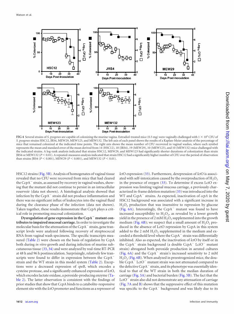
HSC12 strains (Fig. 5B). Analysis of homogenates of vaginal tissuerevealed that no CFU were recovered from mice that had clearedthe CcpA� strain, as assessed by recovery in vaginal washes, show-ing that the mutant did not continue to persist in an intracellularreservoir (data not shown). A histological analysis showed thatinfection by the CcpA� strain did not produce inflammation andthere was no significant influx of leukocytes into the vaginal fluidduring the clearance phase of the infection (data not shown).Taken together, these results demonstrate that CcpA plays a crit-ical role in promoting mucosal colonization.
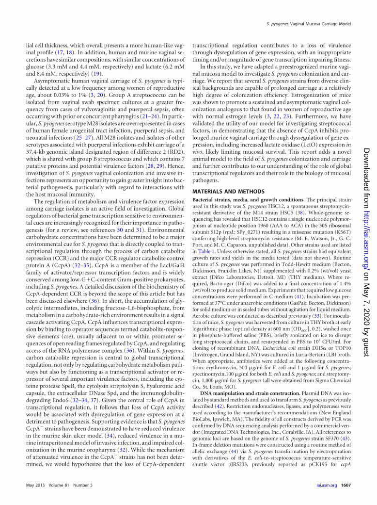
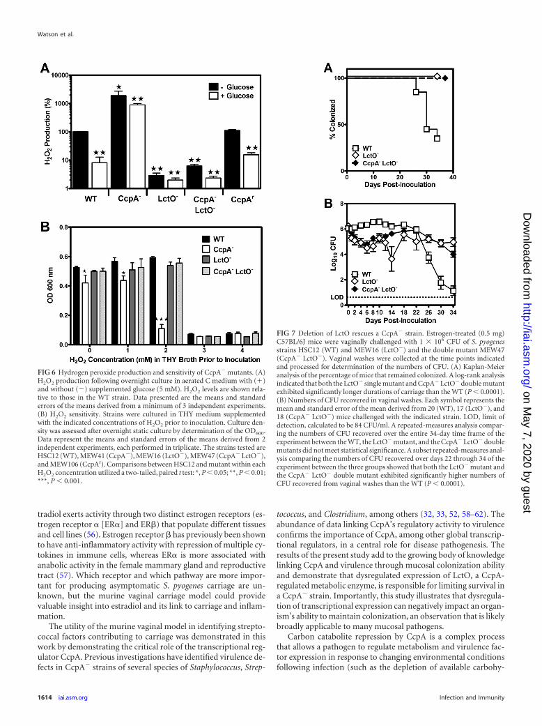
Dysregulation of gene expression in the CcpA� mutant con-tributes to impaired mucosal carriage. In order to investigate themolecular basis for the attenuation of the CcpA� strain, gene tran-script levels were analyzed following recovery of streptococcalRNA from vaginal wash specimens. The specific transcripts mea-sured (Table 2) were chosen on the basis of regulation by CcpAboth during in vitro growth and during infection of murine sub-cutaneous tissue (33, 34) and were analyzed by real-time RT-PCRat 48 h and 96 h postinoculation. Surprisingly, relatively few tran-scripts were found to differ in expression between the CcpA�
strain and the WT strain in this model system (Table 2). Excep-tions were a decreased expression of speB, which encodes acysteine protease, and a significantly enhanced expression of lctO,which encodes lactate oxidase, a peroxide-producing enzyme (Ta-ble 2). The latter observation is consistent with the findings ofprior studies that show that CcpA binds to a catabolite-responsiveelement site with the lctO promoter and functions as a repressor of
lctO expression (33). Furthermore, derepression of lctO is associ-ated with self-intoxication caused by the overproduction of H2O2
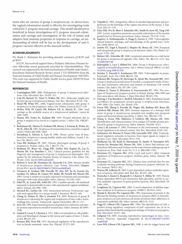
in the presence of oxygen (33). To determine if excess LctO ex-pression was limiting vaginal mucosa carriage, a previously char-acterized in-frame deletion mutation (33) was introduced into theWT and CcpA� strains. As expected, inactivation of ccpA in theHSC12 background was associated with a significant increase inH2O2 production that was insensitive to repression by glucose(Fig. 6A). Interestingly, the CcpA� mutant was found to haveincreased susceptibility to H2O2, as revealed by a lower growthyield in the presence of 2 mM H2O2 supplemented into the growthmedium (Fig. 6B); we suspect that a small amount of H2O2 pro-duced in the absence of LctO repression by CcpA in this systemadded to the 2 mM H2O2 supplemented in the medium and ex-ceeded a threshold level where the CcpA� strain was differentiallyinhibited. Also as expected, the inactivation of lctO by itself or inthe CcpA� strain background (a double CcpA� LctO� mutantstrain) abrogated both peroxide production in aerated cultures(Fig. 6A) and the CcpA� strain’s increased sensitivity to 2 mMH2O2 (Fig. 6B). When analyzed in preestrogenized mice, the dou-ble CcpA� LctO� mutant strain was not attenuated compared tothe defective CcpA� strain, and its phenotype was essentially iden-tical to that of the WT strain in both the median duration ofcarriage (Fig. 5A) and bacterial burden (Fig. 5B). The fact that theLctO� strain also did not demonstrate any attenuation of carriage(Fig. 7A and B) shows that the suppressive effect of this mutationwas specific to the CcpA� background and was likely due to its
FIG 4 Several strains of S. pyogenes are capable of colonizing the murine vagina. Estradiol-treated mice (0.5 mg) were vaginally challenged with 1 106 CFU ofS. pyogenes strains HSC12, JRS4, MEW29, MEW123, and MEW132. The left axis of each panel shows the results of a Kaplan-Meier analysis of the percentage ofmice that remained colonized at the indicated time points. The right axis shows the mean number of CFU recovered in vaginal washes, where each symbolrepresents the mean and standard error of the mean derived from 14 (HSC12), 18 (JRS4), 19 (MEW29), 10 (MEW123), and 10 (MEW132) mice challenged withthe indicated strains. A log-rank analysis indicated that strains HSC12, MEW29, and MEW123 had significantly shorter durations of colonization than strainJRS4 or MEW132 (P � 0.01). A repeated-measures analysis indicated that strain HSC12 had a significantly higher number of CFU over the period of observationthan strains JRS4 (P � 0.001), MEW29 (P � 0.001), and MEW132 (P � 0.01).
Watson et al.
1612 iai.asm.org Infection and Immunity
on May 7, 2020 by guest
http://iai.asm.org/
Dow
nloaded from
ability to alleviate overexpression of LctO. Thus, these data dem-onstrate that CcpA makes a critical contribution to mucosal car-riage by its ability to downregulate a gene whose overexpressiondecreases fitness in the mucosal environment.
DISCUSSION
Investigation of factors critical to mucosal colonization and car-riage in S. pyogenes has been hampered by the fact that it is ahuman-restricted pathogen with no known animal or environ-mental reservoir. Previous animal models for S. pyogenes havesuffered from the fact that the number of strains which can func-tion in those models and/or reproduce the disease phenotype waslimited (10, 51, 53, 54). This work presents a novel animal modelof S. pyogenes mucosal colonization and carriage which will po-tentially advance the field of streptococcal host-pathogen interac-tions at the mucosal surface. Importantly, the preestrogenizedmurine vaginal colonization model is capable of supporting atleast five diverse S. pyogenes strains, including the M14 HSC12,M6 JRS4, M1 SF370, and M49 NZ131 strains and an M28 isolate,MEW123, and this will facilitate investigation into factors distin-
guishing carriage ability between different strains. There are likelymultiple differences between these strains in terms of expressionof various adhesins, exotoxins, and other potential virulence fac-tors that could account for their variable phenotypes in vaginalmucosa carriage. As just one example, strains HSC12 and SF370display oxygen-induced expression of the fibronectin-binding ad-hesin protein F (prtF) and do not express this protein under an-aerobic conditions; in contrast, JRS4 has a mutation in the regu-latory gene rofA which permits constitutive expression of proteinF, even anaerobically (39).
Another advantage of using a murine vaginal model to study S.pyogenes colonization was the relative ease of access to the tissuesto monitor the mucosal immune response. In this model, an in-flux of vaginal leukocytes was found to be a marker of inflamma-tion, with higher leukocyte percentages being noted in vaginalwashes from BALB/c mice than those from C57BL/6J mice. Aprevious study (55) also reported a significantly higher vaginalfluid leukocyte response from BALB/c mice than C57BL/6J mice,when both were similarly colonized with N. gonorrhoeae, support-ing a difference between the two mouse strains in the innate im-mune response that may be common to several mucosal patho-gens. Therefore, the vaginal carriage model may be useful ininvestigating other bacterial factors that interact with the innateresponse to mucosal pathogens, and using BALB/c mice as thehost in this model may help highlight factors affecting inflamma-tion. Furthermore, this model may help explain the inverse rela-tionship between estrogen availability and symptomatic S. pyo-genes vaginal disease in humans. For unclear reasons,vulvovaginitis tends to be more common in prepubertal girls,postmenopausal women, and women with other hypoestrogenconditions (20, 22–24). In our model, estradiol supplementationwas associated with asymptomatic carriage, which seems to mimicthe situation in women of reproductive age vaginally colonizedwith S. pyogenes. Estradiol supplementation is known to promotethe estrus phase of the murine estrous cycle, a time associated withvaginal epithelium proliferation and thickening, production ofglycogen, and a minimal presence of inflammatory cells (15). Es-
TABLE 2 Only a subset of CcpA-regulated genes is dysregulated in theCcpA� mutant during vaginal carriage
Transcriptb
Relative transcript expressiona
48 h P value 96 h P value
speB 0.07 0.06 0.010 0.48 0.35 0.116lctO 5.76 2.18 0.001 4.58 1.09 0.053hasA 0.94 0.10 0.545 0.40 0.05 0.203sagA 0.62 0.12 0.140 0.72 0.24 0.303malM 1.04 0.27 0.873 1.13 0.53 0.772cfa 1.59 0.72 0.122 0.43 0.30 0.077ackA 0.85 0.22 0.200 0.77 0.70 0.463arcA 1.82 1.70 0.506 2.51 3.04 0.267a Relative transcript expression is expressed as expression for the CcpA�
strain/expression for HSC12, with the 2���CT value normalized using the transcriptlevel of recA (46). Values represent means standard errors of the means of threeindependent experiments, each analyzed in duplicate. Samples are from 48 h and 96 hpostinoculation. For each experimental group (WT and CcpA�) vaginal washes fromsix mice were included in the analysis.b The recA (SPy_2116), speB (SPy_2039), lctO (SPy_0414), hasA (SPy_2200), sagA(SPy_0738), malM (SPy_1292), cfa (SPy_1273), ackA (SPy_0109), and arcA (SPy_1543)transcripts were analyzed on the basis of previous work showing that these genes areCcpA regulated (32, 33, 34).
FIG 5 Deletion of CcpA limits the duration of mucosal carriage. Estrogen-treated (0.5 mg) C57BL/6J mice were vaginally challenged with 1 106 CFU ofS. pyogenes strains HSC12 (WT) and MEW41 (CcpA�) and revertant strainMEW106 (CcpAr). Vaginal washes were collected at the time points indicatedand processed for determination of the number of CFU. (A) Kaplan-Meieranalysis of the percentage of mice that remained colonized. Log-rank analysisindicated that the CcpA� mutant was significantly attenuated compared to theWT and CcpAr strains (P � 0.001). (B) Numbers of CFU recovered in vaginalwashes. Each symbol represents the mean and standard error of the meanderived from 20 (WT), 19 (CcpA�), and 5 (CcpAr) mice challenged with theindicated strains. LOD, limit of detection, calculated to be 84 CFU/ml. A re-peated-measures analysis indicated that the CcpA� mutant was significantlyattenuated compared to the WT and CcpAr strains (P � 0.001).
S. pyogenes Vaginal Mucosa Carriage Model
May 2013 Volume 81 Number 5 iai.asm.org 1613
on May 7, 2020 by guest
http://iai.asm.org/
Dow
nloaded from
tradiol exerts activity through two distinct estrogen receptors (es-trogen receptor � [ER�] and ER�) that populate different tissuesand cell lines (56). Estrogen receptor � has previously been shownto have anti-inflammatory activity with repression of multiple cy-tokines in immune cells, whereas ER� is more associated withanabolic activity in the female mammary gland and reproductivetract (57). Which receptor and which pathway are more impor-tant for producing asymptomatic S. pyogenes carriage are un-known, but the murine vaginal carriage model could providevaluable insight into estradiol and its link to carriage and inflam-mation.
The utility of the murine vaginal model in identifying strepto-coccal factors contributing to carriage was demonstrated in thiswork by demonstrating the critical role of the transcriptional reg-ulator CcpA. Previous investigations have identified virulence de-fects in CcpA� strains of several species of Staphylococcus, Strep-
tococcus, and Clostridium, among others (32, 33, 52, 58–62). Theabundance of data linking CcpA’s regulatory activity to virulenceconfirms the importance of CcpA, among other global transcrip-tional regulators, in a central role for disease pathogenesis. Theresults of the present study add to the growing body of knowledgelinking CcpA and virulence through mucosal colonization abilityand demonstrate that dysregulated expression of LctO, a CcpA-regulated metabolic enzyme, is responsible for limiting survival ina CcpA� strain. Importantly, this study illustrates that dysregula-tion of transcriptional expression can negatively impact an organ-ism’s ability to maintain colonization, an observation that is likelybroadly applicable to many mucosal pathogens.
Carbon catabolite repression by CcpA is a complex processthat allows a pathogen to regulate metabolism and virulence fac-tor expression in response to changing environmental conditionsfollowing infection (such as the depletion of available carbohy-
FIG 6 Hydrogen peroxide production and sensitivity of CcpA� mutants. (A)H2O2 production following overnight culture in aerated C medium with (�)and without (�) supplemented glucose (5 mM). H2O2 levels are shown rela-tive to those in the WT strain. Data presented are the means and standarderrors of the means derived from a minimum of 3 independent experiments.(B) H2O2 sensitivity. Strains were cultured in THY medium supplementedwith the indicated concentrations of H2O2 prior to inoculation. Culture den-sity was assessed after overnight static culture by determination of the OD600.Data represent the means and standard errors of the means derived from 2independent experiments, each performed in triplicate. The strains tested areHSC12 (WT), MEW41 (CcpA�), MEW16 (LctO�), MEW47 (CcpA� LctO�),and MEW106 (CcpAr). Comparisons between HSC12 and mutant within eachH2O2 concentration utilized a two-tailed, paired t test: *, P � 0.05; **, P � 0.01;***, P � 0.001.
FIG 7 Deletion of LctO rescues a CcpA� strain. Estrogen-treated (0.5 mg)C57BL/6J mice were vaginally challenged with 1 106 CFU of S. pyogenesstrains HSC12 (WT) and MEW16 (LctO�) and the double mutant MEW47(CcpA� LctO�). Vaginal washes were collected at the time points indicatedand processed for determination of the numbers of CFU. (A) Kaplan-Meieranalysis of the percentage of mice that remained colonized. A log-rank analysisindicated that both the LctO� single mutant and CcpA� LctO� double mutantexhibited significantly longer durations of carriage than the WT (P � 0.0001).(B) Numbers of CFU recovered in vaginal washes. Each symbol represents themean and standard error of the mean derived from 20 (WT), 17 (LctO�), and18 (CcpA� LctO�) mice challenged with the indicated strain. LOD, limit ofdetection, calculated to be 84 CFU/ml. A repeated-measures analysis compar-ing the numbers of CFU recovered over the entire 34-day time frame of theexperiment between the WT, the LctO� mutant, and the CcpA� LctO� doublemutants did not meet statistical significance. A subset repeated-measures anal-ysis comparing the numbers of CFU recovered over days 22 through 34 of theexperiment between the three groups showed that both the LctO� mutant andthe CcpA� LctO� double mutant exhibited significantly higher numbers ofCFU recovered from vaginal washes than the WT (P � 0.0001).
Watson et al.
1614 iai.asm.org Infection and Immunity
on May 7, 2020 by guest
http://iai.asm.org/
Dow
nloaded from

drates) or upon encountering a new environment or stage in theinfectious process. In support of this, S. pyogenes CcpA� strainsrecovered from the relatively confined environment of the murineskin ulcer model showed that dysregulation was not a static all-onor all-off process, but even among genes that were strongly regu-lated by CcpA in vitro, there were rather dynamic fluctuations inthe magnitude of differences between gene expression patternsbetween wild-type and CcpA� strains (34). Some of these fluctu-ations may have been due to changing carbohydrate availability inthe ulcer tissue bed; others may be due to the influence of addi-tional factors, including LacD.1, which function as coregulatoryproteins along with CcpA for some gene transcripts, including thecysteine protease SpeB (33, 34).
The present study identifies an increase in the transcription ofLctO from vaginal washes colonized by CcpA� strains; however,beyond LctO there were relatively few differences in transcriptexpression identified from vaginal washes of mice colonized withthe CcpA� strain and mice colonized with the HSC12 parentstrain at the time points examined (Table 2). This could be due toseveral reasons. S. pyogenes organisms colonizing the murine vag-inal epithelium could be relatively metabolically inactive with alow level of transcriptional activity that could make finding com-parative differences difficult. Alternatively, the little to no differ-ence in transcript expression between CcpA� strains and theHSC12 parent strain could suggest that available carbohydratelevels in the murine vaginal mucosa are not sufficient to signalCcpA-mediated regulation to a significant degree to promote dif-ferences. It has previously been determined that estradiol-treatedmurine and human vaginal secretions both have glucose concen-trations of 3 to 4 mM, which calculates to about 0.072% (wt/vol)(19). Prior experiments by our group showed a significant reduc-tion in lctO transcription in vitro with �0.1% glucose in C me-dium (33); thus, it is possible that in the murine vaginal model theglucose concentrations were too low to show significant differ-ences for many genes normally regulated by CcpA. S. pyogenesHSC12 can utilize a variety of carbohydrate sources, most ofwhich are ultimately converted to glycolytic intermediates whichactivate CcpA-mediated repression of LctO. Vaginal secretionscontain monosaccharides, including glucose, mannose, and glu-cosamine, in addition to oligosaccharides, including maltose,maltotriose, and maltotetraose (63, 64). Carbon catabolite repres-sion in strain HSC12 can be induced by glucose, maltose, malto-triose, pullulan, galactose, and mannose (data not shown); strainHSC12 cannot directly utilize glycogen or lactose, although otherbacterial species present in the vaginal flora may degrade thesesugars into forms that S. pyogenes strain HSC12 can utilize forcarbon catabolite repression. Another possibility is that in the mu-rine vaginal environment, there are perhaps additional coregula-tory proteins (LacD.1, among other possibilities) that could beacting to help coordinate gene expression in the absence of inputfrom CcpA. In our experiments, however, even in a CcpA� strain,dysregulation of gene expression on a global scale did not suffi-ciently explain the attenuation in carriage more so than dysregu-lation of a single gene, as observed when inactivation of LctO inthe CcpA� background prolonged carriage of the double mutantstrain.
When in the presence of glycolytic intermediates, CcpA bindsto the lctO promoter within a catabolite-responsive element se-quence located upstream of the lctO ATG translation start site,blocking lctO transcription. In the CcpA� strain, LctO transcrip-
tion is largely unchecked, producing abundant amounts of perox-ide that accumulate to lethal concentrations (33). Inactivation ofLctO in the CcpA� strain prevented hydrogen peroxide produc-tion, and this resulted in a prolonged in vivo duration of carriageof the CcpA� LctO� strain. In S. pyogenes, lactate oxidase is theprincipal enzyme producing significant levels of hydrogen perox-ide; the enzyme NADH oxidase also contributes a small and rela-tively insignificant amount of detectable peroxide (65); this factmay explain why H2O2 production in the LctO� and CcpA�
LctO� strains was low but not undetectable (Fig. 6A). Amongboth clinical isolates and lab-generated mutants, inactivation ofLctO activity and reduced hydrogen peroxide production havepreviously been demonstrated to prolong stationary-phase sur-vival under aerobic conditions (33, 65, 66). It was surprising tofind such a strong phenotype related to peroxide production in themurine vaginal compartment, as traditional thought is that thevaginal environment is of relatively low oxygen tension (67).However, in humans, vaginal lactobacillus species producing hy-drogen peroxide are thought to be associated with vaginal healthand a reduced risk of acquiring disease, such as bacterial vaginitis(68–70). By this, the vaginal compartment presumably has suffi-cient available oxygen for lactobacillus and, likewise, S. pyogenes tohave the capacity to produce peroxide. Alternatively, there may bedifferences in the oxygen tension present in the murine vaginalenvironment and that present in the human vaginal environment.Oxygen may have been further introduced into the murine vaginalcompartment in our model via repeat vaginal specimen collectionfor culture; mechanical insertion of contraceptive diaphragms ortampons has previously been shown to transiently increase thevaginal oxygen content in humans (67, 71).
The present study is not the first to find that CcpA� strains areattenuated in mucosal colonization and carriage. CcpA� strains ofStreptococcus pneumoniae were previously demonstrated to be at-tenuated for carriage in the murine nasopharynx (62), just as S.pyogenes was attenuated for carriage in the murine oropharynx(32). Neither of these prior studies identified specific defects thatcould complement the CcpA� strain phenotype like the double�ccpA �lctO mutations were capable of doing in this study. In S.pneumoniae, H2O2 production under aerobic conditions is mostlydue to the activity of pyruvate oxidase (SpxB); under glucose lim-itation, the pneumococcal LctO also shows peroxide-generatingactivity. In S. pneumoniae, LctO was also found to be upregulatedin CcpA� strains; however, SpxB was not influenced by ccpA de-letion, as determined by microarray analysis (62, 72). Pneumo-coccal stains producing peroxide have competitive advantageswithin mixed bacterial populations, presumably by inhibiting thegrowth of peroxide-susceptible species (73, 74). However, S.pneumoniae SpxB mutants were found to be defective in animalmodels of nasopharyngeal colonization, pneumonia, and sepsis,although factors in addition to peroxide generation alone mayhave contributed to this phenotype (74–77). In our model of S.pyogenes vaginal colonization, we noted no significant defect incarriage of strains with a mutation of lctO. Altogether, these resultssuggest that regulated hydrogen peroxide generation by severalstreptococcal species is critical for mucosal colonization and vir-ulence.
Asymptomatic mucosal carriage with S. pyogenes is a conceptthat remains poorly understood, in terms of both the basic scienceunderlying the microbiology and immunology occurring at themucosal surface and the clinical science on how to manage pa-
S. pyogenes Vaginal Mucosa Carriage Model
May 2013 Volume 81 Number 5 iai.asm.org 1615
on May 7, 2020 by guest
http://iai.asm.org/
Dow
nloaded from
tients who are carriers of group A streptococcus. As shown here,the vaginal colonization model is effective for investigating traitscritical for S. pyogenes mucosal carriage. This model should provebeneficial in future investigations of S. pyogenes mucosal coloni-zation and carriage and investigation of the role of innate andadaptive host immune properties in response to carriage, an un-derstanding of which will be key to the development of anti-S.pyogenes vaccines effective at the mucosal surface.
ACKNOWLEDGMENTS
We thank C. Kietzman for providing plasmid constructs pCK195 andpCK037.
M.E.W. has received support from a Pediatric Infectious Diseases So-ciety fellowship award generously provided by GlaxoSmithKline. Thisresearch was supported by the National Institutes of Health under Ruth L.Kirschstein National Research Service award 2 T32 HD043010 from theNational Institute of Child Health and Human Development (NICHD).This work was supported by Public Health Service grant AI070759 fromthe National Institutes of Health.
REFERENCES1. Cunningham MW. 2000. Pathogenesis of group A streptococcal infec-
tions. Clin. Microbiol. Rev. 13:470 –511.2. Cole JN, Barnett TC, Nizet V, Walker MJ. 2011. Molecular insight into
invasive group A streptococcal disease. Nat. Rev. Microbiol. 9:724 –736.3. Mead PB, Winn WC. 2000. Vaginal-rectal colonization with group A
streptococci in late pregnancy. Infect. Dis. Obstet. Gynecol. 8:217–219.4. McKee WM, Di Caprio JM, Roberts CE, Jr, Sherris JC. 1966. Anal
carriage as the probable source of a streptococcal epidemic. Lancet ii:1007–1009.
5. Stamm WE, Feeley JC, Facklam RR. 1978. Wound infections due togroup A streptococcus traced to a vaginal carrier. J. Infect. Dis. 138:287–292.
6. Berkelman RL, Martin D, Graham DR, Mowry J, Freisem R, Weber JA,Ho JL, Allen JR. 1982. Streptococcal wound infections caused by a vaginalcarrier. JAMA 247:2680 –2682.
7. Stromberg A, Schwan A, Cars O. 1988. Throat carrier rates of beta-hemolytic streptococci among healthy adults and children. Scand. J. In-fect. Dis. 20:411– 417.
8. Tanz RR, Shulman ST. 2007. Chronic pharyngeal carriage of group Astreptococci. Pediatr. Infect. Dis. J. 26:175–176.
9. Shulman ST, Bisno AL, Clegg HW, Gerber MA, Kaplan EL, Lee G,Martin JM, Van Beneden C. 2012. Clinical practice guideline for thediagnosis and management of group A streptococcal pharyngitis: 2012update by the Infectious Diseases Society of America. Clin. Infect. Dis.55:e66 – e102. doi:10.1093/cid/cis629.
10. Roberts S, Scott JR, Husmann LK, Zurawski CA. 2006. Murine modelsof Streptococcus pyogenes infection. Curr. Protoc. Microbiol. Chapter9:Unit 9D.5. doi:10.1002/9780471729259.mc09d05s02.
11. Virtaneva K, Graham MR, Porcella SF, Hoe NP, Su H, Graviss EA,Gardner TJ, Allison JE, Lemon WJ, Bailey JR, Parnell MJ, Musser JM.2003. Group A streptococcus gene expression in humans and cynomolgusmacaques with acute pharyngitis. Infect. Immun. 71:2199 –2207.
12. Fidel PL, Jr, Lynch ME, Sobel JD. 1993. Candida-specific cell-mediatedimmunity is demonstrable in mice with experimental vaginal candidiasis.Infect. Immun. 61:1990 –1995.
13. Meysick KC, Garber GE. 1992. Interactions between Trichomonas vagi-nalis and vaginal flora in a mouse model. J. Parasitol. 78:157–160.
14. Cheng Q, Nelson D, Zhu S, Fischetti VA. 2005. Removal of group Bstreptococci colonizing the vagina and oropharynx of mice with a bacte-riophage lytic enzyme. Antimicrob. Agents Chemother. 49:111–117.
15. Jerse AE. 1999. Experimental gonococcal genital tract infection and opac-ity protein expression in estradiol-treated mice. Infect. Immun. 67:5699 –5708.
16. Galand P, Leroy F, Chretien J. 1971. Effect of oestradiol on cell prolifer-ation and histological changes in the uterus and vagina of mice. J. Endo-crinol. 49:243–252.
17. Brenner RM, West NB. 1975. Hormonal regulation of the reproductivetract in female mammals. Annu. Rev. Physiol. 37:273–302.
18. Tripathi G. 1984. Antagonistic effects of estradiol dipropionate and pro-gesterone on the histology of the vagina and uterus of the mouse. J. Exp.Zool. 232:151–155.
19. Exley RM, Wu H, Shaw J, Schneider MC, Smith H, Jerse AE, Tang CM.2007. Lactate acquisition promotes successful colonization of the murinegenital tract by Neisseria gonorrhoeae. Infect. Immun. 75:1318 –1324.
20. Jaquiery A, Stylianopoulos A, Hogg G, Grover S. 1999. Vulvovaginitis:clinical features, aetiology, and microbiology of the genital tract. Arch.Dis. Child. 81:64 – 67.
21. Anteby EY, Yagel S, Hanoch J, Shapiro M, Moses AE. 1999. Puerperaland intrapartum group A streptococcal infection. Infect. Dis. Obstet. Gy-necol. 7:276 –282.
22. Meltzer MC, Schwebke JR. 2008. Lactational amenorrhea as a risk factorfor group A streptococcal vaginitis. Clin. Infect. Dis. 46:e112– e115. doi:10.1086/587748.
23. Rahangdale L, Lacy J, Hillard PA. 2008. Group A Streptococcus vulvo-vaginitis in breastfeeding women. Am. J. Obstet. Gynecol. 199:e4 – e5. doi:10.1016/j.ajog.2008.02.045.
24. Stricker T, Navratil F, Sennhauser FH. 2003. Vulvovaginitis in prepu-bertal girls. Arch. Dis. Child. 88:324 –326.
25. Eriksson BK, Norgren M, McGregor K, Spratt BG, Normark BH. 2003.Group A streptococcal infections in Sweden: a comparative study of inva-sive and noninvasive infections and analysis of dominant T28 emm28isolates. Clin. Infect. Dis. 37:1189 –1193.
26. Colman G, Tanna A, Efstratiou A, Gaworzewska ET. 1993. The sero-types of Streptococcus pyogenes present in Britain during 1980-1990 andtheir association with disease. J. Med. Microbiol. 39:165–178.
27. Chuang I, Van Beneden C, Beall B, Schuchat A. 2002. Population-basedsurveillance for postpartum invasive group A streptococcus infections,1995-2000. Clin. Infect. Dis. 35:665– 670.
28. Green NM, Zhang S, Porcella SF, Nagiec MJ, Barbian KD, Beres SB,LeFebvre RB, Musser JM. 2005. Genome sequence of a serotype M28strain of group A streptococcus: potential new insights into puerperalsepsis and bacterial disease specificity. J. Infect. Dis. 192:760 –770.
29. Zhang S, Green NM, Sitkiewicz I, Lefebvre RB, Musser JM. 2006.Identification and characterization of an antigen I/II family protein pro-duced by group A streptococcus. Infect. Immun. 74:4200 – 4213.
30. Cases I, de Lorenzo V. 2005. Promoters in the environment: transcrip-tional regulation in its natural context. Nat. Rev. Microbiol. 3:105–118.
31. Seshasayee AS, Bertone P, Fraser GM, Luscombe NM. 2006. Transcrip-tional regulatory networks in bacteria: from input signals to output re-sponses. Curr. Opin. Microbiol. 9:511–519.
32. Shelburne SA, III, Keith D, Horstmann N, Sumby P, Davenport MT,Graviss EA, Brennan RG, Musser JM. 2008. A direct link between car-bohydrate utilization and virulence in the major human pathogen group AStreptococcus. Proc. Natl. Acad. Sci. U. S. A. 105:1698 –1703.
33. Kietzman CC, Caparon MG. 2010. CcpA and LacD.1 affect temporalregulation of Streptococcus pyogenes virulence genes. Infect. Immun. 78:241–252.
34. Kietzman CC, Caparon MG. 2011. Distinct time-resolved roles for twocatabolite-sensing pathways during Streptococcus pyogenes infection. In-fect. Immun. 79:812– 821.
35. Warner JB, Lolkema JS. 2003. CcpA-dependent carbon catabolite repres-sion in bacteria. Microbiol. Mol. Biol. Rev. 67:475– 490.
36. Deutscher J, Kuster E, Bergstedt U, Charrier V, Hillen W. 1995. Proteinkinase-dependent HPr/CcpA interaction links glycolytic activity to car-bon catabolite repression in gram-positive bacteria. Mol. Microbiol. 15:1049 –1053.
37. Loughman JA, Caparon MG. 2006. A novel adaptation of aldolase regu-lates virulence in Streptococcus pyogenes. EMBO J. 25:5414 –5422.
38. Hanski E, Horwitz PA, Caparon MG. 1992. Expression of protein F, thefibronectin-binding protein of Streptococcus pyogenes JRS4, in heterolo-gous streptococcal and enterococcal strains promotes their adherence torespiratory epithelial cells. Infect. Immun. 60:5119 –5125.
39. Fogg GC, Gibson CM, Caparon MG. 1994. The identification of rofA, apositive-acting regulatory component of prtF expression: use of an mgamma delta-based shuttle mutagenesis strategy in Streptococcus pyogenes.Mol. Microbiol. 11:671– 684.
40. Caligioni CS. 2009. Assessing reproductive status/stages in mice. Curr.Protoc. Neurosci. Appendix 4:Appendix 4I. doi:10.1002/0471142301.nsa04is48.
41. Lyon WR, Gibson CM, Caparon MG. 1998. A role for trigger factor and
Watson et al.
1616 iai.asm.org Infection and Immunity
on May 7, 2020 by guest
http://iai.asm.org/
Dow
nloaded from

an rgg-like regulator in the transcription, secretion and processing of thecysteine proteinase of Streptococcus pyogenes. EMBO J. 17:6263– 6275.
42. Caparon MG, Stephens DS, Olsen A, Scott JR. 1991. Role of M proteinin adherence of group A streptococci. Infect. Immun. 59:1811–1817.
43. Ferretti JJ, McShan WM, Ajdic D, Savic DJ, Savic G, Lyon K, PrimeauxC, Sezate S, Suvorov AN, Kenton S, Lai HS, Lin SP, Qian Y, Jia HG,Najar FZ, Ren Q, Zhu H, Song L, White J, Yuan X, Clifton SW, Roe BA,McLaughlin R. 2001. Complete genome sequence of an M1 strain ofStreptococcus pyogenes. Proc. Natl. Acad. Sci. U. S. A. 98:4658 – 4663.
44. Perez-Casal J, Price JA, Maguin E, Scott JR. 1993. An M protein with asingle C repeat prevents phagocytosis of Streptococcus pyogenes: use of atemperature-sensitive shuttle vector to deliver homologous sequences tothe chromosome of S. pyogenes. Mol. Microbiol. 8:809 – 819.
45. Ryan PL, Baum DL, Lenhart JA, Ohleth KM, Bagnell CA. 2001. Expres-sion of uterine and cervical epithelial cadherin during relaxin-inducedgrowth in pigs. Reproduction 122:929 –937.
46. Schmittgen TD, Livak KJ. 2008. Analyzing real-time PCR data by thecomparative C(T) method. Nat. Protoc. 3:1101–1108.
47. Brenot A, King KY, Caparon MG. 2005. The PerR regulon in peroxideresistance and virulence of Streptococcus pyogenes. Mol. Microbiol. 55:221–234.
48. Gibson CM, Mallett TC, Claiborne A, Caparon MG. 2000. Contributionof NADH oxidase to aerobic metabolism of Streptococcus pyogenes. J. Bac-teriol. 182:448 – 455.
49. Sheen TR, Jimenez A, Wang NY, Banerjee A, van Sorge NM, Doran KS.2011. Serine-rich repeat proteins and pili promote Streptococcus agalactiaecolonization of the vaginal tract. J. Bacteriol. 193:6834 – 6842.
50. Scott JR, Guenthner PC, Malone LM, Fischetti VA. 1986. Conversion ofan M-group A streptococcus to M� by transfer of a plasmid containing anM6 gene. J. Exp. Med. 164:1641–1651.
51. Skinner JM, Caro-Aguilar IC, Payne AM, Indrawati L, Fontenot J,Heinrichs JH. 2011. Comparison of rhesus and cynomolgus macaques ina Streptococcus pyogenes infection model for vaccine evaluation. Microb.Pathog. 50:39 – 47.
52. Seidl K, Goerke C, Wolz C, Mack D, Berger-Bachi B, Bischoff M. 2008.Staphylococcus aureus CcpA affects biofilm formation. Infect. Immun. 76:2044 –2050.
53. Neely MN, Pfeifer JD, Caparon M. 2002. Streptococcus-zebrafish modelof bacterial pathogenesis. Infect. Immun. 70:3904 –3914.
54. Medina E. 2010. Murine model of cutaneous infection with Streptococcuspyogenes. Methods Mol. Biol. 602:395– 403.
55. Packiam M, Veit SJ, Anderson DJ, Ingalls RR, Jerse AE. 2010. Mousestrain-dependent differences in susceptibility to Neisseria gonorrhoeae in-fection and induction of innate immune responses. Infect. Immun. 78:433– 440.
56. Straub RH. 2007. The complex role of estrogens in inflammation. Endocr.Rev. 28:521–574.
57. Cvoro A, Tatomer D, Tee MK, Zogovic T, Harris HA, Leitman DC.2008. Selective estrogen receptor-beta agonists repress transcription ofproinflammatory genes. J. Immunol. 180:630 – 636.
58. Seidl K, Stucki M, Ruegg M, Goerke C, Wolz C, Harris L, Berger-BachiB, Bischoff M. 2006. Staphylococcus aureus CcpA affects virulence deter-minant production and antibiotic resistance. Antimicrob. Agents Che-mother. 50:1183–1194.
59. Wen ZT, Burne RA. 2002. Functional genomics approach to identifyinggenes required for biofilm development by Streptococcus mutans. Appl.Environ. Microbiol. 68:1196 –1203.
60. Antunes A, Martin-Verstraete I, Dupuy B. 2011. CcpA-mediated repres-sion of Clostridium difficile toxin gene expression. Mol. Microbiol. 79:882–899.
61. Zheng L, Chen Z, Itzek A, Herzberg MC, Kreth J. 2012. CcpA regulatesbiofilm formation and competence in Streptococcus gordonii. Mol. OralMicrobiol. 27:83–94.
62. Iyer R, Baliga NS, Camilli A. 2005. Catabolite control protein A (CcpA)contributes to virulence and regulation of sugar metabolism in Streptococ-cus pneumoniae. J. Bacteriol. 187:8340 – 8349.
63. Rajan N, Cao Q, Anderson BE, Pruden DL, Sensibar J, Duncan JL,Schaeffer AJ. 1999. Roles of glycoproteins and oligosaccharides found inhuman vaginal fluid in bacterial adherence. Infect. Immun. 67:5027–5032.
64. Sumawong V, Gregoire AT, Johnson WD, Rakoff AE. 1962. Identifica-tion of carbohydrates in the vaginal fluid of normal females. Fertil. Steril.13:270 –280.
65. Seki M, Iida K, Saito M, Nakayama H, Yoshida S. 2004. Hydrogenperoxide production in Streptococcus pyogenes: involvement of lactate ox-idase and coupling with aerobic utilization of lactate. J. Bacteriol. 186:2046 –2051.
66. Saito M, Ohga S, Endoh M, Nakayama H, Mizunoe Y, Hara T, YoshidaS. 2001. H(2)O(2)-nonproducing Streptococcus pyogenes strains: survivalin stationary phase and virulence in chronic granulomatous disease. Mi-crobiology 147:2469 –2477.
67. Wagner G, Levin RJ, Bohr L. 1988. Diaphragm insertion increases hu-man vaginal oxygen tension. Am. J. Obstet. Gynecol. 158:1040 –1043.
68. Al-Mushrif S, Jones BM. 1998. A study of the prevalence of hydrogenperoxide generating lactobacilli in bacterial vaginosis: the determinationof H2O2 concentrations generated, in vitro, by isolated strains and thelevels found in vaginal secretions of women with and without infection. J.Obstet. Gynaecol. 18:63– 67.
69. Wilks M, Wiggins R, Whiley A, Hennessy E, Warwick S, Porter H,Corfield A, Millar M. 2004. Identification and H(2)O(2) production ofvaginal lactobacilli from pregnant women at high risk of preterm birth andrelation with outcome. J. Clin. Microbiol. 42:713–717.
70. Klebanoff SJ, Hillier SL, Eschenbach DA, Waltersdorph AM. 1991.Control of the microbial flora of the vagina by H2O2-generating lactoba-cilli. J. Infect. Dis. 164:94 –100.
71. Wagner G, Bohr L, Wagner P, Petersen LN. 1984. Tampon-inducedchanges in vaginal oxygen and carbon dioxide tensions. Am. J. Obstet.Gynecol. 148:147–150.
72. Carvalho SM, Kloosterman TG, Kuipers OP, Neves AR. 2011. CcpAensures optimal metabolic fitness of Streptococcus pneumoniae. PLoS One6:e26707. doi:10.1371/journal.pone.0026707.
73. Pericone CD, Overweg K, Hermans PW, Weiser JN. 2000. Inhibitoryand bactericidal effects of hydrogen peroxide production by Streptococcuspneumoniae on other inhabitants of the upper respiratory tract. Infect.Immun. 68:3990 –3997.
74. Regev-Yochay G, Trzcinski K, Thompson CM, Lipsitch M, Malley R.2007. SpxB is a suicide gene of Streptococcus pneumoniae and confers aselective advantage in an in vivo competitive colonization model. J. Bac-teriol. 189:6532– 6539.
75. Spellerberg B, Cundell DR, Sandros J, Pearce BJ, Idanpaan-Heikkila I,Rosenow C, Masure HR. 1996. Pyruvate oxidase, as a determinant ofvirulence in Streptococcus pneumoniae. Mol. Microbiol. 19:803– 813.
76. Orihuela CJ, Gao G, Francis KP, Yu J, Tuomanen EI. 2004. Tissue-specific contributions of pneumococcal virulence factors to pathogenesis.J. Infect. Dis. 190:1661–1669.
77. Ramos-Montanez S, Tsui HC, Wayne KJ, Morris JL, Peters LE, ZhangF, Kazmierczak KM, Sham LT, Winkler ME. 2008. Polymorphism andregulation of the spxB (pyruvate oxidase) virulence factor gene by a CBS-HotDog domain protein (SpxR) in serotype 2 Streptococcus pneumoniae.Mol. Microbiol. 67:729 –746.
S. pyogenes Vaginal Mucosa Carriage Model
May 2013 Volume 81 Number 5 iai.asm.org 1617
on May 7, 2020 by guest
http://iai.asm.org/
Dow
nloaded from