Multiplexed Bead-Based Mesofluidic System for Gene Diagnosis and Genotyping
7
Multiplexed Bead-Based Mesofluidic System for Gene Diagnosis and Genotyping Sheng-Quan Jin, † Bang-Ce Ye,* ,† Hao Huo, † Ai-Jun Zeng, ‡ Cheng-Ke Xie, ‡ Bing-Qiang Ren, ‡ and Hui-Jie Huang ‡ Lab of Biosystems and Microanalysis, State Key Laboratory of Bioreactor Engineering, East China University of Science & Technology, Shanghai, 200237, China, and Shanghai Institute of Optics and Fine Mechanics, Chinese Academy of Sciences, Shanghai 201800, China We have developed a novel multiplexed bead-based me- sofluidic system (MBMS) based on the specific recogni- tion events on the surface of a series of microbeads (diameter 250 µm) arranged in polydimethylsiloxane (PDMS) microchannels (diameter 300 µm) with the predetermined order and assembled an apparatus imple- menting automatically the high-throughput bead-based assay and further demonstrated its feasibility and flex- ibility of gene diagnosis and genotyping, such as -thalas- semia mutation detection and HLA-DQA genotyping. The apparatus, consisting of bead-based mesofluidic PDMS chip, liquid-processing module, and fluorescence detec- tion module, can integrate the procedure of automated- sampling, hybridization reactions, washing, and in situ fluorescence detection. The results revealed that MBMS is fast, has high sensitivity, and can be automated to carry out parallel and multiplexed genotyping and has the potential to open up new routes to flexible, high-through- put approaches for bioanalysis. The completion of the Human Genome Project has inspired new efforts in the area of mutation detection and genotyping for gene diagnostics. Gene analysis is very critical to clinical diagnosis, for the essential of individual inherited difference is on the gene level, and is more accurate than serotyping and cell typing. Conventional methods used for gene diagnosis include restriction fragment length polymorphism (RFLP), 1,2 reverse dot blot hy- bridization (RDB), 3-6 and Southern blot. 7 Most of these methods are relatively complex, time-consuming, and cumbersome. In the past decade, DNA microarray become a powerful tool for rapid and high-throughput detection of genes and has been applied successfully for gene diagnosis. 8-14 However traditional microar- rays have either limitations with respect to slow reaction kinetics or in certain cases difficult integration and automation of opera- tions. Recently much effort has been devoted to develop bead- based assays where encoded micrometer-sized beads are used for attaching probe biomolecules. The suspension assay using microbeads provides a powerful, flexible, and high-throughput platform, which offers many advantages over alternative methods. Methods for multiplexing such assays involve encoding the beads, either spatially (as is the case in a planar microarray) or by spectra (such as fluorescence spectrum, Raman spectrum, reflection spectrum, or absorbance spectrum), 15-17 graphical pattern 18 or barcode tags. 19,20 A recent review gave the detailed discussion about bead-encoding technologies. 21 So far, numerous well- established bead-based assays have been developed for sequenc- ing, gene expression, and genotyping in the past decade. Sydney Brenner et al. described a gene expression analysis approach by massively parallel signature sequencing (MPSS) on microbead arrays. 22 Millions of template-containing microbeads (diameter 5 µm), assembling a planar array in a microfluidic chamber, can be simultaneously tracked through successive cycles of ligation, probing, and cleavage to generate a time series of spatially * To whom correspondence should be addressed. Phone: 0086-21-64252094. Fax: 0086-21-64252094. E-mail: [email protected]. † East China University of Science & Technology. ‡ Chinese Academy of Sciences. (1) Ota, M.; Seki, T.; Fukushima, H.; Tsuji, K.; Inoko, H. Tissue Antigens 1992, 39, 187–202. (2) Old, J. M.; Petrou, M.; Modell, B.; Weatherall, D. J. Br. J. Haematol. 1984, 57, 255–263. (3) Sutcharitchan, P.; Saiki, R.; Huisman, T. H.; Kutlar, A.; McKie, V.; Erlich, H.; Embury, S. H. Blood 1995, 86, 1580–5. (4) Buyse, I.; Decorte, R.; Baens, M.; Cuppens, H.; Semana, G.; Emonds, M. P.; Marynen, P.; Cassiman, J. J. Tissue Antigens 1992, 41, 1–14. (5) Cai, S. P.; Wall, J.; Kan, Y. W.; Chehab, F. F. Hum. Mutat. 1994, 3, 59–63. (6) Li, D. Z.; Liao, C.; Li, J.; Huang, Y. N.; Xie, X. M.; Wei, J. X.; Wu, S. Q. Hemoglobin 2006, 30, 365–370. (7) Old, J.; Petrou, M.; Varnavides, L.; Layton, M.; Modell, B. Prenatal Diagn. 2000, 20, 986–991. (8) Ye, B. C.; Zhang, Z. F.; Lei, Z. S. Genet. Test. 2007, 11, 75–83. (9) Lu, Y.; Kham, S. K. Y.; Tan, P. L.; Quah, T. C.; Heng, C. K.; Yeoh, A. E. J. Genet. Test. 2005, 9, 212–219. (10) van Moorsel, C. H. M.; van Wijngaarden, E. E.; Fokkema, I.; den Dunnen, J. T.; Roos, D.; van Zwieten, R.; Giordano, P. C.; Harteveld, C. L. Eur. J. Hum. Genet. 2004, 12, 567–573. (11) Chan, K. M.; Wong, M. S.; Chan, T. K.; Chan, V. Br. J. Haematol. 2004, 124, 232–239. (12) Lee, K. R.; Park, E.; Moon, S. H.; Kim, J. M.; Kwon, O. J.; Kim, M. H.; Sohn, Y. H.; Ko, S. Y.; Oh, H. B. Tissue Antigens 2008, 72, 568–577. (13) Zhang, F.; Hu, S. W.; Huang, J.; Wang, H.; Wen, Z.; Geng, Y. Y.; Wang, S. Q. Pharmacogenomics 2006, 7, 973–985. (14) Zhang, F.; Hu, S. W.; Huang, J.; Wang, H.; Zhang, W.; Geng, Y. Y.; Wang, S. Q. Tissue Antigens 2005, 65, 467–473. (15) Pregibon, D. C.; Toner, M.; Doyle, P. S. Science 2007, 315, 1393–1396. (16) Eastman, P. S.; Ruan, W.; Doctolero, M.; Nuttall, R.; de Feo, G.; Park, J. S.; Chu, F.; Cooke, P.; Gray, J. W.; Li, S.; Chen, F. F. Nano Lett. 2006, 6, 1059–1064. (17) Han, M.; Gao, X.; Su, J. Z.; Nie, S. Nat. Biotechnol. 2001, 19, 631–635. (18) Zhi, Z.; Morita, Y.; Hasan, Q.; Tamiya, E. Anal. Chem. 2003, 75, 4125– 4131. (19) Li, Y.; Thi, Y.; Luo, D. Nat. Biotechnol. 2005, 23, 885–889. (20) Geiss, G. K.; Bumgarner, R. E.; Birditt, B.; Dahl, T.; Dowidar, N.; Dunaway, D. L.; Fell, H. P.; Ferree, S.; George, R. D.; Grogan, T.; James, J. J.; Maysuria, M.; Mitton, J. D.; Oliveri, P.; Osborn, J. L.; Peng, T.; Ratcliffe, A. L.; Webster, P. J.; Davidson, E. H.; Hood, L. Nat. Biotechnol. 2008, 26, 317–325. (21) Birtwell, S.; Morgan, H. Integr. Biol. 2009, 1, 345–362. Anal. Chem. 2010, 82, 9925–9931 10.1021/ac1024792 2010 American Chemical Society 9925 Analytical Chemistry, Vol. 82, No. 23, December 1, 2010 Published on Web 10/29/2010
Transcript of Multiplexed Bead-Based Mesofluidic System for Gene Diagnosis and Genotyping

Multiplexed Bead-Based Mesofluidic System forGene Diagnosis and Genotyping
Sheng-Quan Jin,† Bang-Ce Ye,*,† Hao Huo,† Ai-Jun Zeng,‡ Cheng-Ke Xie,‡ Bing-Qiang Ren,‡ andHui-Jie Huang‡
Lab of Biosystems and Microanalysis, State Key Laboratory of Bioreactor Engineering, East China University ofScience & Technology, Shanghai, 200237, China, and Shanghai Institute of Optics and Fine Mechanics,Chinese Academy of Sciences, Shanghai 201800, China
We have developed a novel multiplexed bead-based me-sofluidic system (MBMS) based on the specific recogni-tion events on the surface of a series of microbeads(diameter 250 µm) arranged in polydimethylsiloxane(PDMS) microchannels (diameter 300 µm) with thepredetermined order and assembled an apparatus imple-menting automatically the high-throughput bead-basedassay and further demonstrated its feasibility and flex-ibility of gene diagnosis and genotyping, such as -thalas-semia mutation detection and HLA-DQA genotyping. Theapparatus, consisting of bead-based mesofluidic PDMSchip, liquid-processing module, and fluorescence detec-tion module, can integrate the procedure of automated-sampling, hybridization reactions, washing, and in situfluorescence detection. The results revealed that MBMSis fast, has high sensitivity, and can be automated to carryout parallel and multiplexed genotyping and has thepotential to open up new routes to flexible, high-through-put approaches for bioanalysis.
The completion of the Human Genome Project has inspirednew efforts in the area of mutation detection and genotyping forgene diagnostics. Gene analysis is very critical to clinical diagnosis,for the essential of individual inherited difference is on the genelevel, and is more accurate than serotyping and cell typing.Conventional methods used for gene diagnosis include restrictionfragment length polymorphism (RFLP),1,2 reverse dot blot hy-bridization (RDB),3-6 and Southern blot.7 Most of these methodsare relatively complex, time-consuming, and cumbersome. In thepast decade, DNA microarray become a powerful tool for rapid
and high-throughput detection of genes and has been appliedsuccessfully for gene diagnosis.8-14 However traditional microar-rays have either limitations with respect to slow reaction kineticsor in certain cases difficult integration and automation of opera-tions. Recently much effort has been devoted to develop bead-based assays where encoded micrometer-sized beads are usedfor attaching probe biomolecules. The suspension assay usingmicrobeads provides a powerful, flexible, and high-throughputplatform, which offers many advantages over alternative methods.Methods for multiplexing such assays involve encoding the beads,either spatially (as is the case in a planar microarray) or by spectra(such as fluorescence spectrum, Raman spectrum, reflectionspectrum, or absorbance spectrum),15-17 graphical pattern18 orbarcode tags.19,20 A recent review gave the detailed discussionabout bead-encoding technologies.21 So far, numerous well-established bead-based assays have been developed for sequenc-ing, gene expression, and genotyping in the past decade. SydneyBrenner et al. described a gene expression analysis approach bymassively parallel signature sequencing (MPSS) on microbeadarrays.22 Millions of template-containing microbeads (diameter 5µm), assembling a planar array in a microfluidic chamber, can besimultaneously tracked through successive cycles of ligation,probing, and cleavage to generate a time series of spatially
* To whom correspondence should be addressed. Phone: 0086-21-64252094.Fax: 0086-21-64252094. E-mail: [email protected].
† East China University of Science & Technology.‡ Chinese Academy of Sciences.
(1) Ota, M.; Seki, T.; Fukushima, H.; Tsuji, K.; Inoko, H. Tissue Antigens 1992,39, 187–202.
(2) Old, J. M.; Petrou, M.; Modell, B.; Weatherall, D. J. Br. J. Haematol. 1984,57, 255–263.
(3) Sutcharitchan, P.; Saiki, R.; Huisman, T. H.; Kutlar, A.; McKie, V.; Erlich,H.; Embury, S. H. Blood 1995, 86, 1580–5.
(4) Buyse, I.; Decorte, R.; Baens, M.; Cuppens, H.; Semana, G.; Emonds, M. P.;Marynen, P.; Cassiman, J. J. Tissue Antigens 1992, 41, 1–14.
(5) Cai, S. P.; Wall, J.; Kan, Y. W.; Chehab, F. F. Hum. Mutat. 1994, 3, 59–63.(6) Li, D. Z.; Liao, C.; Li, J.; Huang, Y. N.; Xie, X. M.; Wei, J. X.; Wu, S. Q.
Hemoglobin 2006, 30, 365–370.(7) Old, J.; Petrou, M.; Varnavides, L.; Layton, M.; Modell, B. Prenatal Diagn.
2000, 20, 986–991.
(8) Ye, B. C.; Zhang, Z. F.; Lei, Z. S. Genet. Test. 2007, 11, 75–83.(9) Lu, Y.; Kham, S. K. Y.; Tan, P. L.; Quah, T. C.; Heng, C. K.; Yeoh, A. E. J.
Genet. Test. 2005, 9, 212–219.(10) van Moorsel, C. H. M.; van Wijngaarden, E. E.; Fokkema, I.; den Dunnen,
J. T.; Roos, D.; van Zwieten, R.; Giordano, P. C.; Harteveld, C. L. Eur. J.Hum. Genet. 2004, 12, 567–573.
(11) Chan, K. M.; Wong, M. S.; Chan, T. K.; Chan, V. Br. J. Haematol. 2004,124, 232–239.
(12) Lee, K. R.; Park, E.; Moon, S. H.; Kim, J. M.; Kwon, O. J.; Kim, M. H.;Sohn, Y. H.; Ko, S. Y.; Oh, H. B. Tissue Antigens 2008, 72, 568–577.
(13) Zhang, F.; Hu, S. W.; Huang, J.; Wang, H.; Wen, Z.; Geng, Y. Y.; Wang,S. Q. Pharmacogenomics 2006, 7, 973–985.
(14) Zhang, F.; Hu, S. W.; Huang, J.; Wang, H.; Zhang, W.; Geng, Y. Y.; Wang,S. Q. Tissue Antigens 2005, 65, 467–473.
(15) Pregibon, D. C.; Toner, M.; Doyle, P. S. Science 2007, 315, 1393–1396.(16) Eastman, P. S.; Ruan, W.; Doctolero, M.; Nuttall, R.; de Feo, G.; Park, J. S.;
Chu, F.; Cooke, P.; Gray, J. W.; Li, S.; Chen, F. F. Nano Lett. 2006, 6,1059–1064.
(17) Han, M.; Gao, X.; Su, J. Z.; Nie, S. Nat. Biotechnol. 2001, 19, 631–635.(18) Zhi, Z.; Morita, Y.; Hasan, Q.; Tamiya, E. Anal. Chem. 2003, 75, 4125–
4131.(19) Li, Y.; Thi, Y.; Luo, D. Nat. Biotechnol. 2005, 23, 885–889.(20) Geiss, G. K.; Bumgarner, R. E.; Birditt, B.; Dahl, T.; Dowidar, N.; Dunaway,
D. L.; Fell, H. P.; Ferree, S.; George, R. D.; Grogan, T.; James, J. J.; Maysuria,M.; Mitton, J. D.; Oliveri, P.; Osborn, J. L.; Peng, T.; Ratcliffe, A. L.; Webster,P. J.; Davidson, E. H.; Hood, L. Nat. Biotechnol. 2008, 26, 317–325.
(21) Birtwell, S.; Morgan, H. Integr. Biol. 2009, 1, 345–362.
Anal. Chem. 2010, 82, 9925–9931
10.1021/ac1024792 2010 American Chemical Society 9925Analytical Chemistry, Vol. 82, No. 23, December 1, 2010Published on Web 10/29/2010

localized signals for gene expression. Illumina has also developeda multiplex automated bead-based platform for gene expressionprofiling and genotyping. The beads (diameter 3 µm) with ∼80bp oligonucleotides, consisting of an address code (∼30 bp) anda 50 bp gene-specific sequence, are randomly deposited intocavities on a glass slide. The resultant array can be decoded todetermine which bead is in which cavity.23,24 Combined with themicrofluidic device, these automated bead-based arrays platformsoffer high capability for multiplexing assays, particularly wellsuited for basic research of functional genomics in some sophis-ticated laboratories. However, customized bead-microarrays withlimited multiplexing capability, focusing on only small groups ofspecific desirable SNP or gene panels, are in demand in moreand more research laboratories and clinics to meet their specificpurposes, especially when the SNP or gene panels have clinicalapplications. In fact, a large number of specific SNP or gene setscan be employed for clinical diagnosis and routine clinicalpractices. Generally, these bead-based arrays with lower encodingcapacities (approximately 102 codes) may often be sufficient forgenetic diagnosis and immunoassays and have advantages notonly in their flexibility in SNP and gene inclusion but also intheir low cost for fabrication and operation. Furthermore,application-targeted bead-arrays in microfluidic or mesofluidicchannels can easily be integrated into devices for biochemicalresearch and point-of-care medical diagnostic applications (suchas Luminex Corporation’s xMAP using 100 color-encodedbeads).25,26 However, fluorescence spectrum encoding methodhas some limitations: dyes tend to be quenched or bleached, andthe fluorescence of beads can interfere with the signal fromdetection reaction. Very recently, we developed a novel designstrategy of multiplexed bead-based mesofluidic system (MBMS)for bioanalysis. We exploited the mesofluidic system based onthe specific recognition event on the surface of a series ofmicrobeads arranged in PDMS microchannels to achieve multi-plexing detection without the need for color-coding. The systemshave been successfully applied for determination of foodbornepathogens27 and drug residues.28 Although more sample volume(∼10 µL) is needed, mesofluidic chip (channel diameter, >100 µm)analysis provides better fluidics control and maneuverability thanthe microfluidic system (channel diameter, <10 µm). It allowsaccurate control of the required conditions on the beads and theflow of reagents to facilitate the hybridization reactions using aperistaltic pump and an injection pump, such as a standard flowinjection analysis (FIA) unit. In this paper, we further fabricateda bead-based mesofluidic instrument prototype integrated with acustom-built optical setup. The instrument was assembled with
three functional modules: the bead-based mesofluidic chips, liquid-processing module controlling the sampling and washing with aperistaltic pump, syringe pump and valves, and in situ fluorescencedetection module and, thus, provided an automatic high-through-put platform for gene diagnosis. -Thalassemia mutation detectionand HLA-DQA genotyping were used to evaluate the systemperformance. Thalassemia is endemic to many regions over theworld. The screening of severe determinant of thalassemia is ofcritical importance in management and control of thalassemia.This requires an accurate identification of genotypes at high riskfor thalassemia in a large population. The human leukocyteantigen (HLA) region is the most known polymorphic region inthe human genome. Characterization and identification of HLAgenotypes is crucial for transplantation, as well as for antigenpresentation, autoimmune disease, and many others areas ofclinical interest. It should be highly desirable to develop a methodthat can provide simple, practical, and high-throughput routineof thalassemia diagnosis and HLA-DQA genotyping for clinicalsamples. First, a set of specific oligonucleotide probes weredesigned corresponding to -thalassemia mutation points or HLA-DQA genotypes. The glass microbeads precoated with thesedifferent probes were arranged into the PDMS microchannels withthe predetermined-order. Then the fluorescence-labeled PCRproducts of target genes amplified from the studied samples wereinfused into microchannels, hybridized, and captured by thecorresponding probes on the beads. The beads were in situscanned and the fluorescence intensities were used to identifygenotypes. The overall operations, including sampling, hybridiza-tion reactions, washing, and fluorescence detection can beautomatically performed and completed within 1 h. The MBMSwas tested with the real clinical samples for -thalassemia mutationdetection and HLA-DQA genotyping and demonstrated its feasibil-ity of gene diagnosis.
MATERIALS AND METHODSReagents and Materials. Glass microbeads (average diameter
250 µm), 3-aminopropyltrimethoxysilane (APTMS), 2,4,6-trini-trobenzene sulfonic acid solution (TNBS), and 1,4-phenylenediisothiocyanate (PDITC) were purchased from Sigma-Aldrich (St.Louis, MO). PDMS was from Dow Corning (Midland, MI). Otherchemicals and solvents were analytical grade and purchased fromSinopharm Chemical Reagent Co. Ltd. (Shanghai, China). Allreagents were directly used without additional purification. Thewater was double-distilled and purified to 18.2 MΩ with Milli-Qwater purification system (Millipore, Bedford, MA). Oligonucle-otide probes and Cy5-labeled primers were purchased fromSongon Inc. (Shanghai, China), synthesized by standard phos-phoramidite chemistry, and purified by reverse-phase HPLC.
Bead Modification and Oligonucleotide Immobilization.The modification of glass microbeads and immobilization ofoligonucleotide probes were performed according to our reportedliterature method.29 As-prepared probe-precoated microbeads werestored in a vacuum at room temperature.
MBMS System and Apparatus. The configuration of theMBMS system, schematically shown in Figure 1A, was built inthe laboratory. It is composed of three parts: a mesofluidic chip,a liquid-processing module, and a fluorescence detection unit. The
(22) Brenner, S.; Johnson, M.; Bridgham, J.; Golda, G.; Lloyd, D. H.; Johnson,D.; Luo, S.; McCurdy, S.; Foy, M.; Ewan, M.; Roth, R.; George, D.; Eletr,S.; Albrecht, G.; Vermaas, E.; Williams, S. R.; Moon, K.; Burcham, T.; Pallas,M.; DuBridge, R. B.; Kirchner, J.; Fearon, K.; Mao, J.; Corcoran, K. Nat.Biotechnol. 2000, 18, 630–634.
(23) Gunderson, K. L.; Kruglyak, S.; Graige, M. S.; Garcia, F.; Kermani, B. G.;Zhao, C.; Che, D.; Dickinson, T.; Wickham, E.; Bierle, J.; Doucet, D.;Milewski, M.; Yang, R.; Siegmund, C.; Haas, J.; Zhou, L.; Oliphant, A.; Fan,J. B.; Barnard, S.; Chee, M. S. Genome Res. 2004, 14, 870–877.
(24) Gunderson, K. L. Methods Mol. Biol. 2009, 529, 197–213.(25) Ligler, F. S. Anal. Chem. 2009, 81, 519–526.(26) Yager, P.; Domingo, G. J.; Gerdes, J. Annu. Rev. Biomed. Eng. 2008, 10,
107.(27) Jin, S. Q.; Yin, B. C.; Ye, B. C. Appl. Environ. Microbiol. 2009, 75, 6647–
6654.(28) Zhang, D.; Zuo, P.; Ye, B. C. J. Agric. Food. Chem. 2008, 56, 9862–9867. (29) Sheng, H.; Ye, B. C. Appl. Biochem. Biotechnol. 2009, 152, 54–65.
9926 Analytical Chemistry, Vol. 82, No. 23, December 1, 2010

core component was the mesofluidic reaction chamber made byPDMS, which was filled with probe-precoated microbeads inPDMS microchannels. The bead-based mesofluidic chip wasfabricated by PDMS and a slide.27 PDMS was mixed well withcuring agent at a ratio of 10:1 (w/w), and then the mixture waspoured onto the fluoroalkyl silanes fumigated mesofluidic mold.The master with the PDMS was placed in a vacuum for ap-proximately 15 min to remove air bubbles from the PDMS andwas placed on an 80 °C hot plate for 1 h to cure. After the PDMScured on the mold, it was lifted off the mold. Then the PDMSdevice was bonded to a glass slide using an oxygen plasma bonder(PDC-002, Harrick Sci. Corp.). The mesofluidic chamber de-vice was connected to the liquid-processing module, includinga programmable multichannel peristaltic pump (KH-07550,ColeParmer), syringe pump (TJ-2A/L0107-2A, Longer, China), andvalves. Probe-immobilized beads were pumped into the PDMSmicrochannels (diameter 300 µm) in predetermined order usingthe peristaltic pump.
The experimental MBMS setup for in situ fluorescencescanning consisted of a custom-built optical unit. The structureof fluorescence detection and the data processing module is shownin Figure 1B. It contains a fluorescence excitation unit, afluorescence and scattered light detection unit, and x-axis andy-axis scanning stages. The fluorescence excitation unit and thefluorescence and scattered light detection unit were fixed on thex-axis scanning stage. The laser beam is focused on the beads inthe microchannel, and the fluorescence and scattered light werecollected by a photomultiplier tube detector (CR131-01 PMT,Hamamatsu Photonics K.K.). With the movement of the x-axisscanning stage, the fluorescence and scattered light detection unitcan obtain the signal of all the beads in the microchannel in onedimension. With the longitudinal scanning by the y-axis stage, thescattered light image and the fluorescence image were obtainedand overlaid. The scattered light image was used as a backgroundto position the beads, and the fluorescence intensities werecalculated after signal recording.
The entire reaction process in the MBMS was as follows: First,the triple valve 2 was switched to connect the bead-basedmesofluidic chip and syringe pump, then the electrical machinerywas employed to move the sampling needle to immerse into the
sample, which was drawn by the syringe pump into the bead-based mesofluidic chip and was kept the flow back and forth in alittle range to react 30 min (the fluid was not allow to reach thetriple valve 2, avoiding the contamination). After the reaction, thesample was discarded by the syringe pump. Second, the triplevalve 1 was switched to connect the washings and peristaltic pump,the triple valve 2 was turned to connect the bead-based mesofluidicchip and peristaltic pump, and washings were pumped by theperistaltic pump to rinse the channels to wash away the nonspe-cific adsorption, then switch the triple valve 1 and the air waspumped to dry the channels. Repeat the first step, take suctionsample 2 to reaction, and repeat the second step and then washingcan be performed. The reaction and washing of biologicalsubstances on the beads were realized by this process.
All operations of the MBMS were controlled by the homemadesoftware. After scanning, two images, the one-dimensional chro-matogram as well as a two-dimensional fluorescent image, willbe displayed. The fluorescence intensity of the microbeads willbe calculated by the custom software, and results will be given inan Excel document that shows the data corresponding to thechromatogram.
Detection of -Thalassamia Mutation. Eight mutation siteswere chosen to investigate by the MBMS. The design of the probeset corresponding to these mutations, primers, and PCR amplifica-tion were described in detail in our previous papers.8,30 Thearchived genomic DNA samples of various -thalassemia geno-types were from the First Affiliated Hospital of Gongxi MedicalUniversity. The genotypes of these patient samples were deter-mined previously by reverse dot-blot hybridization and direct genesequencing. Assay specificity was evaluated by analysis of het-erozygous and homozygous samples for each mutation. Whereactual homozygous mutant samples were not available, the assaywas tested directly on recombinant plasmids containing therelevant cloned mutant allele.
The microbeads precoated with -thalassamia probes werearranged in PDMS microchannels with an order of (-28)/(-29)W,-28M, -29M, 17W, 17M, HbE26W, HbE26M, IVS(I-1)W, IVS-(I-1)M, (41-42)W, (41-42)M, 43W, 43M, IVS(II-654)W, and
(30) Ye, B. C.; Liu, H. Q.; Zhang, Z. F.; Lei, Z. S.; Gao, J. L. Haematologica 2004,89, 1010–2.
Figure 1. The operation principle of mesofluidic module (A) and the structure of fluorescence detection and data processing module (B) of theMBMS.
9927Analytical Chemistry, Vol. 82, No. 23, December 1, 2010
IVS(II-654)M (Table S1 in the Supporting Information). All beadsand channels were rinsed by a peristaltic pump with 1% BSA, 0.2%SDS, and distilled water at 20 µL/min for 3 min, respectively.Hybridization solution (5 × SSC-0.2% SDS, 50 µL) containing Cy5-labeled PCR products was injected to react with probes im-mobilized on the surface of the microbeads. The hybridizationtime (the retain time of PCR products in microchannels) can becontrolled through flow velocity using the syrings. After reaction,1 × SSC-0.03% SDS, 0.2 × SSC, 0.05 × SSC were pumped into thechannel to wash for 3 min, respectively. The mesofluidic chip wasin situ scanned, and the fluorescence intensities of microbeadswere used to identify mutations.
Genotyping of DQA1 Gene. The MBMS was also employedto identify genotypes of the DQA1 gene. The design of the probeset corresponding to all alleles of exon2 of DQA1 gene, primers,and PCR amplification were described in detail in our previouspapers.31 The microbeads attached with the DQA probe set werearranged in PDMS microchannels with an order of A1, A2, B1,B2, B3, C1, C2, D1, D2, D3, E1, E2, F1, F2, F3, F4, G1, G2, G3,H1, H2, H3, H4, H5, I1, I2, I3, I4, J1, J2, and Pos (Table S2 in theSupporting Information). The entire analysis process was sameas the detection of -thalassamia mutations.
RESULTS AND DISCUSSIONDiscrimination of Point Mutations. Figure 1 depicts a
schematic of an experimental apparatus that combines the FIAsystem with a bead-based mesofluidic chip and in situ fluorescencescanner unit for multiplexed analysis. In the configuration ofmesofluidic chip, PDMS/slide microchannels fabrication, and
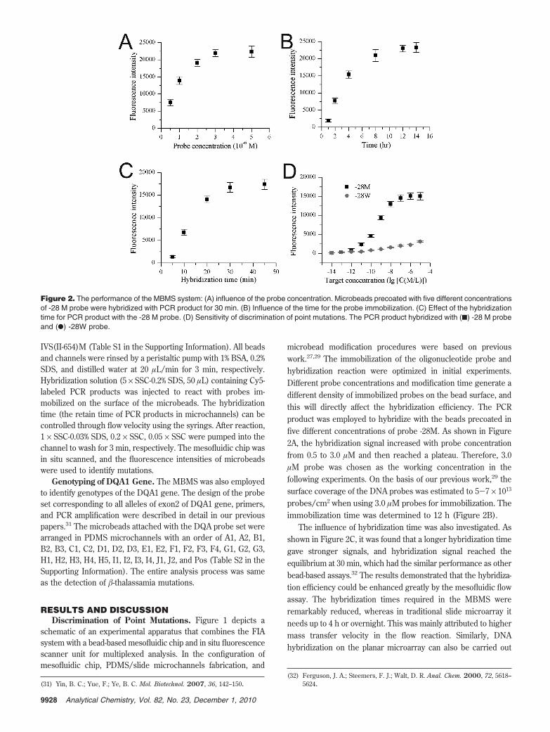
microbead modification procedures were based on previouswork.27,29 The immobilization of the oligonucleotide probe andhybridization reaction were optimized in initial experiments.Different probe concentrations and modification time generate adifferent density of immobilized probes on the bead surface, andthis will directly affect the hybridization efficiency. The PCRproduct was employed to hybridize with the beads precoated infive different concentrations of probe -28M. As shown in Figure2A, the hybridization signal increased with probe concentrationfrom 0.5 to 3.0 µM and then reached a plateau. Therefore, 3.0µM probe was chosen as the working concentration in thefollowing experiments. On the basis of our previous work,29 thesurface coverage of the DNA probes was estimated to 5-7 × 10¹³
probes/cm2 when using 3.0 µM probes for immobilization. Theimmobilization time was determined to 12 h (Figure 2B).
The influence of hybridization time was also investigated. Asshown in Figure 2C, it was found that a longer hybridization timegave stronger signals, and hybridization signal reached theequilibrium at 30 min, which had the similar performance as otherbead-based assays.32 The results demonstrated that the hybridiza-tion efficiency could be enhanced greatly by the mesofluidic flowassay. The hybridization times required in the MBMS wereremarkably reduced, whereas in traditional slide microarray itneeds up to 4 h or overnight. This was mainly attributed to highermass transfer velocity in the flow reaction. Similarly, DNAhybridization on the planar microarray can also be carried out
(31) Yin, B. C.; Yue, F.; Ye, B. C. Mol. Biotechnol. 2007, 36, 142–150.(32) Ferguson, J. A.; Steemers, F. J.; Walt, D. R. Anal. Chem. 2000, 72, 5618–
5624.
Figure 2. The performance of the MBMS system: (A) influence of the probe concentration. Microbeads precoated with five different concentrationsof -28 M probe were hybridized with PCR product for 30 min. (B) Influence of the time for the probe immobilization. (C) Effect of the hybridizationtime for PCR product with the -28 M probe. (D) Sensitivity of discrimination of point mutations. The PCR product hybridized with (9) -28 M probeand (b) -28W probe.
9928 Analytical Chemistry, Vol. 82, No. 23, December 1, 2010
actively through fluidic flow to achieve mixing and circulation,resulting in a much shorter reaction time.33,34
The sensitivity of discrimination of point mutations is of greatimportance for genetic diagnosis. Evaluation of the sensitivity ofthe MBMS system and apparatus was carried out via a seriesdilution experiment using -28(A > G) point mutation of the HBBgene as a model. A pair of probes was designed for -28(A > G)site, one complementary to the wild sequence (-28W) and one tothe variant (-28M). In the analysis of point mutation and geno-typing, it is assumed that the strength of fluorescent signal ofeach microbead is representative of the amount of labeled DNAassociated with that microbead. The amount of labeled DNA oneach microbead relies upon the stability of the duplex betweenlabeled DNA and probe immobilized on that microbead. The wild-to mutant signal ratios (W/M), wild type probe (denoted as W)to mutant probe (denoted as M) can be employed to detect thegenotypes. For heterozygous samples, two oligonucleotide probesin a group could be perfectly matched with the PCR product, eachmatched with a different allele. For homozygous samples, onlyone probe in each group would be perfectly matched with thePCR product. The probes should be carefully selected to achievesignificant discrimination of single-base mismatch.
The dilutions of the HBB gene PCR product were used toinvestigate the sensitivity of the discrimination of point mutations.Figure 2D showed a correlation of the signal intensity with theconcentration of PCR targets. The fluorescence intensity discrimi-nated the point mutation evident at 10-11 M, and the mutant-to-wild signal ratios exceeded 5:1. The data demonstrated thatMBMS has the ability to resolve single-base mismatches atthe concentration of 10 pM, which had the similar sensitivityas the fiber-optic DNA random microbead (diameter 3.1 µm)array with dye-encoding (detection limit of 10 pM in 30 min).32
Recently, Zhang et al. described a novel microfluidic devicewith a microbead array for genotyping HBV using quantumdot as labels, and the sensitivity was estimated to 4 pM.35
However, the microfluidic system without microbeads, in whichprobes were directly immobilized on the slide surface inmicrochannels, only exhibited the sensitivity of ∼10 nM (1.4ng/µL PCR product of 260 bp) for gene detection.36 Further-more, a series of five repetitive measurements with 3 µM DNAtarget was used for investigating the accuracy of the bead-basedmesofluidic system and obtained a coefficient of variation (CV)of ∼10%, demonstrating an excellent reproducibility of theassay.
Detection of -Thalassamia Mutation. The -thalassemiaresults from more than 200 different HBB gene mutations. Despitethis marked molecular heterogeneity, the prevalent moleculardefects are limited in each at-risk population. In south China, eightmutations usually account for the large majority of HBB disease-causing alleles, including -28(A > G), -29(A > G), 17(A > T),HbE26(G > A), IVS(I-1)(G > T), (41-42)(delTCTT), 43(G >T), IVS(II-654)(C > T). The probe set corresponding to thesemutations was designed and optimized. Some clinical samples and
eight plasmids (as homozygous samples) were used as the knownsamples to validate the new system. Every sample was tested intriplicate. The results show high fluorescent signal on themicrobeads containing the probes, which perfectly match to targetgene sequence. Very weak fluorescent signal is found on themicrobeads containing related probes, which single-base mismatchto corresponding positions. All results were correct, as comparedwith the known sequence, and W/M (or M/W) signal ratiosmostly exceeded 5:1, which can be considered as a threshold todetect and discriminate mutations. For the detection of pointmutation of blood samples, the W/M signal ratios obtainedproduced unequivocal assignment of samples genotype. All probesin each pair/group served as the control for each other, offeringunequivocal and specific discrimination of each genotype.
To demonstrate how the MBMS system could be used togenotype blind samples, we conducted an equivalency study of40 clinical samples whose HBB genes were sequenced. In all setsof experiments for each mutation, the W/M ratios were employedto distinguish single-base mismatch, allowing assignment of wild-type, heterozygous, or homozygous status to each sample asexpected. No false-positive or false-negative result was observedin any case. The MBMS detection results were coincident withstandard sequencing. Among the 40 samples, 26 mutant heterozy-gotes (2 double-heterozygotes) and 1 mutant homozygote werefound. (41-42)(delTCTT) was found in 8 out of the 40 (29.6%),the total number of -thalassemia mutated alleles, followed by 7cases of -28(A > G) (25.9%). The relative frequencies of thedifferent thalassemia genotypes of all 40 samples are shown inTable 1.
Genotyping of HLA-DQA1 Gene. As a further illustrationof the utility of the MBMS, the genotyping of HLA-DQA1 wasinvestigated. There are 28 HLA-DQA genotypes (including sub-genotypes) according to gene sequence of DQA exon 2 from theIMGT/HLA sequence database (release 2.11.0). A total of 31oligonucleotide probes were designed for all polymorphisms in thesecond exon of the DQA gene (Table S2 in the Supporting Information).The probes were carefully selected to achieve a significant discriminationof single-base mismatch. All genotypes and hybridization patterns of allprobes were listed in Figure 3. All probes were divided into 10 groupsfrom A group to J group; a gray box indicated a positive signal ofhybridization, and a white box indicated a negative signal of hybridiza-tion. Because of the identical gene sequence in the target region, somesubgenotypes could not be differentiated, which were DQA*010101,DQA*010102, DQA*010401, DQA*010402, and DQA*0105; DQA*010201andDQA*010202;DQA*030101,DQA*0302,andDQA*0303;DQA*040101,
(33) Situma, C.; Hashimoto, M.; Soper, S. A. Biomol. Eng. 2006, 23, 213–231.(34) Wei, C. W.; Cheng, J. Y.; Huang, C. T.; Yen, M. H.; Young, T. H. Nucleic
Acids Res. 2005, 33, e78.(35) Zhang, H.; Xu, T.; Li, C. W.; Yang, M. S. Biosens. Bioelectron. 2010, 25,
2402–2407.(36) Wang, L.; Li, P. C. J. Agric. Food Chem. 2007, 55, 10509–10516.
Table 1. The Allele Frequence of -ThalassemiaMutation in All 40 Samples
mutations heterozygote homozygotecompound
heterozygoteallele
frequence
-28 6 1 0 25.9%-29 1 0 1 7.4%17 2 0 1 11.1%HbE26 2 0 0 7.4%IVS(I-1) 1 0 0 3.7%(41-42) 8 0 0 29.6%43 1 0 1 7.4%IVS(II-654) 3 0 1 14.8%total 24 1 4(÷2) 27wild type 13 13
9929Analytical Chemistry, Vol. 82, No. 23, December 1, 2010

DQA*040102, DQA*0402, DQA*0403N, and DQA*0404; DQA*050101,DQA*050102, DQA*0503, and DQA*0505; DQA*060101 and DQA*0602.Although the method does not perform DQA high-resolutiongenotyping, it could successfully differentiate DQA specificity.Moreover, the resolution could be easily developed by adding newprobe sequences descriptive of novel polymorphisms withoutsignificantly increased costs.
The microbeads precoated with 31 probes were sequenced inthe microchannel. The PCR product (229 bp) of the second exonof the DQA1 gene was injected into the microchannel andhybridized with probes on the surface of the beads. Genotypes ofDQA were identified by the positive and negative signals in eachprobe group. For a homozygous sample, only one probe in eachgroup would be perfectly matched to the PCR products; forheterozygous one, there were two positive signals in a group andeach matched with a different allele. The highest signal(s) in eachgroup was chosen as the positive signal(s), and an intensity ratioof 3:1 was employed as the threshold to categorize the positiveand negative signals. Generally, the intensity ratio of a positivesignal(s) and negative signal(s) in each group exceeded 3:1, whichdepend on the physical characteristics of the probes, conditionsof hybridization, and washing. As a heterozygote, the thresholdwas decided as smaller than 1.5. After identification of positivesignal(s) in each group, compared with hybridization signalpatterns of HLA-DQA genotypes (Figure 3), the matched geno-type(s) was the genotype(s) of the sample.
A total of 32 samples, whose DQA1 alleles had been typedpreviously by direct sequencing, were tested using the MBMSmethod. All genomic DNA were derived from peripheral bloodlymphocyte of irrelative individuals. The positive probes of thesample were categorized according to the threshold, and thegenotype of the sample was determined from the hybridizationsignal patterns of HLA-DQA genotypes. The results of 31 samples
were completely consistent with the direct sequencing results.Only one heterozygous sample was determined as a homozygoteby MBMS. Also, most frequency genotypes are DQA*03 (includ-ing DQA*030101, DQA*0302, and DQA*0303) and DQA*05(including DQA*050101, DQA*050102, DQA*0503, and DQA*0505),which is in accordance with DQA1 genotypes frequencies of thepopulation in China. This equivalency study demonstrated thatthe MBMS method could get satisfactory results to these het-erozygous combinations and homozygous samples within the
Figure 4. The analysis of hybridization data for sample DQA1*0311.Each peak corresponds to one microbead above, which was im-mobilized with the corresponding probe.
Figure 3. Hybridization signal patterns of all 30 probes for all HLA-DQA genotypes. Black boxes indicate positive signals of hybridization, andwhite boxes indicate negative signals.
9930 Analytical Chemistry, Vol. 82, No. 23, December 1, 2010

medium resolution. The results of analysis of sample DQA1*0311was shown in Figure 4.
CONCLUSIONSIn this study, a MBMS system was developed, which contains
a bead-based mesofluidic PDMS chip, liquid-processing module,and fluorescence detection module, and integrated automated-sampling, hybridization reactions, washing, and in situ fluores-cence scanning. The combination of a probe-modified microbeadsarray in mesofluidic devices should give new, flexible, high-throughput approaches. With this MBMS, all of the analysisprocedure can be carried out automately, and the whole of theanalysis could be done within an hour. All of the process wasconducted in microchannels, which reduce the sample volumeand avoid the liquid evaporation and cross contamination. Thismethod is fast, sensitive, and high-throughput and can be carriedout automatically. With this MBMS, the analysis of -thalassemiaand genotyping of HLA-DQA were performed and have a goodcoincidence with standard sequencing. It is demonstrated thatMBMS has the capability to be applied for the effective gene
diagnosis and has the potential in biological fundamental research,food safety, and environmental monitoring.
ACKNOWLEDGMENTThis work was financially supported by NSF (20627005,
21075040), the Shanghai Project (Grant 09JC1404100), the SKLBE(Grant 2060204), Program for New Century Excellent Talents(Grant NCET-07-0287), and the Fundamental Research Funds forthe Central Universities.
SUPPORTING INFORMATION AVAILABLEAddition information on the sequences and names of probes
and primers for -thalassemia analysis and the sequences andnames of probes for HLA-DQA gene typing. This material isavailable free of charge via the Internet at http://pubs.acs.org.
Received for review July 8, 2010. Accepted October 18,2010.
AC1024792
9931Analytical Chemistry, Vol. 82, No. 23, December 1, 2010