Multiple sclerosis in research studying swayback lambs: an ...
7
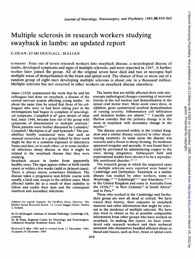
Journal of Neurology, Neurosurgery, and Psychiatry 1985;48:859-865 Multiple sclerosis in research workers studying swayback in lambs: an updated report G DEAN, El MCDOUGALL, M ELIAN SUMMARY Four out of seven research workers into swayback disease, a neurological disease of lambs, developed symptoms and signs of multiple sclerosis, and were reported in 1947. A further worker later joined the group. All of the original seven have died and two at necropsy had multiple areas of demyelination in the brain and spinal cord. The chance of four or more out of a random group of eight men developing multiple sclerosis is about one in a thousand million. Multiple sclerosis has not occurred in other workers on swayback disease elsewhere. Innes (1939) summarised the work that he and his colleagues had done on swayback, a disease of the central nervous system affecting young lambs.' At about the same time he noted that three of his col- leagues who were or had been taking part in the investigations had begun to complain of neurologi- cal symptoms. Campbell et a12 gave details of what had, since 1946, become four out of the group with symptoms of disseminated or multiple sclerosis.2 These patients were further discussed by Lumsden,3 Campbell,4 McAlpine et al- and Symonds.6 The pos- sibilities briefly considered were that such an unusual occurrence in a group linked by their work might be due to a common exposure to laboratory fumes and dust, or to each other, or to some inciden- tal infectious sheep disease or that it might be related to the swayback disease that they were studying. Swayback occurs in lambs from apparently healthy ewes. The signs appear either at birth (acute cases) or within a few weeks (mild or delayed cases). There is always ataxia, sometimes blindness. The disease takes a progressive non-febrile course with usually a fatal end, except in the mildest cases. Most affected lambs die as a result of their inability to follow and suckle their dam and the consequent inanition and secondary infections. Address for reprint requests: Dr Geoffrey Dean, Director, The Medico-Social Research Board. 73. Lower Baggot Street, Dublin 2. Ireland. Dr El McDougall, Institute of Animal Pathology, Cambridge UK, (retired) Dr M Elian, Regional Centre for Neurology and Neurosurgery, Oldchurch Hospital, Romford, Essex, UK. Received 8 May 1984 and in revised form 17 December 1984. Accepted 24 December 1984 The lambs that are mildly affected show only mic- roscopic pathological changes consisting of neuronal lesions in the red nucleus and demyelination in the spinal cord motor tract. More acute cases show, in addition, gross symmetrical cerebral demyelination of varying severity. Inflammatory cell infiltration and inclusion bodies are absent.7-'3 Cancilla and Barlow consider that the primary change is in the axon in swayback with secondary change in the myelin." The disease occurred widely in the United King- dom and a similar disease occurred in other sheep- rearing countries. In some areas the disease was enzootic and a threat to sheep farming. Its incidence appeared irregular and sporadic. It was found that it could be prevented by administering copper to the ewes during pregnancy. Subsequent field and experimental studies have shown it to be a reproduc- ible nutritional disorder.'0-13 The research group in which the suspected cases of multiple sclerosis were reported were based in Cambridge and Derbyshire. Swayback or a similar disease was studied by other workers, some at Weybridge,'4- 16 Edinburgh8 "17 and Aberdeen,'0 18 19 in the United Kingdom and some in Australia from the 1930s,202' in New Zealand,22 in South Africa23 and in Peru.24 Those who worked in the Cambridge and Derby- shire group are now all dead or retired. We have traced their history, their exposure to swayback material and other information that might be relev- ant to the incidence of multiple sclerosis. We have also tried to obtain as far as possible comparable information from other groups who have worked on swayback. In making this report, we have consi- dered only research workers and their technical assistants who themselves handled affected sheep or blood and tissues, such as liver, brain or spinal cord, 859 guest. Protected by copyright. on March 16, 2022 by http://jnnp.bmj.com/ J Neurol Neurosurg Psychiatry: first published as 10.1136/jnnp.48.9.859 on 1 September 1985. Downloaded from
Transcript of Multiple sclerosis in research studying swayback lambs: an ...
Journal of Neurology, Neurosurgery, and Psychiatry 1985;48:859-865
Multiple sclerosis in research workers studyingswayback in lambs: an updated reportG DEAN, El MCDOUGALL, M ELIAN
SUMMARY Four out of seven research workers into swayback disease, a neurological disease oflambs, developed symptoms and signs of multiple sclerosis, and were reported in 1947. A furtherworker later joined the group. All of the original seven have died and two at necropsy hadmultiple areas of demyelination in the brain and spinal cord. The chance of four or more out of arandom group of eight men developing multiple sclerosis is about one in a thousand million.Multiple sclerosis has not occurred in other workers on swayback disease elsewhere.
Innes (1939) summarised the work that he and hiscolleagues had done on swayback, a disease of thecentral nervous system affecting young lambs.' Atabout the same time he noted that three of his col-leagues who were or had been taking part in theinvestigations had begun to complain of neurologi-cal symptoms. Campbell et a12 gave details of whathad, since 1946, become four out of the group withsymptoms of disseminated or multiple sclerosis.2These patients were further discussed by Lumsden,3Campbell,4 McAlpine et al- and Symonds.6 The pos-sibilities briefly considered were that such anunusual occurrence in a group linked by their workmight be due to a common exposure to laboratoryfumes and dust, or to each other, or to some inciden-tal infectious sheep disease or that it might berelated to the swayback disease that they werestudying.Swayback occurs in lambs from apparentlyhealthy ewes. The signs appear either at birth (acutecases) or within a few weeks (mild or delayed cases).There is always ataxia, sometimes blindness. Thedisease takes a progressive non-febrile course withusually a fatal end, except in the mildest cases. Mostaffected lambs die as a result of their inability tofollow and suckle their dam and the consequentinanition and secondary infections.
Address for reprint requests: Dr Geoffrey Dean, Director, TheMedico-Social Research Board. 73. Lower Baggot Street, Dublin2. Ireland.
Dr El McDougall, Institute of Animal Pathology, Cambridge UK,(retired)Dr M Elian, Regional Centre for Neurology and Neurosurgery,Oldchurch Hospital, Romford, Essex, UK.
Received 8 May 1984 and in revised form 17 December 1984.Accepted 24 December 1984
The lambs that are mildly affected show only mic-roscopic pathological changes consisting of neuronallesions in the red nucleus and demyelination in thespinal cord motor tract. More acute cases show, inaddition, gross symmetrical cerebral demyelinationof varying severity. Inflammatory cell infiltrationand inclusion bodies are absent.7-'3 Cancilla andBarlow consider that the primary change is in theaxon in swayback with secondary change in themyelin."The disease occurred widely in the United King-
dom and a similar disease occurred in other sheep-rearing countries. In some areas the disease wasenzootic and a threat to sheep farming. Its incidenceappeared irregular and sporadic. It was found that itcould be prevented by administering copper to theewes during pregnancy. Subsequent field andexperimental studies have shown it to be a reproduc-ible nutritional disorder.'0-13The research group in which the suspected cases
of multiple sclerosis were reported were based inCambridge and Derbyshire. Swayback or a similardisease was studied by other workers, some atWeybridge,'4- 16 Edinburgh8 "17 and Aberdeen,'0 18 19
in the United Kingdom and some in Australia fromthe 1930s,202' in New Zealand,22 in South Africa23and in Peru.24Those who worked in the Cambridge and Derby-
shire group are now all dead or retired. We havetraced their history, their exposure to swaybackmaterial and other information that might be relev-ant to the incidence of multiple sclerosis. We havealso tried to obtain as far as possible comparableinformation from other groups who have worked onswayback. In making this report, we have consi-dered only research workers and their technicalassistants who themselves handled affected sheep orblood and tissues, such as liver, brain or spinal cord,
859
guest. Protected by copyright.
on March 16, 2022 by
http://jnnp.bmj.com
/J N
eurol Neurosurg P
sychiatry: first published as 10.1136/jnnp.48.9.859 on 1 Septem
ber 1985. Dow
nloaded from
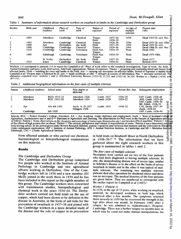
Table 1 Summary ofinformation about research workers on swayback in lambs in the Cambridge and Derbyshire group
Worker Birth year Childhood Place of Type of Nature of Period of 1st sign of Present stateresidence work work exposure exposure multiple 1981
sclerosis
1 1905 Aberdeen Cambridge Chemical Tissues 1937-46 1938 Dead 1953 D. cert. Nec.Field 1938-39
2 1903 Aberdeen Cambridge Chemical Tissues 1934-36 1938 Dead 1955 D. cert. Obit.3 1908 Ayr Derbyshire An. husb. Field 1937-39 1939 Dead 1954 D. cert.4 1906 Cambridge Cambridge Chemical Tissues 1934-46 1944 Dead 1966 D. cert. Nec.5 1910 Cambridge Cambridge Veterinary Field Aut. 1934-39 None Dead 1982
Tissues6 1903 Edinburgh Cambridge Veterinary Field Aut. 1934-39 None Dead 1974 Obit.
Tissues7 N. Yorks. Derbyshire An. husb. Field 1937-39 None Dead 19778 1915 Essex Cambridge Chemical Tissues 1939-44 None Retired 1975
Workers 1-4 correspond to patients 1-4 in report by Campbell et al.2 Place of work refers to the swayback investigations. Type of work: An. husb. =Animal husbandry (advisory work, field survey, field trials organisation). Nature of exposure: Tissues = preparing and examining blood and tissues fromaffected lambs. Field = handling the ewes and lambs in the field. Aut. = carrying out necropsies on affected lambs. Period of exposure is mainly fromCampbell et al.2 Present state is followed bv D. cert. = death certificate or obit. = obituary as sources of information. Nec. = necropsy carried out. Theobituaries consulted were: workers I and 2, Aberdeen University Review (1953-4) 35,325 and (1955-6) 36.329. Worker 6, J Pathol (1976) 119,187-191.
Table 2 Additional biographical information on the four cases ofmultiple sclerosis
Patient Childhood residence School years First degree or PhD Rowett Res. Inst. Subsequent employmentdiplomas
1 Aberdeen RGG 1919-23 Aberdeen 1926 Camb. 1937 1926-28 IAP, Camb. 1928-482 Aberdeen RGC 1915-20 Aberdeen 1924 Camb. 1928 1924-25 ANI, Camb. 1925-26
IAP, Camb. 1926-36MI, Edinb. 1936-55
3 Ayr AA left 1925 Auch. A, D 1927 Camb. 1935 1930-32 CAI, Derby 1937-39Glasgow 1930 Auch. 1939-54
4 Cambridge left 1920 IAP. Camb. 1926-46
Schools, RGC = Robert Gordon's College, Aberdeen, AA = Ayr Academy. Under diplomas and employment, Auch. = West of Scotland College ofAgriculture, Auchencruive and A and D = Diplomas in Agriculture and Dairying. The dissertations for PhD were in the Facultv of Agriculture and wereas follows: Patient I Some observations on the chemistry and toxicology of buttercups (Ranunculus spp.) and bracken (Pteris aquilina).' Patient 2 A studyof the effects of high protein diet on sheep.' Patient 3 "lie calcium, phosphorus and vitamin L) requirements of swine.' Patient 4 was a laboratory assistantfrom leaving school. In the period given under Rowett Research Institute, patient 3 spent his time between that Institute and the University in Aberdeen.Under subsequent employment, IAP = Institute of Animal Pathology, ANI = Animal Nutrition Institute, in Cambridge and Ml = Moredun Institute,Edinburgh; CAI = County Agricultural Institute.
from affected animals or who carried out chemical,haematological or histopathological examinationson this material.
Results
The Cambridge and Derbyshire GroupThe Cambridge and Derbyshire group comprisedfive people who worked at the Institute of AnimalPathology in Cambridge and two agriculturalofficers working in Derbyshire. One of the Cam-bridge workers left in 1936 and a new member (ElMcD) joined in the work there in 1939 and he hasbeen included in this report as the eighth member ofthis group. The Cambridge workers were concernedwith transmission studies, histopathological andchemical work in the years 1934-44. The Derby-shire workers carried out field trials on the use ofcopper, which had been shown to be deficient in thedisease in Australia, in the form of salt licks for theprevention of swayback in 1937-38 and joined withthe Cambridge workers in a more detailed study ofthe disease and the role of copper in its prevention
in field trials on Bradwell Moor in North Derbyshirein 1938-39.25-28 The information that we havegathered about the eight research workers in thisgroup is summarised in tables 1 and 2.
The four cases of multiplesclerosisNecropsies were carried out on two of the four patientswho had been diagnosed as having multiple sclerosis. Inone, the demyelinating disease was of severe type, similarto Schilder's disease or to the effect on the brain of severeswayback in lambs and in the other it was typical of mul-tiple sclerosis. The remaining two multiple sclerosispatients died after operation for duodenal ulcers and therewas no necropsy. The medical histories of the four patientsare given below. They are numbered to correspond withthe earlier report by Campbell et al (1947).2Worker 1 (Patient 1)In 1938, at the age of 33 years, while working on swaybackmaterial, he developed weakness in both legs whichimproved after a few months. The symptoms returnedmore severely in 1939 but he recovered the strength in hislegs after about one month. In February 1940, after arelapse, he was admitted to Addenbrooke's Hospital,Cambridge. There was a further relapse six months later atwhich time he could not make manual manipulations, his
Dean, McDougall, Elian860guest. P
rotected by copyright. on M
arch 16, 2022 byhttp://jnnp.bm
j.com/
J Neurol N
eurosurg Psychiatry: first published as 10.1136/jnnp.48.9.859 on 1 S
eptember 1985. D
ownloaded from
Multiple sclerosis in research workers studying swayback in lambs: an updated report
gait was staggering and his speech was affected. Heimproved after four weeks in bed. He continued withnumerous attacks and periodical impairment of vision inone or both eyes, with some remissions, until his admissionto the Radcliffe Infirmary, Oxford, in 1947, under the careof Dr Ritchie Russell, by which time he was disorientated,mentally confused and incapable of sustained conversa-tion. He was temperamentally aggressive but there waseuphoria. He had slurring of speech, incoordination of hisarms and legs, diminution of vibration sense in his legs,spasticity of all his limbs, the plantar responses were exten-sor, his knee and ankle tendon reflexes were brisk and theabdominal reflexes were absent. He also had a pleural effu-sion following a previous haemothorax resulting from afall. He gave his age as 32 when he was, in fact, 42. Effortsto help him walk with a walking aid were not successful.Copper estimations were undertaken and they indicated alow excretion of copper. The WR was negative and theLange gold curve was 34444310. There was no increase inCSF cells or protein. He was admitted to MorningfieldHospital in Aberdeen in July, 1951 with "inability to walkwithout staggering, incontinence of urine and generallyunable to do things". He was mentally confused. There wasno nystagmus but he had a constant static tremor". Hewas very childish and aggressive, otherwise the examina-tion was the same as in 1947.He died at Morningfield Hospital on the 11 November,
1953, aged 48 and necropsy was undertaken by Dr HGRichmond. His body was wasted and he had a deepexcavating bedsore. On opening the skull there was anincrease in subarachnoid fluid of gelatinous character overboth cerebral hemispheres, the total weight of the brainwas 1,150 g, there was obvious atrophy of the gyri, withwidening of the sulci in all lobes of the brain, possibly moremarked in the frontal and pyramidal regions. Incision intothe lateral ventricles showed clear cerebrospinal fluid, thearteries at the base of the skull were normal and the pituit-ary gland was normal in size. The brain and spinal cord, anumber of posterior root ganglia and the left Gasserianganglion were removed and sent to Dr Charles Lumsden,who at that time was at Maida Vale Hospital, London. Hisdeath was certified as due to encephalomyelitis.
Professor Lumsden died in 1974 and, unfortunately, inspite of an intensive search no records can be found of thisnecropsy at Maida Vale Hospital, at Professor Lumsden'sdepartment of pathology at Leeds or at his home. Noreport was sent to Professor Young, Professor of Pathologyat Aberdeen nor to Dr Richmond who did the necropsy,nor does Professor Lumsden's widow have any relevantrecords. Fortunately, Professor Lumsden wrote to DrAMG Campbell from Maida Vale about the necropsy inMay 1957. In the letter he said: "Though I would not sayvery much about it meantime until it has been fixed morethoroughly, examined and processed for celloidin sections,etc there is no doubt there are extensive foci of demyelina-tion in the cerebrum. As far as I could tell, the cerebellumwas negative as well as mid-brain and medulla. The cordhas only been cut so far at three levels and I think there israther a lot of secondary- degeneration. Certainly, thelesion in the cerebrum is complicated by atrophy whichmay account for the relatively poor demarcation of thedemyelination" .
A report by Professor Lumsden was published about thispatient's necropsy in 1958.3 In it he says "Since relationbetween swayback' and human disease is implied in thisdiscussion, I shall begin by referring to the report ofCampbell et al (1947)2 which has naturally received prom-inence in the world literature on demyelinating disease.It will be remembered that four out of seven researchworkers on swayback were thought to be affected by adisease indistinguishable from multiple sclerosis. Sinceover 10 years have now elapsed, ample time would seem tohave occurred for re-assessment of this report. In thisinstance, some fresh information would seem to be aparamount need. The clinically most typical case of thefour was the only one known to me; this patient eventuallysuccumbed to multiple sclerosis which, I think by a curiouscoincidence only, happened to be of a rather " transitional"type with diffuse cerebral demyelination and with plaquesof the more typical discrete type only in the optic pathway,cerebellum, brain-stem and cord. I hope to report andillustrate the pathological findings in this case elsewhere infull detail. As far as I can ascertain, this case was purelycoincidental and has no relevance whatsoever to theswayback problem. If we were to learn that the diagnosis ofmultiple sclerosis is no longer tenable in the remainingcases cited in the 1947 report, I think it would be of realvalue in the difficult field of multiple sclerosis research".Professor Lumsden reported to Professor Young that thenecropsy findings were "very similar to that of Schilder'sdisease".
Worker 2 (Patient 2)In June 1938, at the age of 35 years, for a period of twodays this worker complained of a peculiar sensation in hislegs and found walking an effort and could not chase theball at tennis. This cleared up but later in the same yearthere was weakness in both legs, the right leg was moreaffected than the left. The third, fourth and fifth fingers ofthe right hand also felt numb. On examination inDecember 1938, his legs had recovered but his fingerswere still numb and this was affecting his writing. Therewere no other abnormal symptoms. There was marked lossof two point discrimination in the third, fourth and fifthfingers of his right hand. There was no weakness or ataxia.The tendon reflexes were equal and brisk. The abdominalreflexes were brisk and the plantar reflexes were flexor, theoptic discs were normal and there was no nystagmus, theWasserman reaction was negative. In 1939, he complainedof blurring of vision, which was diagnosed as retrobulbarneuritis, affecting both eyes. His vision recovered in aboutsix weeks. Since then he had been well except for occa-sional numbness in the right leg and symptoms due to pep-tic ulcer. He died following an operation for duodenalulcer at the age of 52 at Edinburgh in December, 1955. Nonecropsy was undertaken.The report by Campell et al ( 1947)2 states that his sister
"had died from disseminated sclerosis". None of the othermembers of the family can now remember her having dis-seminated sclerosis but rather tuberculosis. She died in1927 aged- 29 years and the diagnosis on her deathcertificate is "pulmonary tuberculosis". So far no evidenceexcept for the statement in the 1947 paper can be foundthat she-also had disseminated sclerosis.
861
guest. Protected by copyright.
on March 16, 2022 by
http://jnnp.bmj.com
/J N
eurol Neurosurg P
sychiatry: first published as 10.1136/jnnp.48.9.859 on 1 Septem
ber 1985. Dow
nloaded from

862
Although this research worker's symptoms were neververy severe, the story of weakness in his legs in 1938, ofnumbness in his fingers affecting his writing and ofretrobulbar neuritis in 1939 and then of minor attacks ofparaesthesia after this time makes the diagnosis of prob-able multiple sclerosis acceptable.
Worker 3 (Patient 3)In August 1939, at the age of 31, while still in Derbyshirethis patient complained of slight weakness in the right legand a change of sensation in the left leg. On examinationhis tendon reflexes were exaggerated and the right plantarresponse was extensor. There was difficulty in eliciting theabdominal reflexes. He recovered but in 1945 he com-
plained of haziness in the vision of his left eye and he was
seen by an ophthalmologist who diagnosed retrobulbarneuritis and considered it was due to disseminatedsclerosis. On examination in 1945, the plantar responses
were then flexor on both sides. The abdominal reflexescould not be obtained. There was a slight shakiness of theright eye on looking to the right but no sustained nystag-mus. There was pallor of the temporal half of the left discwhich had not been present when he was seen previouslyby the same physician in 1939. In view of his history, theeye symptoms and signs, the extensor plantar response inthe right leg in 1939 and the absence of abdominal reflexesin 1945, it was considered that he was suffering from dis-seminated sclerosis. His brother still remembers that in1939 he was in bed for three weeks with weakness in hislegs and that eye symptoms diagnosed as retrobulbarneuritis, developed while he was living in Ayr. His brotheralso remembers how he used to walk with a swaying gaitand that he could not walk in a straight line.
In 1947, he fell from the platform of a bus in Ayr andinjured his head but he was conscious on admission tohospital. His widow reports that his walking slowlydeteriorated after this so that he had to retire in December,1953. During the last two years of his life his walkingdeteriorated more rapidly, although he could still walkunaided. His widow reports that there was mental deterio-ration in the last year of his life. He died at Ayr Hospital atthe age of 46 years in 1954 after an operation for a perfo-rated duodenal ulcer. No necropsy was undertaken. Prob-able multiple sclerosis would appear to have been the cor-rect diagnosis.
Worker 4 (Patient 4)In 1945, at the age of 39 years, this patient noticed that hisleft leg was inclined to drag. This lasted for a few days andthen it completely recovered. In 1946, he noticed that hewas dragging his right leg and his walking was unsteady.This also improved and when he was examined by aneurologist there was no obvious weakness in his limbs buthis knee and ankle tendon reflexes were exaggerated, moreso on the left where there was also ankle clonus. Bothplantar responses were extensor. Although there was no
gross weakness or ataxia, attempts to hop indicated some
weakness and loss of balance, particularly on the left. Theabdominal reflexes could be obtained. In 1950, he was
thoroughly investigated at the National Hospital, London,by a number of neurologists, including Dr Elkington, SirCharles Symonds, Sir Francis Walshe and Dr MacDonald
Dean, McDougall, Elian
Critchley. The first three considered that he had dissemi-nated sclerosis and Dr Critchley though it was humanswayback. At this time his legs were weak and spastic withclonus of both knees and ankles, the abdominal reflexeswere absent, the plantar responses were extensor. The cra-nial nerves were normal. A psychological report showedsigns of slight but definite organic intellectual impairmentwith difficulty in retention and learning. During 1950, hehad periodic blurring of vision and difficulty in maintainingan erection. By 1953, his vision had become very poor sohe could only read the headlines in the newspaper. In 1952his WR was positive and he had an abnormal Lange goldcurve (5543222100). The CSF cells were slightly increasedwith a weakly positive CSF globulin test.He died in December 1966, at the age of 60 and had a
full necropsy at Addenbrooke's Hospital. His brainweighed 1,215 g, there was clear differentiation betweenthe grey and the white matter of the brain. Scattered in thebrain substance there were firm pink plaques, principallyaround the ventricles, the spinal cord surface showed simi-lar plaques or pink areas of irregular lengths. There wasmoderate symmetrical dilation of the ventricles and someof the cerebral plaques were 2 x 1 cm, in cross section.Cortical and subcortical plaques were not conspicuous.There was generalised reduction of the white matter andthinning of the corpus callosum. The medulla ribbon wasslightly thinner than normal for the patient's age, the cere-bellum contained only one or two small grey patches up to1 mm in diameter in the white matter. In the medulla therewere two gelatinous grey areas lateral to the hypoglossalnuclei, the pons showed a grey patch in the left brachiumconjunctivum and a similar less well defined area on theright. The spinal dura was normal. Scattered throughoutthe length of the cord were numerous grey patches inhaphazard distribution. These were most numerous andextensive in the cervical cord but the thoracic and lumbarcord were also affected. In one mid-thoracic section almostthe entire cut section was involved. Microscopically, thebrain and cord showed a number of plaques of gliosis withsome perivascular round cell infiltration. Special stainsshowed lipid accumulation and loss of myelin in these areas(Dr Peter R Millard).
In 1981, the slides were re-examined by Professor IngridAllen of Queen's University, Belfast, who reported: ''Sec-tions have been studied from grey matter, deep white mat-ter, basal ganglia, pons, medulla, cerebellum and spinalcord. In all the sections studied there are discrete areas ofdemyelination of some duration. The meninges are notinflamed. There is no abnormality of the cortical grey mat-ter. In the white matter there is very diffuse gliosis, markedperivascular inflammation which, outside the areas ofdemyelination, consists largely of a lymphocytic reaction.The areas of demyelination are relatively discrete and aresurrounded by intense glial reaction. Within the demyeli-nated areas astrocytes are present, blood vessels are thick-ened and there is a mixed perivascular inflammatory reac-tion. Some of the most striking areas of demyelination areadjacent to the ventricles. Sections from the pons, medullaand cerebullum show similar lesions. Sections from thespinal cord show very marked demyelination with the pre-sence of neutral fat. Overall, the picture is that of aninflammatory demyelinating disease. The histology is quite
guest. Protected by copyright.
on March 16, 2022 by
http://jnnp.bmj.com
/J N
eurol Neurosurg P
sychiatry: first published as 10.1136/jnnp.48.9.859 on 1 Septem
ber 1985. Dow
nloaded from

863Multiple sclerosis in research workers studying swayback in lambs: an updated report
,.W *7
Fig 1 Spinal cord demyelination (H-aematoxylin and
Eosin. X 9-5.) (Patient 4)
~~~~~~~~A
Fig 2 Plaque in brain. (Luxol fast bluelHaematoxylin andEosin. x 7-5.) (Patient 4)
Fig 3 Deep white matter. (Luxol fast bluelHaematoxylinand Eosin. x 240.) (Patient 4)
compatible with some form of multiple sclerosis."With this history and the clinical and the necropsy
findings, the undoubted diagnosis is multiple sclerosis.When he died, Dr Millard of Cambridge sent part of thebrain to Dr PA Palsson of the Institute of ExperimentalPathology, University of Iceland, Reykjavik. A prepara-tion was injected into five sheep intracerebrally in 1967 tosee if it would induce scrapie. The last of the sheep were
killed in 1979 and examined. No sign or evidence ofscrapie was found (Personal communication 1982).
-NhpA
"VE,~ ~ ~ V
Fig 4 Pons. (Luxol fast bluelHaematoxyln and Eosin. X100.) (Patient 4)
Inset: Axis cylinder stain. (Palmgren. x 600.)
Other groups who have worked on swaybackInformation was obtained by correspondence aboutresearch workers in other groups concerned with the studyof swayback. Twelve of these were at Weybridge, eight inEdinburgh, five in Aberdeen, nineteen in Australia andNew Zealand and six in South Africa. The Weybridgegroup carried out much of their field work on sheep fromthe same area of Derbyshire as the Cambridge and Derby-shire group had carried out their field trials. All groupscontained members who had handled fresh brain material.None of the fifty workers in these other groups has shownsigns of multiple sclerosis; only one showed a neurologicaldisease (motor neuron disease).
Discussion
It is difficult to find a common cause that could applyto all four cases of multiple sclerosis as well as notapplying to those who did not develop the disease.As gangue from old lead mines is common on Brad-well Moor3' where the Derbyshire field trials werecarried out, it is possible that there could have beenshort exposure to lead containing dust in thelaboratory from the drying and powdering of sam-ples of tissue or pasture. Symonds suggested thatthey might have been exposed to basicencephalitogenic protein.62930 The possibility of arelation between the human and sheep diseases hasbeen tested by intracerebral injection of materialfrom cases of multiple sclerosis into sheep.3234Although some cases of scrapie were reported atfirst, it has not proved possible to confirm this andtests by Palsson with the brain of patient 4 failed toproduce scrapie. Visna, which occurred naturally inIceland until 1965, has not been reported in theUnited Kingdom.The occurrence of four cases of multiple sclerosis
guest. Protected by copyright.
on March 16, 2022 by
http://jnnp.bmj.com
/J N
eurol Neurosurg P
sychiatry: first published as 10.1136/jnnp.48.9.859 on 1 Septem
ber 1985. Dow
nloaded from

864or a multiple sclerosis-like illness in the Cambridgeand Derbyshire group and the similarity of thesymptoms and necropsy findings of one of them toSchildeies disease and the anatomical similarity ofthe latter to acute swayback disease might suggest a
relation between the two diseases. However, a simi-larity need not imply a causal relation. The absenceof multiple sclerosis in the survey of fifty otherworkers on swayback, twelve of whom were con-
cerned at a later period with material from the samepart of Derbyshire as the earlier workers, gives no
support to any strong correlation between multiplesclerosis and working on swayback disease. It isperhaps relevant that the symptoms in workers 1, 2and 3 all started in 1938-39.Two out of the four multiple sclerosis patients,
those who developed multiple sclerosis in 1938/39,originated from the Grampian area of north-eastScotland where there is a higher prevalence of mul-tiple sclerosis than in England and Wales and wherethe multiple sclerosis prevalence in middle-agedmen, 35-44 years, is 200 (200.4) per 100,000, or 1in 500. (Phadke, JG and Downie AW. Personalcommunication). Using this high prevalence rate theBinomial Probability Distribution shows that thechance of four or more out of a random group ofeight men having multiple sclerosis is about 1 in 109,or 1 in a thousand million. The only linking factorthat has been ascertained is that this group of eightveterinary research workers were working togetherwith the brains of lambs that had a disease of thecentral nervous system.
It is concluded from a review of the history andnecropsy findings that four out of eight, not seven,veterinary research workers into a disease of theCNS in lambs, swayback disease, developed mul-tiple sclerosis or a multiple sclerosis-like illness. Thisstrange happening is very unlikely to be coincidentaland, with the reported epidemic of multiple sclerosisin the Faroe Islands after World War 235 supportsthe concept that there is a major environmental fac-tor, probably an infective factor, in multiplesclerosis. It remains an important clue to the cause
of the disease.
We are indebted to the numerous former workerson swayback, to their colleagues and relatives in theUnited Kingdom, Australia, New Zealand andSouth Africa, who helped us with this research. Wealso thank Mr JP Brophy, Regional AdministrativeOffice, Perth, for his help in tracing what happenedto workers on swayback disease in Australia. Dr CSTreip, consultant neuropathologist, Addenbrooke'sHospital, Cambridge, provided pathology reports,blocks and slides about the necropsy on patient 4.We also thank the doctors and records staff of
Dean, McDougall, Elian
numerous hospitals who have searched their files forold records about the four multiple sclerosispatients. Mr AV Vincent, Trinity College, Dublin,provided statistical advice. Miss Hilda McCoughlinassisted with the study and in the preparation of thereport.
References
Innes JRM. Swayback: a demyelinating disease of lambswith affinities to Schilderes encephalitis and its preven-tion by copper. J Neurol Psychiatry 1939;2:323-34.
2 Campbell AMG, Daniel P, Porter RJ, Russell WR,Smith HV, Innes JRM. Disease of the nervous systemoccurring among research workers on swayback inlambs. Brain 1947;70:50-8.
Lumsden CE. Demyelinating disease: The present situa-tion. Proc R Soc Med 1958;51:753-4.
4Campbell AMG. Veterinary workers and disseminatedsclerosis. J Neurol Neurosurg Psychiatry 1963;26:514-5.
McAlpine D, Lumsden CE, Acheson ED. Multiplesclerosis: a Reappraisal. 2nd Ed. Edinburgh. ChurchillLivingstone. 1972.
6 Symonds CP. Multiple sclerosis and the swayback story.Lancet 1975; 1: 155-6.
7Innes JRM, Saunders LZ. Comparative Neuropathology.Chap. XIV, London, Academic Press. 1962.
Barlow RM, Purvis D, Butler EJ, McIntyre IJ. Swaybackin south-east Scotland. i. Field aspects. ii. Clinical,pathological and biochemical aspects. J Comp Pathol1960;70:396-410, 411-27.
9 Barlow RM. Further observations on swayback. Transi-tional pathology. J Comp Pathol 1963;73:51-60.
'0 Fell BF, Mills CF, Boyne R. Cytochrome oxidase defi-ciency in the motor neurones of copper deficientlambs; a histochemical study. Res Vet Sci1965;6: 170-1.
Cancilla PA, Barlow RM. Structural changes in the cen-tral nervous system in swayback (enzootic ataxia) oflambs. IV. Electron microscopy of the white matter ofthe spinal cord. Acta Neuropathol (Berl.),1968;11:294-300.
12 Smith RM, Fraser FJ, Russell GR, Robertson JS. Enzoo-tic ataxia in lambs: appearance of lesions in the spinalcord during foetal development. J Comp Pathol1977;87: 119-28.
3 Smith RM, Fraser FJ, Robertson JS. Enzootic ataxia inlambs: Absence of detectable neuronal pathology infoetal and neonatal spinal cord. J Comp Pathol1978;88:401-8.
'4 Hunter AH, Eden A, Green HH. Contributions to thestudy of swayback in lambs. i. Field experiments. JComp Pathol Ther 1945;55: 19-28.
"Eden A, Hunter H, Green HH. Contributions to thestudy of swayback in lambs. ii. Blood copper. J CompPathol Ther 1945;55:29-40.
16 Allcroft R, Clegg FG, Uvarov 0. Prevention ofswayback in lambs. Vet Rec 1959;71:884-9.
7 Butler EJ, Barlow RM. Copper deficiency in relation to
guest. Protected by copyright.
on March 16, 2022 by
http://jnnp.bmj.com
/J N
eurol Neurosurg P
sychiatry: first published as 10.1136/jnnp.48.9.859 on 1 Septem
ber 1985. Dow
nloaded from
Multiple sclerosis in research workers studying swayback in lambs: an updated report
swayback in lambs. 1. Effect of molybdate and sul-phate supplement during pregnancy. J Comp Pathol1963;73:208-13.
18 Mills CF, Fell BF. Demyelination in lambs born of ewesmaintained on high intakes of sulphate and molyb-date. Nature 1960; 185: 20-2.
9 Mills CF, Williams RB. Copper concentration and cyt-rochrome oxidase and ribonuclease activities in thebrains of copper deficient lambs. Biochem J1962;85: 629-32.
20 Bennetts HW, Beck AB. Enzootic ataxia and copperdeficiency of sheep in Western Australia. Bull Councilfor Scientific Industrial Research 1942; 147:1-52.
21 McDonald IW. Enzootic ataxia of lambs in SouthAustralia. Aust Vet J 1942; 18:165-72.
22 Cunningham IJ. Copper deficiency in cattle and sheep inpeat lands. NZ J Sci Technol 1946; 27:381-96.
23 Schulz KCA, van der Merwe PK, van Rensburg P, SwartJS. Studies in demyelinating diseases of sheep associ-ated with copper deficiency. Onderstepoort J Vet Res1951;25:35-7.
24 Tabusso ME. Vida Agricola (Lima), 1941-2, 18, 719-27, 789-94, 875-84, 943-9 and 19, 123-6 (Quoted inref. 7).
25 Dunlop G, Wells HE. "Warfa" (swayback) in lambs inNorth Derbyshire and its prevention by adding coppersupplements to the diet of the ewes during gestation.Vet Rec 1938; 1175-82.
26 Dunlop G, Innes JRM, Shearer GD, Wells HE.Swayback studies in North Deryshire. J Comp Pathol
Ther 1939;52:259-65.27 Innes JRM, Shearer GD. Swayback: A demyelinating
disease of lambs with affinities to Schilder'sencephalitis in man. J Comp Pathol Ther 1940;53: 1-41.
28 Shearer GD, Innes JRM, McDougall EI. Swaybackstudies in North Derbyshire. ii. The relationship of thestorage of copper and lead in the body tissues to thecausation of swayback. Vet J 1941; 96:309-22.
29 Carnegie PR, Lumsden CE. Encephalitogenic peptidesfrom spinal cord. Nature 1966;209: 1354.
30 Carnegie PR. Properties, structure and possibleneuroceptor role of the encephalitogenic protein ofhuman brain. Nature 1971;229:25.
3' Ford TD, Rieuwerts JH. Lead Mining in the Peak Dis-trict. Peak Park Joint Planning Board, Bakewell,Derbyshire. 1975.
32 Sutherland JM, Wilson DR, Disseminated sclerosis inman and experimentation with sheep. Glasgow Med J1951;32:302-10.
33 Palsson PA, Pattison IH, Field EJ. Slow, latent andtemperate virus infections. NINDB Mono. No. 2, pp.49-54, Washington Public Health Service, PublicationNo. 1378. 1965.
34 Dick G, McAlister JJ, McKeown F, Campbell AMG.Multiple sclerosis and scrapie. J Neurol NeurosurgPsychiatry 1965;28: 560-2.
Kirtzke JF, Hyllested K. Multiple sclerosis in the FaroeIslands. 1. Clinical and epidemiological features. AnnNeurol 1978;5:6-21.
865
guest. Protected by copyright.
on March 16, 2022 by
http://jnnp.bmj.com
/J N
eurol Neurosurg P
sychiatry: first published as 10.1136/jnnp.48.9.859 on 1 Septem
ber 1985. Dow
nloaded from