Multiple Pigment Cell Types Contribute to the Black, Blue ... · Blue, and Orange Ornaments ......
10
Multiple Pigment Cell Types Contribute to the Black, Blue, and Orange Ornaments of Male Guppies (Poecilia reticulata) Verena A. Kottler 1 *, Iris Koch 2 , Matthias Flo ¨ tenmeyer 2 , Hisashi Hashimoto 3 , Detlef Weigel 1 , Christine Dreyer 1 * 1 Department of Molecular Biology, Max Planck Institute for Developmental Biology, Tu ¨ bingen, Germany, 2 Max Planck Institute for Developmental Biology, Tu ¨ bingen, Germany, 3 Bioscience and Biotechnology Center, Nagoya University, Nagoya, Japan Abstract The fitness of male guppies (Poecilia reticulata) highly depends on the size and number of their black, blue, and orange ornaments. Recently, progress has been made regarding the genetic mechanisms underlying male guppy pigment pattern formation, but we still know little about the pigment cell organization within these ornaments. Here, we investigate the pigment cell distribution within the black, blue, and orange trunk spots and selected fin color patterns of guppy males from three genetically divergent strains using transmission electron microscopy. We identified three types of pigment cells and found that at least two of these contribute to each color trait. Further, two pigment cell layers, one in the dermis and the other in the hypodermis, contribute to each trunk spot. The pigment cell organization within the black and orange trunk spots was similar between strains. The presence of iridophores in each of the investigated color traits is consistent with a key role for this pigment cell type in guppy color pattern formation. Citation: Kottler VA, Koch I, Flo ¨ tenmeyer M, Hashimoto H, Weigel D, et al. (2014) Multiple Pigment Cell Types Contribute to the Black, Blue, and Orange Ornaments of Male Guppies (Poecilia reticulata). PLoS ONE 9(1): e85647. doi:10.1371/journal.pone.0085647 Editor: Kevin McGraw, Arizona State University, United States of America Received September 19, 2013; Accepted November 27, 2013; Published January 22, 2014 Copyright: ß 2014 Kottler et al. This is an open-access article distributed under the terms of the Creative Commons Attribution License, which permits unrestricted use, distribution, and reproduction in any medium, provided the original author and source are credited. Funding: This work was supported by a Gottfried Wilhelm Leibniz Award of the Deutsche Forschungsgemeinschaft and funds from the Max Planck Society to DW. The funders had no role in study design, data collection and analysis, decision to publish, or preparation of the manuscript. Competing Interests: The authors have declared that no competing interests exist. * E-mail: [email protected] (VAK); [email protected] (CD) Introduction The spectacular orange, yellow, white, and black along with the blue to green iridescent colors of male guppies (Poecilia reticulata) have attracted the attention of biologists and hobby breeders for almost a century [1–5]. The guppy is a small live-bearing freshwater fish native to northeastern South America. Guppy populations have been studied most extensively on the island of Trinidad, where male coloration, as well as other traits, such as body shape and life history characteristics, covary with predation intensity [6,7]. Mate choice experiments have demonstrated that guppy females, which are camouflaged by an inconspicuous reticulate pattern [4,8], prefer males with high amounts of orange and iridescent pigments [6,9,10]. Both orange and iridescent orna- ments can indicate a male’s current physical condition and genetic quality. The orange spots contain two types of pigments, carotenoids, which are obtained from the food, mainly from unicellular algae, and pteridines, which are synthesized de novo [11,12]. Orange pigments therefore reflect a male’s foraging efficiency and ability to synthesize pteridines [11–13]. Pteridine production within the orange spots of wild guppy males varies with carotenoid availability; for instance, males produce less pteridines in habitats in which carotenoids are scarce, leading to a relatively constant pteridines to carotenoids ratio, and hence orange hue, across populations [11,12]. A recent study has shown that female guppy preference for this specific orange hue causes this pattern [14]. Iridescent ornaments increase the risk of being noticed by predators and hence provide information on a male’s capability to evade these [6,9,15]. Males also intensify their black pigmentation during courtship, which might emphasize orange areas [6,16]. The amount and size of male ornaments is highly heritable and a substantial portion is inherited in a Y-linked manner from the father [2,5,8,17,18]. Studies have demonstrated that guppy females favor males with rare or novel color patterns over males with familiar phenotypes, suggesting that negative frequency- dependent selection contributes to the maintenance of male color polymorphisms within guppy populations [19–23]. While the selection pressures driving male color patterns have been well studied, little is known about the morphology of male ornaments. Five pigment cell types have been described in the skin of the guppy: black melanophores, orange to yellow xanthophores, red erythrophores, blue to green iridescent iridophores (Figure 1), and white leucophores [4,8,24–28]. The pigment organelles of melanophores, xanthophores, and erythrophores contain light- absorbing pigment colors, namely eumelanin and carotenoids/ pteridines, respectively [11,29]. The thin guanine crystals found in organelles within iridophores produce glittering blue, green, and silvery structural colors by thin film interference and refraction of incident light waves [26,28,30]. Leucophores appear whitish by scattering light in various directions; their pigment granules might contain uric acid [28,30–32]. The precursors of vertebrate chromatophores migrate from the neural crest to various regions within the body [33,34]. There is PLOS ONE | www.plosone.org 1 January 2014 | Volume 9 | Issue 1 | e85647
Transcript of Multiple Pigment Cell Types Contribute to the Black, Blue ... · Blue, and Orange Ornaments ......
Multiple Pigment Cell Types Contribute to the Black,Blue, and Orange Ornaments of Male Guppies (Poeciliareticulata)Verena A. Kottler1*, Iris Koch2, Matthias Flotenmeyer2, Hisashi Hashimoto3, Detlef Weigel1,
Christine Dreyer1*
1 Department of Molecular Biology, Max Planck Institute for Developmental Biology, Tubingen, Germany, 2 Max Planck Institute for Developmental Biology, Tubingen,
Germany, 3 Bioscience and Biotechnology Center, Nagoya University, Nagoya, Japan
Abstract
The fitness of male guppies (Poecilia reticulata) highly depends on the size and number of their black, blue, and orangeornaments. Recently, progress has been made regarding the genetic mechanisms underlying male guppy pigment patternformation, but we still know little about the pigment cell organization within these ornaments. Here, we investigate thepigment cell distribution within the black, blue, and orange trunk spots and selected fin color patterns of guppy males fromthree genetically divergent strains using transmission electron microscopy. We identified three types of pigment cells andfound that at least two of these contribute to each color trait. Further, two pigment cell layers, one in the dermis and theother in the hypodermis, contribute to each trunk spot. The pigment cell organization within the black and orange trunkspots was similar between strains. The presence of iridophores in each of the investigated color traits is consistent with akey role for this pigment cell type in guppy color pattern formation.
Citation: Kottler VA, Koch I, Flotenmeyer M, Hashimoto H, Weigel D, et al. (2014) Multiple Pigment Cell Types Contribute to the Black, Blue, and OrangeOrnaments of Male Guppies (Poecilia reticulata). PLoS ONE 9(1): e85647. doi:10.1371/journal.pone.0085647
Editor: Kevin McGraw, Arizona State University, United States of America
Received September 19, 2013; Accepted November 27, 2013; Published January 22, 2014
Copyright: � 2014 Kottler et al. This is an open-access article distributed under the terms of the Creative Commons Attribution License, which permitsunrestricted use, distribution, and reproduction in any medium, provided the original author and source are credited.
Funding: This work was supported by a Gottfried Wilhelm Leibniz Award of the Deutsche Forschungsgemeinschaft and funds from the Max Planck Society toDW. The funders had no role in study design, data collection and analysis, decision to publish, or preparation of the manuscript.
Competing Interests: The authors have declared that no competing interests exist.
* E-mail: [email protected] (VAK); [email protected] (CD)
Introduction
The spectacular orange, yellow, white, and black along with the
blue to green iridescent colors of male guppies (Poecilia reticulata)
have attracted the attention of biologists and hobby breeders for
almost a century [1–5]. The guppy is a small live-bearing
freshwater fish native to northeastern South America. Guppy
populations have been studied most extensively on the island of
Trinidad, where male coloration, as well as other traits, such as
body shape and life history characteristics, covary with predation
intensity [6,7].
Mate choice experiments have demonstrated that guppy
females, which are camouflaged by an inconspicuous reticulate
pattern [4,8], prefer males with high amounts of orange and
iridescent pigments [6,9,10]. Both orange and iridescent orna-
ments can indicate a male’s current physical condition and genetic
quality. The orange spots contain two types of pigments,
carotenoids, which are obtained from the food, mainly from
unicellular algae, and pteridines, which are synthesized de novo
[11,12]. Orange pigments therefore reflect a male’s foraging
efficiency and ability to synthesize pteridines [11–13]. Pteridine
production within the orange spots of wild guppy males varies with
carotenoid availability; for instance, males produce less pteridines
in habitats in which carotenoids are scarce, leading to a relatively
constant pteridines to carotenoids ratio, and hence orange hue,
across populations [11,12]. A recent study has shown that female
guppy preference for this specific orange hue causes this pattern
[14]. Iridescent ornaments increase the risk of being noticed by
predators and hence provide information on a male’s capability to
evade these [6,9,15]. Males also intensify their black pigmentation
during courtship, which might emphasize orange areas [6,16].
The amount and size of male ornaments is highly heritable and a
substantial portion is inherited in a Y-linked manner from the
father [2,5,8,17,18]. Studies have demonstrated that guppy
females favor males with rare or novel color patterns over males
with familiar phenotypes, suggesting that negative frequency-
dependent selection contributes to the maintenance of male color
polymorphisms within guppy populations [19–23].
While the selection pressures driving male color patterns have
been well studied, little is known about the morphology of male
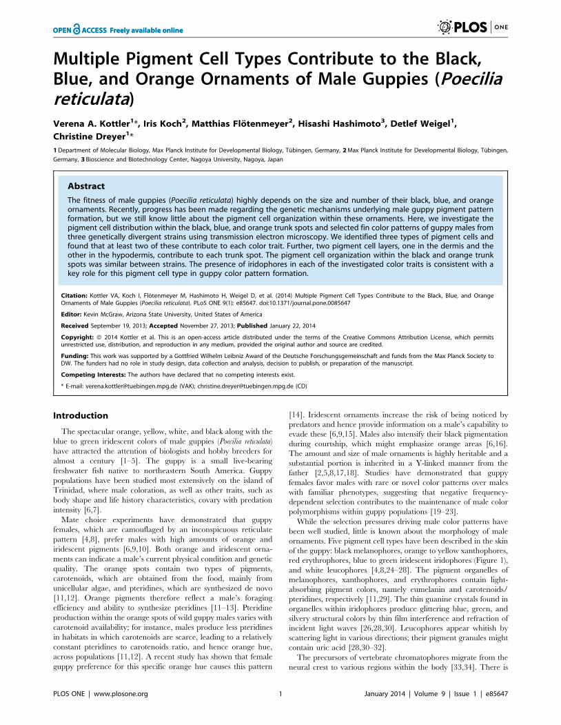
ornaments. Five pigment cell types have been described in the skin
of the guppy: black melanophores, orange to yellow xanthophores,
red erythrophores, blue to green iridescent iridophores (Figure 1),
and white leucophores [4,8,24–28]. The pigment organelles of
melanophores, xanthophores, and erythrophores contain light-
absorbing pigment colors, namely eumelanin and carotenoids/
pteridines, respectively [11,29]. The thin guanine crystals found in
organelles within iridophores produce glittering blue, green, and
silvery structural colors by thin film interference and refraction of
incident light waves [26,28,30]. Leucophores appear whitish by
scattering light in various directions; their pigment granules might
contain uric acid [28,30–32].
The precursors of vertebrate chromatophores migrate from the
neural crest to various regions within the body [33,34]. There is
PLOS ONE | www.plosone.org 1 January 2014 | Volume 9 | Issue 1 | e85647
increasing evidence that both short- and long-range interactions
between different types of pigment cells are required for proper
migration, differentiation, and survival of their precursors. For
example, during zebrafish (Danio rerio) pigment pattern develop-
ment, iridophores stimulate xanthophore precursors to migrate to
the prospective interstripe regions, but inhibit melanoblast
localization to these areas, which then accumulate in the adjacent
stripes [35]. Similarly, zebrafish xanthophores promote stripe
development by short-range inhibitory and long-range stimulating
interactions with melanophores [36–40]. Xanthophore-melano-
phore interactions are also crucial for the development of the
male-specific pattern of the guppy, as male guppies lacking
xanthophores due to a mutation in colony-stimulating factor 1 receptor a
(csf1ra) also have severe melanophore localization defects [8]. To
unravel such interactions in the guppy, it is critical to understand
how different pigment cell types are organized within the male
ornaments, which can be assessed best by transmission electron
microscopy (TEM) [41–44]. Previous TEM studies on guppy
pigment cells focused on the identification of different chromato-
phore types in the tail fins of adult males and on the development
of lateral iridophores on the trunk [24–28]. The guppies
investigated in these prior studies were obtained from pet shops.
Unfortunately, the precise position of the ornaments on the guppy
body was not documented, making it difficult to relate these TEM
images to the macroscopically visible ornaments.
Here, we describe the pigment cell distribution, for which we
subsequently use the term ultrastructure, within the blue, black,
and orange trunk spots and fin color patterns of male wild-type
guppies from three genetically divergent strains. TEM revealed
that several chromatophore types contribute to each ornament.
We could, however, not identify any leucophore. Our compre-
hensive study on pigment cell distribution in the skin of male
guppies provides a foundation from which the natural variation in
the placement and expression of male ornaments can be studied.
Results and Discussion
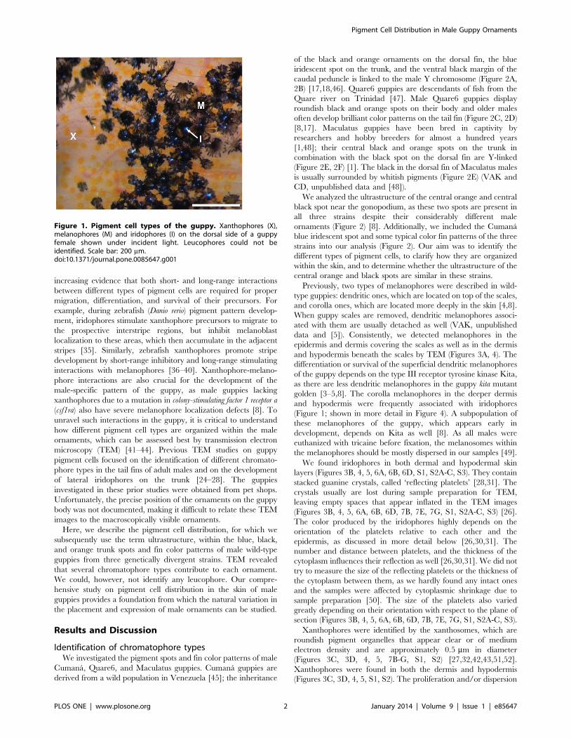
Identification of chromatophore typesWe investigated the pigment spots and fin color patterns of male
Cumana, Quare6, and Maculatus guppies. Cumana guppies are
derived from a wild population in Venezuela [45]; the inheritance
of the black and orange ornaments on the dorsal fin, the blue
iridescent spot on the trunk, and the ventral black margin of the
caudal peduncle is linked to the male Y chromosome (Figure 2A,
2B) [17,18,46]. Quare6 guppies are descendants of fish from the
Quare river on Trinidad [47]. Male Quare6 guppies display
roundish black and orange spots on their body and older males
often develop brilliant color patterns on the tail fin (Figure 2C, 2D)
[8,17]. Maculatus guppies have been bred in captivity by
researchers and hobby breeders for almost a hundred years
[1,48]; their central black and orange spots on the trunk in
combination with the black spot on the dorsal fin are Y-linked
(Figure 2E, 2F) [1]. The black in the dorsal fin of Maculatus males
is usually surrounded by whitish pigments (Figure 2E) (VAK and
CD, unpublished data and [48]).
We analyzed the ultrastructure of the central orange and central
black spot near the gonopodium, as these two spots are present in
all three strains despite their considerably different male
ornaments (Figure 2) [8]. Additionally, we included the Cumana
blue iridescent spot and some typical color fin patterns of the three
strains into our analysis (Figure 2). Our aim was to identify the
different types of pigment cells, to clarify how they are organized
within the skin, and to determine whether the ultrastructure of the
central orange and black spots are similar in these strains.
Previously, two types of melanophores were described in wild-
type guppies: dendritic ones, which are located on top of the scales,
and corolla ones, which are located more deeply in the skin [4,8].
When guppy scales are removed, dendritic melanophores associ-
ated with them are usually detached as well (VAK, unpublished
data and [5]). Consistently, we detected melanophores in the
epidermis and dermis covering the scales as well as in the dermis
and hypodermis beneath the scales by TEM (Figures 3A, 4). The
differentiation or survival of the superficial dendritic melanophores
of the guppy depends on the type III receptor tyrosine kinase Kita,
as there are less dendritic melanophores in the guppy kita mutant
golden [3–5,8]. The corolla melanophores in the deeper dermis
and hypodermis were frequently associated with iridophores
(Figure 1; shown in more detail in Figure 4). A subpopulation of
these melanophores of the guppy, which appears early in
development, depends on Kita as well [8]. As all males were
euthanized with tricaine before fixation, the melanosomes within
the melanophores should be mostly dispersed in our samples [49].
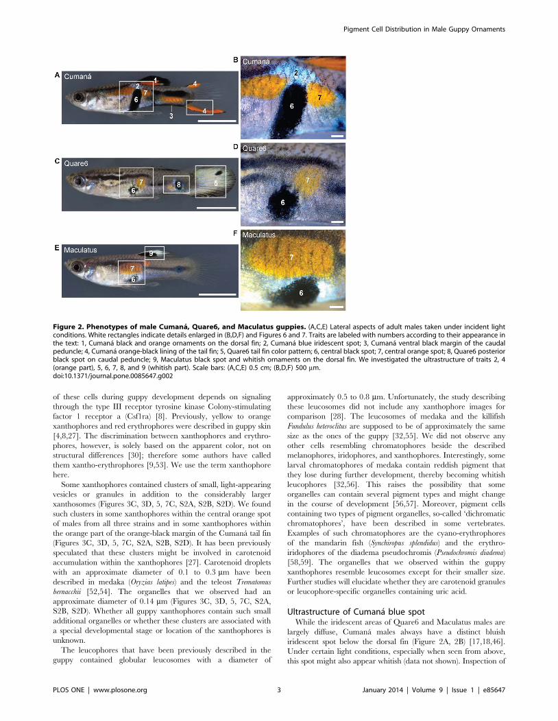
We found iridophores in both dermal and hypodermal skin
layers (Figures 3B, 4, 5, 6A, 6B, 6D, S1, S2A-C, S3). They contain
stacked guanine crystals, called ‘reflecting platelets’ [28,31]. The
crystals usually are lost during sample preparation for TEM,
leaving empty spaces that appear inflated in the TEM images
(Figures 3B, 4, 5, 6A, 6B, 6D, 7B, 7E, 7G, S1, S2A-C, S3) [26].
The color produced by the iridophores highly depends on the
orientation of the platelets relative to each other and the
epidermis, as discussed in more detail below [26,30,31]. The
number and distance between platelets, and the thickness of the
cytoplasm influences their reflection as well [26,30,31]. We did not
try to measure the size of the reflecting platelets or the thickness of
the cytoplasm between them, as we hardly found any intact ones
and the samples were affected by cytoplasmic shrinkage due to
sample preparation [50]. The size of the platelets also varied
greatly depending on their orientation with respect to the plane of
section (Figures 3B, 4, 5, 6A, 6B, 6D, 7B, 7E, 7G, S1, S2A-C, S3).
Xanthophores were identified by the xanthosomes, which are
roundish pigment organelles that appear clear or of medium
electron density and are approximately 0.5 mm in diameter
(Figures 3C, 3D, 4, 5, 7B-G, S1, S2) [27,32,42,43,51,52].
Xanthophores were found in both the dermis and hypodermis
(Figures 3C, 3D, 4, 5, S1, S2). The proliferation and/or dispersion
Figure 1. Pigment cell types of the guppy. Xanthophores (X),melanophores (M) and iridophores (I) on the dorsal side of a guppyfemale shown under incident light. Leucophores could not beidentified. Scale bar: 200 mm.doi:10.1371/journal.pone.0085647.g001
Pigment Cell Distribution in Male Guppy Ornaments
PLOS ONE | www.plosone.org 2 January 2014 | Volume 9 | Issue 1 | e85647
of these cells during guppy development depends on signaling
through the type III receptor tyrosine kinase Colony-stimulating
factor 1 receptor a (Csf1ra) [8]. Previously, yellow to orange
xanthophores and red erythrophores were described in guppy skin
[4,8,27]. The discrimination between xanthophores and erythro-
phores, however, is solely based on the apparent color, not on
structural differences [30]; therefore some authors have called
them xantho-erythrophores [9,53]. We use the term xanthophore
here.
Some xanthophores contained clusters of small, light-appearing
vesicles or granules in addition to the considerably larger
xanthosomes (Figures 3C, 3D, 5, 7C, S2A, S2B, S2D). We found
such clusters in some xanthophores within the central orange spot
of males from all three strains and in some xanthophores within
the orange part of the orange-black margin of the Cumana tail fin
(Figures 3C, 3D, 5, 7C, S2A, S2B, S2D). It has been previously
speculated that these clusters might be involved in carotenoid
accumulation within the xanthophores [27]. Carotenoid droplets
with an approximate diameter of 0.1 to 0.3 mm have been
described in medaka (Oryzias latipes) and the teleost Trematomus
bernacchii [52,54]. The organelles that we observed had an
approximate diameter of 0.14 mm (Figures 3C, 3D, 5, 7C, S2A,
S2B, S2D). Whether all guppy xanthophores contain such small
additional organelles or whether these clusters are associated with
a special developmental stage or location of the xanthophores is
unknown.
The leucophores that have been previously described in the
guppy contained globular leucosomes with a diameter of
approximately 0.5 to 0.8 mm. Unfortunately, the study describing
these leucosomes did not include any xanthophore images for
comparison [28]. The leucosomes of medaka and the killifish
Fundulus heteroclitus are supposed to be of approximately the same
size as the ones of the guppy [32,55]. We did not observe any
other cells resembling chromatophores beside the described
melanophores, iridophores, and xanthophores. Interestingly, some
larval chromatophores of medaka contain reddish pigment that
they lose during further development, thereby becoming whitish
leucophores [32,56]. This raises the possibility that some
organelles can contain several pigment types and might change
in the course of development [56,57]. Moreover, pigment cells
containing two types of pigment organelles, so-called ‘dichromatic
chromatophores’, have been described in some vertebrates.
Examples of such chromatophores are the cyano-erythrophores
of the mandarin fish (Synchiropus splendidus) and the erythro-
iridophores of the diadema pseudochromis (Pseudochromis diadema)
[58,59]. The organelles that we observed within the guppy
xanthophores resemble leucosomes except for their smaller size.
Further studies will elucidate whether they are carotenoid granules
or leucophore-specific organelles containing uric acid.
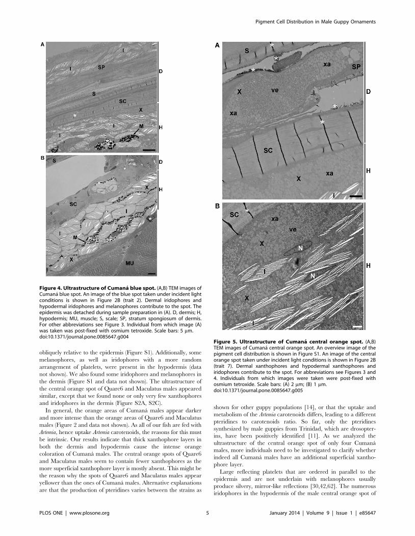
Ultrastructure of Cumana blue spotWhile the iridescent areas of Quare6 and Maculatus males are
largely diffuse, Cumana males always have a distinct bluish
iridescent spot below the dorsal fin (Figure 2A, 2B) [17,18,46].
Under certain light conditions, especially when seen from above,
this spot might also appear whitish (data not shown). Inspection of
Figure 2. Phenotypes of male Cumana, Quare6, and Maculatus guppies. (A,C,E) Lateral aspects of adult males taken under incident lightconditions. White rectangles indicate details enlarged in (B,D,F) and Figures 6 and 7. Traits are labeled with numbers according to their appearance inthe text: 1, Cumana black and orange ornaments on the dorsal fin; 2, Cumana blue iridescent spot; 3, Cumana ventral black margin of the caudalpeduncle; 4, Cumana orange-black lining of the tail fin; 5, Quare6 tail fin color pattern; 6, central black spot; 7, central orange spot; 8, Quare6 posteriorblack spot on caudal peduncle; 9, Maculatus black spot and whitish ornaments on the dorsal fin. We investigated the ultrastructure of traits 2, 4(orange part), 5, 6, 7, 8, and 9 (whitish part). Scale bars: (A,C,E) 0.5 cm; (B,D,F) 500 mm.doi:10.1371/journal.pone.0085647.g002
Pigment Cell Distribution in Male Guppy Ornaments
PLOS ONE | www.plosone.org 3 January 2014 | Volume 9 | Issue 1 | e85647
TEM images revealed that this spot is formed by two sheets of
iridophores, one of which is located in the stratum spongiosum of
the dermis and the other in the hypodermis (Figure 4). Just below
the hypodermal iridophores, on top of the muscles, we found
melanophores whose appendices frequently protruded into the
iridophore layer (Figure 4). The melanophores did not form a
complete sheet; in some areas the iridophores were in direct
contact with the underlying muscle layer (Figure 4). Melanophores
were also present within the dermal iridophore layer (data not
shown). The hypodermal as well as the dermal iridophore sheet
contained some xanthophores, too (Figure 4 and data not shown).
The reflecting platelets of the iridophores appeared to be
randomly distributed and were tilted in different directions at
some locations, whereas they looked more organized at other
locations (Figure 4).
A previous study on the development of iridophores on the
lateral trunk of fancy guppies reported that all reflecting platelets
had an angle of approximately 15–30u relative to the surface of the
fish, thought to account for the blue-green reflection with a
wavelength of 496 nm [26]. The light blue coloration of the
common surgeonfish (Paracanthurus hepatus) is derived from a double
layer of iridophores in which the reflective platelets are oriented
almost in parallel relative to the fish surface; the iridophores are
located on top of melanophores [60]. Ordered iridophores above
melanophores have also been observed in the blue skin of the blue-
green damselfish (Chromis viridis) and the lizard Plestiodon latiscutatus
[44,61]. In contrast, randomly arranged reflecting platelets usually
seem to produce a whitish coloration, e.g. in the white spots of the
domino damsel (Dascyllus trimaculatus) [41]. We found that both
disordered and ordered reflecting platelets are present within the
bluish to whitish spot of Cumana males. The appearance of this
spot is dynamic and depends on the angle of the incident light and
the movement of the fish. The bluish coloration is presumably
derived from the platelets that are arranged in parallel, while the
whitish color comes from the disordered platelets. Interestingly,
melanophores contribute to the ultrastructure of the blue
ornament of the Cumana guppy like in the common surgeonfish,
the blue-green damselfish, and P. latiscutatus. Even within the
stratum compactum, a melanophore was found in close contact
with an iridophore (Figure 4B). We suspect that the melanophores
modulate the reflection of the iridophores.
Ultrastructure of central orange spotWe detected large accumulations of xanthophores in the
stratum spongiosum of the dermis and hypodermis within the
Cumana central orange spot (Figures 5, S1). These xanthophores
frequently contained clusters of the small vesicles or granules
described above (Figure 5). Between the xanthophores in the
hypodermis were numerous iridophores, with reflecting platelets
aligned in parallel (Figures 5B, S1). They were arranged slightly
Figure 3. TEM images of guppy chromatophore types. (A) Melanophore on top of a scale in the dermis. Melanophores can be recognized bytheir dark-appearing pigment organelles, the eumelanin-containing melanosomes. (B) Melanophores, xanthophores, and iridophores in thehypodermis of the central orange spot of a Maculatus male. (C,D) Dermal xanthophores within the central orange spot of a Cumana male. BL, basallamina demarcating the boundary between the epidermis and dermis; E, epidermis; EO, external osseous layer of scale; I, iridophore; IF, internalfibrillary plate of scale; M, melanophore; MR, microridges of the epidermis; N, nucleus; SC, stratum compactum of dermis; ve, small vesicles orgranules described in the text; X, xanthophore; xa, xanthosomes. Asterisks exemplarily mark artifacts caused by sample preparation; inflated emptyspaces within iridophores are not marked. Individuals from which images (B-D) were taken were post-fixed with osmium tetroxide. Scale bars: 2 mm.doi:10.1371/journal.pone.0085647.g003
Pigment Cell Distribution in Male Guppy Ornaments
PLOS ONE | www.plosone.org 4 January 2014 | Volume 9 | Issue 1 | e85647
obliquely relative to the epidermis (Figure S1). Additionally, some
melanophores, as well as iridophores with a more random
arrangement of platelets, were present in the hypodermis (data
not shown). We also found some iridophores and melanophores in
the dermis (Figure S1 and data not shown). The ultrastructure of
the central orange spot of Quare6 and Maculatus males appeared
similar, except that we found none or only very few xanthophores
and iridophores in the dermis (Figure S2A, S2C).
In general, the orange areas of Cumana males appear darker
and more intense than the orange areas of Quare6 and Maculatus
males (Figure 2 and data not shown). As all of our fish are fed with
Artemia, hence uptake Artemia carotenoids, the reasons for this must
be intrinsic. Our results indicate that thick xanthophore layers in
both the dermis and hypodermis cause the intense orange
coloration of Cumana males. The central orange spots of Quare6
and Maculatus males seem to contain fewer xanthophores as the
more superficial xanthophore layer is mostly absent. This might be
the reason why the spots of Quare6 and Maculatus males appear
yellower than the ones of Cumana males. Alternative explanations
are that the production of pteridines varies between the strains as
shown for other guppy populations [14], or that the uptake and
metabolism of the Artemia carotenoids differs, leading to a different
pteridines to carotenoids ratio. So far, only the pteridines
synthesized by male guppies from Trinidad, which are drosopter-
ins, have been positively identified [11]. As we analyzed the
ultrastructure of the central orange spot of only four Cumana
males, more individuals need to be investigated to clarify whether
indeed all Cumana males have an additional superficial xantho-
phore layer.
Large reflecting platelets that are ordered in parallel to the
epidermis and are not underlain with melanophores usually
produce silvery, mirror-like reflections [30,42,62]. The numerous
iridophores in the hypodermis of the male central orange spot of
Figure 4. Ultrastructure of Cumana blue spot. (A,B) TEM images ofCumana blue spot. An image of the blue spot taken under incident lightconditions is shown in Figure 2B (trait 2). Dermal iridophores andhypodermal iridophores and melanophores contribute to the spot. Theepidermis was detached during sample preparation in (A). D, dermis; H,hypodermis; MU, muscle; S, scale; SP, stratum spongiosum of dermis.For other abbreviations see Figure 3. Individual from which image (A)was taken was post-fixed with osmium tetroxide. Scale bars: 5 mm.doi:10.1371/journal.pone.0085647.g004
Figure 5. Ultrastructure of Cumana central orange spot. (A,B)TEM images of Cumana central orange spot. An overview image of thepigment cell distribution is shown in Figure S1. An image of the centralorange spot taken under incident light conditions is shown in Figure 2B(trait 7). Dermal xanthophores and hypodermal xanthophores andiridophores contribute to the spot. For abbreviations see Figures 3 and4. Individuals from which images were taken were post-fixed withosmium tetroxide. Scale bars: (A) 2 mm; (B) 1 mm.doi:10.1371/journal.pone.0085647.g005
Pigment Cell Distribution in Male Guppy Ornaments
PLOS ONE | www.plosone.org 5 January 2014 | Volume 9 | Issue 1 | e85647
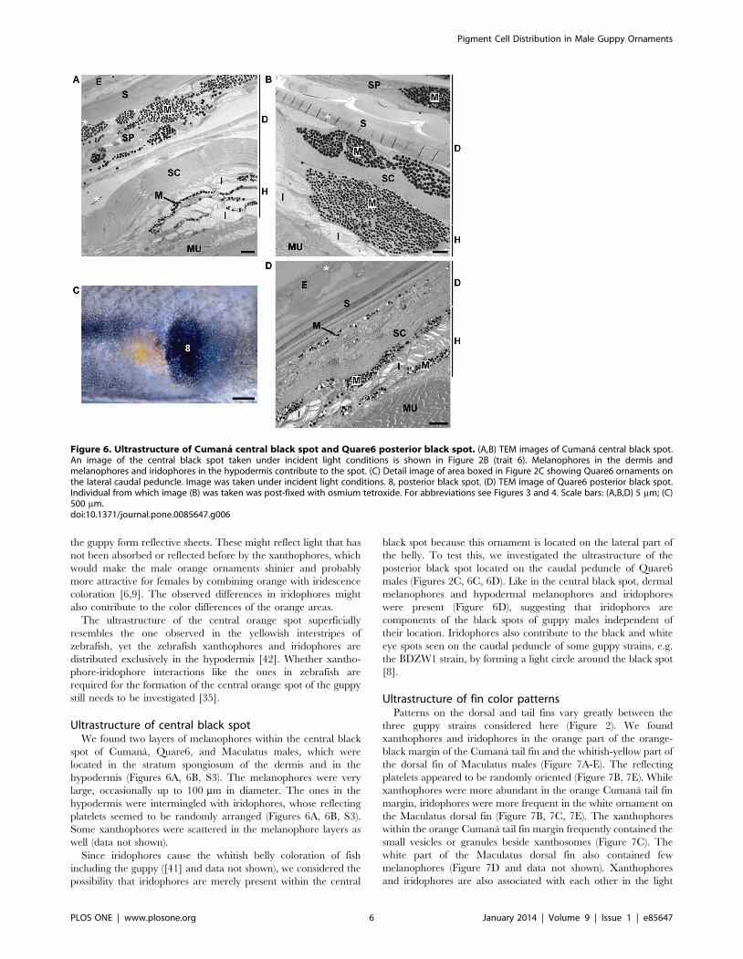
the guppy form reflective sheets. These might reflect light that has
not been absorbed or reflected before by the xanthophores, which
would make the male orange ornaments shinier and probably
more attractive for females by combining orange with iridescence
coloration [6,9]. The observed differences in iridophores might
also contribute to the color differences of the orange areas.
The ultrastructure of the central orange spot superficially
resembles the one observed in the yellowish interstripes of
zebrafish, yet the zebrafish xanthophores and iridophores are
distributed exclusively in the hypodermis [42]. Whether xantho-
phore-iridophore interactions like the ones in zebrafish are
required for the formation of the central orange spot of the guppy
still needs to be investigated [35].
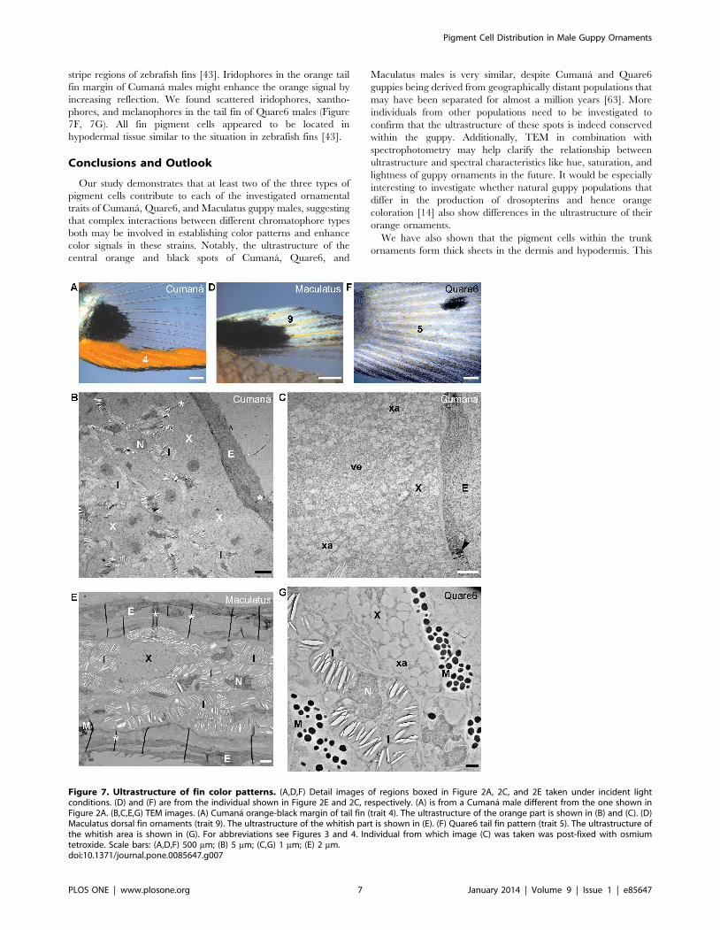
Ultrastructure of central black spotWe found two layers of melanophores within the central black
spot of Cumana, Quare6, and Maculatus males, which were
located in the stratum spongiosum of the dermis and in the
hypodermis (Figures 6A, 6B, S3). The melanophores were very
large, occasionally up to 100 mm in diameter. The ones in the
hypodermis were intermingled with iridophores, whose reflecting
platelets seemed to be randomly arranged (Figures 6A, 6B, S3).
Some xanthophores were scattered in the melanophore layers as
well (data not shown).
Since iridophores cause the whitish belly coloration of fish
including the guppy ([41] and data not shown), we considered the
possibility that iridophores are merely present within the central
black spot because this ornament is located on the lateral part of
the belly. To test this, we investigated the ultrastructure of the
posterior black spot located on the caudal peduncle of Quare6
males (Figures 2C, 6C, 6D). Like in the central black spot, dermal
melanophores and hypodermal melanophores and iridophores
were present (Figure 6D), suggesting that iridophores are
components of the black spots of guppy males independent of
their location. Iridophores also contribute to the black and white
eye spots seen on the caudal peduncle of some guppy strains, e.g.
the BDZW1 strain, by forming a light circle around the black spot
[8].
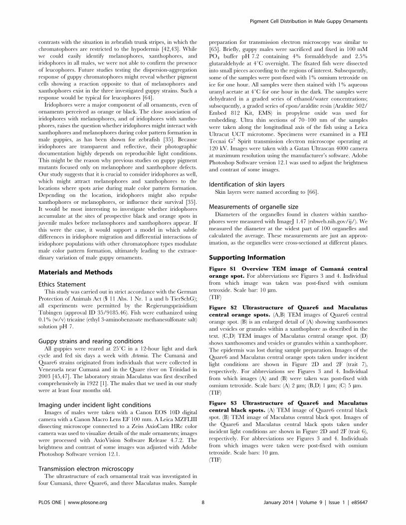
Ultrastructure of fin color patternsPatterns on the dorsal and tail fins vary greatly between the
three guppy strains considered here (Figure 2). We found
xanthophores and iridophores in the orange part of the orange-
black margin of the Cumana tail fin and the whitish-yellow part of
the dorsal fin of Maculatus males (Figure 7A-E). The reflecting
platelets appeared to be randomly oriented (Figure 7B, 7E). While
xanthophores were more abundant in the orange Cumana tail fin
margin, iridophores were more frequent in the white ornament on
the Maculatus dorsal fin (Figure 7B, 7C, 7E). The xanthophores
within the orange Cumana tail fin margin frequently contained the
small vesicles or granules beside xanthosomes (Figure 7C). The
white part of the Maculatus dorsal fin also contained few
melanophores (Figure 7D and data not shown). Xanthophores
and iridophores are also associated with each other in the light
Figure 6. Ultrastructure of Cumana central black spot and Quare6 posterior black spot. (A,B) TEM images of Cumana central black spot.An image of the central black spot taken under incident light conditions is shown in Figure 2B (trait 6). Melanophores in the dermis andmelanophores and iridophores in the hypodermis contribute to the spot. (C) Detail image of area boxed in Figure 2C showing Quare6 ornaments onthe lateral caudal peduncle. Image was taken under incident light conditions. 8, posterior black spot. (D) TEM image of Quare6 posterior black spot.Individual from which image (B) was taken was post-fixed with osmium tetroxide. For abbreviations see Figures 3 and 4. Scale bars: (A,B,D) 5 mm; (C)500 mm.doi:10.1371/journal.pone.0085647.g006
Pigment Cell Distribution in Male Guppy Ornaments
PLOS ONE | www.plosone.org 6 January 2014 | Volume 9 | Issue 1 | e85647
stripe regions of zebrafish fins [43]. Iridophores in the orange tail
fin margin of Cumana males might enhance the orange signal by
increasing reflection. We found scattered iridophores, xantho-
phores, and melanophores in the tail fin of Quare6 males (Figure
7F, 7G). All fin pigment cells appeared to be located in
hypodermal tissue similar to the situation in zebrafish fins [43].
Conclusions and Outlook
Our study demonstrates that at least two of the three types of
pigment cells contribute to each of the investigated ornamental
traits of Cumana, Quare6, and Maculatus guppy males, suggesting
that complex interactions between different chromatophore types
both may be involved in establishing color patterns and enhance
color signals in these strains. Notably, the ultrastructure of the
central orange and black spots of Cumana, Quare6, and
Maculatus males is very similar, despite Cumana and Quare6
guppies being derived from geographically distant populations that
may have been separated for almost a million years [63]. More
individuals from other populations need to be investigated to
confirm that the ultrastructure of these spots is indeed conserved
within the guppy. Additionally, TEM in combination with
spectrophotometry may help clarify the relationship between
ultrastructure and spectral characteristics like hue, saturation, and
lightness of guppy ornaments in the future. It would be especially
interesting to investigate whether natural guppy populations that
differ in the production of drosopterins and hence orange
coloration [14] also show differences in the ultrastructure of their
orange ornaments.
We have also shown that the pigment cells within the trunk
ornaments form thick sheets in the dermis and hypodermis. This
Figure 7. Ultrastructure of fin color patterns. (A,D,F) Detail images of regions boxed in Figure 2A, 2C, and 2E taken under incident lightconditions. (D) and (F) are from the individual shown in Figure 2E and 2C, respectively. (A) is from a Cumana male different from the one shown inFigure 2A. (B,C,E,G) TEM images. (A) Cumana orange-black margin of tail fin (trait 4). The ultrastructure of the orange part is shown in (B) and (C). (D)Maculatus dorsal fin ornaments (trait 9). The ultrastructure of the whitish part is shown in (E). (F) Quare6 tail fin pattern (trait 5). The ultrastructure ofthe whitish area is shown in (G). For abbreviations see Figures 3 and 4. Individual from which image (C) was taken was post-fixed with osmiumtetroxide. Scale bars: (A,D,F) 500 mm; (B) 5 mm; (C,G) 1 mm; (E) 2 mm.doi:10.1371/journal.pone.0085647.g007
Pigment Cell Distribution in Male Guppy Ornaments
PLOS ONE | www.plosone.org 7 January 2014 | Volume 9 | Issue 1 | e85647
contrasts with the situation in zebrafish trunk stripes, in which the
chromatophores are restricted to the hypodermis [42,43]. While
we could easily identify melanophores, xanthophores, and
iridophores in all males, we were not able to confirm the presence
of leucophores. Future studies testing the dispersion-aggregation
response of guppy chromatophores might reveal whether pigment
cells showing a reaction opposite to that of melanophores and
xanthophores exist in the three investigated guppy strains. Such a
response would be typical for leucophores [64].
Iridophores were a major component of all ornaments, even of
ornaments perceived as orange or black. The close association of
iridophores with melanophores, and of iridophores with xantho-
phores, raises the question whether iridophores might interact with
xanthophores and melanophores during color pattern formation in
male guppies, as has been shown for zebrafish [35]. Because
iridophores are transparent and reflective, their photographic
documentation highly depends on reproducible light conditions.
This might be the reason why previous studies on guppy pigment
mutants focused only on melanophore and xanthophore defects.
Our study suggests that it is crucial to consider iridophores as well,
which might attract melanophores and xanthophores to the
locations where spots arise during male color pattern formation.
Depending on the location, iridophores might also repulse
xanthophores or melanophores, or influence their survival [35].
It would be most interesting to investigate whether iridophores
accumulate at the sites of prospective black and orange spots in
juvenile males before melanophores and xanthophores appear. If
this were the case, it would support a model in which subtle
differences in iridophore migration and differential interactions of
iridophore populations with other chromatophore types modulate
male color pattern formation, ultimately leading to the extraor-
dinary variation of male guppy ornaments.
Materials and Methods
Ethics StatementThis study was carried out in strict accordance with the German
Protection of Animals Act (1 11 Abs. 1 Nr. 1 a und b TierSchG);
all experiments were permitted by the Regierungsprasidium
Tubingen (approval ID 35/9185.46). Fish were euthanized using
0.1% (w/v) tricaine (ethyl 3-aminobenzoate methanesulfonate salt)
solution pH 7.
Guppy strains and rearing conditionsAll guppies were reared at 25uC in a 12-hour light and dark
cycle and fed six days a week with Artemia. The Cumana and
Quare6 strains originated from individuals that were collected in
Venezuela near Cumana and in the Quare river on Trinidad in
2003 [45,47]. The laboratory strain Maculatus was first described
comprehensively in 1922 [1]. The males that we used in our study
were at least four months old.
Imaging under incident light conditionsImages of males were taken with a Canon EOS 10D digital
camera with a Canon Macro Lens EF 100 mm. A Leica MZFLIII
dissecting microscope connected to a Zeiss AxioCam HRc color
camera was used to visualize details of the male ornaments; images
were processed with AxioVision Software Release 4.7.2. The
brightness and contrast of some images was adjusted with Adobe
Photoshop Software version 12.1.
Transmission electron microscopyThe ultrastructure of each ornamental trait was investigated in
four Cumana, three Quare6, and three Maculatus males. Sample
preparation for transmission electron microscopy was similar to
[65]. Briefly, guppy males were sacrificed and fixed in 100 mM
PO4 buffer pH 7.2 containing 4% formaldehyde and 2.5%
glutaraldehyde at 4uC overnight. The fixated fish were dissected
into small pieces according to the regions of interest. Subsequently,
some of the samples were post-fixed with 1% osmium tetroxide on
ice for one hour. All samples were then stained with 1% aqueous
uranyl acetate at 4uC for one hour in the dark. The samples were
dehydrated in a graded series of ethanol/water concentrations;
subsequently, a graded series of epon/araldite resin (Araldite 502/
Embed 812 Kit, EMS) in propylene oxide was used for
embedding. Ultra thin sections of 70–100 nm of the samples
were taken along the longitudinal axis of the fish using a Leica
Ultracut UCT microtome. Specimens were examined in a FEI
Tecnai G2 Spirit transmission electron microscope operating at
120 kV. Images were taken with a Gatan Ultrascan 4000 camera
at maximum resolution using the manufacturer’s software. Adobe
Photoshop Software version 12.1 was used to adjust the brightness
and contrast of some images.
Identification of skin layersSkin layers were named according to [66].
Measurements of organelle sizeDiameters of the organelles found in clusters within xantho-
phores were measured with ImageJ 1.47 (rsbweb.nih.gov/ij/). We
measured the diameter at the widest part of 100 organelles and
calculated the average. These measurements are just an approx-
imation, as the organelles were cross-sectioned at different planes.
Supporting Information
Figure S1 Overview TEM image of Cumana centralorange spot. For abbreviations see Figures 3 and 4. Individual
from which image was taken was post-fixed with osmium
tetroxide. Scale bar: 10 mm.
(TIF)
Figure S2 Ultrastructure of Quare6 and Maculatuscentral orange spots. (A,B) TEM images of Quare6 central
orange spot. (B) is an enlarged detail of (A) showing xanthosomes
and vesicles or granules within a xanthophore as described in the
text. (C,D) TEM images of Maculatus central orange spot. (D)
shows xanthosomes and vesicles or granules within a xanthophore.
The epidermis was lost during sample preparation. Images of the
Quare6 and Maculatus central orange spots taken under incident
light conditions are shown in Figure 2D and 2F (trait 7),
respectively. For abbreviations see Figures 3 and 4. Individual
from which images (A) and (B) were taken was post-fixed with
osmium tetroxide. Scale bars: (A) 2 mm; (B,D) 1 mm; (C) 5 mm.
(TIF)
Figure S3 Ultrastructure of Quare6 and Maculatuscentral black spots. (A) TEM image of Quare6 central black
spot. (B) TEM image of Maculatus central black spot. Images of
the Quare6 and Maculatus central black spots taken under
incident light conditions are shown in Figure 2D and 2F (trait 6),
respectively. For abbreviations see Figures 3 and 4. Individuals
from which images were taken were post-fixed with osmium
tetroxide. Scale bars: 10 mm.
(TIF)
Pigment Cell Distribution in Male Guppy Ornaments
PLOS ONE | www.plosone.org 8 January 2014 | Volume 9 | Issue 1 | e85647
Acknowledgments
We thank Makoto Goda for helping to identify the basal lamina; Felix
Breden for the Cumana guppy strain; David Reznick for the Quare6 guppy
strain, and Manfred Schartl for the Maculatus guppy strain.
Author Contributions
Conceived and designed the experiments: VAK CD. Performed the
experiments: VAK IK MF. Analyzed the data: VAK IK MF HH. Wrote
the paper: VAK. Provided helpful comments on the manuscript: IK MF
HH DW CD. Oversaw the experimental design and data analysis: DW.
References
1. Winge O (1922) One-sided masculine and sex-linked inheritance in Lebistes
reticulatus. Journal of Genetics 12: 145–162.
2. Winge O (1927) The location of eighteen genes in Lebistes reticulatus. Journal ofGenetics 18: 1–43.
3. Haskins CP, Druzba JP (1938) Note on anomalous inheritance of sex-linked
color factors in the Guppy. Am Nat 72: 571–574.
4. Goodrich HB, Josephson ND, Trinkaus JP, Slate JM (1944) The cellular
expression and genetics of two new genes in Lebistes reticulatus. Genetics 29: 584–
592.
5. Winge O, Ditlevsen E (1947) Colour inheritance and sex determination in
Lebistes. Heredity 1: 65–83.
6. Endler JA (1983) Natural and sexual selection on color patterns in poeciliidfishes. Environmental biology of Fishes 9: 173–190.
7. Endler JA (1995) Multiple-trait coevolution and environmental gradients inguppies. Trends Ecol Evol 10: 22–29.
8. Kottler VA, Fadeev A, Weigel D, Dreyer C (2013) Pigment Pattern Formationin the Guppy, Poecilia reticulata, Involves the Kita and Csf1ra Receptor Tyrosine
Kinases. Genetics 194: 631–646.
9. Kodric-Brown A (1985) Female preference and sexual selection for male
coloration in the guppy (Poecilia reticulata). Behavioral Ecology and Sociobiology17: 199–205.
10. Houde AE (1987) Mate choice based upon naturally occurring color-patternvariation in a guppy population. Evolution: 1–10.
11. Grether GF, Hudon J, Endler JA (2001) Carotenoid scarcity, synthetic pteridinepigments and the evolution of sexual coloration in guppies (Poecilia reticulata).
Proceedings of the Royal Society of London Series B: Biological Sciences 268:
1245–1253.
12. Grether GF, Cummings ME, Hudon J (2005) Countergradient variation in thesexual coloration of guppies (Poecilia reticulata): drosopterin synthesis balances
carotenoid availability. Evolution 59: 175–188.
13. Endler JA (1980) Natural selection on color patterns in Poecilia reticulata.
Evolution 34: 76–91.
14. Deere KA, Grether GF, Sun A, Sinsheimer JS (2012) Female mate preference
explains countergradient variation in the sexual coloration of guppies (Poecilia
reticulata). Proc Biol Sci 279: 1684–1690.
15. Endler JA (1987) Predation, light intensity and courtship behaviour in Poecilia
reticulata (Pisces: Poeciliidae). Animal Behaviour 35: 1376–1385.
16. Brooks R (1996) Melanin as a visual signal amplifier in male guppies.Naturwissenschaften 83: 39–41.
17. Tripathi N, Hoffmann M, Dreyer C (2008) Natural variation of maleornamental traits of the guppy, Poecilia reticulata. Zebrafish 5: 265–278.
18. Tripathi N, Hoffmann M, Willing EM, Lanz C, Weigel D, et al. (2009) Geneticlinkage map of the guppy, Poecilia reticulata, and quantitative trait loci analysis of
male size and colour variation. Proc Biol Sci 276: 2195–2208.
19. Eakley AL, Houde AE (2004) Possible role of female discrimination against
’redundant’ males in the evolution of colour pattern polymorphism in guppies.Proc Biol Sci 271 Suppl 5: S299–301.
20. Farr JA (1977) Male rarity or novelty, female choice behavior, and sexualselection in the guppy, Poecilia reticulata Peters (Pisces: Poeciliidae). Evolution:
162–168.
21. Farr JA (1980) Social behavior patterns as determinants of reproductive success
in the guppy, Poecilia reticulata Peters (Pisces: Poeciliidae): an experimental studyof the effects of intermale competition, female choice, and sexual selection.
Behaviour: 38–91.
22. Hughes KA, Du L, Rodd FH, Reznick DN (1999) Familiarity leads to female
mate preference for novel males in the guppy, Poecilia reticulata. Anim Behav 58:
907–916.
23. Hughes KA, Houde AE, Price AC, Rodd FH (2013) Mating advantage for raremales in wild guppy populations. Nature 503: 108–110.
24. Fujii R (1966) A functional interpretation of the fine structure in themelanophore of the guppy, Lebistes reticulatus. Annot Zool Japon 39: 185–192.
25. Fujii R, Taguchi S (1970) Ultrastructure of nerve-melanophore relationships inthe guppy, Lebistes reticulatus. Annot Zool Japon 43: 123–131.
26. Gundersen R, Rivera E (1982) An ultrastructural study of the development ofthe dermal iridophores and structural pigmentation in Poecilia reticulata (Peters).
Journal of Morphology 172: 349–359.
27. Takeuchi IK (1975) Electron microscopic study on erythrophores of the guppy,
Lebistes reticulatus Peters. Annot Zool Japon 48: 242–251.
28. Takeuchi IK (1976) Electron microscopy of two types of reflecting chromato-
phores (iridophores and leucophores) in the guppy, Lebistes reticulatus Peters. CellTissue Res 173: 17–27.
29. Braasch I, Schartl M, Volff JN (2007) Evolution of pigment synthesis pathwaysby gene and genome duplication in fish. BMC Evol Biol 7: 74.
30. Fujii R (1993) Cytophysiology of fish chromatophores. International Review ofCytology 143: 191–255.
31. Fujii R (2000) The regulation of motile activity in fish chromatophores. Pigment
Cell Res 13: 300–319.
32. Hama T (1975) Chromatophores and iridocytes. Medaka (Killifish): Biology and
Strains: 138–153.
33. Kelsh RN, Harris ML, Colanesi S, Erickson CA (2009) Stripes and belly-spots --
a review of pigment cell morphogenesis in vertebrates. Semin Cell Dev Biol 20:90–104.
34. Sauka-Spengler T, Bronner-Fraser M (2008) A gene regulatory network
orchestrates neural crest formation. Nat Rev Mol Cell Biol 9: 557–568.
35. Patterson LB, Parichy DM (2013) Interactions with Iridophores and the Tissue
Environment Required for Patterning Melanophores and Xanthophores during
Zebrafish Adult Pigment Stripe Formation. PLoS Genet 9: e1003561.
36. Parichy DM, Ransom DG, Paw B, Zon LI, Johnson SL (2000) An orthologue of
the kit-related gene fms is required for development of neural crest-derived
xanthophores and a subpopulation of adult melanocytes in the zebrafish, Danio
rerio. Development 127: 3031–3044.
37. Parichy DM, Turner JM (2003) Temporal and cellular requirements for Fms
signaling during zebrafish adult pigment pattern development. Development
130: 817–833.
38. Maderspacher F, Nusslein-Volhard C (2003) Formation of the adult pigment
pattern in zebrafish requires leopard and obelix dependent cell interactions.
Development 130: 3447–3457.
39. Nakamasu A, Takahashi G, Kanbe A, Kondo S (2009) Interactions between
zebrafish pigment cells responsible for the generation of Turing patterns. Proc
Natl Acad Sci U S A 106: 8429–8434.
40. Inaba M, Yamanaka H, Kondo S (2012) Pigment pattern formation by contact-
dependent depolarization. Science 335: 677.
41. Goda M, Fujii R (2001) Coloration and chromatophores of the domino damsel,Dascyllus trimaculatus. Zoolog Sci 18: 165–174.
42. Hirata M, Nakamura K, Kanemaru T, Shibata Y, Kondo S (2003) Pigment cell
organization in the hypodermis of zebrafish. Dev Dyn 227: 497–503.
43. Hirata M, Nakamura K, Kondo S (2005) Pigment cell distributions in different
tissues of the zebrafish, with special reference to the striped pigment pattern. Dev
Dyn 234: 293–300.
44. Kuriyama T, Miyaji K, Sugimoto M, Hasegawa M (2006) Ultrastructure of thedermal chromatophores in a lizard (Scincidae: Plestiodon latiscutatus) with
conspicuous body and tail coloration. Zoolog Sci 23: 793–799.
45. Alexander HJ, Breden F (2004) Sexual isolation and extreme morphologicaldivergence in the Cumana guppy: a possible case of incipient speciation. Journal
of Evolutionary Biology 17: 1238–1254.
46. Tripathi N, Hoffmann M, Weigel D, Dreyer C (2009) Linkage analysis revealsthe independent origin of Poeciliid sex chromosomes and a case of atypical sex
inheritance in the guppy (Poecilia reticulata). Genetics 182: 365–374.
47. Reznick D, Endler JA (1982) The impact of predation on life history evolution in
Trinidadian guppies (Poecilia reticulata). Evolution 36: 160–177.
48. Schmidt J (1920) Racial investigations. IV. The genetic behaviour of a secondary
sexual character. C R trav Labor Carlsberg 14.
49. Sheets L, Ransom DG, Mellgren EM, Johnson SL, Schnapp BJ (2007) Zebrafish
melanophilin facilitates melanosome dispersion by regulating dynein. Curr Biol
17: 1721–1734.
50. Morrison RL (1995) A transmission electron microscopic (TEM) method fordetermining structural colors reflected by lizard iridophores. Pigment Cell Res 8:
28–36.
51. Matsumoto J (1965) Studies on fine structure and cytochemical properties oferythrophores in swordtail, Xiphophorus helleri, with special reference to their
pigment granules (pterinosomes). The Journal of cell biology 27: 493–504.
52. Obika M (1993) Formation of pterinosomes and carotenoid granules inxanthophores of the teleost Oryzias latipes as revealed by the rapid-freezing and
freeze-substitution method. Cell Tissue Res 271: 81–86.
53. Goodrich H, Hill G, Arrick MS (1941) The chemical identification of gene-
controlled pigments in Platypoecilus and Xiphophorus and comparisons withother tropical fish. Genetics 26: 573.
54. Obika M, Meyer-Rochow VB (1990) Dermal and epidermal chromatophores of
the Antarctic teleost Trematomus bernacchii. Pigment Cell Res 3: 33–37.
55. Menter DG, Obika M, Tchen T, Taylor JD (1979) Leucophores and iridophores
of Fundulus heteroclitus: biophysical and ultrastructural properties. Journal of
Morphology 160: 103–119.
56. Hama T (1970) On the coexistence of drosopterin and purine (drosopterino-
some) in the leucophore of Oryzias latipes (teleostean fish) and the effect of
phenylthiourea and melamine. Chemistry and biology ofpteridines (K Iwai, M
Akino, M Goto, Y Iwanami, eds): 391–398.
Pigment Cell Distribution in Male Guppy Ornaments
PLOS ONE | www.plosone.org 9 January 2014 | Volume 9 | Issue 1 | e85647
57. Oliphant LW, Hudon J (1993) Pteridines as reflecting pigments and components
of reflecting organelles in vertebrates. Pigment Cell Research 6: 205–208.
58. Goda M, Fujiyoshi Y, Sugimoto M, Fujii R (2013) Novel Dichromatic
Chromatophores in the Integument of the Mandarin Fish Synchiropus splendidus.
Biol Bull 224: 14–17.
59. Goda M, Ohata M, Ikoma H, Fujiyoshi Y, Sugimoto M, et al. (2011)
Integumental reddish-violet coloration owing to novel dichromatic chromato-
phores in the teleost fish, Pseudochromis diadema. Pigment Cell Melanoma Res
24: 614–617.
60. Goda M, Toyohara J, Visconti A, Oshima N, Fujii R (1994) The Blue
Coloration of the Common Surgeonfish, Paracanthurus hepatus-I. Morphological
Features of Chromatophores. Zoolog Sci 11: 527–535.
61. Fujii R, Kasukawa H, Miyaji K, Oshima N (1989) Mechanisms of Skin
Coloration and Its Changes in the Blue-Green Damselfish, Chromis viridis. ZoologSci 6: 477–486.
62. Fujii R (1993) Coloration and chromatophores. The physiology of fishes: 535–
562.63. Magurran AE (2005) Evolutionary ecology: the Trinidadian guppy. New York:
Oxford University Press. xi, 206 p.64. Iga T (1978) The mode of action of potassium ions on the leukophores of a
freshwater teleost, Oryzias latipes. Journal of Experimental Zoology 205: 413–421.
65. Harris MP, Rohner N, Schwarz H, Perathoner S, Konstantinidis P, et al. (2008)Zebrafish eda and edar mutants reveal conserved and ancestral roles of
ectodysplasin signaling in vertebrates. PLoS Genet 4: e1000206.66. Hawkes JW (1974) The structure of fish skin. Cell Tissue Res 149: 159–172.
Pigment Cell Distribution in Male Guppy Ornaments
PLOS ONE | www.plosone.org 10 January 2014 | Volume 9 | Issue 1 | e85647


![Hydrogen Sulfide Protects Retinal Pigment Epithelial Cells from … · 2020. 8. 20. · human retinal pigment epithelial cell inflammation by inhi-biting ROS formation [12], but](https://static.fdocuments.in/doc/165x107/60dbb5335e46af67e64b77cb/hydrogen-sulfide-protects-retinal-pigment-epithelial-cells-from-2020-8-20-human.jpg)