Multiple Pelvic and Lower Abdominal GSW w/ Retained Bullet ...
33
Multiple Pelvic and Lower Abdominal GSW w/ Retained Bullet Fragments Samuel Dugger 11/13/2019 Diagnostic Radiology - RAD 4001 Dr. Pritish Bawa
Transcript of Multiple Pelvic and Lower Abdominal GSW w/ Retained Bullet ...

Multiple Pelvic and Lower Abdominal GSW w/ Retained
Bullet Fragments
Samuel Dugger
11/13/2019
Diagnostic Radiology - RAD 4001
Dr. Pritish Bawa

McGovern Medical School
Clinical History
• 24 y/o M no PMH presented 10/28 w/ multiple GSW to lower abdomen + perineum
• Ambulated after being shot multiple times, no LOC• GCS 15, c/o severe genital pain• VS: 96.8F, HR 68, 100/70, RR 21, SpO2 92%• Two entry wounds to lower abdomen, dorsal base of penis w/ deformity of
penis, and left buttock• FAST negative, limited echocardiogram w/o abnormality• Labs: lytes wnl, nl plasma EtOH, UDS +benzo, +cannabis, +opiates, CBC 15.3
WBC, 14.9 Hg, 325 Plt

McGovern Medical School
Clinical History (cont.)
• Trauma surgery performed rigid proctoscopy, ex lap, EGD, hepatorrhaphy, abdominal closure
• Ballistic injury at suprapubic midline, dorsal base of penis midline, ventrolateral surface of penis near base, left scrotum
• 400 mL free intraperitoneal blood in pelvis• Liver laceration at segment IVb, anterior and posterior + laceration to caudate lobe —>
bleeding controlled w/ electrocautery• Bullet trajectory adjacent to lesser curvature —> EGD + insufflation —> no blood in stomach,
no air leak• No rectal injury, retroperitoneal hematoma, bowel injury, stomach injury, diaphragmatic
injury

McGovern Medical School
Clinical History (cont.)
• Uro consult intraop• L scrototesticular clot• Hemorrhage from l spermatic cord and avulsion of l epididymal tail w/
successful repair• Corpus cavernosa injury —> Bilateral corporotomies repaired w/ intact tunic• Intact urethra, bladder
• Post-op CT w/ contrast chest/abd/pelv

McGovern Medical School
Initial CXR, AP, Supine, 10/28
Legend:
= Bullet fragment

McGovern Medical School
Initial Abdominal Film, AP, Supine, 10/28
???
Legend:
= Bullet fragment
= subcuteneous edema

McGovern Medical School
CT Thorax, Axial

McGovern Medical School
CT Thorax, Coronal

McGovern Medical School
CT Thorax, Sagittal
McGovern Medical School
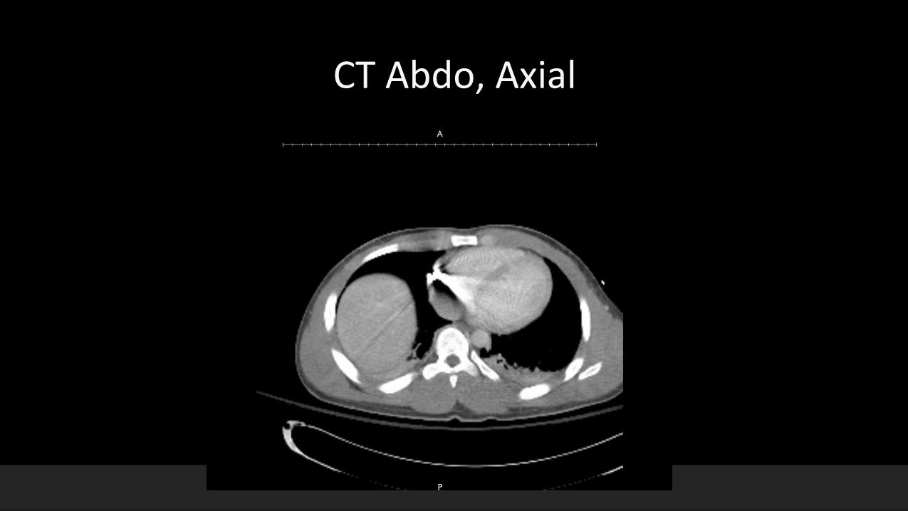
CT Abdo, Axial

McGovern Medical School
Pelvis XR, AP, supine, 10/28
Legend:
= Laparotomy staples
= Bullet fragment
= small bullet fragment
= Foley catheter

McGovern Medical School
L Pelvis XR, AP, 10/31 (Post Surgical)
Legend:
= Laparotomy staples
= skin staples
= surgical drain
= small bullet fragment

McGovern Medical School
F/u CXR, AP, semierect, 10/29
Legend:
= Bullet fragment

McGovern Medical School
Serial CXRs, AP, semierect, 10/30
Legend:
= Bullet fragment

McGovern Medical School
Key Findings• Serial CXRs: Bullet fragment in area of right atrium at T10
• Abdominal XR: Bullet fragments above pelvis and left femoral head, subcutaneous edema from gunshot wound near left hip
• CT Chest/Abdo/Pelvis w/ Contrast: 10 mm bullet fragment abutting the right atrium/right ventricle along atrioventricular groove, 4-5 cm (grade 2) liver laceration in segment IVb, scrotal hematoma, 9mm bullet fragment in left hip joint space w/o definite displaced fx, mild comminution of left inferior pubic ramus, some low-density fluid in upper abdomen likely 2/2 ventral laparotomy, subcutaneous emphysema in pelvis, left perineum, scrotum + small-volume free intraperitoneal air 2/2 bullet injury and/or ventral laparotomy

McGovern Medical School
Key Findings (cont.)
• Pre-operative Pelvic XR: 9 mm bullet fragment over left femoral head, smaller bullet fragment medial to the left trochanter
• Post-operative L Pelvis XR: interval removal of 9mm bullet fragment over left femoral head, surgical drain in left thigh/gluteal region, unchanged small bullet fragment medial to left trochanter

McGovern Medical School
Differential Diagnosis
• N/A

McGovern Medical School
Discussion
• Couinaud classification of hepatic segments (1)
• Divides liver into 8 functional segments, each with their own dual vascular inflow, biliary drainage, and lymphatic drainage
• Vertical planes divided by right, middle, and left hepatic veins
• Horizontal planes divided by portal plane at bifurcation of portal vein

McGovern Medical School
Liver Injury Grading, American Association for the Surgery of Trauma (2)
Grade I – Subcapsular hematoma <10 percent surface area. Parenchymal laceration <1 cm in depth (image 1).
Grade II – Subcapsular hematoma 10 to 50 percent surface area; intraparenchymal hematoma <10 cm in diameter (image 3). Laceration: 1 to 3 cm parenchymal
depth and ≤10 cm in length.
Grade III – Subcapsular hematoma >50 percent of surface area; ruptured subcapsular or parenchymal hematoma. Intraparenchymal hematoma >10 cm.
Laceration >3 cm in depth. Any injury in the presence of a liver vascular injury or active bleeding contained within liver parenchyma.
Grade IV – Parenchymal disruption involving 25 to 75 percent of a hepatic lobe. Active bleeding extending beyond the liver parenchyma into the peritoneum.
Grade V – Parenchymal disruption of >75 percent of a hepatic lobe. Juxtahepatic venous injury to include retrohepatic vena cava and central major hepatic veins.

McGovern Medical School
Examples of Liver LacerationsGrade I Grade II Grade III
Grade IV Grade V

McGovern Medical School
Gunshot Imaging (3)
Primary objective of imaging: determine path of projectile(s), assess which tissues have been injured, estimate severity, determine need for additional studies
Mechanism of gunshots:
• Kinetic energy: Rifles > Handguns, greater wounding potential due to increased acceleration time• Rifling = spiral grooves in gun —> bullet spins while traveling to target• Upon hitting target, non-deformed bullets tend to tumble (rotate about short axis) w/ proximal end
(more mass) leading —> bullets often point towards entry wound• Tumbling is decreased or even eliminated if bullet deforms upon entry• Low-velocity weapons tend to use pure lead bullets, while medium- to high- velocity weapons often have
metal jacket (full or semijacket) to prevent deformation in gun barrel• Semijacketed bullets have often have a exposed lead tip to deform on impact• Others have open cavity at tip (hollow-point) and transform into mushroom shape on impact• Hollow-point bullets decelerate more quickly leading to greater energy delivery to smaller amount of
tissue and overall greater tissue damage

McGovern Medical School
Gunshot Imaging (cont.)
Mechanism of injury
• Damage is most severe in friable soft tissue, such as liver and brain• Temporary cavitation of tissue remote to bullet track can also cause injury (more likely with high-velocity
weapons)• Bones alter course of bullets, slow them down, and increase deformation/fragmentation
• Carefully examine for beveling of bone towards direction of travel (trauma physicians cannot reliably distinguish between entry and exit wounds)
• Semijacketed, hollow-point, non-jacketed, and soft-point bullets often leave a trail of metal fragments• Low-velocity weapons fired at medium- to long- range is less likely to be fatal
• Less kinetic energy —> bullet travels deeply in subcutaneous tissue but tends to stop at fascia, esp. when entering at shallow angle to skin
• High-velocity fragmentation leads to “lead snowstorm” with conical distribution• Shotguns
• Severe at close range —> combined mass of multiple pellets• Contact injuries result in further damage from combustion gas
• Often superficial at long range• Pellets can be made of lead or ferromagnetic steel (distinguishable on radiograph —> lead deforms)
Image source: http://mcadams.posc.mu.edu/medical.htm

McGovern Medical School
High-velocity rifle injury to hip Shotgun injury to shoulder

McGovern Medical School
Expended Bullet Images
Legend:
1 - soft-point 0.44 magnum2,3 - hollow-point 0.38 special4,5 - hollow-point semi-jacketed 9-mm6 - solid-point fully jacketed 9-mm7 - 7.62-mm fully jacket rifle8 - jacket fragment of soft-point rifle9 - lead fragment of soft-point rifle10 - solid-point nonjacketed 0.2211 - hollow-point nonjacketed 0.22

McGovern Medical School
Urogenital Trauma Classification
Urethral Trauma, Goldman (4):
Type I: posterior urethra stretched but intact
Type II: urethra disrupted at the membranous-prostatic junction above the urogenital diaphragm
Type III: disruption of the membranous urethra, extending below the urogenital diaphragm and involving the anterior urethra
Type IV: bladder neck injury, with extension into the proximal urethra
Type IVa: injury to the base of the bladder, with periurethral extravasation simulating a type IV urethral injury
Type V: isolated anterior urethral injury
Bladder Trauma, American Association for the Surgery of Trauma (5):
Grade 1: Hematoma, partial thickness laceration
Grade 2: Extraperitoneal bladder wall laceration <2 cm
Grade 3: Extraperitoneal bladder (>2 cm) or intraperitoneal bladder (<2 cm) laceration
Grade 4: Intraperitoneal bladder wall laceration >2 cm
Grade 5: Intraperitoneal or extraperitoneal bladder wall laceration extending into the bladder neck or ureteric orifice

McGovern Medical School
Final Diagnosis
• Retained 10 mm bullet fragment abutting RA/RV• Grade IIb liver laceration, segment IVb w/o extravasation or
pseudoaneurysm• Retained 9 mm bullet fragment w/n posterior l hip joint space• Comminuted l inferior pubic ramus fx• Scrotal hematoma• No bladder or ureteral injury

McGovern Medical School
Outcome
• Cardiology + CV Surgery consulted for bullet in myocardium
• TTE + TEE revealed no foreign body w/n the heart, no functional deficit, no effusion
• Underwent I & D + removal of foreign body L hip 10/30
• Open L ischium fx, open left femoral head fx w/ foreign body, open left hip joint w/ posterior capsulotomy
• Discharged 11/6 on POD#8 after being cleared by PT/OT• F/u in 2 weeks w/ Dr. Gary (orthopedic trauma), Dr. Wang (urology), and Dr.
Meyer (trauma)

McGovern Medical School
ACR appropriateness Criteria
• the ACR recommendation for penetrating injury to the lower abdomen / pelvis w/ suspected lower urinary tract injury lists fluoroscopy retrograde cystography and CT pelvis w/ bladder contrast as “usually appropriate”
• In this case, patient was hemodynamically unstable and immediately taken for operative mgmt + mechanism of injury was GSW

McGovern Medical School

McGovern Medical School
Cost of Imaging• 3 x CXR, CT Chest w/ contrast, CT Abdo w/ contrast, pelvis AP DX,
wrist DX, HIP 1 view DX, NO read hip fluoro, TTE x 2, TEE x 1• ct abdo —> $5,540• ct chest —> $3,936• CXR —> $683 x 3 = $2,049• pelvis AP DX —> $719• wrist —> $771• hip 1 view dx —> $719 (??)• fluoro —> ??• TTE x 2 —> ??• TEE x 1 —> ??

McGovern Medical School
Take Home Points
• Imaging of gunshot wounds is useful for initial management and/or post-operative decision-making
• Different gun and bullet varieties result in markedly different pathology and clinical outcomes
• Trauma classification systems exist for various types of injuries, including liver, ureteral, and bladder

McGovern Medical School
References
1. Vadera, S., & Jones, J. (n.d.). Couinaud classification of hepatic segments. Retrieved November 10, 2019, from https://radiopaedia.org/articles/couinaud-classification-of-hepatic-segments?lang=us.
2. Coccolini, F., Catena, F., Moore, E. E., Ivatury, R., Biffl, W., Peitzman, A., … Ansaloni, L. (2016). WSES classification and guidelines for liver trauma. World Journal of Emergency Surgery, 11(1). doi: 10.1186/s13017-016-0105-2
3. Wilson, A. J. (1999). Gunshot Injuries: What Does a Radiologist Need to Know? RadioGraphics, 19(5), 1358–1368. doi: 10.1148/radiographics.19.5.g99se1713584. Goldman, S. M., Sandler, C. M., Corriere, J. N., & Mcguire, E. J. (1997). Blunt Urethral Trauma. The Journal of Urology, 157(1), 85–89. doi: 10.1097/00005392-
199701000-000285. Bryk, D. J., & Zhao, L. C. (2015). Guideline of guidelines: a review of urological trauma guidelines. BJU International, 117(2), 226–234. doi: 10.1111/bju.130406. American College of Radiology. (2019). American College of Radiology ACR Appropriateness Criteria® Penetrating Trauma–Lower Abdomen and Pelvis.
Retrieved November 10, 2019, from https://acsearch.acr.org/docs/69376/Narrative/.7. Memorial Hermann. (n.d.). Pricing Estimates and Information. Retrieved November 10, 2019, from https://www.memorialhermann.org/patients-
caregivers/pricing-estimates-and-information/.

Questions?