Multiple Chemical Sensitivity (MCS): Chemical action, etiologic
62
Multiple Chemical Sensitivity (MCS): Chemical action, etiologic mechanism and treatment Electromagnetic Field Hypersensitivity (EHS): EMF action and apparent etiologic mechanism Martin L. Pall Professor Emeritus of Biochemistry and Basic Medical Sciences Washington State University thetenthparadigm.org [email protected]
Transcript of Multiple Chemical Sensitivity (MCS): Chemical action, etiologic

Multiple Chemical Sensitivity (MCS): Chemical action, etiologic mechanism and
treatment Electromagnetic Field Hypersensitivity
(EHS): EMF action and apparent etiologic mechanism
Martin L. Pall
Professor Emeritus of Biochemistry and Basic Medical Sciences
Washington State University
thetenthparadigm.org

Much of the evidence that I will be discussing here
comes from 4 of my publications:
1. Pall M. L. 2009 Multiple chemical sensitivity: Toxicological questions
and mechanisms. In General and Applied Toxicology, 3rd Edition, John
Wiley & Sons, pp. 2303-2352.
2. Pall M. L. 2007 “Explaining ‘Unexplained Illness’: Disease Paradigm
for Chronic Fatigue Syndrome, Multiple Chemical Sensitivity,
Fibromyalgia, Post-Traumatic Stress Disorder, Gulf War Syndrome and
Others”, 16 Chapter book, Harrington Park (Haworth) Press.
3. Pall ML. 2013 Pulmonary hypertension is a probable NO/ONOO- cycle
disease: A review. ISRN Hypertension 2013: Article ID 742418, 27
pages.
4. Pall ML. 2013 Electromagnetic fields act via activation of voltage-
gated calcium channels in biology and medicine. J Cell Molec Med,
submitted.

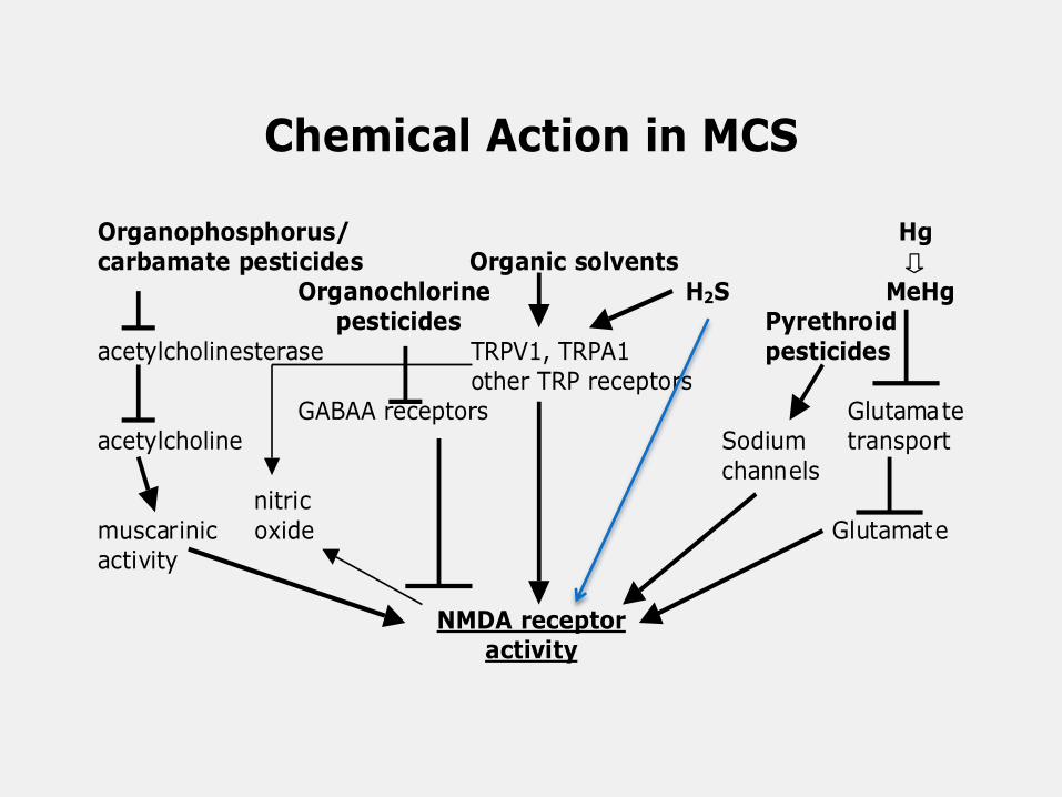
Each of the classes of chemicals implicated in
initiating cases of multiple chemical sensitivity can
act to raise NMDA activity in the body.
Chemical Action in MCS
Organophosphorus/ Hgcarbamate pesticides Organic solvents
Organochlorine H2S MeHg pesticides Pyrethroid
acetylcholinesterase TRPV1, TRPA1 pesticides other TRP receptors
GABAA receptors Glutamateacetylcholine Sodium transport
channels nitric
muscarinic oxide Glutamateactivity
NMDA receptoractivity

The organic solvents and related compounds are
thought to include most of a diverse set of compounds
known as sensory irritants including alkanes,
alkylbenzenes, halogenated benzenes, alcohols,
ketones, ethers, aldehydes including formaldehyde,
isocyanates, and even chlorine and other oxidants. It
can be seen from this, that this group of compounds are
extraordinarily diverse. Much of the sensory irritant
mechanism has been shown to be mediated through the
TRPA1 receptor.

Six other observations supporting an NMDA role in MCS: 1. MCS patients are sensitive to monosodium glutamate and glutamate is the
physiological agonist of the NMDA receptors.
2. An allele of the CCK-B receptor gene that produces increased NMDA
activity is associated with increased prevalence in two studies and therefore
incidence of MCS.
3. The NMDA antagonist dextromethorphan is reported from clinical obser-
vations to produce lowered response to chemical exposures in MCS patients.
4. Bell and others have proposed that neural sensitization has a key role in
MCS and the probable mechanism for such neural sensitization, called long-
term potentiation, is known to involve increased NMDA activity.
5. Elevated NMDA activity has been shown to play an essential role in
several animal models of MCS.
6. Elevated NMDA activity appears to play a role in such related illnesses
as fibromyalgia, chronic fatigue syndrome and post-traumatic stress disorder,
with the most extensive evidence for such a role being found in fibromyalgia
(Pall, 2006 and 2007a).
Compelling evidence for a common toxicological response

Table 1: Genetic polymorphisms involved in
chemical metabolism influencing MCS incidence
Gene Study Function of encoded enzyme
PON1 H; M Detoxification of organophosphates
CYP2D6 M Hydroxylation
NAT2 M; S Acetylation
GSTM1 S Produce glutathione for
conjugation
GSTT1 S Glutathione conjugation
GSTP1 S Glutathione conjugation
UGT1A1 M&S Glucuronidation of chemicals
Studies: H: Haley et al, 1999; M: McKeown-
Eyssen et al, 2004; S: Schnakenberg et al, 2007;
M&S: Mueller and Schnakenberg, 2008
Note: of the Schnakenberg (S) studies, one gene had
p<10-3
, two had p<10-4
and the gene studied in the
M&S study had p<10-4
. The p for all four of these
taken together is p<10-15
.
Note 2: Replication in studies of different
populations will depend on the relevant chemical
exposures of the different populations!

There are other pathways along which toxicants can
act to produce excessive NMDA activity, including
those acting to produce lowered mitochondrial
activity. Among the mitochondrial/energy
metabolism toxicants that have been shown to act at
least in part via excessive NMDA activity are: MPTP,
rotenone, cyanide (although some of its effects
increasing NMDA activity are through another
pathway of action), carbon monoxide and hypoxia.

In summary, we have, then a vast array of TAVENAs (toxicants/toxins in
the body that each act to trigger a common toxic end point- excessive
NMDA activity). These appear to include:
A vast array of organic solvents & related compounds including sensory
irritants
The three major classes of insecticides
Several herbicides
Several fungicides
Several toxic metals
Four classes of antibiotics
A large array of liver toxicants/toxins
Several mitochondrial toxicants/toxins
Several tropical fish/shellfish toxins
Several additional toxicants

Parkinson’s initiators

However there are many other chronic diseases where
cases can be initiated by toxicants acting to produce
excessive NMDA activity, including not only MCS and
Parkinson’s disease, but also Alzheimer’s, amyotrophic
lateral sclerosis (ALS), multiple sclerosis, tinnitus, asthma,
myalgic encephalomyelitis/chronic fatigue syndrome
(ME/CFS), autism and epilepsy.
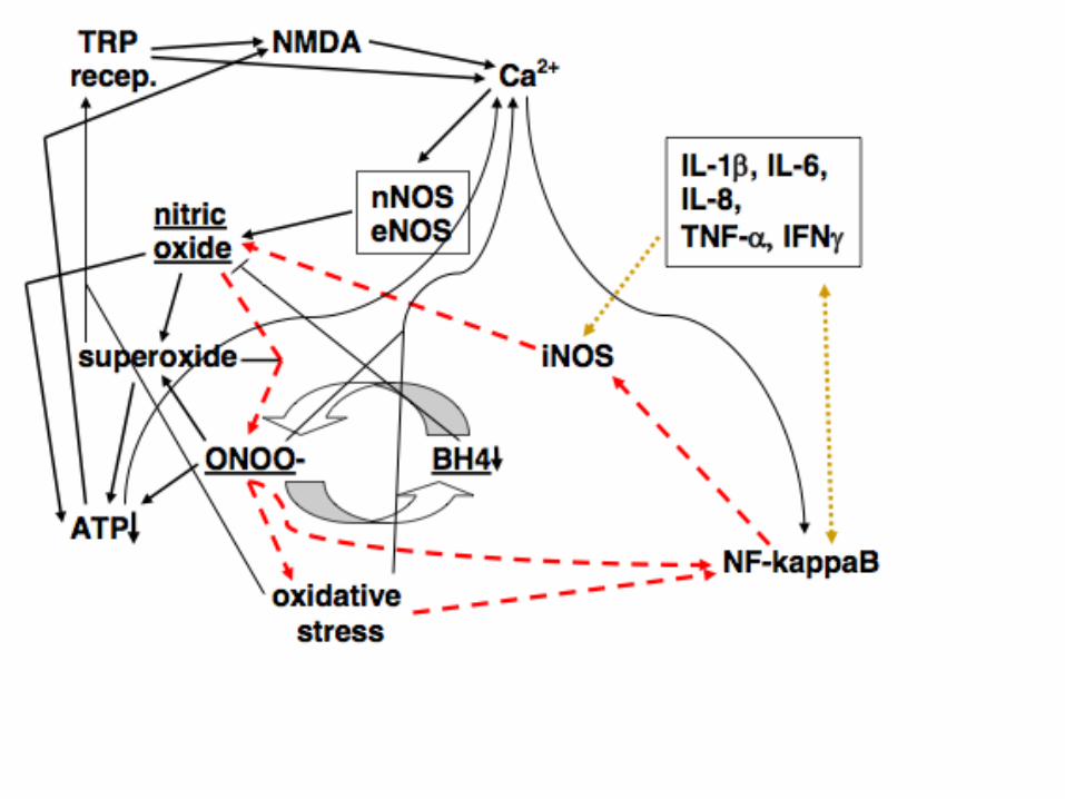
Each of these has also been proposed by the author to be
caused by what is called the NO/ONOO- cycle, a primarily
local biochemical vicious cycle which, depending on where
it is localized in the body, may be able to cause many
different chronic inflammatory diseases. We outline, here,
the properties of the NO/ONOO- cycle in the context of its
proposed role in MCS.

NMDA receptor activation
channels allow calciumentry into cell
nNOS and eNOS activation
nitric oxide increase
react with superoxide to
form peroxynitrite

We think that the etiologic mechanisms of MCS are centered on two interrelated
mechanisms:
1. What is called the NO/ONOO- cycle, a primarily local biochemical vicious
cycle that is initiated by various triggers, including those acting via increased
NMDA activity, and propagates itself over time.
2. And another related mechanism proposed to be involved in MCS by Dr. Iris
Bell and by others, neural sensitization caused by what is known as long-term
potentiation. This can also involves NMDA receptor activity and several other
mechanisms that are part of the NO/ONOO- cycle. Both 1 and 2 are discussed
on some detail in my MCS toxicology review.
Let us first discuss the NO/ONOO- cycle.


Five Principles
1. Cases can be initiated by short-term stressors that increase cycle elements.
2. The chronic phase of illness is produced by the NO/ONOO- cycle. It is
predicted, therefore, that the cycle elements will be elevated in the chronic
phase of illness.
3. The symptoms and signs of illness must be generated by one or more
elements of the cycle.
4. The basic mechanism of the cycle is local and will be localized to different
tissues in different individuals. The reason for this primarily local nature
is that the three compounds involved, NO, superoxide and ONOO-, have
limited half lives in biological tissues. And the mechanisms of the cycle,
those various arrows, act at the level of individual cells. This allows for
great variations in tissue distribution from one patient to another,
producing a huge spectrum of illness. The point here is not that there are
no systemic changes, clearly there are, but rather that the primarily local
mechanisms can generate great variation in diagnosis and in the symptoms
and signs, from one individual to another.
5. NO/ONOO- cycle diseases should be treated by down-regulating the
NO/ONOO- cycle biochemistry, rather than by symptomatic relief. In
other words, we should treat the cause, rather than the symptoms.

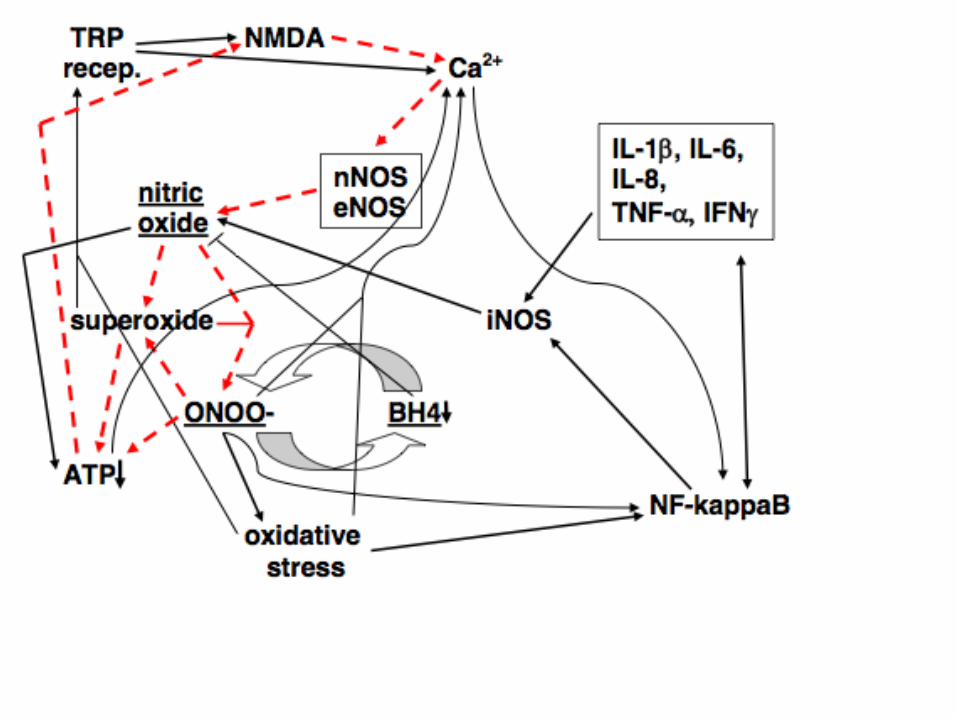
There are 34 distinct, mechanisms that currently make up the
NO/ONOO- cycle models as it was shown in the preceding
figures. These are all copied on subsequent slides and are
all documented in my pulmonary hypertension,
NO/ONOO- cycle review. Of those 31 have reported
substantial pathophysiogical roles. I have added two
additional mechanisms (35&36) which will be discussed
here later.
Thus the only thing truly novel about the NO/ONOO- cycle, is
that when these mechanisms are put into juxtaposition
with each other, as they have been in the preceding
figures, they serve collectively to integrate and explain a
vast array of data about a large number of human
diseases.

1. Extremely rapid, diffusion limited reaction between nitric oxide (NO.) with superoxide (OO.-),
forming peroxynitrite (ONOO-).
2. Peroxynitrite, a potent oxidant, can act mainly through its breakdown products to increase
the activity of the transcription factor NF-kappaB.
3. Peroxynitrite breaks down both before and after reaction with carbon dioxide into the
following free radicals, hydroxyl (HO.), carbonate (CO3.) and NO2 radical (NO2.), each of
which are responsible for a number of consequences produced by peroxynitrite.
4. Peroxynitrite being a potent oxidant produces oxidative stress, an imbalance between
oxidants and antioxidants.
5. Oxidative stress also produces increases in NF-kappaB activity.
6. NF-kappaB produces increased transcription of the inducible nitric oxide synthase (iNOS),
a gene whose transcription is known to be stimulated by NF-kappaB elevation.
7. NF-kappaB also stimulates the transcription of several inflammatory cytokines, including IL-
1 , IL-6, IL-8, TNF- , and IFN .
8. Each of the five cytokines listed in 7 above, act directly and/or indirectly to stimulate the
transcription of the iNOS gene, acting in some cases via the double headed arrow linking it
to NF-kappaB.
9. When iNOS is induced, it produces large amounts of NO.
10. Peroxynitrite inactivates the calcium-ATPase, leading to increased levels of intracellular
calcium.
11. Other oxidants also react with and inactivate the calcium-ATPase as well.
12. Large increases in intracellular calcium raise intramitochondrial calcium, which if large, lead
to increased superoxide generation in the mitochondria and in some cases to apoptotic cell
death.
13. Lowered energy metabolism (decreased energy charge/ATP) also lowers calcium-ATPase
activity, leading to increased levels of intracellular calcium.

14. Intracellular calcium stimulates the nNOS and eNOS forms of nitric oxide synthase,
both of which are calcium dependent enzymes.
15. Increased nNOS and eNOS activity both produce increased NO synthesis.
16. Peroxynitrite oxidizes tetrahydrobiopterin (BH4), depleting BH4 levels.
17. BH4 depletion produces partial uncoupling of the three NO synthases, such that some
of these enzymes produce superoxide in place of NO. Because of the very rapid
reaction of these two compounds to produce peroxynitrite, this partial uncoupling is
expected to produce an increase in peroxynitrite production.
18. Nicking of nuclear DNA by hydroxyl and carbonate radicals, can produce a massive
stimulation of poly ADP-ribosylation of chromosomal proteins, leading, in turn to a
massive depletion of NAD/NADH pools, because NAD is the substrate for such poly
ADP-ribosylation. NADH depletion lowers, in turn, ATP production in the
mitochondrion.
19. Other changes causing ATP depletion come from a cascade of events occurring within
the mitochondrion. The cascade starts with NO, possibly produced by mitochondrial
NO synthase (mtNOS which is thought to be largely a form of nNOS), with NO binding
to cytochrome oxidase, competitively inhibiting the ability of molecular oxygen to bind.
This inhibits the ability of cytochrome oxidase to serve as the terminal oxidase of the
mitochondrial electron transport chain.
20. The action of NO in 18 above, produces increase superoxide production by the electron
transport chain.
21. Peroxynitrite, produced from the combination 18 and 19 above, also acts to produce
increased superoxide from the electron transport chain.

22. Peroxynitrite, superoxide and their products lead to lipid peroxidation of the cardiolipin
in the inner membrane of the mitochondrion. Cardiolipin is highly susceptible to such
peroxidation, because most of the fatty acids that make up its structure in mammals are
polyunsaturated fatty acids, which are much more susceptible to peroxidation than are
other fatty acids .
23. Cardiolipin peroxidation leads to lowered activity of some of the enzymes in the electron
transport chain, leading to further lowering of ATP synthesis.
24. Cardiolipin peroxidation also leads to increased superoxide generation from the
electron transport chain in the mitochondrion.
25. Peroxynitrite produces inactivation of the mitochondrial superoxide dismutase (Mn-
SOD), leading in turn to increased superoxide levels in the mitochondrion.
26. Peroxynitrite, superoxide and nitric oxide all inactivate or inhibit the aconitase enzyme,
lowering citric acid cycle activity and subsequent ATP synthesis.
27. Oxidative stress leads to oxidation of cysteine residues in the enzyme xanthine
reductase, converting it into xanthine oxidase which produces superoxide as a product,
thus increasing superoxide generation.
28. Increased activity of the enzyme NADPH oxidase, which produces superoxide as a
product, is an important part of the inflammatory cascade, and contributes, therefore, to
the cascade by producing increased superoxide.
29. Activity of the NMDA receptors, allow calcium influx into the cell, raising intracellular
calcium levels.
30. Activity of transfer receptor potential (TRP) receptors also allows calcium influx into the
cell, again raising intracellular calcium levels, presumably leading to increased nitric
oxide production.
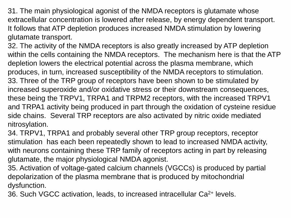
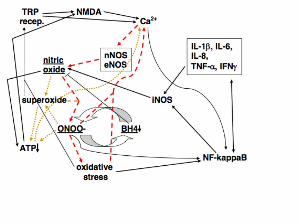
31. The main physiological agonist of the NMDA receptors is glutamate whose
extracellular concentration is lowered after release, by energy dependent transport.
It follows that ATP depletion produces increased NMDA stimulation by lowering
glutamate transport.
32. The activity of the NMDA receptors is also greatly increased by ATP depletion
within the cells containing the NMDA receptors. The mechanism here is that the ATP
depletion lowers the electrical potential across the plasma membrane, which
produces, in turn, increased susceptibility of the NMDA receptors to stimulation.
33. Three of the TRP group of receptors have been shown to be stimulated by
increased superoxide and/or oxidative stress or their downstream consequences,
these being the TRPV1, TRPA1 and TRPM2 receptors, with the increased TRPV1
and TRPA1 activity being produced in part through the oxidation of cysteine residue
side chains. Several TRP receptors are also activated by nitric oxide mediated
nitrosylation.
34. TRPV1, TRPA1 and probably several other TRP group receptors, receptor
stimulation has each been repeatedly shown to lead to increased NMDA activity,
with neurons containing these TRP family of receptors acting in part by releasing
glutamate, the major physiological NMDA agonist.
35. Activation of voltage-gated calcium channels (VGCCs) is produced by partial
depolarization of the plasma membrane that is produced by mitochondrial
dysfunction.
36. Such VGCC activation, leads, to increased intracellular Ca2+ levels.
The NO/ONOO- cycle provides explanations of how
chemically caused excessive NMDA activity can produce
MCS with its chronic local sensitivity to chemical
exposure. The local elevation of the NO/ONOO- cycle in
regions of the body susceptible to chemically-caused
NMDA activation, will be expected to produce, at least
part of the sensitivity response.


It should be noted, however, that chemicals are not the only
stressors that can initiate cases of apparent NO/ONOO-
cycle diseases. They may also be initiated by infections
(probably acting via increased inflammation), by physical
trauma especially to the central nervous system and by
psychological stress (probably both acting via excessive
NMDA activity) and by electromagnetic field exposure
(probably acting via increased intracellular calcium). Some
initiating chemicals may act independently of the NMDA
receptors.

One of the big breakthroughs in our understanding of
MCS came from a comparison of the NO/ONOO- cycle
model of these illnesses with the neural sensitization
model of MCS developed by Dr. Iris Bell (M.D., Ph.D., at
the University of Arizona). Bell argued that the most
important mechanism of MCS was neural sensitization
in the hippocampus region of the brain. This is the
same region that has key functions in learning and
memory. The idea Bell developed was that the
synapses in the brain, the contacts between neurons by
which one stimulates another, may become both
hypersensitive and hyperactive in response to chemical
exposure. The basic idea here is that this process of
neural sensitization which is involved on a very
selective basis in learning and memory, appears to be
activated massively in MCS.

The main mechanism of neural sensitization is known as
long term potentiation (LTP). LTP is known to involve
increased NMDA receptor activity, increased
intracellular calcium nitric oxide and also superoxide.
So one immediately sees major connections between
the NO/ONOO- cycle mechanism and the neural
sensitization mechanism developed by Bell. So by
having chemicals producing increased NMDA activity,
one can see how they could greatly stimulate the long
term potentiation mechanism. Several of the elements
of the NO/ONOO- cycle have roles in LTP, including
NMDA activity, intracellular calcium, nitric oxide and
superoxide.
LTP has been studied predominantly in the brain and
spinal cord. It has been suggested to occur in some
other tissues with NMDA receptors, but this has not
been clearly demonstrated.

MCS has apparent sensitivity responses, not only coming from
the brain, but also from upper and lower respiratory tract
regions, from the skin, GI tract, eye and sometimes other
tissues. Because we are unsure whether LTP occurs in these
peripheral tissues, we are unsure whether it can contribute to
sensitivity responses in those peripheral tissues. But, in
general, it seems likely that MCS sensitivity involves the
NO/ONOO- cycle in these various tissues and also LTP in the
central nervous system (and possibly elsewhere?).
Some other mechanisms may contribute to chemical sensitivity: Nitric oxide (NO), acting to inhibit cytochrome P450 metabolism producing
slowed detoxification and therefore possible increased sensitivity to some
chemicals metabolized in this way.
Oxidants lead to increased TRPV1 and TRPA1 activity, leading to increased
sensitivity to chemicals acting via these receptors.
Peroxynitrite, producing breakdown of the blood brain barrier, leading to increased
chemical access to the brain.

Now, let’s switch over to the effects of electromagnetic fields
(EMFs) on our biology and medicine!!
There has been a great puzzle about how EMFs can influence
our biology, for better or for worse. These EMFs are
composed of low energy photons, with energy per photon
too low to influence the chemistry of the body! How can
they influence our biology through non-thermal effects? And
yet there is a substantial literature reporting that they do.
I have recently solved this important puzzle. EMFs act to
influence the voltage across plasma membranes of cells,
thus activating voltage-gate calcium channels. And it is the
downstream effects of the increased intracellular Ca2+ that
leads to the biological effects of EMF exposure.
I will discuss first some of the evidence supporting this
mechanism and will discuss later how this may lead to
electromagnetic hypersensitivity.

Table 1: EMF Responses Blocked or Lowered by Calcium
Channel Blockers
Ref
#
EMF
type
Calc ium
chan nel
Cell type or
orga nis m
Res pons e
measured
2 Pulsed
magnetic
fields
L-type Human
lymphocytes
Cell
proliferation;
cytokine
production
3 Static
magnetic
field (0.1
T)
L-type Human
polymorphonuclear
leukocytes
Cell migration;
degranulation
5 ELF L-type Rat chromaffin
cells
Differentiation;
catecholamine
release
6 Electric
field
L-type Rat and mouse
bone cells
Increased
Ca2+,
phospholipase
A2, PGE2
7 50 Hz L-type Mytilus (mussel)
immunocytes
Reduced shape
change,
cytotoxicity
8 50 Hz L-type AtT20 D16V,
mouse pituitary
corticotrope-
derived
Ca2+ increase;
cell
morphology,
premature
differentiation
9 50 Hz L-type Neural stem/
progenitor cells
In vitro
differentiation,
neurogenesis
10 Static
magnetic
field
L-type Rat Reduction in
edema
formation
11 NMR L-type Tumor cells Synergistic


22 Very weak
electrical fields
T-
type
Sharks Detection of very weak
magnetic fields in the
ocean
23 Short electric
pulses
L-
type
Human eye Effect on electro-
oculogram
24 Weak static
magnetic field
L-
type
Rabbit Baroreflex sensitivity
25 Weak electric
fields
T-
type
Neutrophils Electrical and ion
dynamics

The finding that EMF exposure acts via activation of VGCCs, provides
for the first time, an answer to the puzzle of how exposure to EMFs
composed of low energy photons can affect our biology and medicine.
The effects of EMFs on the voltage across the plasma membrane can
lead to partial depolarization and subsequent activation of of VGCCs,
leading to very rapid increases in intracellular Ca2+. Because increased
intracellular Ca2+ can act, in turn, to stimulate NO synthesis, such NO
increase may also have an important role.
Pilla recently showed that such EMF exposure can lead to almost
instantaneous increases in both intracellular Ca2+ and also of NO
synthesis (all occurring in less than 5 seconds):
Pilla AA. Electromagnetic fields instantaneously modulate nitric oxide
signaling in challenged biological systems. Biochem Biophys Res
Commun. 2012;426:330-3.

Most responses physiological responses to Ca2+ and NO, act as follows:
NO increasing levels of cGMP, leading in turn to stimulation of the
cGMP-dependent protein kinase (protein kinase G).
In contrast, most pathophysiological effects of NO are mediated through
its role as a precursor of peroxynitrite (ONOO-), leading to free radical
generation and oxidative stress.
There are a series of therapeutic effects of EMFs, raising the question of
how these might act. And there are a series of pathophysiologic effects
of EMFs, raising the question of how these might act. I took what is
probably the best documented example of each of these to determine
apparent answers to these questions.
I found that the therapeutic effects of EMFs in stimulating bone growth,
act via EMF stimulation of osteoblasts probably via NO, cGMP and
increased protein kinase G.
I also found that the pathophysiogic effects of EMF exposure, inducing
single strand breaks in cellular DNA, probably acts via increased NO,
ONOO- and oxidative stress. Each of these results, then tend to confirm
our preconceived notions of what mechanisms are likely to be involved!

What about EMF hypersensitivity (EHS)?? Anecdotal reports claim a number of
similarities to MCS: These have high levels of co-morbity- that this they often
occur together in the same patients. Physicians have reported that they both
appear to respond to the same therapeutic approaches, approaches that I will
argue may work by lowering the NO/ONOO- cycle. The symptoms of each vary
quite a bit from patient to patient. Both appear to occur following previous
exposure, chemical exposure in the case of MCS and EMF exposure in the case
of EHS. The basic question that I am raising here is whether EHS is produced
by the NO/ONOO- cycle and by long-term potentiation (LTP), as we think MCS
is?
There is, in fact, a substantial literature showing that VGCC stimulation can lead
to LTP, in much the same way that NMDA stimulation does. This is not
surprising, given the fact that the downstream effects of VGCC stimulation are
similar if not identical to those of NMDA stimulation.
A second question is whether VGCC elevation acts as part of NO/ONOO- cycle
as does NMDA elevation? I argue here that VGCC elevation does act as part of
the NO/ONOO- cycle, because lowered mitochondrial function/ATP levels lead
to partial depolarization of the plasma membrance and therefore VGCC
stimulation. Such VGCC stimulation, acts, in turn to increase intracellular
Ca2+,an important element of the cycle.


Therapy: How can we treat and hopefully cure NO/ONOO- cycle
diseases? There are many agents that have been used to treat
proposed NO/ONOO- cycle diseases that can be shown to lower cycle
elements but we don’t have time to review this large literature here.
Some of this is discussed in Chapter 15 of my book. But in general,
there has not been any extensive study of combinations of agents
aimed specifically at lowering the entire cycle and presumably this is
what we need!
(PLEASE NOTE: I am a PhD, not an MD or ND and none of what I say
here should be viewed as medical advice)
Let’s look again at the various parts of the cycle, as it has been
proposed, to see why it is predicted to be so robust and what our
challenges are in down-regulating the cycle.


It can be seen from the above, that the one element of the cycle
that occurs in each of the five component cycles, shown above is
peroxynitrite (ONOO-) and therefore a peroxynitrite scavenger may
be expected to be particularly useful in treatment. One agent that is
a powerful peroxynitrite scavenger is 5-methyltetrahydrofolate (5-
MTHF) (see Rezk, FEBS Lett 2003;555:601–605; Antoniades,
Circulation 2006;114:1193–1201) . So, in principle, by using
sufficient 5-MTHF, one should be able to be able to cure
NO/ONOO- cycle disease. But it is more complicated than that. I
have received information from four different sources, that most
patients with the ME/CFS, MCS and fibromyalgia group of diseases,
do not tolerate well doses above 300 micrograms per day of 5-
MTHF. Why should that be?? When there is a lot of peroxynitrite
present, particularly in the GI tract, then much of the 5-MTHF is
oxidized presumably to the dihydro form, 5-MDHF which breaks
down further to products that are lost from the folate pool.
Presumably this leads not only to loss of reduced folates but also
accumulation of some toxic product.

So how can we avoid both loss of 5-MTHF and accumulation of a
toxic product of 5-MTHF oxidation? Most likely by using high doses
of ascorbate (vitamin C). Ascorbate is both a peroxynitrite
scavenger, although one needs high concentrations to be
reasonably effective and scavenging peroxynitrite will, of course,
lower 5-MTHF oxidation. Furthermore, high doses of ascorbate will
reduce the 5-MDHF oxidation product back to 5-MTHF thus
simultaneously lowering peroxynitrite mediated loss of 5-MTHF and
greatly lowering accumulation of toxic oxidation products. In
general by using high dose ascorbate along with substantial
amounts of 5-MTHF, one should be able to much more effectively
lower peroxynitrite than by using either one alone.
What dose of ascorbate should be used? If 5-MTHF is taken orally,
then perhaps 1 to 2 g of oral ascorbate should be taken
simultaneously. And as the tolerance of this combination becomes
clear, it may be possible to repeat it two or three times per day.


One of the mechanisms that leads to mitochondrial dysfunction in the NO/ONOO-
cycle is the massive stimulation of poly (ADP-ribose) polymerase (PARP)activity in
the nucleus in response to DNA nicking by hydroxyl radical and other radical products
of peroxynitrite, leading, in turn to a massive depletion of NAD/NADH in the cell. This
depletion occurs because NAD is the substrate for this enzyme. And the SIRT1
enzyme is an NAD dependent deacetylase whose activity is strongly dependent in
vivo on NAD levels in the cell. Consequently, it is essential to restore NAD levels
before resveratrol can possibly be effective in treatment of and possibly cure of
NO/ONOO- cycle diseases.
The best way to do this may be to use substantial doses of nicotinic acid, possibly
using low flush niacin, to help restore NAD pools. It may also be useful to
simultaneously use D-ribose, which is converted to PRPP which reacts enzymatically
with nicotinic acid or nicotinamide to generate NMN and NAD. I don’t think one
should use nicotinamide here for NAD generation because nicotinamide inhibits
SIRT1 activity itself!
Let me just add one thing. I wonder whether Abram Hoffer’s treatment of
schizophrenic patients with high dose nicotinic acid may have worked via increased
SIRT1 activity.

Another promising agent is the resveratrol, a phenolic compound which acts via more
than one pathway, but where a single pathway appears to be central to the important
favorable effects for lowering the NO/ONOO- cycle:
Resveratrol SIRT1 lowered superoxide via at least five
mechanisms (induction of all three superoxide dismutases, lowered NADPH oxidase,
lowered mitochondrial superoxide generation), improved mitochondrial function,
lowered NF-kappaB activity, lowered iNOS induction, increased BH4 production and
consequent improved NOS coupling, lowered excessive NMDA activity (via two
mechanisms), lowered peroxynitrite, lowered oxidative stress and lowered
intracellular calcium levels. Essentially, the whole NO/ONOO- cycle is lowered by
resveratrol raising SIRT1 activity!!
So is resveratrol the long awaited magic bullet to cure NO/ONOO- cycle diseases??
It probably is a good preventive agent, but curing such diseases is another matter.
I suspect you knew that this was too good to be true, but why and how can we get
around the limitations? What is the problem and how can we get around it?

Let’s go on to some other agents that are often used to treat proposed NO/ONOO-
cycle diseases.
One of these is magnesium. Marginal or more severe magnesium deficiencies are
common in many countries, due to the low magnesium levels in highly process foods
and due to soil magnesium depletion due to intensive agriculture. Magnesium has a
crucial role in regulating NMDA receptor activity due to the role of magnesium ions in
blocking the channel of these receptors that can open to allow calcium influx. It
follows from this that those with magnesium deficiencies are at great risk for
generating NO/ONOO- cycle disease due to excessive NMDA activity.
Another agent used to treat these diseases is fish oil and similar lipids containing long
chain omega-3 fatty acids DHA and EPA. These have anti-inflammatory activity,
lowering the inflammatory effects of arachidonic acid-derived eicosanoids produced
in excessive amounts when our diets have excessive omega-6 fatty acids, as is
typical in most of our diets.
Phospholipids are also used to treat these diseases and may act, at least in part, by
helping restore the oxidized cardiolipin the the inner membrane in the mitochondrion.
It is possible that phosphatidyl serine may be particularly effective here, although we
don’t know that, because there is a transporter that specifically transports
phosphatidyl serine into the inner mitochondrial membrane.

L-carnitine/acetyl-L-carnitine (ALC) are other agents often used for treatment, with
ALC being more active at least in part because it is transported more efficiently in the
body. Until recently, I have assumed that the main mechanism of action of these
compounds is to stimulate mitochondrial function, given the well established role of
carnitine in fatty acid transport into mitochondria. However, recently, there has been
established another mechanism that may turn out to be more important here, a
mechanism that lowers excessive NMDA activity.
Glutamate is the main physiological agonist of the NMDA receptors and glutamate
release stimulates not only the NMDA receptors but also the AMPA and kainate
receptors and the metabotropic receptors. AMPA and kainate receptor stimulation
produce still more NMDA receptor activity by depolarizing the plasma membrane, but
the metabotropic receptor stimulation lowers the NMDA response. It has been shown
that ALC/carnitine stimulate one of the metabotropic receptors, mGluR2, causing it to
be more susceptible to glutamate stimulation, which lowers, in turn the response of
the NMDA receptors.
However, as with many agents, ALC/carnitine may be a mixed blessing. Many NMDA
antagonists lead to increased production of NMDA receptor when used chronically.
Does this occur with ALC/carnitine?? Furthermore ALC/carnitine can produce at least
a modest increase in NF-kappaB activity, so that could be a problem.

We discussed earlier the use of 5-MTHF together with high dose ascorbate
to scavenge peroxynitrite. These should work effectively in an aqueous
environment but not in the lipid phase of cells, where the acid form,
peroxynitrous acid has substantial solubility. Carotenoids act however in the
lipid phase as peroxynitrous acid scavengers.
When they do so, the cis-double bonds within carotenoids appear to have a
special role, changing from cis to trans in the process. Natural carotenoids
have some cis-double bonds. For example natural beta-carotene has
roughly one cis-double bond per two molecules, whereas synthetic beta-
carotene is essentially all trans. This may be important because most if not
all clinical trials on beta-carotene have used synthetic beta-carotene. Other
natural carotenoids, including lycopene and lutein/zeaxanthin, may have
special roles in this process of peroxynitrous acid scavenging.
Agents that lower NF-kappaB activity include a number of chain breaking
antioxidants, including phenolic and thiol antioxidants. It is unclear whether
using these is adequate in lowering NF-kappaB activity, such that using
herbal or pharmaceutical agents recognized to lower NF-kappaB activity via
other mechanisms may also be important.

Sauna therapy has been used to treat several proposed NO/ONOO- cycle
diseases. It has often been assumed to be acting via a detoxification
mechanism. While some detoxification has been shown to occur of stored
toxicants in the body, typically over a period of weeks, patients often report much
more rapid symptomatic improvement. There is no published evidence, to my
knowledge, showing that lowering of toxicants in the body is the main
mechanism of symptomatic improvement.
I have argued that the main mechanism of symptomatic improvement in
response to sauna therapy is produced by increased BH4 availability. The rate
limiting enzyme in the de novo synthesis of BH4 is GTP cyclohydrolase I
(GTPCH-I). This enzyme has been shown to be increased by two
consequences of sauna therapy: induction of the heat shock protein Hsp90 and
increased blood flow shear in the vasculature. And both of these lead to
decreased nitric oxide synthase uncoupling which is produced by increased
availability of BH4. Increased BH4 production in the heated regions of the body
and in the vasculature should raise levels of circulating BH4, thus feeding
tissues of the body with BH4 depletion, whether they are directly impacted by
sauna treatment or not.
Sauna therapy via this mechanism may well be useful in the treatment of cases
of many NO/ONOO- cycle diseases, whether these cases are characterized by
elevated levels of toxicants in the body, or not.

Agents that raise the levels of reduced glutathione (GSH) in the body
should be useful in the treatment of NO/ONOO- cycle diseases,
lowering the oxidative stress that is one of the features of the cycle. It
is common for glutathione, both reduced (GSH) and total glutathione
(GSH + GSSG) to be depleted in tissues under oxidative stress.
There are a number of agents that may be useful in helping restore
GSH pools. These include a precursor of GSH de novo synthesis, N-
acetylcysteine, -lipoic acid, liposomal GSH or inhaled, nebulized or
nasal spray GSH or oral acetylated GSH. It has been argued that
sublingual GSH is also useful.
Agents that stimulate glutathione reductase (which uses NADPH to
reduce GSSG to GSH), such as high dose riboflavin or niacin, may be
useful and also possibly agents that increase the generation of
NADPH via the pentose phosphate shunt, such as high dose thiamine
may also be useful.

“Vitamin E” may be useful but may also be damaging, depending on the form and
dosage used and the patient cohort studied. Synthetic (all rac) -tocopherol, the
usual form studied in clinical trials at high doses (400IU/day or more) induces an
enzyme (CYP4F2) which degrades all the other forms of vitamin E including -
tocopherol, -tocopherol, -tocotrienol, -tocotrienol, -tocotrienol and -tocotrienol.
Consequently, high doses of -tocopherol leads to a deficiency in all of these other
forms of vitamin E. This might be OK if -tocopherol had all of the activities of these
other forms, but it is very clear that it does not.
& -tocopherol and tocotrienol all scavenge NO2 radical, an important breakdown
product of peroxynitrite, but -tocopherol does not. -tocopherol has important anti-
inflammatory effects, acting to lower cyclooxygenase activity much more than
-tocopherol. Excitotoxicity caused by excessive NMDA activity works, in part via
excessive activity of the 12-lipoxygenase enzyme; this enzyme is potently inhibited by
-tocotrienol which greatly lowers NMDA excitotoxicity but this property is not shown
by -tocopherol. & -tocotrienols have some anticancer properties with much higher
activities than do the tocopherols. Tocotrienols have been shown to have higher
antioxidant activities in membranes than do the similar tocopherols. Some cell types
have been shown to have much higher transport activity, concentrating tocotrienols
much more than tocopherols. And there is some evidence suggesting that
tocotrienols may be more effective in protecting mitochondria than are tocopherols.

None of these observations negate important roles for -tocopherol. But they do
suggest that when high dose synthetic -tocopherol is used in clinical trials, the
accompanying loss of other forms of vitamin E is likely to have important negative
consequences and may therefore be responsible for the many disappointing
responses in such clinical trials.
In the context of the NO/ONOO- cycle, the roles of these other forms of vitamin E in
scavenging NO2 radical, lowering inflammatory responses, lowering NMDA-induced
excitotoxicity and in protecting mitochondrial activities are all reasons not to use such
high dose synthetic -tocopherol in treatment of NO/ONOO- cycle diseases. It also
suggests that use of 400 IU/day nutritional supplements of synthetic -tocopherol
may make us more susceptible to some NO/ONOO- cycle diseases, rather than less
susceptible. My own view, therefore, is that NO/ONOO- cycle diseases when treated
with protocols using vitamin E, should be treated using modest doses of natural -
tocopherol , much higher doses of -tocopherol and substantial doses of -
tocotrienols.

The last agent I wish to discuss is high-dose hydroxocobalamin form of
vitamin B-12. Hydroxocobalamin when reduced in the body from the cobalt
(III) form to the cobalt (II) form is a potent nitric oxide scavenger. It has also
been recently reported to be both a superoxide scavenger and a
peroxynitrite scavenger, but it is unclear to me whether these last two
activities are likely to be physiologically important. Other forms of vitamin B-
12 may also serve as precursors of hydroxocobalamin.
Ellis and Nasser published a placebo-controlled study showing efficacy of
hydroxocobalamin IM injections (5 mg/twice a week) in ME/CFS-like patients
back in 1973. Both IM and IV injections have been used clinically, as have
hydroxocobalamin nasal spray and nebulized inhaled hydroxocobalamin.
Oral hydroxocobalamin is probably of very limited value due to absorption
being limited by the availability of intrinsic factor. Sublingual B-12 has been
suggested to be useful, but increased sublingual absorption has not been
confirmed in published studies, to my knowledge.
I think that hydroxocobalamin is likely to be a very useful, well tolerated
agent for the treatment of NO/ONOO- cycle diseases.

In summary, we have some 21 chronic inflammatory diseases
characterized by elevation of other elements of the NO/ONOO-
cycle, most of which have a good fit to the five principles
underlying the cycle.
None of these diseases can be cured and in most cases can
even be effectively treated by conventional allopathic medicine.
It is my view, as a PhD biochemist, that naturopathic medicine
is much better equipped to deal effectively with these diseases.
If this view is correct, you are in THE most important part of
medicine. But in order to show that, I very desperately need
your help! So you should take my talk also as a challenge – to
join me to show that naturopathic medicine is where the action
is in the treatment of chronic inflammatory disease.

High dose ascorbate can be viewed as a useful therapeutic agent in a
different context – it can lower both sides of what is called the central
couplet, as seen in Fig 2C. That is it can not only lower peroxynitrite, as
discussed immediately above, but it can also lower loss of peroxynitrite
mediated oxidation of BH4 by a second mechanism. When peroxynitrite
oxidizes BH4, it produces BH3 which can be reduced back to BH4 by
ascorbate – however again, one needs fairly high doses for this to be
effective. So again if oral ascorbate is used, something on the order of 1 to
2 g doses may be needed. IV ascorbate can, of course, generate much
higher levels and so may be still much more effective.
There is a third mechanism that may be useful but that is probably only
going to contribute when high doses of IV ascorbate are used. Ascorbate
being a reducing agent can reduce molecular oxygen to H2O2, which
induces the rate limiting enzyme in BH4 de novo biosynthesis, GTP
cyclohydrolase 1. This may act, then to increase de novo BH4 synthesis.
H2O2 is of course an oxidant so here, one needs to be concerned about
going too high, since oxidative stress has a major role in the NO/ONOO-
cycle.

Summary:
1. 7 classes of chemicals implicated in MCS all act via excessive NMDA
activity.
2. 6 other types of evidence also implicate excessive NMDA activity in
MCS.
3. Genetics of susceptibility show that genes involved in chemical
metabolism influence susceptibility to MCS.
4. #1-3 show beyond doubt that MCS is a real disease involving chemical
exposure.
5. MCS is thought to be a NO/ONOO- cycle disease, with the cycle
causing it to be chronic and with chemicals acting to initiate of elevate
the cycle through their action raising NMDA activity.

6. Long-term potentiation (LTP) also has a probable role in MCS in the
brain and possibly in some other tissues.
7. Other chronic, possible NO/ONOO- cycle diseases may be initiated by
chemicals acting via excessive NMDA activity but also by other
stressors acting a various ways.
8. EMF exposure acts by activating voltage-gated calcium channels
(VGCCs) leading in turn to increased intracellular Ca2+ and NO.
9. These may act, in turn, to produce electromagnetic hypersensitivity
(EHS) via the NO/ONOO- cycle and also LTP (similar mechanisms to
MCS).
10. The major goal in treatment is to lower the NO/ONOO- cycle.
11. The robust nature of the NO/ONOO- cycle makes this a major
challenge.