Multiple cerebral infarction associated with neuroleptic...
4
Behavioural Neurology (1994),7,185-187 Multiple cerebral infarction associated with neuroleptic-induced lupus anticoagulant J.D.E. Laugharne University Department of Psychiatry, Northern General Hospital, Herries Road, Sheffield S5 7AU, UK A 39 year old schizophrenic woman with a 20 year history of neuroleptic treatment suffered bilateral cerebral infarcts and coagulation studies revealed a lupus anticoagulant (LA). She highlights the poorly recognized risk of thrombosis in patients on neuroleptics who develop LA. In this case the LA may have been induced by long-term therapy with haloperidol, a novel and potentially important association. Keywords: Cerebral infarction - Lupus anticoagulant - Neuroleptics - Haloperidol INTRODUCTION The relatively high incidence of immunological abnor- malities induced by phenothiazines, especially chlor- promazine, is not an area with which many psychia- trists are familiar. However, the fact that such abnor- malities do occur has been well documented (Zarrabi et ai., 1979; Canosos and Sise, 1982) and of particular interest is the increased occurrence of the lupus antico- agulant (LA) in patients on phenothiazines. The LA is an antiphospholipid antibody, usually an IgG or IgM, which prolongs in vitro measurement of the partial thromboplastin time (PTT) but causes a hyper- coagulable state in vivo (Boxer et ai., 1976; Shapiro et ai., 1981), thereby increasing the risk of thrombotic episodes. The patient described here had a history of treatment with various neuro1eptics and sustained multiple cerebral infarcts, in association with a circu- lating lupus anticoagulant. CASE REPORT The patient, a 39 year old woman, was first diagnosed as schizophrenic at the age of 19 years. Following this diagnosis she was treated with various antipsy- chotic agents on an intermittent basis, including triflu- operazine, thioridazine and flupenthixol depot injec- tions. She received chlorpromazine between 1980 and 1983, at an average dose of 400 mg/ day but has not been prescribed this drug since then. From 1985 onwards she was maintained on haloperidol, initially in oral form, and from 1987 as a depot at 300 mg monthly. There is no history of any other drugs previously associated with LA. In December 1991 the patient, residing at the time © 1994 Rapid Communications of Oxford Ltd on a psychiatric rehabilitation unit, developed right- sided limb and facial weakness, from which she made a rapid spontaneous recovery. One month later she experienced sudden onset facial weakness, more marked on the left than the right side, with accompa- nying dysphagia and dysarthria. Examination re- vealed signs of a pseudobulbar palsy, mild weakness of the right lower limb with a positive Babinski sign on the right side and no sensory deficit. There was no splenomegaly or adenopathy. An urgent magnetic resonance imaging (MRI) scan showed the presence of bilateral parietal infarcts (Fig. 1). Clinical recovery from this episode was slow but steady until 2 months later when, whilst being investi- gated for the cause of these infarcts, she developed a further episode of left-sided hemiparesis, with a wors- ening of her dysarthria and dysphagia which has improved only minimally. A clotting screen gave a prothrombin time (PT) of 14.1 s (control time: 10.5-14.0 s) and a PTT of 51.5 s (control: 28-39 s), which was suggestive of the pres- ence of LA. A dilute Russell's viper venom time (DRVVT) ratio of 1.9 (normal value 0.9-1.09) with a correction to 1.34 on addition of frozen-thawed platelets strongly supported the diagnosis (Creagh and Greaves, 1991). Anticardiolipin antibodies were normal. Antinuclear factor (ANF) was positive with a titre of 20, but all other autoantibodies tested for, including dsDNA, n-RNP, SM, SCL70, Ro and La, were negative. Erythrocyte sedimentation rate (ESR) was raised at 87 mm/h but serum urea and electro- lytes, glucose and liver function tests were unremark- able as were serum cholesterol and triglycerides. Total Behavioural Neurology. Vo17 . 1994 185
Transcript of Multiple cerebral infarction associated with neuroleptic...
Behavioural Neurology (1994),7,185-187
Multiple cerebral infarction associated with neuroleptic-induced lupus anticoagulant
J.D.E. Laugharne
University Department of Psychiatry, Northern General Hospital, Herries Road, Sheffield S5 7AU, UK
A 39 year old schizophrenic woman with a 20 year history of neuroleptic treatment suffered bilateral cerebral infarcts and coagulation studies revealed a lupus anticoagulant (LA). She highlights the poorly recognized risk of thrombosis in patients on neuroleptics who develop LA. In this case the LA may have been induced by long-term therapy with haloperidol, a novel and potentially important association.
Keywords: Cerebral infarction - Lupus anticoagulant - Neuroleptics - Haloperidol
INTRODUCTION
The relatively high incidence of immunological abnormalities induced by phenothiazines, especially chlorpromazine, is not an area with which many psychiatrists are familiar. However, the fact that such abnormalities do occur has been well documented (Zarrabi et ai., 1979; Canosos and Sise, 1982) and of particular interest is the increased occurrence of the lupus anticoagulant (LA) in patients on phenothiazines. The LA is an antiphospholipid antibody, usually an IgG or IgM, which prolongs in vitro measurement of the partial thromboplastin time (PTT) but causes a hypercoagulable state in vivo (Boxer et ai., 1976; Shapiro et ai., 1981), thereby increasing the risk of thrombotic episodes. The patient described here had a history of treatment with various neuro1eptics and sustained multiple cerebral infarcts, in association with a circulating lupus anticoagulant.
CASE REPORT
The patient, a 39 year old woman, was first diagnosed as schizophrenic at the age of 19 years. Following this diagnosis she was treated with various antipsychotic agents on an intermittent basis, including trifluoperazine, thioridazine and flupenthixol depot injections. She received chlorpromazine between 1980 and 1983, at an average dose of 400 mg/ day but has not been prescribed this drug since then. From 1985 onwards she was maintained on haloperidol, initially in oral form, and from 1987 as a depot at 300 mg monthly. There is no history of any other drugs previously associated with LA.
In December 1991 the patient, residing at the time
© 1994 Rapid Communications of Oxford Ltd
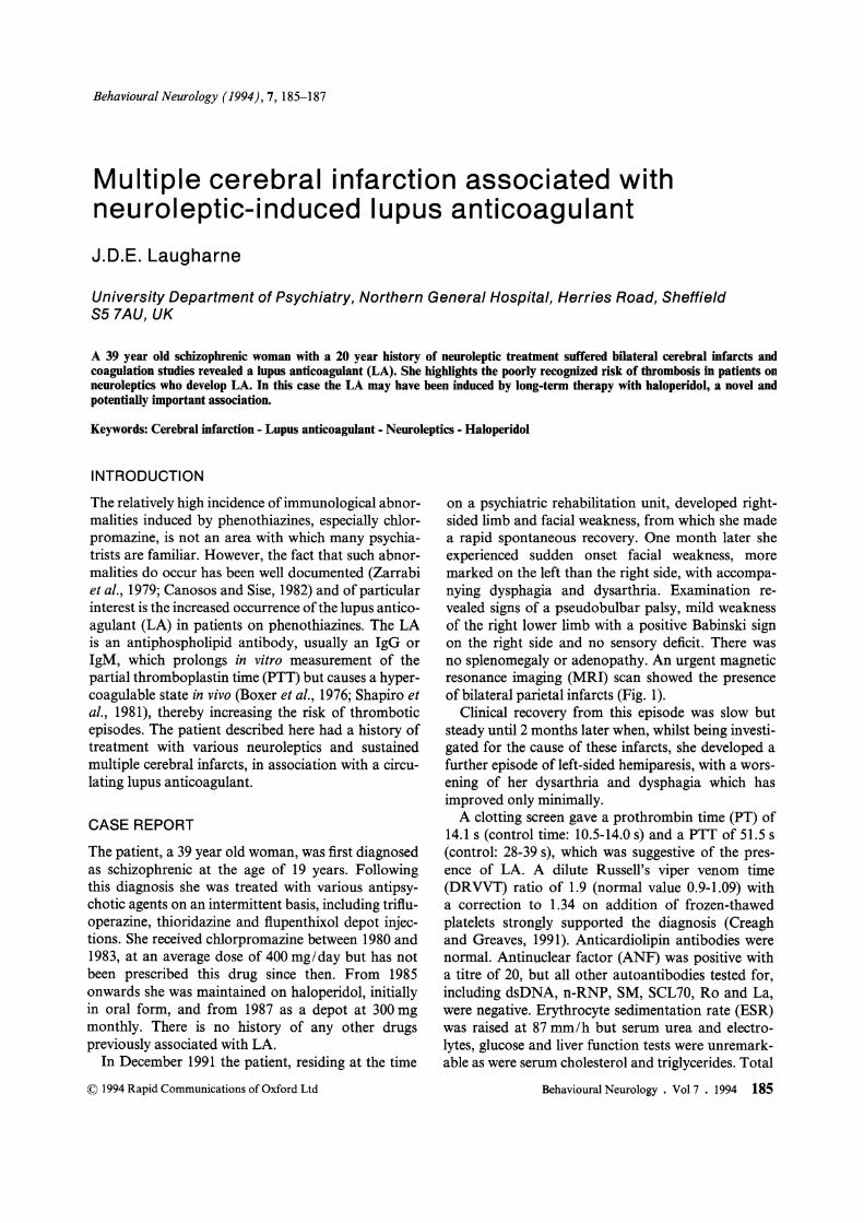
on a psychiatric rehabilitation unit, developed rightsided limb and facial weakness, from which she made a rapid spontaneous recovery. One month later she experienced sudden onset facial weakness, more marked on the left than the right side, with accompanying dysphagia and dysarthria. Examination revealed signs of a pseudobulbar palsy, mild weakness of the right lower limb with a positive Babinski sign on the right side and no sensory deficit. There was no splenomegaly or adenopathy. An urgent magnetic resonance imaging (MRI) scan showed the presence of bilateral parietal infarcts (Fig. 1).
Clinical recovery from this episode was slow but steady until 2 months later when, whilst being investigated for the cause of these infarcts, she developed a further episode of left-sided hemiparesis, with a worsening of her dysarthria and dysphagia which has improved only minimally.
A clotting screen gave a prothrombin time (PT) of 14.1 s (control time: 10.5-14.0 s) and a PTT of 51.5 s (control: 28-39 s), which was suggestive of the presence of LA. A dilute Russell's viper venom time (DRVVT) ratio of 1.9 (normal value 0.9-1.09) with a correction to 1.34 on addition of frozen-thawed platelets strongly supported the diagnosis (Creagh and Greaves, 1991). Anticardiolipin antibodies were normal. Antinuclear factor (ANF) was positive with a titre of 20, but all other autoantibodies tested for, including dsDNA, n-RNP, SM, SCL70, Ro and La, were negative. Erythrocyte sedimentation rate (ESR) was raised at 87 mm/h but serum urea and electrolytes, glucose and liver function tests were unremarkable as were serum cholesterol and triglycerides. Total
Behavioural Neurology. Vo17 . 1994 185

FIG. 1. Single coronal T1 weighted image from gadoliniumOTPA enhanced MRI shows gyral swelling bilaterally and adjacent gyral enhancement (arrowed) consistent with luxury perfusion in subacute infarctions.
serum protein and IgG and IgM fractions were within normal limits. Syphilis serology was negative. ECG and cardiac echogram were normal and Doppler studies showed evidence of minimal bilateral carotid artery disease, with less than 20% loss of lumen diameter and with good vertebral artery flow.
Neuroleptics were discontinued following the first ischaemic episode and have not been administered since. Following the third episode, the patient was commenced on aspirin 300 mg daily and has suffered no further such insult to date.
DISCUSSION
LA was first described in patients with systemic lupus erythematosus (SLE) (Conley and Hartmann, 1952) where it is found in around 10'% of cases (Canosos and Sise, 1982), and also occurs in a range of disorders, including neoplasms, rheumatoid arthritis, autoimmune haemolytic anaemia and in women with a history of recurrent spontaneous abortions as well as in healthy individuals (EI-Mallakh et al., 1988). It has also been reported to be induced by a variety of
186 Behavioural Neurology. Vol7 . 1994
J.D.E. LAUGHARNE
drugs, including procainamide, quinidine, phenytoin, valproic acid and phenothiazines (EI-Mallakh et aI., 1988).
Several workers have demonstrated immunological changes in patients on phenothiazines. Canosos and Sise (1982) reported a group of 42 patients treated with chlorpromazine (CPZ) or another phenothiazine but with a history of CPZ use. Thirty-eight per cent of these patients had LA, 33% had antinuclear antibodies and 46% had elevated levels of serum IgM, whereas 53 controls showed no such abnormalities. Zarrabi et al. (1979) described a group of schizophrenic patients, in whom 75% of those who had received CPZ for longer than 2.5 years or were currently on a different phenothiazine but had previously taken CPZ for more than 2.5 years, had developed raised levels of IgM and circulating inhibitors of coagulation. Fifty per cent of their patients who had LA also had splenomegaly (which was not found in this patient).
LA is known to predispose to thrombotic events. Triplett et at. (1988) reported a thrombotic event in one of four patients with LA attributed to CPZ treatment and observed an increased risk ofthrombosis in patients with drug-induced LA, compared with those with non-drug-induced LA. EI-Mallakh et al. (1988) described a 35 year old woman who suffered multiple lucunar cerebral infarctions secondary to a CPZ-associated LA. In a retrospective study to determine the frequency of thrombosis in patients with the LA Mueh et al. (1980) described eight patients with phenothiazine-induced LA, three of whom had a history of a thrombotic event of which one was a cerebral infarct. Although EI-Mallakh et at. (1988) suggest that thrombotic events are apparently a rarity in phenothiazine-induced LA, the literature is scanty and Triplett et at. (1988) and Mueh et at. (1980) suggest otherwise.
On the current available evidence it would appear that LA is induced by phenothiazines and not by other groups of neuroleptics (Zarrabi et al., 1979) and that CPZ is by far the most commonly implicated agent. Canosos and Sise (1982) comment that the presence of LA was observed as early as 3 months after starting CPZ therapy, but that the autoantibodies were more frequent in patients treated over a longer period. Although most patients with LA in their study had been exposed to a cumulative dose of CPZ greater than 1000 mg, LA development was observed with a minimum dose of 48 mg indicating that in some patients relatively low exposure to CPZ can be immunogenic. They also noted that the persistence of immunological abnormalities, long after discontinuation of CPZ in patients maintained on other

CEREBRAL INFARCTION ASSOCIATED WITH NEUROLEPTIC-INDUCED LA
phenothiazines, suggested a cross-reaction between these compounds which perpetuated the antigenicity induced by the CPZ.
The patient reported here is of particular interest in that, although she had received chlorpromazine over a 3 year period in the past, her treatment during the 7 years prior to the first infarct was with haloperidol alone. The only previous case in the literature of haloperidol possibly being implicated as a cause of LA was one of the eight patients described by Mueh et al. (1980), who was apparently treated concurrently with haloperidol and thioridazine prior to an LArelated thrombotic event. The importance of halo peridol in this case has been called into doubt (EI-Mallakh et al., 1988). The long period of time which had elapsed between my patient last receiving chlorpromazine and her first ischaemic event strongly raises the possibility that haloperidol and presumably other agents of the butyrophenone group may be capable of inducing LA. An alternative explanation is that haloperidol is capable, like non-CPZ phenothiazines, of perpetuating the antigenicity induced by CPZ, but this seems unlikely in view of the marked difference in the molecular structure of these drugs.
In regard to the treatment of neuroleptic-induced LA, no clear management strategy or sense of longterm outcome has yet emerged (Kaslow et al., 1992). Discontinuation of the neuroleptic and commencement of anticoagulant therapy (heparin, warfarin or aspirin) have proved successful in patients with thrombotic complications, as with this patient. In this particular case aspirin was prescribed rather than warfarin due to difficulty ensuring the patient's cooperation with regular venepuncture for clotting function evaluation.
In conclusion, clinicians should be aware that phenothiazines, particularly CPZ, commonly induce immunological abnormalities including LA which can result in thrombotic events including infarction, the rarity of which is open to question.
This case also raises the possibility that other neuroleptics, in particular haloperidol, may also be capable of inducing LA.
Acknowledgements The author wishes to thank Dr M. Peet, Mr P. Pratt, Dr M. Greaves and Dr A. Mason for their valuable comments and Mrs B. Nesbitt for secretarial assistance.
REFERENCES Boxer M, Ellman L and Carvalho A (1976) The lupus
anticoagulant. Arthritis and Rheumatism, 19, 1244-1248. Canosos Rand Sise H (1982) Chlorpromazine-induced
lupus anticoagulant and associated immunologic abnormalities. American Journal of Haematology, 13, 121-129.
Conley CL and Hartmann RC (1952) A hemorrhagic disorder caused by circulating anticoagulants in patients with disseminated lupus erythematosus. Journal of Clinical Investigations, 31, 621-622.
Creagh MD and Greaves M (1991) Lupus anticoagulant. Blood Reviews, 5, 162-167.
El-Mallakh R, Donaldson J, Kranzler Hand Raly A (1988) Phenothiazine-associated lupus anticoagulant and thrombotic disease. Psychosomatics, 29, 109-122.
Kaslow KA, Rosse RB, Zeller JA, Nagy MS and Deutsch SI (1992) Journal of Clinical Psychiatry, 53, 103-104 (letter).
Mueh JR, Herbst KD and Rapaport SI (1980) Thrombosis in patients with the lupus anticoagulant. Annals of Internal Medicine, 92,156-159.
Shapiro SS, Thiagarajan P and DeMarco L (1981) Mechanism of action of the lupus anticoagulant. Annals of the New York Academy of Sciences, 370, 359-365.
Triplett DA, Brandt JT, Musgrave KA and Orr CA (1988) The relationship between lupus anticoagulants and antibodies to phospholipid. Journal of the American Medical Association, 259, 550-554.
Zarrabi M, Zucker S and Miller F (1979) Immunologic and coagulation disorders in chlorpromazine-treated patients. Annals of Internal Medicine, 991, 194-199.
(Received 15 March 1994; accepted as revised 26 August 1994)
Behavioural Neurology. Vol 7 . 1994 187

Submit your manuscripts athttp://www.hindawi.com
Stem CellsInternational
Hindawi Publishing Corporationhttp://www.hindawi.com Volume 2014
Hindawi Publishing Corporationhttp://www.hindawi.com Volume 2014
MEDIATORSINFLAMMATION
of
Hindawi Publishing Corporationhttp://www.hindawi.com Volume 2014
Behavioural Neurology
EndocrinologyInternational Journal of
Hindawi Publishing Corporationhttp://www.hindawi.com Volume 2014
Hindawi Publishing Corporationhttp://www.hindawi.com Volume 2014
Disease Markers
Hindawi Publishing Corporationhttp://www.hindawi.com Volume 2014
BioMed Research International
OncologyJournal of
Hindawi Publishing Corporationhttp://www.hindawi.com Volume 2014
Hindawi Publishing Corporationhttp://www.hindawi.com Volume 2014
Oxidative Medicine and Cellular Longevity
Hindawi Publishing Corporationhttp://www.hindawi.com Volume 2014
PPAR Research
The Scientific World JournalHindawi Publishing Corporation http://www.hindawi.com Volume 2014
Immunology ResearchHindawi Publishing Corporationhttp://www.hindawi.com Volume 2014
Journal of
ObesityJournal of
Hindawi Publishing Corporationhttp://www.hindawi.com Volume 2014
Hindawi Publishing Corporationhttp://www.hindawi.com Volume 2014
Computational and Mathematical Methods in Medicine
OphthalmologyJournal of
Hindawi Publishing Corporationhttp://www.hindawi.com Volume 2014
Diabetes ResearchJournal of
Hindawi Publishing Corporationhttp://www.hindawi.com Volume 2014
Hindawi Publishing Corporationhttp://www.hindawi.com Volume 2014
Research and TreatmentAIDS
Hindawi Publishing Corporationhttp://www.hindawi.com Volume 2014
Gastroenterology Research and Practice
Hindawi Publishing Corporationhttp://www.hindawi.com Volume 2014
Parkinson’s Disease
Evidence-Based Complementary and Alternative Medicine
Volume 2014Hindawi Publishing Corporationhttp://www.hindawi.com