MultimodalImaginginthePrenatalDiagnosisof ... TuberousSclerosisComplex MariyaGusman,1 SabahServaes,2...
4
Hindawi Publishing Corporation Case Reports in Pediatrics Volume 2012, Article ID 925646, 3 pages doi:10.1155/2012/925646 Case Report Multimodal Imaging in the Prenatal Diagnosis of Tuberous Sclerosis Complex Mariya Gusman, 1 Sabah Servaes, 2 Tamara Feygin, 2 Karl Degenhardt, 3 and Monica Epelman 2 1 School of Medicine, Drexel University, Philadelphia, PA 19129, USA 2 Division of Radiology, The Children’s Hospital of Philadelphia, 34th and Civic Center boulevard, Philadelphia, PA 19104, USA 3 Division of Cardiology, The Children’s Hospital of Philadelphia, Philadelphia, PA 19104, USA Correspondence should be addressed to Sabah Servaes, [email protected] Received 29 June 2012; Accepted 4 September 2012 Academic Editors: N. L. Shapiro, E. Veneselli, and V. C. Wong Copyright © 2012 Mariya Gusman et al. This is an open access article distributed under the Creative Commons Attribution License, which permits unrestricted use, distribution, and reproduction in any medium, provided the original work is properly cited. Tuberous sclerosis complex (TSC) is an autosomal dominant disorder in which benign hamartomas develop in multiple organ systems. Increasingly, stigmata of the disease, such as cardiac rhabdomyomas, are detected on routine prenatal ultrasound. Such a finding should prompt additional imaging studies in order to confirm diagnosis and to identify potential complications, which vary greatly from patient to patient. Early diagnosis allows for accurate parental counseling, coordination of high-level perinatal care, and subspecialty followup. We present a case of TSC in utero wherein access to and use of multiple imaging modalities confirmed diagnosis and allowed the patient to receive optimal care prior to birth. 1. Introduction Tuberous sclerosis complex (TSC) is an autosomal dominant neurocutaneous disorder associated with benign hamar- tomas that can develop in almost any organ system. There is tremendous variability in the combination of organ systems affected and the degree of severity. Clinical diagnosis of TSC can be made based on the presence of major and minor features as defined by the Tuberous Sclerosis Consensus Conference [1]. The presence of two major features is sufficient for a definite diagnosis of TSC. Major features that may be discovered with prenatal imaging include cardiac rhabdomyomas, cortical tubers, subependymal nodules, and renal angiomyolipomas. The ability to detect each of these lesions differs between imaging modalities. Suspicion of TSC in a fetus often begins with the finding of multiple intracardiac masses on routine ultrasound, sug- gesting rhabdomyomas. Fetal echocardiography can further delineate these masses and assess their effects on ventricular function, obstruction of blood flow, and arrhythmias. Cor- tical tubers and subependymal nodules are best visualized prenatally by ultrasound or MRI. Renal angiomyolipomas can be suggested by findings of fat-containing lesions with sonographic or MR imaging. We present a case of TSC that was diagnosed prenatally with multiple imaging modalities. 2. Case The mother was a 24-year-old G2P1001 with no family history of TSC or congenital defects. Fetal ultrasound at 35 weeks and 6 days demonstrated multiple intracardiac masses (Figure 1(a)), which were further visualized by fetal echocar- diography (Figures 1(b) and 1(c)). The masses are seen within the ventricular myocardium and most likely represent rhabdomyomas. The masses were relatively stable in size, and ventricular function remained normal on serial echocardiog- raphy. The masses did not cause ventricular outflow or inflow obstruction. There was no evidence of fetal tachyarrhythmia. Fetal ultrasound also revealed a renal lesion (Figure 2(a)). Given its increased echogenicity and solid appearance, this is most likely an angiomyolipoma. The lesion was confirmed on fetal MRI (Figures 2(b) and 2(c)). All imaging modalities were negative for neurological findings. The baby was born by spontaneous vaginal delivery at a children’s hospital as a precaution against any cardiac com- plications. The possibility of arrhythmias or the development
Transcript of MultimodalImaginginthePrenatalDiagnosisof ... TuberousSclerosisComplex MariyaGusman,1 SabahServaes,2...
Hindawi Publishing CorporationCase Reports in PediatricsVolume 2012, Article ID 925646, 3 pagesdoi:10.1155/2012/925646
Case Report
Multimodal Imaging in the Prenatal Diagnosis ofTuberous Sclerosis Complex
Mariya Gusman,1 Sabah Servaes,2 Tamara Feygin,2 Karl Degenhardt,3 and Monica Epelman2
1 School of Medicine, Drexel University, Philadelphia, PA 19129, USA2 Division of Radiology, The Children’s Hospital of Philadelphia, 34th and Civic Center boulevard, Philadelphia, PA 19104, USA3 Division of Cardiology, The Children’s Hospital of Philadelphia, Philadelphia, PA 19104, USA
Correspondence should be addressed to Sabah Servaes, [email protected]
Received 29 June 2012; Accepted 4 September 2012
Academic Editors: N. L. Shapiro, E. Veneselli, and V. C. Wong
Copyright © 2012 Mariya Gusman et al. This is an open access article distributed under the Creative Commons AttributionLicense, which permits unrestricted use, distribution, and reproduction in any medium, provided the original work is properlycited.
Tuberous sclerosis complex (TSC) is an autosomal dominant disorder in which benign hamartomas develop in multiple organsystems. Increasingly, stigmata of the disease, such as cardiac rhabdomyomas, are detected on routine prenatal ultrasound. Sucha finding should prompt additional imaging studies in order to confirm diagnosis and to identify potential complications, whichvary greatly from patient to patient. Early diagnosis allows for accurate parental counseling, coordination of high-level perinatalcare, and subspecialty followup. We present a case of TSC in utero wherein access to and use of multiple imaging modalitiesconfirmed diagnosis and allowed the patient to receive optimal care prior to birth.
1. Introduction
Tuberous sclerosis complex (TSC) is an autosomal dominantneurocutaneous disorder associated with benign hamar-tomas that can develop in almost any organ system. There istremendous variability in the combination of organ systemsaffected and the degree of severity. Clinical diagnosis of TSCcan be made based on the presence of major and minorfeatures as defined by the Tuberous Sclerosis ConsensusConference [1]. The presence of two major features issufficient for a definite diagnosis of TSC. Major features thatmay be discovered with prenatal imaging include cardiacrhabdomyomas, cortical tubers, subependymal nodules, andrenal angiomyolipomas. The ability to detect each of theselesions differs between imaging modalities.
Suspicion of TSC in a fetus often begins with the findingof multiple intracardiac masses on routine ultrasound, sug-gesting rhabdomyomas. Fetal echocardiography can furtherdelineate these masses and assess their effects on ventricularfunction, obstruction of blood flow, and arrhythmias. Cor-tical tubers and subependymal nodules are best visualizedprenatally by ultrasound or MRI. Renal angiomyolipomascan be suggested by findings of fat-containing lesions withsonographic or MR imaging.
We present a case of TSC that was diagnosed prenatallywith multiple imaging modalities.
2. Case
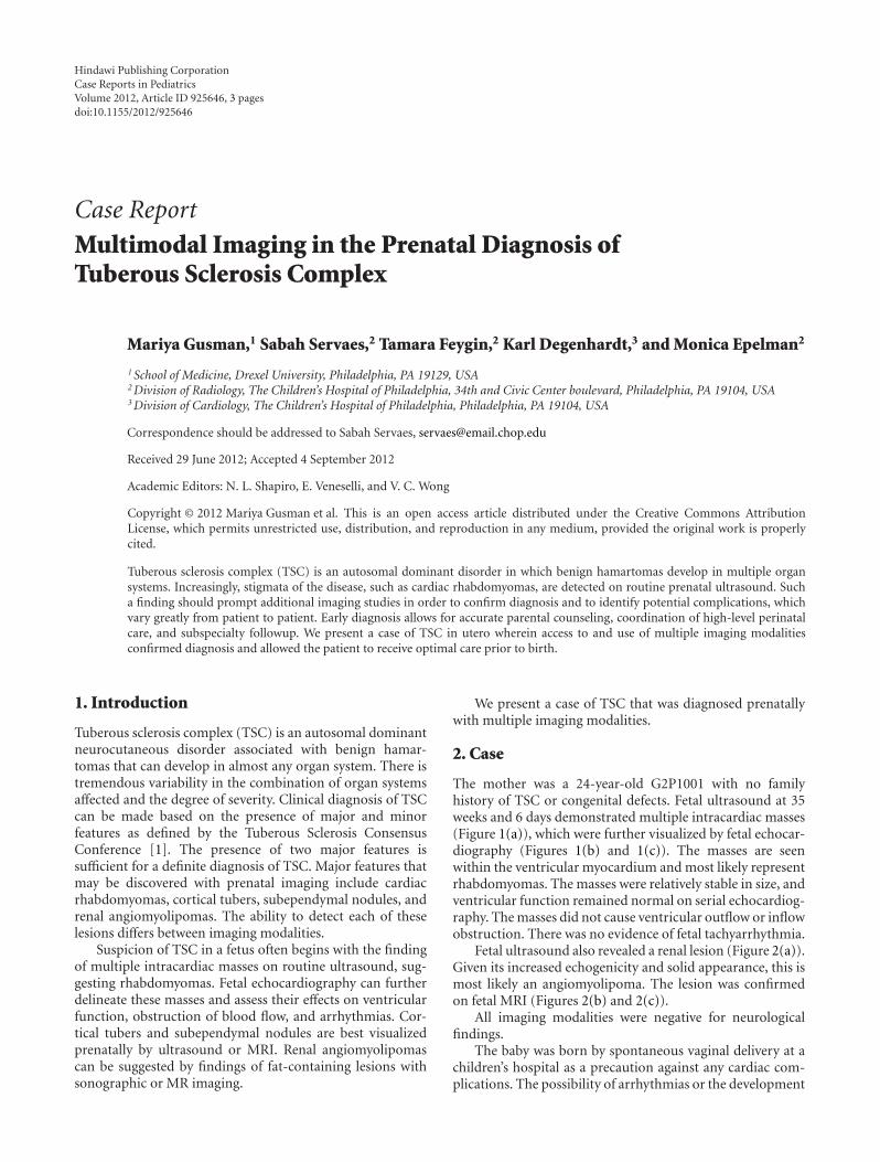
The mother was a 24-year-old G2P1001 with no familyhistory of TSC or congenital defects. Fetal ultrasound at 35weeks and 6 days demonstrated multiple intracardiac masses(Figure 1(a)), which were further visualized by fetal echocar-diography (Figures 1(b) and 1(c)). The masses are seenwithin the ventricular myocardium and most likely representrhabdomyomas. The masses were relatively stable in size, andventricular function remained normal on serial echocardiog-raphy. The masses did not cause ventricular outflow or inflowobstruction. There was no evidence of fetal tachyarrhythmia.
Fetal ultrasound also revealed a renal lesion (Figure 2(a)).Given its increased echogenicity and solid appearance, this ismost likely an angiomyolipoma. The lesion was confirmedon fetal MRI (Figures 2(b) and 2(c)).
All imaging modalities were negative for neurologicalfindings.
The baby was born by spontaneous vaginal delivery at achildren’s hospital as a precaution against any cardiac com-plications. The possibility of arrhythmias or the development

2 Case Reports in Pediatrics
LV
LA
RV
RA
Heart
(a)
LV
LA
RV
RA
(b)
LV
LA RV
RA
(c)
Figure 1: Multiple intracardiac masses (arrows) were detected onroutine ultrasound (a), prompting a more thorough investigation.Fetal echocardiography ((b) and (c)) identified five masses that werewithin the ventricular myocardium although only two (arrows) canbe seen in this plane. The masses were of uniform echogenicity andwere more echogenic than the surrounding myocardium, consistentwith the typical appearance of cardiac rhabdomyomas.
of obstruction to flow with the transition to postnatal cir-culation was of particular concern [2]. The perinatal coursewas uneventful. Postnatal echocardiography confirmed goodventricular function without inflow or outflow obstruction.Around three weeks of age, the baby developed prematureventricular contractions. Holter monitoring showed signif-icant ventricular ectopy with an episode of nonsustainedventricular tachycardia. The ectopy responded well to theinitiation of a beta-blocker. By three months of age, there
Right kidney, sagittal
(a)
MS
K K
(b)
ML
L L
K K
(c)
Figure 2: (a) On ultrasound, a single small, echogenic lesion wasdetected within the parenchyma of the upper pole of the rightkidney, consistent with an angiomyolipoma. (b) T-2-weighted MRIimage, in the transverse plane, at the level of the top of the rightkidney demonstrates a high-signal lesion within the upper pole(arrow). K, kidneys; MS, maternal spine. (c) TruFISP coronal imagethrough the kidneys shows another view of the same lesion (arrow)K, kidney; ML, maternal liver; L, lung.
was echocardiographic evidence of a decrease in the size ofthe ventricular masses.
In the neonatal population, TSC is most often clinicallyassociated with seizures and dermatological findings. Todate, our patient has not experienced seizures and has madenormal developmental milestones, which is consistent withhis lack of neurological findings on imaging. Dermatological

Case Reports in Pediatrics 3
findings are limited to a single hypopigmented macule on thelower abdomen.
3. Discussion
TSC may be diagnosed prenatally in the presence of two ofthe following: cardiac rhabdomyomas, cortical tubers, sub-ependymal nodules, and renal angiomyolipomas.
Cardiac rhabdomyomas are the most commonly foundfetal cardiac tumor [3, 4] and are also the most commonfetal finding in TSC. Larger numbers of fetal cardiacrhabdomyomas combined with a family history of TSC aremore predictive of a TSC diagnosis [4]. However, only one-third of TSC cases are in children with a family history of TSC[5]. In our patient, the appearance of cardiac rhabdomyomason ultrasound led to further imaging workup. Had thediagnosis not been made prenatally, the infant may have hadmore significant arrhythmias. The natural history of cardiacrhabdomyomas is for regression to occur in the first years oflife. Had the diagnosis not been made prenatally, it may havebeen delayed by several years.
Subependymal nodules and cortical tubers, while not themost common presenting sign, are a very frequent findingin infants receiving a TSC diagnosis (93% and 88%, resp.).Chen et al. demonstrated that ultrafast MRI can discoversuch lesions even when they are not apparent on a detailedsonographic examination of the fetal brain [6]. Because ourpatient underwent fetal MRI to evaluate his renal findings,we are confident that he did not have any subependymalor cortical masses. Consistent with this, our patient has hadnormal development without clinical evidence of seizures.
Renal findings, although a common manifestation of thedisease, usually begin to form and increase in number duringchildhood and adolescence [7, 8]. There are a handful ofreports of infants with severe renal disease. Only four priorarticles report prenatal renal findings [9]. Our case is notablefor the presence of this rare finding, which, along with thefinding of multiple rhabdomyomas, confirmed the diagnosisof TSC.
Pediatricians and pediatric subspecialists should be awareof prenatal imaging findings relevant to TSC, as the presenceof more than one finding is sufficient for TSC diagnosis.A suspicion of TSC based on an ultrasound finding of acardiac, neurological, or renal mass should prompt furtherimaging. We utilized fetal echocardiography and MRI towardthis end. Multimodal imaging allows clinicians to prepareboth themselves and the family for potential complications,and arrange for early subspecialty care.
References
[1] E. S. Roach, F. J. DiMario, R. S. Kandt, and H. Northrup, “Tub-erous sclerosis consensus conference: recommendations fordiagnostic evaluation,” Journal of Child Neurology, vol. 14, no.6, pp. 401–407, 1999.
[2] S. C. Degueldre, P. Chockalingam, Y. Mivelaz et al., “Consid-erations for prenatal counselling of patients with cardiac rhab-domyomas based on their cardiac and neurologic outcomes,”Cardiology in the Young, vol. 20, no. 1, pp. 18–24, 2010.
[3] Y. Yinon, D. Chitayat, S. Blaser et al., “Fetal cardiac tumors: asingle-center experience of 40 cases,” Prenatal Diagnosis, vol. 30,no. 10, pp. 941–949, 2010.
[4] A. S. Chao, A. Chao, T. H. Wang et al., “Outcome of antenatallydiagnosed cardiac rhabdomyoma: case series and a meta-ana-lysis,” Ultrasound in Obstetrics and Gynecology, vol. 31, no. 3,pp. 289–295, 2008.
[5] V. M. Rose, K. S. Au, G. Pollom, E. S. Roach, H. R. Prashner,and H. Northrup, “Germ-line mosaicism in tuberous sclerosis:how common?” American Journal of Human Genetics, vol. 64,no. 4, pp. 986–992, 1999.
[6] C. P. Chen, Y. P. Liu, J. K. Huang et al., “Contribution ofultrafast magnetic resonance imaging in prenatal diagnosis ofsonographically undetected cerebral tuberous sclerosis associ-ated with cardiac rhabdomyomas,” Prenatal Diagnosis, vol. 25,no. 6, pp. 523–524, 2005.
[7] K. A. Casper, L. F. Donnelly, B. Chen, and J. J. Bissler, “Tuberoussclerosis complex: renal imaging findings,” Radiology, vol. 225,no. 2, pp. 451–456, 2002.
[8] F. J. O’Callaghan, M. J. Noakes, C. N. Martyn, and J. P. Osborne,“An epidemiological study of renal pathology in tuberoussclerosis complex,” British Journal of Urology International, vol.94, no. 6, pp. 853–857, 2004.
[9] A. Gedikbasi, K. Oztarhan, V. Ulker et al., “Prenatal sonograph-ic diagnosis of tuberous sclerosis complex,” Journal of ClinicalUltrasound, vol. 39, no. 7, pp. 427–430, 2011.

Submit your manuscripts athttp://www.hindawi.com
Stem CellsInternational
Hindawi Publishing Corporationhttp://www.hindawi.com Volume 2014
Hindawi Publishing Corporationhttp://www.hindawi.com Volume 2014
MEDIATORSINFLAMMATION
of
Hindawi Publishing Corporationhttp://www.hindawi.com Volume 2014
Behavioural Neurology
EndocrinologyInternational Journal of
Hindawi Publishing Corporationhttp://www.hindawi.com Volume 2014
Hindawi Publishing Corporationhttp://www.hindawi.com Volume 2014
Disease Markers
Hindawi Publishing Corporationhttp://www.hindawi.com Volume 2014
BioMed Research International
OncologyJournal of
Hindawi Publishing Corporationhttp://www.hindawi.com Volume 2014
Hindawi Publishing Corporationhttp://www.hindawi.com Volume 2014
Oxidative Medicine and Cellular Longevity
Hindawi Publishing Corporationhttp://www.hindawi.com Volume 2014
PPAR Research
The Scientific World JournalHindawi Publishing Corporation http://www.hindawi.com Volume 2014
Immunology ResearchHindawi Publishing Corporationhttp://www.hindawi.com Volume 2014
Journal of
ObesityJournal of
Hindawi Publishing Corporationhttp://www.hindawi.com Volume 2014
Hindawi Publishing Corporationhttp://www.hindawi.com Volume 2014
Computational and Mathematical Methods in Medicine
OphthalmologyJournal of
Hindawi Publishing Corporationhttp://www.hindawi.com Volume 2014
Diabetes ResearchJournal of
Hindawi Publishing Corporationhttp://www.hindawi.com Volume 2014
Hindawi Publishing Corporationhttp://www.hindawi.com Volume 2014
Research and TreatmentAIDS
Hindawi Publishing Corporationhttp://www.hindawi.com Volume 2014
Gastroenterology Research and Practice
Hindawi Publishing Corporationhttp://www.hindawi.com Volume 2014
Parkinson’s Disease
Evidence-Based Complementary and Alternative Medicine
Volume 2014Hindawi Publishing Corporationhttp://www.hindawi.com