Multiligament Knee Injuries and Their Treatment Knee Injuries and Their Treatment Demetris Delos,...
8
Multiligament Knee Injuries and Their Treatment Demetris Delos, MD, Russell F. Warren, MD, and Robert G. Marx, MD, MSc, FRCSC Multiligament knee injuries are rare but potentially devastating. They require expeditious and thorough evaluation and treatment for best outcomes. Management consists of a careful history and physical examination, including a complete assessment of the neuro- vascular status with ankle-brachial indexes magnetic resonance angiography. Imaging should include plain radiographs, stress radiography as necessary, and possibly computed tomography scans if fracture is suspected. Magnetic resonance imaging is the modality of choice for evaluating the ligaments and soft tissues. Once evaluation is complete, the decision to proceed with surgery versus conservative management is made. We favor surgery in patients who are able to tolerate the procedure, are relatively high functioning, and are not obese. Surgery is performed at 10 to 14 days if repair is possible or it is delayed until at least 4 to 6 weeks from the time of injury if reconstructions are to be performed to minimize the risk of arthrofibrosis. We prefer allografts for our reconstructions rather than autografts to minimize morbidity and operative time. After surgery, patients may be anti- coagulated with either aspirin or coumadin. With extensive, focused rehabilitation, patients can return to heavy labor and/or sports beginning at 9 to 12 months postoperatively. Oper Tech Sports Med 18:219-226 © 2010 Elsevier Inc. All rights reserved. KEYWORDS knee, ligament, reconstruction, augmentation, posterolateral corner A lthough multiligament knee injuries are uncommon, failure to diagnose and treat them appropriately can lead to devastating outcomes. Careful and thorough clinical eval- uation is critical as is timely, appropriate treatment. The fol- lowing is an overview of our approach to these injuries and their treatment. Diagnosis History In our practice, patients are usually referred from other cen- ters rather than seen in the emergency room. Regardless of the manner of presentation, however, a careful and complete history is mandatory. This includes an understanding of the mechanism of injury and the level of energy of the forces involved. A knee dislocation with subsequent reduction should always be considered a multiligament injured knee and should be evaluated as such. Comorbidities, especially a history of diabetes, peripheral neuropathy, or vascular insuf- ficiency, should be elicited during the history. It is also im- portant to gain a sense of the individual’s overall well-being and activity level. Physical Examination Multiligament knee injuries are usually the result of high- energy forces; therefore, other injuries should first be ruled out. In the emergency room scenario, this often means a full trauma evaluation is performed under the auspices of the trauma surgery, orthopedic surgery, and emergency depart- ments. During the secondary survey, the limb is inspected for gross deformity and alignment. A subluxated or frankly dis- located knee joint should be relocated promptly. It is impor- tant to also consider patellar dislocations in the setting of multiligamentous knee injuries. The skin should be exam- ined for color and integrity. A thorough neurovascular exam- ination is performed, including palpation of the popliteal, dorsalis pedis, and posterior tibial arteries as well as deter- mining whether there are motor or sensory changes. Peroneal nerve injury is not uncommon, especially with lateral injury. 1 In the office setting, we do not routinely perform ankle- brachial indices (ABIs) because most of our patients are seen upon referral and typically have already had an angiogram or magnetic resonance angiography (MRA) performed. There is prospective literature to support the use of ABIs and serial examinations in the initial evaluation period. 2 However, it should be noted that a recent retrospective study reported Department of Orthopaedic Surgery, Hospital for Special Surgery, New York, NY. Address reprint requests to Robert G. Marx, MD, MSc, FRCSC, Hospital for Special Surgery, 535 East 70 Street, New York, NY 10021. E-mail: [email protected] 219 1060-1872/10/$-see front matter © 2010 Elsevier Inc. All rights reserved. doi:10.1053/j.otsm.2010.09.002
Transcript of Multiligament Knee Injuries and Their Treatment Knee Injuries and Their Treatment Demetris Delos,...
MD
Atult
DHItthmisahfi
D
A
1d
ultiligament Knee Injuries and Their Treatmentemetris Delos, MD, Russell F. Warren, MD, and Robert G. Marx, MD, MSc, FRCSC
Multiligament knee injuries are rare but potentially devastating. They require expeditiousand thorough evaluation and treatment for best outcomes. Management consists of acareful history and physical examination, including a complete assessment of the neuro-vascular status with ankle-brachial indexes � magnetic resonance angiography. Imagingshould include plain radiographs, stress radiography as necessary, and possibly computedtomography scans if fracture is suspected. Magnetic resonance imaging is the modality ofchoice for evaluating the ligaments and soft tissues. Once evaluation is complete, thedecision to proceed with surgery versus conservative management is made. We favorsurgery in patients who are able to tolerate the procedure, are relatively high functioning,and are not obese. Surgery is performed at 10 to 14 days if repair is possible or it is delayeduntil at least 4 to 6 weeks from the time of injury if reconstructions are to be performed tominimize the risk of arthrofibrosis. We prefer allografts for our reconstructions rather thanautografts to minimize morbidity and operative time. After surgery, patients may be anti-coagulated with either aspirin or coumadin. With extensive, focused rehabilitation, patientscan return to heavy labor and/or sports beginning at 9 to 12 months postoperatively.Oper Tech Sports Med 18:219-226 © 2010 Elsevier Inc. All rights reserved.
KEYWORDS knee, ligament, reconstruction, augmentation, posterolateral corner
pa
PMeottmgltmiidmnIbumpe
lthough multiligament knee injuries are uncommon,failure to diagnose and treat them appropriately can lead
o devastating outcomes. Careful and thorough clinical eval-ation is critical as is timely, appropriate treatment. The fol-
owing is an overview of our approach to these injuries andheir treatment.
iagnosisistory
n our practice, patients are usually referred from other cen-ers rather than seen in the emergency room. Regardless ofhe manner of presentation, however, a careful and completeistory is mandatory. This includes an understanding of theechanism of injury and the level of energy of the forces
nvolved. A knee dislocation with subsequent reductionhould always be considered a multiligament injured kneend should be evaluated as such. Comorbidities, especially aistory of diabetes, peripheral neuropathy, or vascular insuf-ciency, should be elicited during the history. It is also im-
epartment of Orthopaedic Surgery, Hospital for Special Surgery, NewYork, NY.
ddress reprint requests to Robert G. Marx, MD, MSc, FRCSC, Hospital forSpecial Surgery, 535 East 70 Street, New York, NY 10021. E-mail:
060-1872/10/$-see front matter © 2010 Elsevier Inc. All rights reserved.oi:10.1053/j.otsm.2010.09.002
ortant to gain a sense of the individual’s overall well-beingnd activity level.
hysical Examinationultiligament knee injuries are usually the result of high-
nergy forces; therefore, other injuries should first be ruledut. In the emergency room scenario, this often means a fullrauma evaluation is performed under the auspices of therauma surgery, orthopedic surgery, and emergency depart-ents. During the secondary survey, the limb is inspected for
ross deformity and alignment. A subluxated or frankly dis-ocated knee joint should be relocated promptly. It is impor-ant to also consider patellar dislocations in the setting ofultiligamentous knee injuries. The skin should be exam-
ned for color and integrity. A thorough neurovascular exam-nation is performed, including palpation of the popliteal,orsalis pedis, and posterior tibial arteries as well as deter-ining whether there are motor or sensory changes. Peronealerve injury is not uncommon, especially with lateral injury.1
n the office setting, we do not routinely perform ankle-rachial indices (ABIs) because most of our patients are seenpon referral and typically have already had an angiogram oragnetic resonance angiography (MRA) performed. There isrospective literature to support the use of ABIs and serialxaminations in the initial evaluation period.2 However, it
hould be noted that a recent retrospective study reported219
f4Mhldi
cdpbcitvFa
coflntfgnglrtctt
ap3rcpteve
IItogouprms
to
bmtspopfKi
Fd
220 D. Delos, R.F. Warren, and R.G. Marx
alse-negative rates for ABIs and angiograms of 67% and0%, respectively.3 Early experience at our institution withRA has shown that it correlates well with angiography and
olds potential to be an important tool for diagnosing arterialesions.1,4 We perform MRA routinely for patients with kneeislocation or bicruciate injury who have not had a vascular
maging study before referral to our facility.Ligament examination should include testing anterior cru-
iate ligament (ACL) integrity with the Lachman, anteriorrawer, and pivot shift tests although in the acutely injuredatient the pivot shift and drawers tests are of limited utilityecause of restricted motion. It is important to note that inases of multiligament rupture the tibiofemoral relationships compromised, which can affect the ability of the examinero accurately judge translation and rotation as well as varus/algus stability if injuries exist on both sides of the joint.urthermore, in the acutely injured knee, significant swellingnd pain can limit range of motion, particularly knee flexion.
Posterior cruciate ligament (PCL) examination should in-lude the posterior drawer test at 90° of knee flexion and 45°f hip flexion, the posterior sag test (at approximately 70° ofexion), and the quadriceps active test (though this is usuallyot possible in the acutely injured knee). In the normal knee,he medial tibial plateau is usually 1 cm anterior to the medialemoral condyle. We devised the following simplified clinicalrading system to be used with the posterior drawer test:ormal, no loss of tibial offset; grade A, slight loss of tibial offset;rade B, flush with femur; and grade C, the tibia can be trans-ated posterior to the femur. We have found that grade C inju-ies, according to this system, are usually associated with injuryo the posterolateral corner or medial side of the knee.5 Medialollateral ligament (MCL) and lateral collateral ligament (LCL)esting should be performed with valgus and varus stressing ofhe knee, respectively, at 0° and 30° knee flexion.
Posteromedial corner testing can be performed with thenteromedial drawer test. It is also important to examine theosterolateral corner using the external rotatory (dial) test at0° and 90° with the patient in the prone position. We alsoely on the posterolateral spin test to detect posterolateralorner injury. With the knee flexed at 30° or 90° and theatient supine, the step-off of the lateral tibial plateau fromhe lateral femoral condyle can be palpated with the examin-r’s thumb and compared with the contralateral side. Theirtue of this test, compared with the dial test, is that it avoidsrror because of rotation of the tibia, ankle, or foot.6
magingmaging should first consist of obtaining a radiographic series ofhe knee (anteroposterior/lateral/oblique/merchant). Alignmentf the knee is carefully examined, and the characteristic radio-raphic signs of soft-tissue injury are looked for (effusion, Seg-nd fracture, an posterior sag). It is important to always doc-ment reduction of the joint because an unstable knee isrone to recurrent subluxation or frank dislocation. Weeklyadiographs are indicated to confirm that the reduction isaintained if the patient is treated nonoperatively or before
urgery. If the tibia shows interval displacement (usually pos- r
eriorly) on radiographs, an external fixator may be appliedr an open procedure may be necessary.Stress testing is a useful adjunct to standard radiographs,
oth in the office and intraoperatively. Stress radiographyay be performed at 30° of flexion for varus and valgus laxity
esting. Greater than 5 to 6 mm of widening should promptuspicion of ligamentous injury; �9 to 10 mm suggests com-lete rupture. It is important to always consider that both sidesf the joint may be affected, and, therefore, widening may beresent both medially and laterally. One of us (RFW) also per-orms TELOS stress radiography (Telos Stress Device; METAX,upplungs und Dichtungstechnik GmbH, Marburg, Germany)
n the office as warranted. If fracture is suspected, a computed
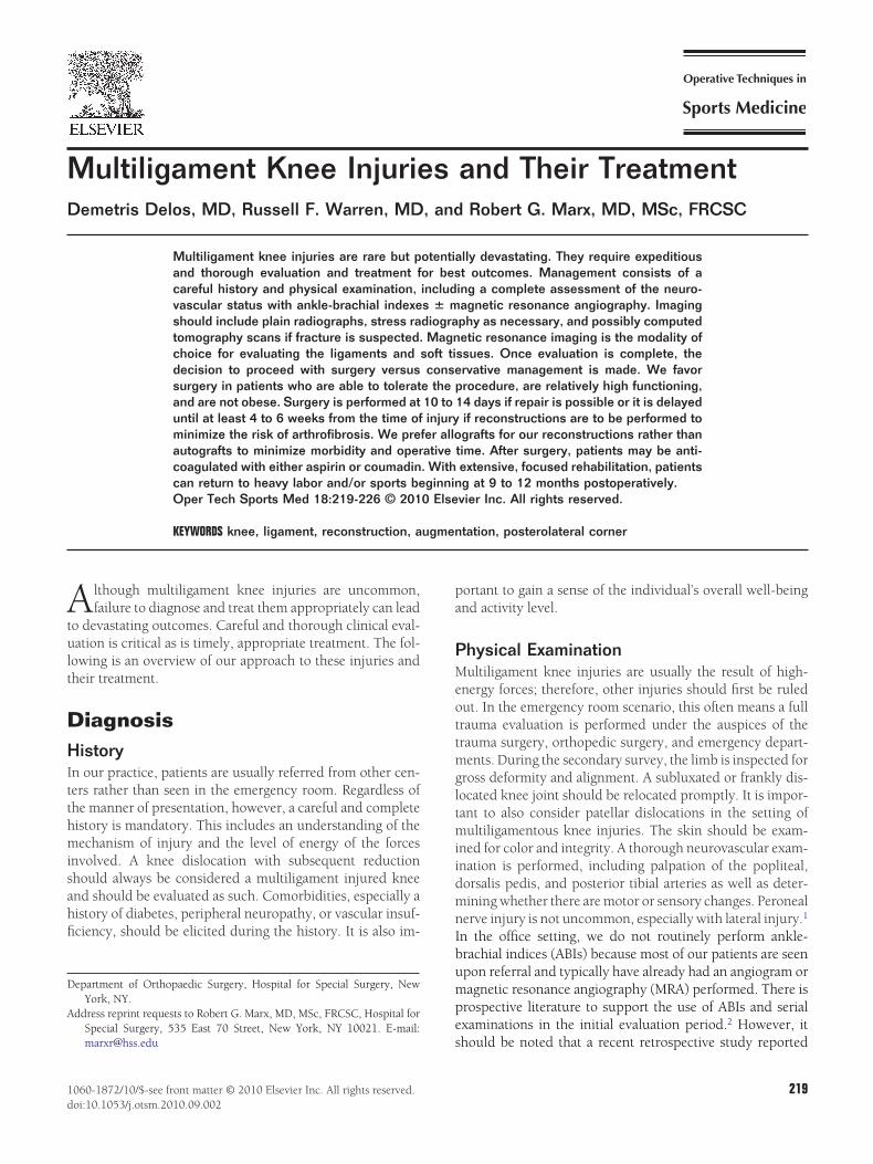
igure 1 (A) Coronal magnetic resonance image showing both me-ial and lateral collateral ligament injuries. (B) Sagittal magnetic
esonance image showing bicruciate ligament rupture.
th
tuapwas
PSWmdcocwpos
ACEatwi
of
utttsicb(wt
TTotawaosl
mavtt
Multiligament knee injuries and their treatment 221
omography scan is recommended. For soft-tissue evaluation,owever, MRI is our modality of choice (Fig. 1).ABIs, angiograms, and MRA are usually performed before
he patient has been referred to us. If not, we have the patientndergo MRA. Early data from our institution showed 100%greement between conventional angiograms and MRA inatients who had both studies performed although numbersere limited.1 We ensure that every patient we operate on has
n imaging study performed preoperatively to document thetatus of the circulation of the injured extremity.
atientelection/Preoperative Planninge favor surgical treatment for patients with multiple liga-ent-injured knees. However, not all patients are good can-idates for multiligament knee surgery. Relative contraindi-ations to surgery include morbid obesity and advanced ager extremely limited preinjury function. Patients with medi-al contraindications to surgery are treated conservativelyith initial immobilization, bracing, and rehabilitation. Foratients who present late with a grossly stable knee, we rec-mmend treating with rehabilitation and subsequent recon-truction if necessary.
cute Versushronic Considerations
valuation in the acute scenario should focus on diagnosingnd treating injuries that threaten life and limb, such as frac-ures, neurovascular injuries, and open injuries. In cases inhich surgery must be performed acutely (ie, open fractures/
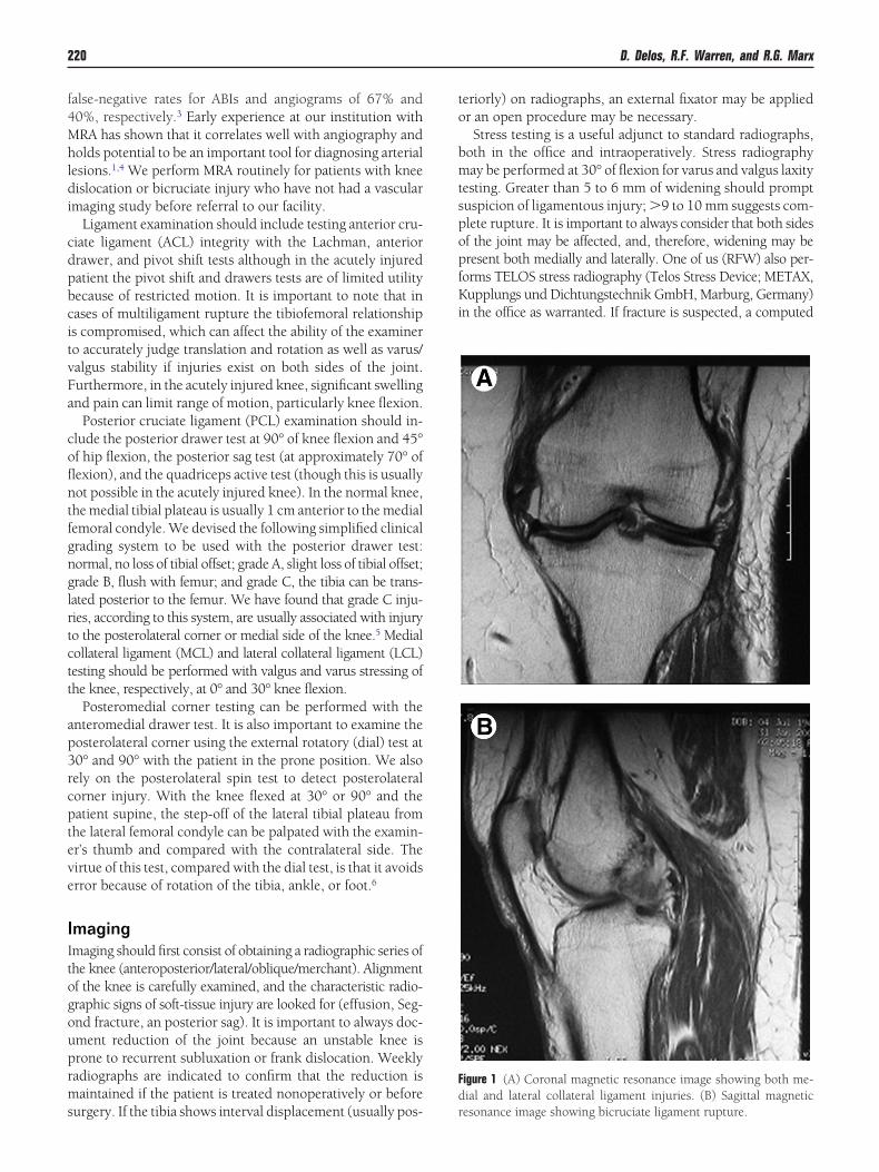
Figure 2 (A) Anteroposterior and (B) lateral radiographsfailed ACL and posterolateral corner surgery.
njuries with vascular compromise/irreducible dislocations), p
ther pathologies, such as meniscal or articular cartilage de-ects, can be definitively addressed at a later date.
For the patient who presents in the chronic scenario, eval-ation should focus on a more thorough understanding ofhe overall status of the joint. Limb alignment, articular car-ilage integrity, and the state of the menisci should be inves-igated. Long-leg radiographs should be obtained. Gaithould be examined for evidence of dynamic thrust. Depend-ng on the extent of the pathology, surgery may be staged. Forases of severe deformity, we now favor an opening-wedgeiplanar high tibial osteotomy to address both the coronalvarus/valgus) and sagittal planes (tibial slope). If this is done,e prefer to perform the ligament reconstructions at a later
ime (Fig. 2).
iming of Surgeryhe optimal timing of surgery varies. One of us (RFW) willften perform surgery at approximately 10 days after injuryo minimize scar formation and swelling7,8 and allow a morenatomic dissection. Thus, if indicated, soft-tissue repairith augmentation can be attempted, with reconstruction as
n alternative if the tissue is not amenable to repair. Based onur own experience, and as reported by Shelbourne et al,9
oft-tissue avulsions off the fibula that are repaired early canead to excellent long-term outcomes.
The other author (RGM) generally prefers waiting a mini-um of 4 to 6 weeks before surgery to minimize the risk of
rthrofibrosis. In most cases, he prefers reconstructions iniew of data indicating lower rates of failure for reconstruc-ions versus repairs.10-13 According to a recent report fromhe Mayo Clinic,11 the rate of failure for reconstructions com-
g closing-wedge high tibial osteotomy performed after
showinared with repairs of the fibular collateral ligament/postero-
lparnfc
nfdate
acarss
SJuvoi
catRcsettma
odpewPivdsamtsP6
tdpfiPaooEaatpssgptffoec
bdploptcgt
Fp
222 D. Delos, R.F. Warren, and R.G. Marx
ateral corner was 6% versus 40%, respectively. This sup-orts the results of an earlier prospective study by Stannard etl10 who noted a 37% failure rate of repairs versus 9% ofeconstructions. A recent systematic review also reported sig-ificantly higher rates of knee flexion loss after acute surgeryor multiligament knees (performed within 3 weeks of injury)ompared with chronic or staged procedures.14
Ultimately, the timing of surgery will be determined by aumber of factors, including the vascular status of the af-ected extremity, degree of swelling, soft-tissue lesions/con-ition of skin, and the degree of instability. The decision tottempt repair should be based on the integrity of the softissues, the nature of the soft-tissue injury, and the experi-nce of the surgeon.
On occasion, patients are referred to our institution withn external fixator already applied and/or having had a vas-ular repair performed. For these patients, the external fix-tor is left on for approximately 5 to 6 weeks before it isemoved and rehabilitation is begun. These patients may betable after removal of the fixator, at which point furtherurgery is not necessary.
urgical Techniquesust before the procedure, a thorough ligament examinationnder anesthesia is performed, including the pivot shift/re-erse pivot shift. We prefer to use a tourniquet during thepen portion of the procedure as long as preoperative imag-ng has ruled out vascular injury.
We perform our ACL and PCL reconstructions arthroscopi-ally and strive for an anatomic reconstruction of both the ACLnd PCL footprints. For the ACL reconstruction, we use either aranstibial (RFW) or anteromedial portal approach (RFW andGM)15 depending on whether the transtibial method can re-reate the femoral footprint. For the PCL, we use a transtibialingle-bundle approach that strives to reproduce the anterolat-ral bundle (ALB). Currently, there is yet no good evidence inhe literature showing improved results with double-bundle oribial inlay, both of which are more technically complex anday remove an excessive amount of bone stock. Tibial inlay is
n option, however, in the revision setting.Whether one first addresses the ACL or the PCL will depend
n surgeon preference. One of the authors (RGM) recommendsrilling the ACL femoral socket first endoscopically and thenositioning the ACL guidewire on the tibia to provide a refer-nce point for the tibial PCL tunnel. The other author (RFW)ill often begin with the PCL and then address the ACL. For theCL portion of the procedure, a posteromedial accessory portal
s first established under visualization. With an arthrocare de-ice, the posterior capsule is elevated off the tibia (up to 2.5 cmistally) to ensure the tunnel is low enough for anatomic recon-truction. Then, a combination of shaver and arthrocare devicesre used to debride the tibial stump of the PCL via the postero-edial portal, taking care that the instruments are directed an-
eriorly at all times. For the PCL, we have been using the Acufexystem (Smith and Nephew, Andover, MA). The Acufex directorCL Tibial Aimer is placed through the medial portal at a 60° to
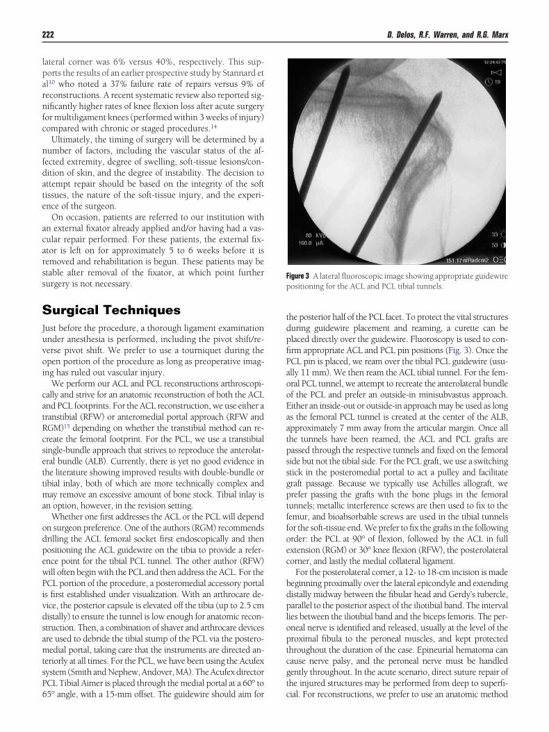
5° angle, with a 15-mm offset. The guidewire should aim for che posterior half of the PCL facet. To protect the vital structuresuring guidewire placement and reaming, a curette can belaced directly over the guidewire. Fluoroscopy is used to con-rm appropriate ACL and PCL pin positions (Fig. 3). Once theCL pin is placed, we ream over the tibial PCL guidewire (usu-lly 11 mm). We then ream the ACL tibial tunnel. For the fem-ral PCL tunnel, we attempt to recreate the anterolateral bundlef the PCL and prefer an outside-in minisubvastus approach.ither an inside-out or outside-in approach may be used as longs the femoral PCL tunnel is created at the center of the ALB,pproximately 7 mm away from the articular margin. Once allhe tunnels have been reamed, the ACL and PCL grafts areassed through the respective tunnels and fixed on the femoralide but not the tibial side. For the PCL graft, we use a switchingtick in the posteromedial portal to act a pulley and facilitateraft passage. Because we typically use Achilles allograft, werefer passing the grafts with the bone plugs in the femoralunnels; metallic interference screws are then used to fix to theemur, and bioabsorbable screws are used in the tibial tunnelsor the soft-tissue end. We prefer to fix the grafts in the followingrder: the PCL at 90° of flexion, followed by the ACL in fullxtension (RGM) or 30° knee flexion (RFW), the posterolateralorner, and lastly the medial collateral ligament.
For the posterolateral corner, a 12- to 18-cm incision is madeeginning proximally over the lateral epicondyle and extendingistally midway between the fibular head and Gerdy’s tubercle,arallel to the posterior aspect of the iliotibial band. The interval
ies between the iliotibial band and the biceps femoris. The per-neal nerve is identified and released, usually at the level of theroximal fibula to the peroneal muscles, and kept protectedhroughout the duration of the case. Epineurial hematoma canause nerve palsy, and the peroneal nerve must be handledently throughout. In the acute scenario, direct suture repair ofhe injured structures may be performed from deep to superfi-
igure 3 A lateral fluoroscopic image showing appropriate guidewireositioning for the ACL and PCL tibial tunnels.
ial. For reconstructions, we prefer to use an anatomic method
antptrtc
va
esiipaflpor
Multiligament knee injuries and their treatment 223
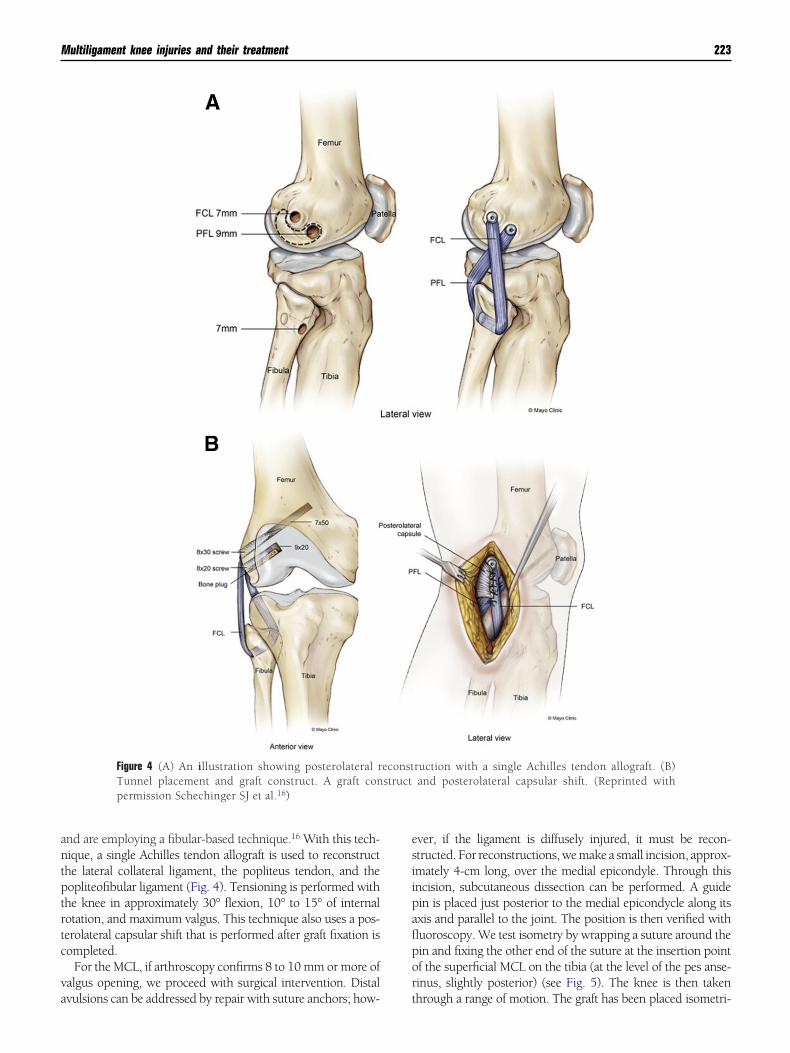
nd are employing a fibular-based technique.16 With this tech-ique, a single Achilles tendon allograft is used to reconstructhe lateral collateral ligament, the popliteus tendon, and theopliteofibular ligament (Fig. 4). Tensioning is performed withhe knee in approximately 30° flexion, 10° to 15° of internalotation, and maximum valgus. This technique also uses a pos-erolateral capsular shift that is performed after graft fixation isompleted.
For the MCL, if arthroscopy confirms 8 to 10 mm or more ofalgus opening, we proceed with surgical intervention. Distal
Figure 4 (A) An illustration showing posterolateral rTunnel placement and graft construct. A graft conpermission Schechinger SJ et al.16)
vulsions can be addressed by repair with suture anchors; how- t
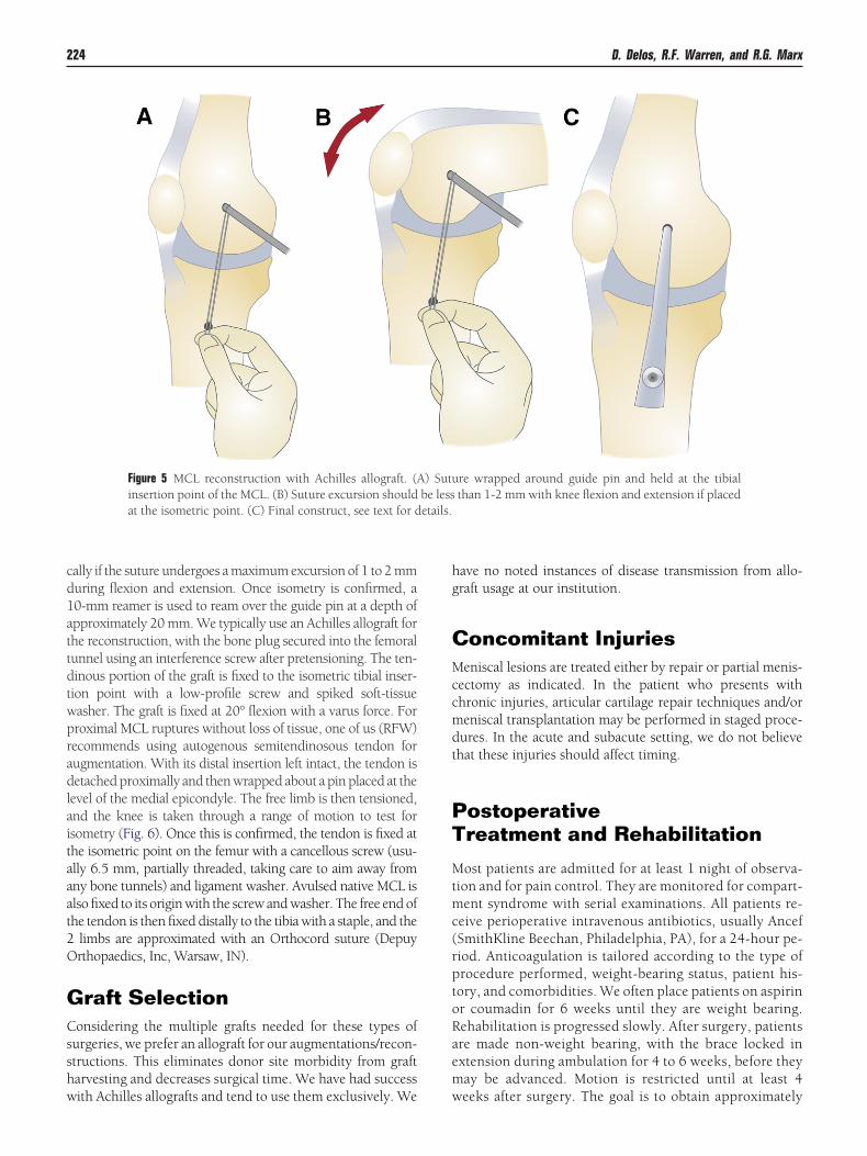
ver, if the ligament is diffusely injured, it must be recon-tructed. For reconstructions, we make a small incision, approx-mately 4-cm long, over the medial epicondyle. Through thisncision, subcutaneous dissection can be performed. A guidein is placed just posterior to the medial epicondycle along itsxis and parallel to the joint. The position is then verified withuoroscopy. We test isometry by wrapping a suture around thein and fixing the other end of the suture at the insertion pointf the superficial MCL on the tibia (at the level of the pes anse-inus, slightly posterior) (see Fig. 5). The knee is then taken
ruction with a single Achilles tendon allograft. (B)and posterolateral capsular shift. (Reprinted with
econststruct
hrough a range of motion. The graft has been placed isometri-
cd1attdtwpradlaitaaat2O
GCsshw
hg
CMccmdt
PT
Mtmc(rptoRaem
etails.
224 D. Delos, R.F. Warren, and R.G. Marx
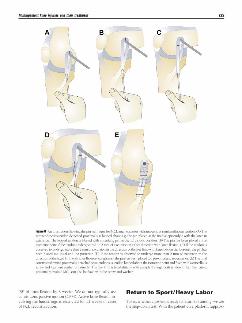
ally if the suture undergoes a maximum excursion of 1 to 2 mmuring flexion and extension. Once isometry is confirmed, a0-mm reamer is used to ream over the guide pin at a depth ofpproximately 20 mm. We typically use an Achilles allograft forhe reconstruction, with the bone plug secured into the femoralunnel using an interference screw after pretensioning. The ten-inous portion of the graft is fixed to the isometric tibial inser-ion point with a low-profile screw and spiked soft-tissueasher. The graft is fixed at 20° flexion with a varus force. Forroximal MCL ruptures without loss of tissue, one of us (RFW)ecommends using autogenous semitendinosous tendon forugmentation. With its distal insertion left intact, the tendon isetached proximally and then wrapped about a pin placed at the
evel of the medial epicondyle. The free limb is then tensioned,nd the knee is taken through a range of motion to test forsometry (Fig. 6). Once this is confirmed, the tendon is fixed athe isometric point on the femur with a cancellous screw (usu-lly 6.5 mm, partially threaded, taking care to aim away fromny bone tunnels) and ligament washer. Avulsed native MCL islso fixed to its origin with the screw and washer. The free end ofhe tendon is then fixed distally to the tibia with a staple, and the
limbs are approximated with an Orthocord suture (Depuyrthopaedics, Inc, Warsaw, IN).
raft Selectiononsidering the multiple grafts needed for these types of
urgeries, we prefer an allograft for our augmentations/recon-tructions. This eliminates donor site morbidity from graftarvesting and decreases surgical time. We have had success
Figure 5 MCL reconstruction with Achilles allograft. (Ainsertion point of the MCL. (B) Suture excursion shouldat the isometric point. (C) Final construct, see text for d
ith Achilles allografts and tend to use them exclusively. We w
ave no noted instances of disease transmission from allo-raft usage at our institution.
oncomitant Injurieseniscal lesions are treated either by repair or partial menis-
ectomy as indicated. In the patient who presents withhronic injuries, articular cartilage repair techniques and/oreniscal transplantation may be performed in staged proce-ures. In the acute and subacute setting, we do not believehat these injuries should affect timing.
ostoperativereatment and Rehabilitation
ost patients are admitted for at least 1 night of observa-ion and for pain control. They are monitored for compart-ent syndrome with serial examinations. All patients re-
eive perioperative intravenous antibiotics, usually AncefSmithKline Beechan, Philadelphia, PA), for a 24-hour pe-iod. Anticoagulation is tailored according to the type ofrocedure performed, weight-bearing status, patient his-ory, and comorbidities. We often place patients on aspirinr coumadin for 6 weeks until they are weight bearing.ehabilitation is progressed slowly. After surgery, patientsre made non-weight bearing, with the brace locked inxtension during ambulation for 4 to 6 weeks, before theyay be advanced. Motion is restricted until at least 4
re wrapped around guide pin and held at the tibialthan 1-2 mm with knee flexion and extension if placed
) Sutube less
eeks after surgery. The goal is to obtain approximately
9cvo
RT
and wa
Multiligament knee injuries and their treatment 225
0° of knee flexion by 8 weeks. We do not typically useontinuous passive motion (CPM). Active knee flexion in-olving the hamstrings is restricted for 12 weeks in cases
Figure 6 An illustration showing the pin technique for MCLsemitendinosus tendon detached proximally is looped aboextension. The looped tendon is labeled with a marking pisometric point if the tendon undergoes �1 to 2 mm of exobserved to undergo more than 2 mm of excursion in the dbeen placed too distal and too posterior. (D) If the tendodirection of the fixed limb with knee flexion (ie, tightens), thconstruct showing proximally detached semitendinosus tenscrew and ligament washer proximally. The free limb is fixproximally avulsed MCL can also be fixed with the screw
f PCL reconstruction. t
eturn to Sport/Heavy Laboro test whether a patient is ready to return to running, we use
entation with autogenous semitendinosus tendon. (A) Theide pin placed at the medial epicondyle with the knee ine 12 o’clock position. (B) The pin has been placed at thein either direction with knee flexion. (C) If the tendon isof the free limb with knee flexion (ie, loosens), the pin hasserved to undergo more than 2 mm of excursion in theas been placed too proximal and too anterior. (E) The finaloped about the isometric point and fixed with a cancellousally with a staple through both tendon limbs. The native,sher.
augmut a guen at thcursionirectionn is obe pin hdon loed dist
he step-down test. With the patient on a platform (approx-
iaatmacho
WCOisgPtwgfst
sctdcwseet
R
1
1
1
1
1
1
1
1
1
1
2
226 D. Delos, R.F. Warren, and R.G. Marx
mately 8-inches high), they are instructed to step forwardnd down toward the ground, alternating between theffected and unaffected limbs. If they cannot perform thisask without loss of balance or control, we do not recom-end they commence running. This test is used to evalu-
te eccentric quadriceps strength, mechanics, and muscleontrol. Most patients are allowed to return to sports oreavy labor at 9 to 12 months postoperatively once rangef motion, proprioception, and strength are sufficient.
hat Have Youhanged Over the Years?
ne of us (RGM) now uses a commercially available tension-ng boot to tension the ACL and PCL grafts during recon-truction.17 After the knee is cycled to allow for settling of theraft and pretensioning, it is placed in 90° of flexion (for theCL graft) or 0° (for the ACL graft), and the boot is tensionedo 9 kg. With the graft under tension, fixation is performedith the implant of choice. Previous studies have shownood results with the tensioning boot,7,18-20 and we haveound it provides for a convenient way to reproducibly ten-ion our grafts and limit graft advancement during fixation,hough manual tensioning is also acceptable.
The senior author (RFW) notes his transition to recon-tructions or augmentations with repair for posterolateralorner injuries over the last several years. Although earlierreatment usually consisted of repairs alone, based on recentata, it seems clear now that repairs with augmentation or re-onstructions provide more reproducible results.10,11 Alongith an increased recognition of these lesions, this has led to
ignificantly better outcomes for what were typically consid-red devastating injuries. However, in the senior author’sxperience, fibular avulsions can heal predictably and do notypically require a reconstruction.
eferences1. Potter HG, Weinstein M, Allen AA, et al: Magnetic resonance imaging of
the multiple-ligament injured knee. J Orthop Trauma 16:330-339,2002
2. Mills WJ, Barei DP, McNair P: The value of the ankle-brachial index fordiagnosing arterial injury after knee dislocation: A prospective study.
J Trauma 56:1261-1265, 20043. McDonough EB Jr, Wojtys EM: Multiligamentous injuries of the kneeand associated vascular injuries. Am J Sports Med 37:156-159, 2009
4. Potter HG: Imaging of the multiple-ligament-injured knee. Clin SportsMed 19:425-441, 2000
5. Patel DV, Allen AA, Warren RF, et al: The nonoperative treatment ofacute, isolated (partial or complete) posterior cruciate ligament-defi-cient knees: An intermediate-term follow-up study. HSS J 3:137-146,2007
6. Marx RG, Shindle MK, Warren RF: Management of posterior cruciateligament injuries. Oper Tech Sports Med 17:162, 2009
7. Fanelli GC, Edson CJ: Arthroscopically assisted combined anterior andposterior cruciate ligament reconstruction in the multiple ligamentinjured knee: 2 to 10-year follow-up. Arthroscopy 18:703-714, 2002
8. Fanelli GC, Giannotti BF, Edson CJ: Arthroscopically assisted com-bined posterior cruciate ligament/posterior lateral complex reconstruc-tion. Arthroscopy 12:521-530, 1996
9. Shelbourne KD, Haro MS, Gray T: Knee dislocation with lateral sideinjury: Results of an en masse surgical repair technique of the lateralside. Am J Sports Med 35:1105-1116, 2007
0. Stannard JP, Brown SL, Farris RC, et al: The posterolateral corner of theknee: Repair versus reconstruction. Am J Sports Med 33:881-888,2005
1. Levy BA, Dajani KA, Morgan JA, et al: Repair versus reconstruction ofthe fibular collateral ligament and posterolateral corner in the multil-igament-injured knee. Am J Sports Med 38:804-809, 2010
2. Mariani PP, Santoriello P, Iannone S, et al: Comparison of surgicaltreatments for knee dislocation. Am J Knee Surg 12:214-221, 1999
3. Richter M, Bosch U, Wippermann B, et al: Comparison of surgicalrepair or reconstruction of the cruciate ligaments versus nonsurgicaltreatment in patients with traumatic knee dislocations. Am J SportsMed 30:718-727, 2002
4. Mook WR, Miller MD, Diduch DR, et al: Multiple-ligament knee inju-ries: a systematic review of the timing of operative intervention andpostoperative rehabilitation. J Bone Joint Surg Am 91:2946-2957, 2009
5. Bedi A, Altchek DW: The “footprint” anterior cruciate ligament tech-nique: An anatomic approach to anterior cruciate ligament reconstruc-tion. Arthroscopy 25:1128-1138, 2009
6. Schechinger SJ, Levy BA, Dajani KA, et al: Achilles tendon allograftreconstruction of the fibular collateral ligament and posterolateral cor-ner. Arthroscopy 25:232-242, 2009
7. Fanelli GC: Evaluation and treatment of the multiple ligament injuredknee. Arthroscopy 19:30-37, 2003 (suppl 1)
8. Fanelli GC, Edson CJ: Combined posterior cruciate ligament-postero-lateral reconstructions with Achilles tendon allograft and biceps femo-ris tendon tenodesis: 2 to 10-year follow-up. Arthroscopy 20:339-345,2004
9. Fanelli GC, Orcutt DR, Edson CJ: The multiple-ligament injuredknee: Evaluation, treatment, and results. Arthroscopy 21:471-486,2005
0. Fanelli GC, Edson CJ, Orcutt DR, et al: Treatment of combined anteriorcruciate-posterior cruciate ligament-medial-lateral side knee injuries. J
Knee Surg 18:240-248, 2005