Multicolor Flow Cytometry - BD Biosciences€¢ Fluorescence minus one (FMO) controls contain ......
47
Multicolor Flow Cytometry: Setup and Optimization on the BD Accuri™ C6 Flow Cytometer Presented by Clare Rogers, MS Senior Marketing Applications Specialist BD Biosciences 23-13660-01 For Research Use Only. Not for use in diagnostic or therapeutic procedures. Class 1 Laser Product.
Transcript of Multicolor Flow Cytometry - BD Biosciences€¢ Fluorescence minus one (FMO) controls contain ......
Multicolor Flow Cytometry:Setup and Optimization on the
BD Accuri™ C6 Flow Cytometer
Presented by Clare Rogers, MSSenior Marketing Applications Specialist
BD Biosciences
23-13660-01
For Research Use Only. Not for use in diagnostic or therapeutic procedures.
Class 1 Laser Product.

Webinar Overview
• Multicolor flow: successful application prerequisiteso Clear definition of your experimental goals
o Careful reagent selection and sample preparation
o Proper cytometer performance, setup, and data collection
o Data analysis: Proper classification of positive and negative populations

• Goalso Identify lymphs, monos, and grans by CD45
(common leukocyte antigen) and SSC properties
o Within the lymphocyte population, determine the percentage of helper (CD3+CD4+) and cytotoxic (CD3+CD8+) T cells
o Within the monocyte population, determine the percentage of CD4+ cells
Example: HPB 4-Color T-Cell Immunophenotyping

Principles of Panel Design: Reagent Selection
Identify required markers and reagents (CD45, 3, 4, 8).
Match fluorochromes by brightness (values from stain index) according to antigen density and distribution (published values or TDS).
Minimize spectral overlap.
Use tandem dyes with consideration of their technical limitations.
Check reagent availability.
Lay out experimental plan with appropriate controls included.
22
11
33
44
55
66

Understand Relative Expression Level for Markers of Interest
• Level of antigen expression on a cell:o Antigen expression can vary
due to cell activation level and functional differences.
o Antigen density can be a range (ie, smeared population).
o Check the literature.
Roederer M, Herzenberg LA, Herzenberg LA. Changes in antigen densities on leukocyte subsets correlate with progression of HIV disease. Int Immunol. 1996;8:1-11.

Pick Possible Fluorochromes Based on Your Instrument Configuration
Detector Position
Filter Options Fluorophore
FL1 533/30 FITC, GFP, YFP, CFSE, Alexa Fluor® 488
510/15 GFP
FL2 585/40 PE, PI, PE-CF594
540/20 YFP
FL3 670 LP PerCP, PE-Cy™5, PerCP-Cy™5.5, PE-Cy™7, PI, 7-AAD
610/20 RFP, PIPE-CF594
FL4 675/25 APC, Alexa Fluor® 647
780/60 APC-H7, APC-Cy7
3-blue, 1-red (Standard Config)

CD45lym
CD3CD8
CD4lym
CD45mon
CD4mon
CD45gran
Antigen density C6 fluor intensity
high
low
PE
APCPerCP-Cy5.5
PE-Cy7FITC
PerCP
Choose Antigen-fluor Pairing Based on Relative Intensities

CD45 PerCP-Cy5.5
CD8 FITC
CD3 APC
CD4 PECD45lym
CD3CD8
CD4lym
CD45mon
CD4mon
CD45gran
Antigen density Fluor intensity
high
low
PE
APCPerCP-Cy5.5
PE-Cy7FITC
PerCP
Choose Antigen-fluor Pairing Based on Relative Intensities

Principles of Panel Design: Reagent Selection
Identify required markers and reagents (CD45, 3, 4, 8).
Match fluorochromes by brightness (values from stain index) according to antigen density and distribution (published values or TDS).
Minimize spectral overlap.
Use tandem dyes with consideration of their technical limitations.
Check reagent availability.
Lay out experimental plan with appropriate controls included.
22
11
33
44
55
66

Fluorescence Spillover
• The single most important factor affecting resolution sensitivity (SI) in multicolor flow cytometry experiments.
• Fluorescence spillover from other channels:o Directly and irreversibly reduces the resolution
sensitivity of that channelo Contributes to background
• This “background” is mathematically accounted for in the process called compensation.

Spillover Irreversibly Decreases Resolution Sensitivity
• To improve resolution (sensitivity) of subpopulations, including dim subpopulations, one must minimize the amount of spillover from other fluorochromes.
Population resolution for a given fluorescence parameter is decreased by increased spread due to spillover from other fluorochromes.
• The dim CD4+ cells, when negative for CD8 (orange ball), are easily resolved from the double negative.
• The same dim CD4+ cells, when positive for CD8, cannot be resolved from CD4–, CD8+ cells.

Spillover Irreversibly Decreases Resolution Sensitivity
This spread is NOT eliminated by compensation.
More colors = more spillover = higher background

Average Compensation Values for Various Fluor Combos: BD Accuri C6
FITC, PE, PerCP-Cy5.5, APC, Compensation dialog layout:
Correct FL1 by: PE PerCP-Cy5.5 APC
3.20 0.00 0.00
Correct FL2 by: FITC PerCP-Cy5.5 APC
7.50 0.00 0.00
Correct FL3 by: FITC PE APC
0.5 19.50 0.80
Correct FL4 by: FITC PE PerCP-Cy5.5
0.00 0.00 12.00

Strategies to Minimize Spillover Issues
• Minimize the potential for spectral overlap • Spillover estimates available in the spectrum viewer

Principles of Panel Design: Reagent Selection
Identify required markers and reagents (CD45, 3, 4, 8).
Match fluorochromes by brightness (values from stain index) according to antigen density and distribution (published values or TDS).
Minimize spectral overlap.
Use tandem dyes with consideration of their technical limitations.
Check reagent availability.
Lay out experimental plan with appropriate controls included.
22
11
33
44
55
66

Use Tandem Dyes with Consideration of their Technical Limitations
• Compensation requirements for tandem dye conjugates can vary.
• Certain tandem dye conjugates (APC-Cy7, PE-Cy7) can degrade with exposure to light, elevated temperature, and fixation.o Minimize exposure to these conditions.o Use BD™ Stabilizing Fixative for final fixation.o Use APC-H7 when possible.
• PE-CF594 (PE-Texas Red® replacement) very stableo FL3 of the C6 (670 LP )

Principles of Panel Design: Reagent Selection
Identify required markers and reagents (CD45, 3, 4, 8).
Match fluorochromes by brightness (values from stain index) according to antigen density and distribution (published values or TDS).
Minimize spectral overlap.
Use tandem dyes with consideration of their technical limitations.
Check reagent availability.
Lay out experimental plan with appropriate controls included.
22
11
33
44
55
66

CD45 PerCP-Cy5.5
CD8 FITC
CD3 APC
CD4 PECD45lym
CD3CD8
CD4lym
CD45mon
CD4mon
CD45gran
Antigen density Fluor intensity
high
low
PE
APCPerCP-Cy5.5
PE-Cy7FITC
PerCP
Availability: Use BD FACSelect™ Multicolor Panel Designer

bdbiosciences.com/research/multicolor
Tools: BD FACSelect Multicolor Panel Designer

Step 1: Select target species Step 2: Select specificities Step 3: Select lasers available or fluors desired Step 4: Search
2211 33
44
Tools: BD FACSelect Multicolor Panel Designer

View available conjugates
Tools: BD FACSelect Multicolor Panel Designer

Select desired clone Close window
Tools: BD FACSelect Multicolor Panel Designer

Review the selected panel
Tools: BD FACSelect Multicolor Panel Designer

Before you perform your first experiment: Optimize your new antibodies!
281
25,000
5 µL Ab/50 µL whole blood (1/10)
275
2,154
1 µL Ab/50 µL whole blood (1/50)
Optimization data: CD4+ to CD4-

Principles of Panel Design: Reagent Selection
Identify required markers and reagents (CD45, 3, 4, 8).
Match fluorochromes by brightness (values from stain index) according to antigen density and distribution (published values or TDS).
Minimize spectral overlap.
Use tandem dyes with consideration of their technical limitations.
Check reagent availability.
Lay out experimental plan with appropriate controls included.
22
11
33
44
55
66

What Controls Do You Need and Why?
• Instrument setup controls (eg, BD™ CompBead particles)
• Gating controls (eg, “fluorescence minus one”:FMO)
• Biological controls (eg, unstimulated samples)
This will allow you to:• Obtain consistent setup and compensation
• Gate populations reproducibly
• Make appropriate biological comparisons and conclusions

Use FMO Controls for Accurate Data Analysis
• Fluorescence minus one (FMO) controls contain all the lineage markers except the one of interest.
• For low-density or smeared populations (eg, activation markers), FMOs allow accurate delineation of positively vs negatively stained cells.

• Goalso Identify lymphs, monos, and grans by CD45
and SSC properties
o Within the lymphocyte population, determine the percentage of helper (CD3+CD4+) and cytotoxic (CD3+CD8+) T cells
o Within the monocyte population, determine the percentage of CD4+ cells
Example: 4-color T-cell Immunophenotyping

Lay Out Your Experimental Plan
Filter 533/30 585/40 670 LP 675/25FL1 FL2 FL3 FL4
Tube FITC PE PerCP‐Cy5.5 APC Purpose1 ‐ ‐ ‐ ‐ Background signal2 CD8 ‐ ‐ ‐ Spillover of FITC3 ‐ CD4 ‐ ‐ Spillover of PE4 ‐ ‐ CD45 ‐ Spillover PerCP‐Cy5.55 ‐ ‐ ‐ CD3 Spillover APC6 ‐ ‐ CD45 CD3 Gating control:FMO (Fluorescence Minus One)7 CD8 ‐ CD45 CD3 FMO8 ‐ CD4 CD45 CD3 FMO9 CD8 CD4 CD45 CD3 Test sample
Population Classifier

Webinar Overview
• Multicolor flow: successful application prerequisiteso Clear definition of your experimental goals
o Careful reagent selection and sample preparation
o Proper cytometer performance, setup, and data collection
o Data analysis: Proper classification of positive and negative populations

The BD Accuri C6 Flow Cytometer: Unique System Attributes
An affordable, full-featured, easy-to-use flow cytometerTwo lasers and six detectors

BD Accuri C6 Features that Simplify Multicolor Analysis
• Four fluorescence detectors• Totally digital system• Easy setup for data collection
o Locked down optical alignmento No laser delay to seto Validate system with beads and collect data
• Predictable fluorescence spillover values• Flexibility in fluorochrome choice using:
o Optional optical filterso Selectable Lasers option

System Innovation: Alignment and Signal Detection are Optimized and Locked Down at Manufacture
FL1
533/30
FL2
585/
40
FL3
670 LP
FL4
675/
25
FSC
SSC488-nm
solid state laser
640-nm diode laser
PMTs for fluorescencedetection
Diodes for scatter detection

FL4 = 675/25
FL3 = 670LP
Allophycocyanin (APC) Calibration Particles (Cat. No. 653145)
FL1 = 533/30 FL2 = 585/40
SPHERO™ Rainbow Calibration Particles (Cat. No. 653144)
FL4 = 675/25
Fluorescence Detection Sensitivity is Pre-optimized,Obviating the Need for Voltage Control

Validate System Performance Before each Experiment, Set Appropriate Threshold, Collect Data
Keep daily bead runs all together in one BD Accuri C6 software file.View data in the Statistics Tab.

Pre-optimizing Voltage and Gain Settings does not Reduce the Fluorescence Detection Range
4-log
5.2-log
6.2-log
Sample: Stained CHO cells plus
non-fluorescent 1.0 micron beads

Fixing Optical Alignment and Pre-optimizing Voltage and Gain Settings Results in Predictable Spillover
Suggested Compensation Values for the BD Accuri C6
FITC PE PerCP PerCP- Cy5.5
PE-Cy7 APC
FL1 (533 BP) ___ 3.2 0.00 0.0 0.50 0.0
FL2 (585 BP) 7.5 ___ 0.00 0.00 1.50 0.0
FL3 (670 BP) 1.0 19.5 ___ ___ ___ 0.8
FL4 (675 BP) 0.0 0.0 3.00 12.00 0.00 ___

Flexible Fluorochrome Choice: Optional Filters
510/15
540/20
565/20
610/20
780/60
User changeable
optical filters
Separation of GFP and YFP signals
FL1: 533/30 FL1: 510/15
Standard Filters GFP/YFP Filters
FL2:
585
/40
FL2:
540
/20
GFPYFP
YFP
GFP
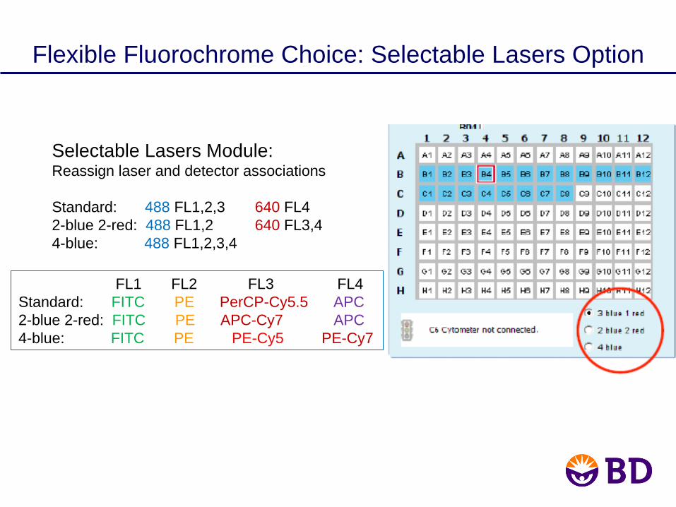
Selectable Lasers Module:Reassign laser and detector associations
Standard: 488 FL1,2,3 640 FL42-blue 2-red: 488 FL1,2 640 FL3,44-blue: 488 FL1,2,3,4
Flexible Fluorochrome Choice: Selectable Lasers Option
FL1 FL2 FL3 FL4Standard: FITC PE PerCP-Cy5.5 APC2-blue 2-red: FITC PE APC-Cy7 APC4-blue: FITC PE PE-Cy5 PE-Cy7

Webinar Overview
• Multicolor flow: successful application prerequisiteso Clear definition of your experimental goals
o Careful reagent selection and sample preparation
o Proper cytometer performance, setup, and data collection
o Data Analysis: Proper classification of positive and negative populations

Using the Controls to Analyze the Data
(1) Apply compensation using single-color controls
FITC PE PerCP-Cy5.5 APC
Lymphgate

Using the Controls to Analyze the Data
(2) Set the gate on the desired population using a primary classifier:
In this case it is the lymphocytes, defined as CD45bright SSClow
Goal 1: Identify lymphocytes, monocytes, and granulocytes

(3) Apply the primary classifier gate to the appropriate FMO plots to determine gate placement.
Using the Controls to Analyze the Data
FMO to set PE background:Contains: FITC, PerCP-Cy5.5, APC
FMO to set FITC background:Contains: PE, PerCP-Cy5.5, APC
PE
FMO (CD45+, CD3+)
CD3-APC (FL4 675/25)
FITC PE
FITC
FITC PE
CD3-APC (FL4 675/25) CD3-APC (FL4 675/25)

Perform Calculations
(4) Subtract the background from the FMO plots to obtain percent positives for each population.
Goal 2: Of the lymphocyte population, what percentage are:
CD3+CD4+: 60.1 - 0.2 = 59.9% CD3+CD8+: 12.7 - 0.1 = 12.6%

Perform Calculations
Goal 3: Of the monocyte population, what percentage are CD4+?
96.13% - 0.3% = 95.83%

Webinar Summary
• Multicolor flow: successful application prerequisites
o Clear definition of experimental goalso Proper cytometer performance, setup, and data
collectionThe BD Accuri C6 is well suited for these applications.Fluorescence detection and the optical bench are optimized at manufacture.Easy to use: validate and collect samples
o Careful reagent selection and sample preparationBD FACSelect multicolor panel designer, along with antigen density and stain index charts, are BD tools that simplify this step.
o Proper classification (analysis) of multiple combinations of positive and negative populations

Acknowledgments
• Maria Jaimes• Mark Edinger• Ming Yan• Alan Stall• Joe Trotter• Skip Maino• Margaret Inokuma• Bob Hoffman
For Research Use Only. Not for use in diagnostic or therapeutic procedures.
Class 1 Laser Product.
Alexa Fluor®
is a registered trademark of Molecular Probes, Inc.
Cy™ is a trademark of Amersham Biosciences Corp. Cy dyes are subject to proprietary rights of Amersham Biosciences Corp and Carnegie Mellon University and are made and sold under license from Amersham Biosciences Corp only for research and in vitro diagnostic use. Any other use requires a commercial sublicense from Amersham Biosciences Corp, 800 Centennial Avenue, Piscataway, NJ 08855-1327, USA.
SPHERO is a trademark of Spherotech, Inc.
BD, BD Logo and all other trademarks are property of Becton, Dickinson and Company. © 2012 BD
Thank you!
• Pat Collins• Joerg Hildmann• Holden Maecker• Mirion Schultz• Barny Abrams• Laurel Nomura• Dennis Sasaki
• Maria Dinkelmann• Stacey Roys• Collin Rich• Leo Ostruszka• BD Accuri Eng. Team