Multi-Scale Attentional Network for Multi-Focal ...
8
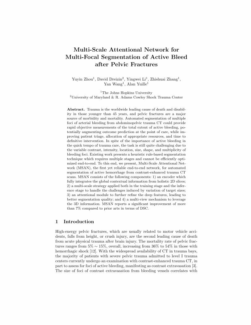
Multi-Scale Attentional Network for Multi-Focal Segmentation of Active Bleed after Pelvic Fractures Yuyin Zhou 1 , David Dreizin 2 , Yingwei Li 1 , Zhishuai Zhang 1 , Yan Wang 1 , Alan Yuille 1 1 The Johns Hopkins University 2 University of Maryland & R. Adams Cowley Shock Trauma Center Abstract. Trauma is the worldwide leading cause of death and disabil- ity in those younger than 45 years, and pelvic fractures are a major source of morbidity and mortality. Automated segmentation of multiple foci of arterial bleeding from abdominopelvic trauma CT could provide rapid objective measurements of the total extent of active bleeding, po- tentially augmenting outcome prediction at the point of care, while im- proving patient triage, allocation of appropriate resources, and time to definitive intervention. In spite of the importance of active bleeding in the quick tempo of trauma care, the task is still quite challenging due to the variable contrast, intensity, location, size, shape, and multiplicity of bleeding foci. Existing work presents a heuristic rule-based segmentation technique which requires multiple stages and cannot be efficiently opti- mized end-to-end. To this end, we present, Multi-Scale Attentional Net- work (MSAN), the first yet reliable end-to-end network, for automated segmentation of active hemorrhage from contrast-enhanced trauma CT scans. MSAN consists of the following components: 1) an encoder which fully integrates the global contextual information from holistic 2D slices; 2) a multi-scale strategy applied both in the training stage and the infer- ence stage to handle the challenges induced by variation of target sizes; 3) an attentional module to further refine the deep features, leading to better segmentation quality; and 4) a multi-view mechanism to leverage the 3D information. MSAN reports a significant improvement of more than 7% compared to prior arts in terms of DSC. 1 Introduction High-energy pelvic fractures, which are usually related to motor vehicle acci- dents, falls from height, or crush injury, are the second leading cause of death from acute physical trauma after brain injury. The mortality rate of pelvic frac- tures ranges from 5% ∼ 15%, overall, increasing from 36% to 54% in those with hemorrhagic shock [12]. With the widespread availability of CT in trauma bays, the majority of patients with severe pelvic trauma admitted to level I trauma centers currently undergo an examination with contrast-enhanced trauma CT, in part to assess for foci of active bleeding, manifesting as contrast extravasation [3]. The size of foci of contrast extravasation from bleeding vessels correlates with
Transcript of Multi-Scale Attentional Network for Multi-Focal ...
Multi-Scale Attentional Network forMulti-Focal Segmentation of Active Bleed
after Pelvic Fractures
Yuyin Zhou1, David Dreizin2, Yingwei Li1, Zhishuai Zhang1,Yan Wang1, Alan Yuille1
1The Johns Hopkins University2University of Maryland & R. Adams Cowley Shock Trauma Center
Abstract. Trauma is the worldwide leading cause of death and disabil-ity in those younger than 45 years, and pelvic fractures are a majorsource of morbidity and mortality. Automated segmentation of multiplefoci of arterial bleeding from abdominopelvic trauma CT could providerapid objective measurements of the total extent of active bleeding, po-tentially augmenting outcome prediction at the point of care, while im-proving patient triage, allocation of appropriate resources, and time todefinitive intervention. In spite of the importance of active bleeding inthe quick tempo of trauma care, the task is still quite challenging due tothe variable contrast, intensity, location, size, shape, and multiplicity ofbleeding foci. Existing work presents a heuristic rule-based segmentationtechnique which requires multiple stages and cannot be efficiently opti-mized end-to-end. To this end, we present, Multi-Scale Attentional Net-work (MSAN), the first yet reliable end-to-end network, for automatedsegmentation of active hemorrhage from contrast-enhanced trauma CTscans. MSAN consists of the following components: 1) an encoder whichfully integrates the global contextual information from holistic 2D slices;2) a multi-scale strategy applied both in the training stage and the infer-ence stage to handle the challenges induced by variation of target sizes;3) an attentional module to further refine the deep features, leading tobetter segmentation quality; and 4) a multi-view mechanism to leveragethe 3D information. MSAN reports a significant improvement of morethan 7% compared to prior arts in terms of DSC.
1 Introduction
High-energy pelvic fractures, which are usually related to motor vehicle acci-dents, falls from height, or crush injury, are the second leading cause of deathfrom acute physical trauma after brain injury. The mortality rate of pelvic frac-tures ranges from 5% ∼ 15%, overall, increasing from 36% to 54% in those withhemorrhagic shock [12]. With the widespread availability of CT in trauma bays,the majority of patients with severe pelvic trauma admitted to level I traumacenters currently undergo an examination with contrast-enhanced trauma CT, inpart to assess for foci of active bleeding, manifesting as contrast extravasation [3].The size of foci of contrast extravasation from bleeding vessels correlates with
2 Y. Zhou et al.
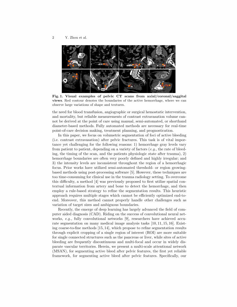
Fig. 1. Visual examples of pelvic CT scans from axial/coronal/saggitalviews. Red contour denotes the boundaries of the active hemorrhage, where we canobserve large variations of shape and textures.
the need for blood transfusion, angiographic or surgical hemostatic intervention,and mortality, but reliable measurements of contrast extravasation volume can-not be derived at the point of care using manual, semi-automated, or shorthanddiameter-based methods. Fully automated methods are necessary for real-timepoint-of-care decision making, treatment planning, and prognostication.
In this paper, we focus on volumetric segmentation of foci of active bleeding(i.e. contrast extravasation) after pelvic fractures. This task is of vital impor-tance yet challenging for the following reasons: 1) hemorrhage gray levels varyfrom patient to patient, depending on a variety of factors (e.g., the rate of bleed-ing, the timing of the scan, and the patients physiologic state after trauma), 2)hemorrhage boundaries are often very poorly defined and highly irregular; and3) the intensity levels are inconsistent throughout the region of a hemorrhagicfocus. Prior works have utilized semi-automated threshold- or region growing-based methods using post-processing software [5]. However, these techniques aretoo time-consuming for clinical use in the trauma radiology setting. To overcomethis difficulty, a method [4] was previously proposed to first utilize spatial con-textual information from artery and bone to detect the hemorrhage, and thenemploy a rule-based strategy to refine the segmentation results. This heuristicapproach requires multiple stages which cannot be efficiently optimized end-to-end. Moreover, this method cannot properly handle other challenges such asvariation of target sizes and ambiguous boundaries.
Recently, the emerge of deep learning has largely advanced the field of com-puter aided diagnosis (CAD). Riding on the success of convolutional neural net-works, e.g ., fully convolutional networks [9], researchers have achieved accu-rate segmentation on many medical image analysis tasks [10, 11, 15, 16]. Exist-ing coarse-to-fine methods [15, 14], which propose to refine segmentation resultsthrough explicit cropping of a single region of interest (ROI) are more suitablefor single connected structures such as the pancreas or liver, while sites of activebleeding are frequently discontinuous and multi-focal and occur in widely dis-parate vascular territories. Herein, we present a multi-scale attentional network(MSAN), for segmenting active bleed after pelvic features, the first yet reliableframework, for segmenting active bleed after pelvic features. Specifically, our
MSAN for Multi-Focal Segmentation of Active Bleed after Pelvic Fractures 3
framework is able to 1) fully exploit contextual information from holistic 2Dslices via using an encoder which is capable of extracting the global contextualinformation across different levels of image features; 2) efficiently handle thevariation of active hemorrhage sizes by adopting multi-scale strategies duringthe training phase and the testing phase; 3) deal with the ambiguous bound-aries by utilizing an attentional mechanism to better enhance the discriminationbetween trauma region and non-trauma region; 4) utilize the aggregation of mul-tiple views (i.e., Coronal, Sagittal and Axial views) to further leverage the 3Dinformation. To assess the effectiveness of our framework, we collect a datasetof 65 patients with pelvic fractures and active hemorrhage with widely varyingdegrees of severity. For each case, every pixel/voxel of active hemorrhage wasmanually labeled by an experienced radiologist. Unlike the previously describedheuristic method which used crude and not widely adopted measurements ofaccuracy such as missegmented area [4], we employed the Dice-Sørensen coeffi-cient (DSC) for evaluation based on pixel/voxel-wise predictions. Experimentalresults demonstrate the superiority of our framework compared with a series of2D/3D state-of-the-art deep learning algorithms.
2 Multi-Scale Attentional Network
2.1 Overall Framework
We denote a 3D CT-scanned image as X with size W × H × L, where eachelement of X indicated the Housefield Unit (HU) of a voxel. The correspondingbinary ground-truth segmentation mask is denoted as Y where yi = 1 indicatesa foreground voxel. Consider a segmentation model M : Z = f(X;Θ), where Mis parameterized by Θ, our goal is to predict a binary output volume Z of thesame dimension as X. We denote Y and Z as the set of foreground voxels in theground-truth and prediction, i.e., Y = {i | yi = 1} and Z = {i | zi = 1}. Theaccuracy of segmentation is evaluated by the Dice-Sørensen coefficient (DSC):
DSC(Y,Z) = 2×|Y∩Z||Y|+|Z| . This metric falls in the range of [0, 1], and DSC = 1
implies a perfect segmentation.Following [15, 14, 11], 3 sets of images, i.e., XC,w (w = 1, 2, . . . ,W ), XS,h
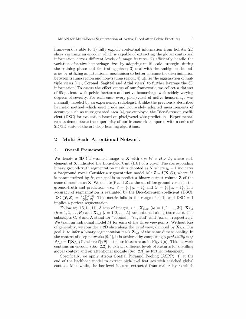
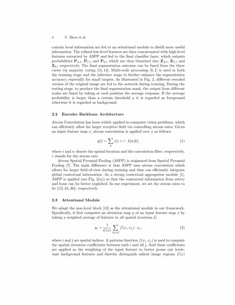
(h = 1, 2, . . . ,H) and XA,l (l = 1, 2, . . . , L) are obtained along three axes. Thesubscripts C, S and A stand for “coronal”, “sagittal” and “axial”, respectively.We train an individual model M for each of the three viewpoints. Without lossof generality, we consider a 2D slice along the axial view, denoted by XA,l. Ourgoal is to infer a binary segmentation mask ZA,l of the same dimensionality. Inthe context of deep networks [9, 1], it is achieved by computing a probability mapPA,l = f [XA,l; θ], where f [·; θ] is the architecture as in Fig. 2(a). This networkcontains an encoder (Sec. 2.2) to extract different levels of features for distillingglobal context and an attentional module (Sec. 2.3) as further refinement.
Specifically, we apply Atrous Spatial Pyramid Pooling (ASPP) [1] at theend of the backbone model to extract high-level features with enriched globalcontext. Meanwhile, the low-level features extracted from earlier layers which
4 Y. Zhou et al.
contain local information are fed to an attentional module to distill more usefulinformation. The refined low-level features are then concatenated with high-levelfeatures extracted by ASPP and fed to the final classifier layer, which outputsprobabilities PA,l, PC,l and PS,l which are then binarized into ZA,l, ZC,l andZS,l respectively. The final segmentation outcome can be fused from the threeviews via majority voting [15, 14]. Multi-scale processing [8, 1] is used in boththe training stage and the inference stage to further enhance the segmentationaccuracy, especially for small targets. As illustrated in Fig. 2, different rescaledversion of the original image are fed to the network during training. During thetesting stage, to produce the final segmentation mask, the output from differentscales are fused by taking at each position the average response. If the averageprobability is larger than a certain threshold ρ it is regarded as foregroundotherwise it is regarded as background.
2.2 Encoder Backbone Architecture
Atrous Convolution has been widely applied in computer vision problems, whichcan efficiently allow for larger receptive field via controlling atrous rates. Givenan input feature map x, atrous convolution is applied over x as follows:
y[i] =∑k
x[i+ r · k]w[k], (1)
where i and w denote the spatial location and the convolution filter, respectively.r stands for the atrous rate.
Atrous Spatial Pyramid Pooling (ASPP) is originated from Spatial PyramidPooling [7]. The main difference is that ASPP uses atrous convolution whichallows for larger field-of-view during training and thus can efficiently integrateglobal contextual information. As a strong contextual aggregation module [1],ASPP is applied (see Fig. 2(a)) so that the contextual information from arteryand bone can be better exploited. In our experiment, we set the atrous rates tobe {12, 24, 36}, respectively.
2.3 Attentional Module
We adapt the non-local block [13] as the attentional module in our framework.Specifically, it first computes an attention map y of an input feature map x bytaking a weighted average of features in all spatial locations L:
yi =1
C(x)
∑∀j∈L
f(xi, xj) · xj , (2)
where i and j are spatial indices. A pairwise function f(xi, xj) is used to computethe spatial attention coefficients between each i and all j. And these coefficientsare applied as the weighting of the input feature to better prune out irrele-vant background features and thereby distinguish salient image regions. C(x)
MSAN for Multi-Focal Segmentation of Active Bleed after Pelvic Fractures 5
…
ResNetBackbone
Group4Feature
Group1Feature
1×1 conv
×
+
×
(a) (b)
NonLocal
Fig. 2. (a) The network architecture structure of MSAN. Low-level features are refinedby an attentional module. Meanwhile ASPP is applied at the end of the backbone modelto extract high-level features with enriched global context. (b) Our implementation ofthe attentional module, where we use nonlocal means [13] as the main operation.
is a normalization function. We use the dot product version in [13] by settingf(xi, xj) = xi
Txj and C(x)=N , where N is the number of pixels in x.Following [13], the attention map y is then processed by a 1×1 convolutional
layer and added to the input feature map x to obtain the final output z, i.e.,z = wy+x, where w is the weight of the convolutional layer. An illustration ourour attentional module can be found in Fig. 2(b).
3 Experiments
3.1 Dataset and Evaluation
We have collected 65 studies were routinely acquired with 64 section or higherMDCT scanners in the trauma bay in either the late arterial or portal venousphase of enhancement. We use 45 cases for training and evaluate the segmen-tation performance on the rest 20 cases. Note that [4] was studied on only 12cases, which, to the best of our knowledge, was the first and only curated datasetwith manual ground truth label masks. Therefore our dataset can be consideredas a valid set for evaluation. The metric we use is DSC, which measures thesimilarity between the prediction voxel set Z and the ground-truth set Y, with
the mathematical form of DSC(Z,Y) = 2×|Z∩Y||Z|+|Y| .
3.2 Implementation details
Our implementations are based on Tensorflow. We used two standard architec-tures, i.e., ResNet-50 and ResNet-101 [6] as backbone models. All our segmen-tation experiments were performed on the whole pelvic CT scan and were runon Tesla V100 GPU. For data pre-processing, following [11], we simply trun-cated the raw intensity values to be within the range of [−80, 320] HU and thennormalized each raw CT case to [0, 255.0]. Random rotation of [0, 15] is used as
6 Y. Zhou et al.
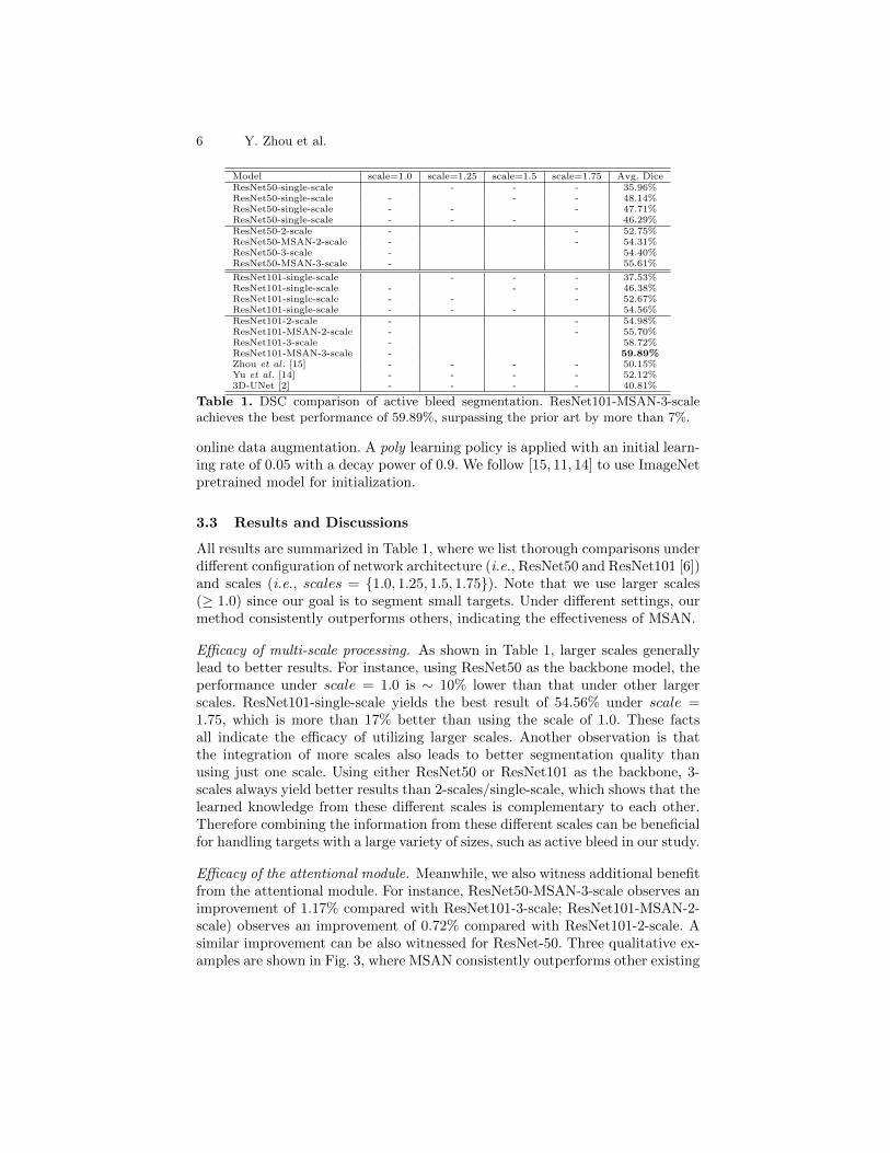
Model scale=1.0 scale=1.25 scale=1.5 scale=1.75 Avg. DiceResNet50-single-scale - - - 35.96%ResNet50-single-scale - - - 48.14%ResNet50-single-scale - - - 47.71%ResNet50-single-scale - - - 46.29%ResNet50-2-scale - - 52.75%ResNet50-MSAN-2-scale - - 54.31%ResNet50-3-scale - 54.40%ResNet50-MSAN-3-scale - 55.61%
ResNet101-single-scale - - - 37.53%ResNet101-single-scale - - - 46.38%ResNet101-single-scale - - - 52.67%ResNet101-single-scale - - - 54.56%ResNet101-2-scale - - 54.98%ResNet101-MSAN-2-scale - - 55.70%ResNet101-3-scale - 58.72%ResNet101-MSAN-3-scale - 59.89%Zhou et al. [15] - - - - 50.15%Yu et al. [14] - - - - 52.12%3D-UNet [2] - - - - 40.81%
Table 1. DSC comparison of active bleed segmentation. ResNet101-MSAN-3-scaleachieves the best performance of 59.89%, surpassing the prior art by more than 7%.
online data augmentation. A poly learning policy is applied with an initial learn-ing rate of 0.05 with a decay power of 0.9. We follow [15, 11, 14] to use ImageNetpretrained model for initialization.
3.3 Results and Discussions
All results are summarized in Table 1, where we list thorough comparisons underdifferent configuration of network architecture (i.e., ResNet50 and ResNet101 [6])and scales (i.e., scales = {1.0, 1.25, 1.5, 1.75}). Note that we use larger scales(≥ 1.0) since our goal is to segment small targets. Under different settings, ourmethod consistently outperforms others, indicating the effectiveness of MSAN.
Efficacy of multi-scale processing. As shown in Table 1, larger scales generallylead to better results. For instance, using ResNet50 as the backbone model, theperformance under scale = 1.0 is ∼ 10% lower than that under other largerscales. ResNet101-single-scale yields the best result of 54.56% under scale =1.75, which is more than 17% better than using the scale of 1.0. These factsall indicate the efficacy of utilizing larger scales. Another observation is thatthe integration of more scales also leads to better segmentation quality thanusing just one scale. Using either ResNet50 or ResNet101 as the backbone, 3-scales always yield better results than 2-scales/single-scale, which shows that thelearned knowledge from these different scales is complementary to each other.Therefore combining the information from these different scales can be beneficialfor handling targets with a large variety of sizes, such as active bleed in our study.
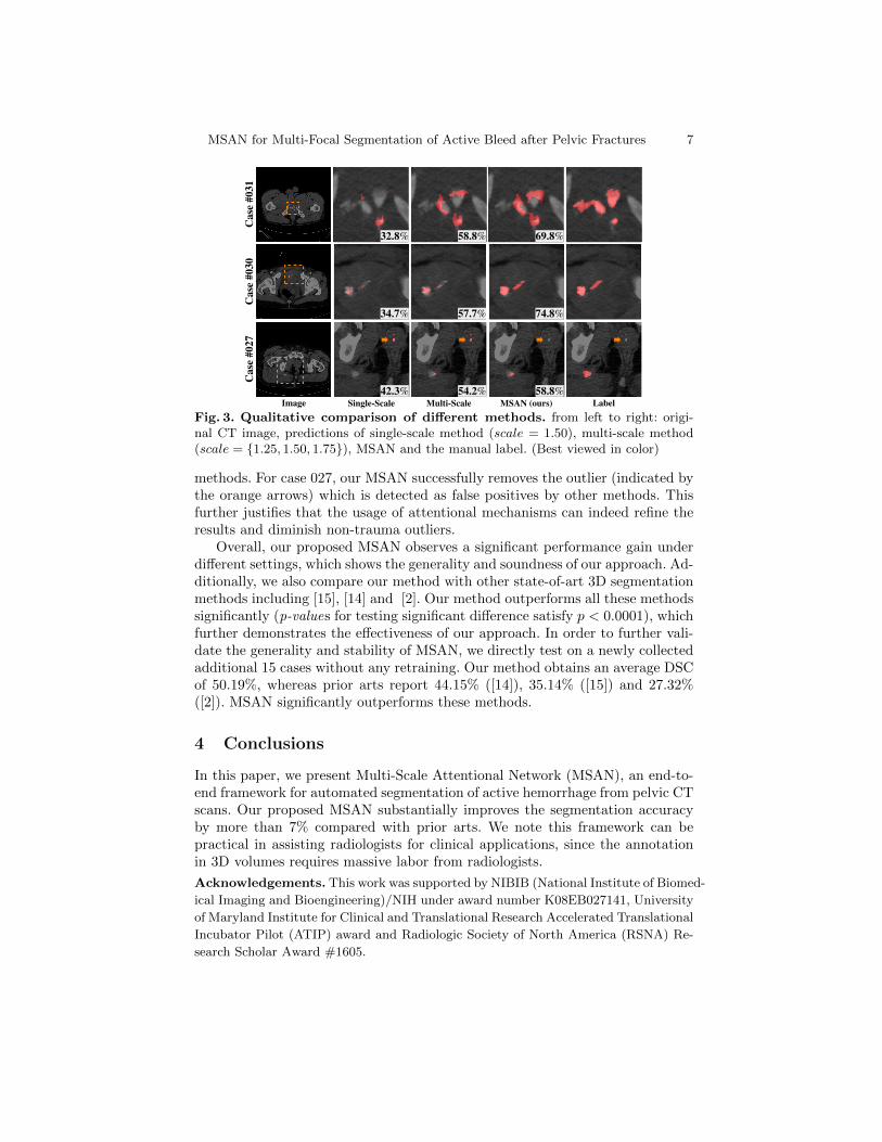
Efficacy of the attentional module. Meanwhile, we also witness additional benefitfrom the attentional module. For instance, ResNet50-MSAN-3-scale observes animprovement of 1.17% compared with ResNet101-3-scale; ResNet101-MSAN-2-scale) observes an improvement of 0.72% compared with ResNet101-2-scale. Asimilar improvement can be also witnessed for ResNet-50. Three qualitative ex-amples are shown in Fig. 3, where MSAN consistently outperforms other existing
MSAN for Multi-Focal Segmentation of Active Bleed after Pelvic Fractures 7
Cas
e#0
3132.8% 58.8% 69.8%
Image
Cas
e#0
30
Single Multi Att Gt34.7% 57.7% 74.8%
Image
Cas
e#0
27
Single-Scale Multi-Scale MSAN (ours) Label42.3% 54.2% 58.8%
Fig. 3. Qualitative comparison of different methods. from left to right: origi-nal CT image, predictions of single-scale method (scale = 1.50), multi-scale method(scale = {1.25, 1.50, 1.75}), MSAN and the manual label. (Best viewed in color)
methods. For case 027, our MSAN successfully removes the outlier (indicated bythe orange arrows) which is detected as false positives by other methods. Thisfurther justifies that the usage of attentional mechanisms can indeed refine theresults and diminish non-trauma outliers.
Overall, our proposed MSAN observes a significant performance gain underdifferent settings, which shows the generality and soundness of our approach. Ad-ditionally, we also compare our method with other state-of-art 3D segmentationmethods including [15], [14] and [2]. Our method outperforms all these methodssignificantly (p-values for testing significant difference satisfy p < 0.0001), whichfurther demonstrates the effectiveness of our approach. In order to further vali-date the generality and stability of MSAN, we directly test on a newly collectedadditional 15 cases without any retraining. Our method obtains an average DSCof 50.19%, whereas prior arts report 44.15% ([14]), 35.14% ([15]) and 27.32%([2]). MSAN significantly outperforms these methods.
4 Conclusions
In this paper, we present Multi-Scale Attentional Network (MSAN), an end-to-end framework for automated segmentation of active hemorrhage from pelvic CTscans. Our proposed MSAN substantially improves the segmentation accuracyby more than 7% compared with prior arts. We note this framework can bepractical in assisting radiologists for clinical applications, since the annotationin 3D volumes requires massive labor from radiologists.
Acknowledgements. This work was supported by NIBIB (National Institute of Biomed-
ical Imaging and Bioengineering)/NIH under award number K08EB027141, University
of Maryland Institute for Clinical and Translational Research Accelerated Translational
Incubator Pilot (ATIP) award and Radiologic Society of North America (RSNA) Re-
search Scholar Award #1605.
8 Y. Zhou et al.
References
1. Chen, L., Papandreou, G., Kokkinos, I., Murphy, K., Yuille, A.: Semantic ImageSegmentation with Deep Convolutional Nets and Fully Connected CRFs. Interna-tional Conference on Learning Representations (2015)
2. Cicek, O., Abdulkadir, A., Lienkamp, S.S., Brox, T., Ronneberger, O.: 3d u-net:learning dense volumetric segmentation from sparse annotation. In: MICCAI. pp.424–432. Springer (2016)
3. Cullinane, D.C., Schiller, H.J., Zielinski, M.D., Bilaniuk, J.W., Collier, B.R., Como,J., Holevar, M., Sabater, E.A., Sems, S.A., Vassy, W.M., et al.: Eastern associationfor the surgery of trauma practice management guidelines for hemorrhage in pelvicfractureupdate and systematic review. Journal of Trauma and Acute Care Surgery71(6), 1850–1868 (2011)
4. Davuluri, P., Wu, J., Tang, Y., Cockrell, C.H., Ward, K.R., Najarian, K., Har-graves, R.H.: Hemorrhage detection and segmentation in traumatic pelvic injuries.Computational and mathematical methods in medicine 2012 (2012)
5. Dreizin, D., Bodanapally, U., Boscak, A., Tirada, N., Issa, G., Nascone, J.W.,Bivona, L., Mascarenhas, D., OToole, R.V., Nixon, E., et al.: Ct prediction modelfor major arterial injury after blunt pelvic ring disruption. Radiology 287(3), 1061–1069 (2018)
6. He, K., Zhang, X., Ren, S., Sun, J.: Deep Residual Learning for Image Recognition.CVPR (2016)
7. He, K., Zhang, X., Ren, S., Sun, J.: Spatial pyramid pooling in deep convolutionalnetworks for visual recognition. IEEE transactions on pattern analysis and machineintelligence 37(9), 1904–1916 (2015)
8. Kamnitsas, K., Ledig, C., Newcombe, V., Simpson, J., Kane, A., Menon, D., Rueck-ert, D., Glocker, B.: Efficient Multi-Scale 3D CNN with Fully Connected CRF forAccurate Brain Lesion Segmentation. arXiv (2016)
9. Long, J., Shelhamer, E., Darrell, T.: Fully Convolutional Networks for SemanticSegmentation. In: CVPR (2015)
10. Ronneberger, O., Fischer, P., Brox, T.: U-Net: Convolutional Networks for Biomed-ical Image Segmentation. MICCAI (2015)
11. Roth, H., Lu, L., Farag, A., Sohn, A., Summers, R.: Spatial Aggregation ofHolistically-Nested Networks for Automated Pancreas Segmentation. MICCAI(2016)
12. Sathy, A.K., Starr, A.J., Smith, W.R., Elliott, A., Agudelo, J., Reinert, C.M.,Minei, J.P.: The effect of pelvic fracture on mortality after trauma: an analysis of63,000 trauma patients. JBJS 91(12), 2803–2810 (2009)
13. Wang, X., Girshick, R., Gupta, A., He, K.: Non-local neural networks. In: CVPR(2018)
14. Yu, Q., Xie, L., Wang, Y., Zhou, Y., Fishman, E.K., Yuille, A.L.: Recurrent saliencytransformation network: Incorporating multi-stage visual cues for small organ seg-mentation. In: CVPR. pp. 8280–8289 (2018)
15. Zhou, Y., Xie, L., Shen, W., Wang, Y., Fishman, E.K., Yuille, A.L.: A fixed-pointmodel for pancreas segmentation in abdominal ct scans. In: MICCAI. pp. 693–701.Springer (2017)
16. Zhu, W., Huang, Y., Zeng, L., Chen, X., Liu, Y., Qian, Z., Du, N., Fan, W.,Xie, X.: Anatomynet: Deep learning for fast and fully automated whole-volumesegmentation of head and neck anatomy. Medical physics 46(2), 576–589 (2019)