Muhammad Atif Qureshi Associate Professor- Medicine Azra Naheed Medical College.
51
SYMPTOMS AND SIGNS IN RESPIRATORY SYSTEM Muhammad Atif Qureshi Associate Professor- Medicine Azra Naheed Medical College
-
Upload
randolf-williamson -
Category
Documents
-
view
231 -
download
2
Transcript of Muhammad Atif Qureshi Associate Professor- Medicine Azra Naheed Medical College.

SYMPTOMS AND SIGNS IN
RESPIRATORY SYSTEM
Muhammad Atif Qureshi
Associate Professor- Medicine
Azra Naheed Medical College
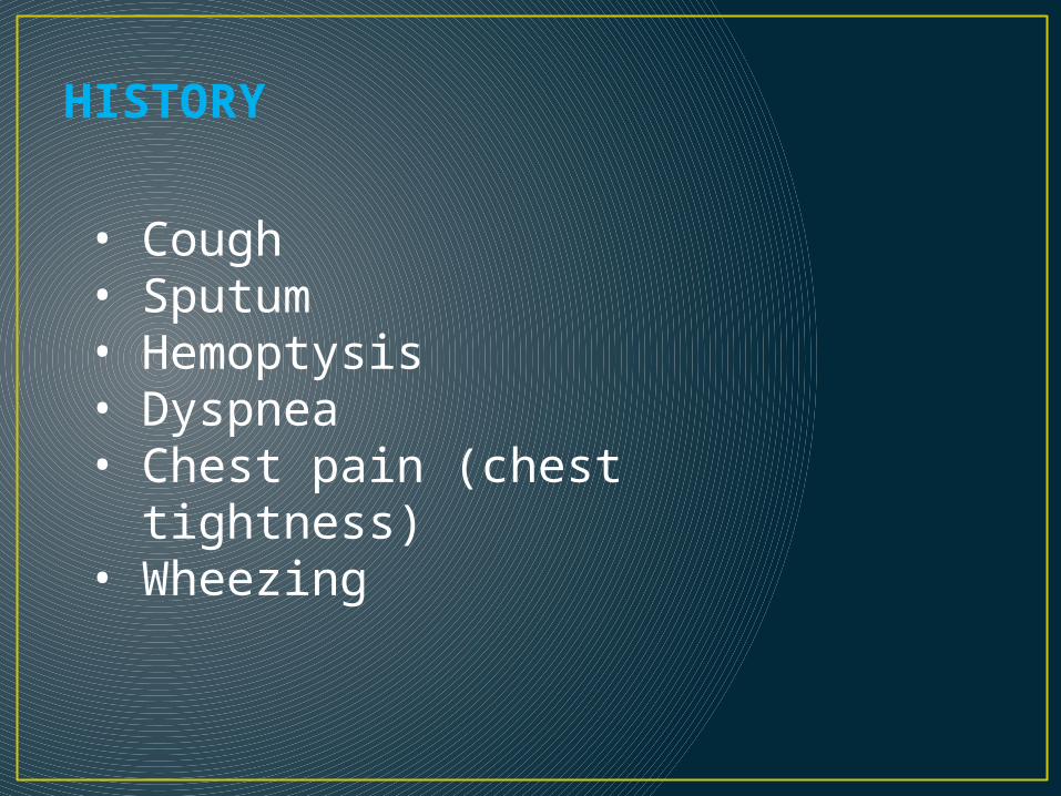
• Cough• Sputum• Hemoptysis• Dyspnea• Chest pain (chest
tightness)• Wheezing
HISTORY

COUGH
Cough reflex arc

TYPES OF COUGH
1. Acute (< 3 wks)
2. Subacute (3-8 wks)
3. Chronic (>8 wks)

CAUSES OF ACUTE COUGH:
1. Acute upper respiratory tract infection.
2. Acute lower respiratory tract infection (pneumonia).
3. Acute exacerbation of underlying chronic pulmonary disease.
4. Pulmonary Embolism (PE).

CAUSES OF SUBACUTE COUGH:
1. Post-infection of upper or lower respiratory tract.
2. Angiotensin Converting Enzyme Inhibitors (ACE-I) medication.

COMMON CAUSES OF CHRONIC COUGH USUALLY WITH A NORMAL CXR:
1. Upper airway cough syndrome (it is related to allergic, non-allergic or vasomotor rhinitis, naso-pharyngitis, & sinusitis. Usually with postnasal drip «PND»)
2. Bronchial Asthma
3. Gastroesophageal reflux disease

OTHER RESPIRATORY CAUSES:
1. Chronic bronchitis (COPD, eosinophilic)2. Bronchiectasis3. Neoplasm4. Interstitial lung disease (ILD)5. Lung abscess6. Obstructive sleep apnea (OSA)7. Tracheobronchial foreign body or mass8. Nasal polyps & others……

NON-RESPIRATORY CAUSES:
Mediastinal: • External tracheal compression ex: enlarged LN• Tumors, cysts, massesCardiac:• LVF• Severe MSENT:• Acute/chronic sinusitis• PND (allergic, or vasomotor rhinitis)

NON-RESPIRATORY CAUSES:
Gastrointestinal Tract:• GERD• Esophageal dysmotility, stricture, or pouch• Esophago-bronchial fistulaCNS: • CVA• MS• MND• Parkinson’s disease

NON-RESPIRATORY CAUSES:
Drugs:• ACE-Inhibitors• Some inhaler preparations can cause cough
Others:• Idiopathic • Ear wax (vagal nerve stimulation)• Psychogenic

• SPUTUM• Bronchiectasis (yellow, green, large amount, more in the morning)
• Lung abscess ( Foul smelling, more on lying on
the other side of lesion)
• Pneumonia( yellowish, streaks of blood)
• Pulmonary edema ( Pink frothy)
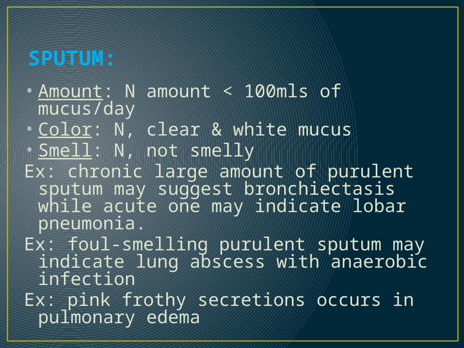
SPUTUM:
• Amount: N amount < 100mls of mucus/day• Color: N, clear & white mucus• Smell: N, not smellyEx: chronic large amount of purulent sputum may
suggest bronchiectasis while acute one may indicate lobar pneumonia.
Ex: foul-smelling purulent sputum may indicate lung abscess with anaerobic infection
Ex: pink frothy secretions occurs in pulmonary edema

HEMOPTYSIS– Massive >200 ml/episode– Frank (fresh, bright red, no sputum)– Causes
Lung cancer ( clots)Tuberculosis, Bronchiectasis ( brisk &
brief)Pulmonary infarctionPneumonia A/V malformation
HEMOPTYSIS:
• It’s a blood-stained sputum• Varies from streaks of blood to massive bleeding
(>100 - 600mls /24 hrs)• It should be investigated thoroughly • Commonest cause is acute infection like exacerbation
of copd but other serious causes should be rolled out• Other causes: PE, Bronchogenic ca., pul TB,
bronchiectasis, lung abscess,

DYSPNOEA:• Defined as: experience of discomfort in breathing or an
awareness of respiratory distress & physiologically its an ↑ in the level & work of breathing.
• Onset: 1. Instantaneous: pneumothorax, PE 2. Min.s – hrs: * Aw disease: (BA, copd exacerbʼn, UAW obstrcʼn) * parenchymal disease: (pneumonia, pul hage, pul edema..) * pul vascular disease: (PE) * cardiac disease: ( MI,……. ) * metabolic acidosis * hyperventilation syndrome.

• TYPES OF DYSPNEA• Paroxysmal dyspnea
• Bronchial asthma (wheezes, no crackles)
• Left ventricular failure (crackles, cardiomegaly)
• Nocturnal dyspnea• Bronchial asthma• GERD• LVF

DYSPNOEA:Subacute (days): * Many of the above plus: * Pl. effusion * lobar collapse * Acute Interstitial pneumonia * SVC obstruct’n * Pul vasculitisChronic (months-years): * COPD & BA * Diffuse parenchymal dis: (IPF, sarcoidosis, bronchiectasis) * Hypoventilat’n:(neuromuscular weakness, chest wall
defor.) * Anemia * Thyrotoxicosis

DYSPNOEA:Severity (grading): Dyspnea can be graded from І – IV based on
the NYHA classification.

DYSPNOEA:
3. Pleura & plural spaces:• Pneumothorax• Pleuritis & serositis• Pleural effusion4. psychogenic/psychosomatic
Wheezing:
It’s a continuous whistling, not diagnostic for asthma & can occur in other resp diseases like copd.

CLINICAL EXAMINATION (SIGNS):
In general appearance, look for:• Respiratory Rate • Respiratory distress• Use of Accessory muscles of respiration.

GENERAL SYSTEM EXAMINATION:
Hands:
1. Clubbing (check respiratory causes)
2. Tar staining
3. Weakness of hand’s small muscles (abduction) Wrist:
4. Pulse: rate & character
5. Flapping tremors (asterixis)
BP:

GENERAL SYSTEM EXAMINATION:Neck: 1. JVP: ↑ in cor-pulmonale & SVC obstruct’n but not
pulsatile.2. LN: enlargement in CA bronchus or metsFace:3. Eye: Horner’s syndrome in CA bronchus4. Tongue: central cyanosis 5. SVC obstruction: plethoric & cyanosed, periorbital
edema, injected conjuctvae

CHEST EXAMINATION:
Inspection:
1. Shape: AP diameter compared to transverse (barrel-chest), pectus excavatum, pectus carinatum, kyphoscoliosis,…. others
2. Symmetry: assessment of upper & lower lobes should be done posteriorly looking for ↓ or delayed chest movement during moderate respirat’n.
3. Scars: from previous operat’n or chest drains or cautery marks or radiotherapy markings.
4. Prominent veins: in case of SVC obstruct’n


KYPHOSCOLIOSIS


CHEST EXAMINATION:Palpation:
1. Trachea: normally central, slight Rt displacement could be N. Check for gross displacement. Tracheal tug means the N distance bet sternal notch & cricoid cartilage is < 3-4 finger breadths & occurs in chest overexpansion as copd.
2. Apex beat: Check for displacement.3. Chest expansion: N expansion ≥ 5cm4. Tactile vocal fremitus (TVF): can be done with
the palm of one hand.


TACTILE VOCAL FREMITUS

PERCUSSION

CHEST EXAMINATION:PERCUSSION:
• Should be done symmetrically (Lt compared with the Rt), posteriorly (the back), anteriorly (the front) & laterally (the sides).
• Supraclavicular area, then clavicles should be percussed directly to evaluate the upper lobes.
• Liver dullness: of the upper edge starting at the 5th rib MCL, resonant note below this area indicates hyper-inflation (copd, severe asthma)
• Cardiac dullness: may be ↓ in hyperinfated chest.

AUSCULTAION

CHEST EXAMINATION:AUSCULTATION: Using the diaphragm of a stethoscope & comment
on the following:1. Breath sounds (BS): • Intensity: N or ↓ as in (consolidation, collapse, pl effusion,
pneumothorax, lung fibrosis)• Quality: Vesicular or bronchial in consolidation• Differentiation between vesicular & bronchial BS: Vesicular: louder &longer on inspiration than expiratory phase &
has no gap between the 2 phasesBronchial: louder &longer on exp phase & has a gap between the
2 phases

ADDED SOUNDS:• Type: Wheezes or Crackles or friction rub• Timing: inspiratory or expiratory• WHEEZES: are continuous musical polyphonic sound,
heard louder on expiration & can be heard on inspiration which may imply severe AW narrowing. High pitched- wheezes are found in BA due to acute/chronic airflow limitation & low pitched in COPD. Localized monophonic wheeze due to fixed AW obstruct’n in CA bronchus.
• CRACKLES: interrupted non-musical inspiratory sound • Crackles may be early, late or pan-inspiratory & fine,
medium or coarse. Ex: late/pan-insp coarse crackles in bronchiectasis, late/pan-insp medium crackles in pul edema , late/pan-insp fine crackles in pul fibrosis

FRICTION RUB:
It’s due to thickened or roughened pleural surfaces rub together as lungs expand & contract & give off a continuous or intermittent grating sound. It indicates pleurisy & may be heard in pneumonia or pulmonary infarction.
VOCAL RESONANCE:
• It’s the ability to transmit sounds.• Ask patients to say 123 (Urdu) or 99 (English) &
listen for the transmitted sound which may be ↓ or ↑ or N (low pitched component of speech heard with booming & high pitched become attenuated).

4. EGOPHONY:
When the patient with consolidation is asked to say ‘e’ it sounds like ‘a’
5. WHISPERING PECTORILOQUY: The whispered speech is heard very loudly over the
consolidated area.

Thank You