Mshhomeobox1(Msx1)-and Msx2-overexpressingbone marrow ... · MSX1 and MSX2. The percentages of MSX1...
15
Msh homeobox 1 (Msx1)- and Msx2-overexpressing bone marrow-derived mesenchymal stem cells resemble blastema cells and enhance regeneration in mice Received for publication, December 28, 2016, and in revised form, April 29, 2017 Published, Papers in Press, May 1, 2017, DOI 10.1074/jbc.M116.774265 Leila Taghiyar ‡§ , Mahdi Hesaraki ‡ , Forough Azam Sayahpour ‡ , Leila Satarian ‡ , Samaneh Hosseini ‡ , Naser Aghdami ‡ , and Mohamadreza Baghaban Eslaminejad ‡1 From the ‡ Department of Stem Cells and Developmental Biology, Cell Science Research Center, Royan Institute for Stem Cell Biology and Technology, Academic Center for Education, Culture and Research (ACECR), Tehran 1665659911, Iran and the § Department of Developmental Biology, University of Science and Culture, Tehran 13145-871, Iran Edited by Xiao-Fan Wang Amputation of the proximal region in mammals is not fol- lowed by regeneration because blastema cells (BCs) and expres- sion of regenerative genes, such as Msh homeobox (Msx) genes, are absent in this animal group. The lack of BCs and positional information in other cells is therefore the main obstacle to ther- apeutic approaches for limb regeneration. Hence, this study aimed to create blastema-like cells (BlCs) by overexpressing Msx1 and Msx2 genes in mouse bone marrow-derived mesen- chymal stem cells (mBMSCs) to regenerate a proximally ampu- tated digit tip. We transduced mBMSCs with Msx1 and Msx2 genes and compared osteogenic activity and expression levels of several Msx-regulated genes (Bmp4, Fgf8, and keratin 14 (K14)) in BlC groups, including MSX1, MSX2, and MSX1/2 (in a 1:1 ratio) with those in mBMSCs and BCs in vitro and in vivo follow- ing injection into the amputation site. We found that Msx gene overexpression increased expression of specific blastemal mark- ers and enhanced the proliferation rate and osteogenesis of BlCs compared with mBMSCs and BCs via activation of Fgf8 and Bmp4. Histological analyses indicated full regrowth of digit tips in the Msx-overexpressing groups, particularly in MSX1/2, through endochondral ossification 6 weeks post-injection. In contrast, mBMSCs and BCs formed abnormal bone and nail. Full digit tip was regenerated only in the MSX1/2 group and was related to boosted Bmp4, Fgf8, and K14 gene expression and to limb-patterning properties resulting from Msx1 and Msx2 over- expression. We propose that Msx-transduced cells that can regenerate epithelial and mesenchymal tissues may potentially be utilized in limb regeneration. Digit tip regeneration is a well-documented example of organ/appendage regeneration in lower vertebrates, such as salamanders, because they exhibit robust regeneration poten- tial in response to amputation anywhere along the proximal/ distal limb axis (1). However, the level of amputation through the terminal phalanx causes various regeneration responses in mammals, such as humans and mice. Amputation in the middle of the terminal phalanx involves complete skeletal restoration, whereas only scar formation occurs following amputation injury at the proximal level (2). Therefore, the main goal of clinical and experimental investigations is to restore lost parts of limbs damaged due to disease or injury. Despite numerous attempts, digit tip regrowth after amputation through terminal phalanges remains challenging. Naturally, blastema cells (BCs) 2 are considered a key mediator of digit tip regeneration so that their removal following proximal amputation leads to scar for- mation in adult mammals (1). BCs are believed to be a heterogeneous population of lineage- restricted progenitor cells derived from fibroblasts of connec- tive tissue (3). There are several main surface biomarkers assigned to BCs: stem cell antigen-1 (Sca-1), endothelial marker (CD31), and vimentin (4). Msh homeobox (Msx) genes are members of the Hox gene family and include 1 Msx1 (Hox7) and Msx2 (Hox8). They are highly conserved among vertebrates and are considered key genes in BCs (5, 6). It has conclusively been shown that the absence of BCs as well as Msx1 and Msx2 genes results in regeneration failure in a proximal amputation of adult mice (7). Additionally, Msx genes are of crucial importance in ecto-mesodermal interactions that mediate cellular prolifera- tion and differentiation during limb formation, apical epithelial cap (AEC) formation, and limb patterning (8). Bensoussan- Trigano et al. (9) have shown that the Prx1-Cre Msx1 null/null Msx2 null/Flox mutants display abnormal digit formation and preaxial polydactyly in fetal mouse digit tip regeneration. Over- expressed Msx1 (Hox7) in hind-limb regeneration in a trans- genic Xenopus model (M1) resulted in a higher proliferation rate in both BCs and apical epithelial cap, thickened wound epithelium, and more regenerated toes in M1 compared with The present research was supported by the Royan Institute and the Iranian Council of Stem Cell Research and Technology (ICSCR). The authors declare that they have no conflicts of interest with the contents of this article. This article contains supplemental Tables 1 and 2 and Figs. 1–3. 1 To whom correspondence should be addressed: Dept. of Stem Cells and Developmental Biology, Cell Science Research Center, Royan Institute for Stem Cell Biology and Technology, 1665659911, ACECR, Tehran, Iran. Tel.: 98-2123562524; Fax: 98-2123562507; E-mail: [email protected]. 2 The abbreviations used are: BC, blastema cell; BMSC, bone marrow-derived mesenchymal stem cell; mBMSC, mouse bone marrow-derived mesenchy- mal stem cell; BlC, blastema-like cell; MSC, mesenchymal stem cell; CFU-F, colony-forming unit fibroblast; ALP, alkaline phosphatase; MTT, 3-(4,5-di- methylthiazol-2-yl)-2,5-diphenyltetrazolium bromide; ICC, immunocyto- chemistry; Nrm. Reg, normal regeneration; Al & Al, Alcian blue and alizarin red S; WPI, weeks post-injection; DPA, days post-amputation; qRT-PCR, quantitative RT-PCR; IRES, internal ribosome entry site. cros ARTICLE 10520 J. Biol. Chem. (2017) 292(25) 10520 –10533 © 2017 by The American Society for Biochemistry and Molecular Biology, Inc. Published in the U.S.A. by guest on May 25, 2020 http://www.jbc.org/ Downloaded from
Transcript of Mshhomeobox1(Msx1)-and Msx2-overexpressingbone marrow ... · MSX1 and MSX2. The percentages of MSX1...

Msh homeobox 1 (Msx1)- and Msx2-overexpressing bonemarrow-derived mesenchymal stem cells resemble blastemacells and enhance regeneration in miceReceived for publication, December 28, 2016, and in revised form, April 29, 2017 Published, Papers in Press, May 1, 2017, DOI 10.1074/jbc.M116.774265
Leila Taghiyar‡§, Mahdi Hesaraki‡, Forough Azam Sayahpour‡, Leila Satarian‡, Samaneh Hosseini‡,Naser Aghdami‡, and Mohamadreza Baghaban Eslaminejad‡1
From the ‡Department of Stem Cells and Developmental Biology, Cell Science Research Center, Royan Institute for Stem CellBiology and Technology, Academic Center for Education, Culture and Research (ACECR), Tehran 1665659911, Iran and the§Department of Developmental Biology, University of Science and Culture, Tehran 13145-871, Iran
Edited by Xiao-Fan Wang
Amputation of the proximal region in mammals is not fol-lowed by regeneration because blastema cells (BCs) and expres-sion of regenerative genes, such as Msh homeobox (Msx) genes,are absent in this animal group. The lack of BCs and positionalinformation in other cells is therefore the main obstacle to ther-apeutic approaches for limb regeneration. Hence, this studyaimed to create blastema-like cells (BlCs) by overexpressingMsx1 and Msx2 genes in mouse bone marrow-derived mesen-chymal stem cells (mBMSCs) to regenerate a proximally ampu-tated digit tip. We transduced mBMSCs with Msx1 and Msx2genes and compared osteogenic activity and expression levels ofseveral Msx-regulated genes (Bmp4, Fgf8, and keratin 14 (K14))in BlC groups, including MSX1, MSX2, and MSX1/2 (in a 1:1ratio) with those in mBMSCs and BCs in vitro and in vivo follow-ing injection into the amputation site. We found that Msx geneoverexpression increased expression of specific blastemal mark-ers and enhanced the proliferation rate and osteogenesis of BlCscompared with mBMSCs and BCs via activation of Fgf8 andBmp4. Histological analyses indicated full regrowth of digit tipsin the Msx-overexpressing groups, particularly in MSX1/2,through endochondral ossification 6 weeks post-injection. Incontrast, mBMSCs and BCs formed abnormal bone and nail.Full digit tip was regenerated only in the MSX1/2 group and wasrelated to boosted Bmp4, Fgf8, and K14 gene expression and tolimb-patterning properties resulting from Msx1 and Msx2 over-expression. We propose that Msx-transduced cells that canregenerate epithelial and mesenchymal tissues may potentiallybe utilized in limb regeneration.
Digit tip regeneration is a well-documented example oforgan/appendage regeneration in lower vertebrates, such assalamanders, because they exhibit robust regeneration poten-tial in response to amputation anywhere along the proximal/
distal limb axis (1). However, the level of amputation throughthe terminal phalanx causes various regeneration responses inmammals, such as humans and mice. Amputation in the middleof the terminal phalanx involves complete skeletal restoration,whereas only scar formation occurs following amputationinjury at the proximal level (2). Therefore, the main goal ofclinical and experimental investigations is to restore lost partsof limbs damaged due to disease or injury. Despite numerousattempts, digit tip regrowth after amputation through terminalphalanges remains challenging. Naturally, blastema cells (BCs)2
are considered a key mediator of digit tip regeneration so thattheir removal following proximal amputation leads to scar for-mation in adult mammals (1).
BCs are believed to be a heterogeneous population of lineage-restricted progenitor cells derived from fibroblasts of connec-tive tissue (3). There are several main surface biomarkersassigned to BCs: stem cell antigen-1 (Sca-1), endothelial marker(CD31), and vimentin (4). Msh homeobox (Msx) genes aremembers of the Hox gene family and include 1 Msx1 (Hox7) andMsx2 (Hox8). They are highly conserved among vertebrates andare considered key genes in BCs (5, 6). It has conclusively beenshown that the absence of BCs as well as Msx1 and Msx2 genesresults in regeneration failure in a proximal amputation of adultmice (7). Additionally, Msx genes are of crucial importance inecto-mesodermal interactions that mediate cellular prolifera-tion and differentiation during limb formation, apical epithelialcap (AEC) formation, and limb patterning (8). Bensoussan-Trigano et al. (9) have shown that the Prx1-Cre Msx1 null/nullMsx2 null/Flox mutants display abnormal digit formation andpreaxial polydactyly in fetal mouse digit tip regeneration. Over-expressed Msx1 (Hox7) in hind-limb regeneration in a trans-genic Xenopus model (M1) resulted in a higher proliferationrate in both BCs and apical epithelial cap, thickened woundepithelium, and more regenerated toes in M1 compared with
The present research was supported by the Royan Institute and the IranianCouncil of Stem Cell Research and Technology (ICSCR). The authors declarethat they have no conflicts of interest with the contents of this article.
This article contains supplemental Tables 1 and 2 and Figs. 1–3.1 To whom correspondence should be addressed: Dept. of Stem Cells and
Developmental Biology, Cell Science Research Center, Royan Institute forStem Cell Biology and Technology, 1665659911, ACECR, Tehran, Iran. Tel.:98-2123562524; Fax: 98-2123562507; E-mail: [email protected].
2 The abbreviations used are: BC, blastema cell; BMSC, bone marrow-derivedmesenchymal stem cell; mBMSC, mouse bone marrow-derived mesenchy-mal stem cell; BlC, blastema-like cell; MSC, mesenchymal stem cell; CFU-F,colony-forming unit fibroblast; ALP, alkaline phosphatase; MTT, 3-(4,5-di-methylthiazol-2-yl)-2,5-diphenyltetrazolium bromide; ICC, immunocyto-chemistry; Nrm. Reg, normal regeneration; Al & Al, Alcian blue and alizarinred S; WPI, weeks post-injection; DPA, days post-amputation; qRT-PCR,quantitative RT-PCR; IRES, internal ribosome entry site.
crosARTICLE
10520 J. Biol. Chem. (2017) 292(25) 10520 –10533
© 2017 by The American Society for Biochemistry and Molecular Biology, Inc. Published in the U.S.A.
by guest on May 25, 2020
http://ww
w.jbc.org/
Dow
nloaded from

WT animals in stage 54 (10). More importantly, BCs haveenabled the process of bone formation as a main process of limbregeneration by triggering a cascade-of-cell-signaling pathway,such as bone morphogenetic proteins (BMPs) and FGFs (11–13). Positional information is one of the key elements in suc-cessful regeneration. It has been proposed that the expressionof region-specific hox genes in early and late blastema tissues ismore likely to be related to positional identity (14). Rao et al.(15) have shown that fibroblastemas of Xenopus limbs ex-press proximal-distal axial patterning genes, including hoxa9,hoxa11, and hoxa13. Indeed, successful limb regeneration hasbeen correlated to the formation of BCs and their related genes.Although transplantation of BCs at the amputation site couldaccelerate wound healing and digit regeneration, availability ofBCs has always been a challenging issue in terms of digit regen-eration. Hence, the use of an alternative available cell sourcewith high similarity to BCs would be a valuable strategy in limbregeneration.
Various cell sources have been isolated and characterized interms of their potential in clinical settings. Fetal limb cells havea tremendous degree of similarity to BCs for limb regeneration,although their administration is hampered due to ethical con-cerns (16). Stem cells, in particular mesenchymal stem cells(MSCs), have tremendous potential in therapeutic approachesdue to their unique characteristics of self-renewal and differen-tiation potential. MSCs are multipotent cells that exist in mostadult tissues (17–19) and have the ability to give rise to multipletissue-forming cell lineages (20 –22). It has been postulated thatMSCs affect cell migration, proliferation, and survival of thesurrounding cells as well as providing anti-scarring propertiesthrough paracrine signaling (23, 24). They may also regulateimmune and inflammatory responses, making them an appro-priate therapeutic candidate for treating inflammatory diseases(25, 26). Thus far, much research has been devoted to the appli-cation of MSCs in various diseased tissues due to their potentregenerative capacity, and they have been used in over 350 clin-ical trials worldwide (27). However, there has been little inves-tigation of MSCs in limb regeneration. Masaki et al. (28) trans-planted bone marrow-derived MSCs (BMSCs) and limb budsinto amputated limbs in neonatal mice and observed the gen-eration of the segmented pattern of bone and cartilage. Inanother study, injection of the hematopoietic stem cells into anamputated digit did not lead to the formation of main struc-tures of the digit, but it contributed to the formation of bloodcells and bone marrow tissue (29). However, a lack of positionalinformation in current efforts that use stem cells is more likelyto be the cause of regeneration failure.
In our previous study, we isolated BCs from neonatal miceand compared their characteristics with mouse BMSCs (mBM-SCs) in vitro. Our study has shown that mBMSCs have similarcharacteristics to BCs in terms of growth and osteogenic activ-ity and can be considered as substitutes for BCs (30). Thus, thepresent study aimed to create blastema-like cells (BlCs)through overexpression of Hox family genes, including Msx1and Msx2, that are involved in positional information in mBM-SCs and regenerate a digit tip by transplantation of BlCs in vivo.Therefore, we transduced mBMSCs with Msx1 and Msx2genes, after which their proliferation and differentiation poten-
tials were examined by 3-(4,5-dimethylthiazol-2-yl)-2,5-diphe-nyltetrazolium bromide (MTT) and real-time PCR in vitro. Todetermine the regenerative potential of BlCs, we injected theminto the amputated proximal digits of adult mice. The resultsdemonstrated that BlCs could completely improve proximaldigit tip amputation compared with mBMSCs and BCs.
Results
Msx1- and Msx2-transduced mBMSCs
To verify the accuracy of gene transduction, third-generationself-inactivating vectors with various fluorescent reporters’genes were constructed as diagrammed in Fig. 1A. Msx1 andMsx2 genes were co-expressed by GFP and tdTomato genes,respectively. Although the majority of cells expressed reportergenes, we needed a pure cell population that absolutelyexpressed Msx1 (GFP�) and Msx2 (tdTomato�). Therefore,the cells were sorted for GFP and tdTomato markers after 72 hof transduction, as seen in Fig. 1B (a and b). ICC analysisshowed significantly lower protein expression levels of endog-enous Msx1 and Msx2 in mBMSCs (� 3–5%) compared withBCs (30 – 40%). Msx1 and Msx2 transduction led to a drasticincrease in expression level of these exogenous genes in BlCs(100%), which was greater than in BCs (Fig. 1C, a and c).
We used RT-PCR to determine the quantitative expressionlevels of Msx1 and Msx2. Fig. 1C shows that the expressionlevels of Msx1 and Msx2 in BCs were �400 –500-fold greaterthan in mBMSCs (**, p � 0.01). These genes were up-regulatedby 5000-fold in BlCs compared with mBMSCs and 4500-fold inBlCs compared with BCs (****, p � 0.0001; Fig. 1C, b and d).Interestingly, endogenous Msx2 and Msx1 genes were up-reg-ulated in the transduced MSX1 and MSX2 groups, respectively.
BC cell-surface marker analysis for BlCs and mBMSCs
To confirm BC phenotype for BlCs (mBMSCs as a controlgroup), cells from each group were analyzed by flow cytometryagainst various surface markers (i.e. Sca1, CD31, and Vim). Asshown in Fig. 1D, the majority of BlCs were positive for Vim(MSX1, 85%; MSX2, 98%), CD31 (MSX1, 45%; MSX2, 35%),and Sca1 (MSX1, 60%; MSX2, 80%), which is comparable withthe BC group.
Cell growth, colony-forming unit fibroblast (CFU-F), and cell-cycle analysis
The CFU-F analysis was performed to examine the prolifer-ation patterns of BlCs, BCs, and mBMSCs in semisolid medium.Supplemental Fig. 1A shows the colonies and average numbersof colonies per culture dish. The numbers of colonies were 80 �5 (mBMSCs), 60 � 5 (B1Cs), 170 � 5 (MSX1), and 140 � 4(MSX2), as seen in supplemental Fig. 1C. The BlCs colonieswere significantly longer compared with mBMSCs and BCs (*,p � 0.05; supplemental Fig. 1D). MTT results indicated thatBlCs had a nonsignficantly greater proliferation rate comparedwith mBMSCs and BCs on day 1. Both MSX1- and MSX2-induced cells had significantly greater proliferation than bothBCs and mBMSCs at 3 and 7 days (supplemental Fig. 1B). Cell-cycle analysis revealed that the accumulation of BCs andBMSCs in G0/G1 phase increased �2-fold compared with
Blastema cell-like formation by Msx1 and Msx2-transduced mBMSCs
J. Biol. Chem. (2017) 292(25) 10520 –10533 10521
by guest on May 25, 2020
http://ww
w.jbc.org/
Dow
nloaded from

Figure 1. Msx1 and Msx2 gene transduction in mBMSCs. A, vector map. Msx1 and Msx2 were co-expressed by GFP and tdTomato, respectively, to followMsx-positive cells in the same vectors. B, the ICC images represent Msx1 and Msx2 genes endogenously expressed in mBMSCs and BCs, as well as exogenousMsx1 (GFP�) and Msx2 (tdTomato�) gene expressions in BlCs cells (a and b). C, real-time PCR analysis shows the gene expression level of Msx1 (b) and Msx2 (d)in BC, mBMSC, and BlC groups. The histograms represent the percentage of Msx1 and Msx2 protein expression levels in BC, mBMSC, and BlC groups (a and c).D, flow cytometry analysis of CD31, Sca1, and Vim cell-surface markers for BlCs, BCs, and mBMSCs. Scale bar, 50 �m. Data are presented as means � S.D. (errorbars). ****, p � 0.0001.
Blastema cell-like formation by Msx1 and Msx2-transduced mBMSCs
10522 J. Biol. Chem. (2017) 292(25) 10520 –10533
by guest on May 25, 2020
http://ww
w.jbc.org/
Dow
nloaded from
MSX1 and MSX2. The percentages of MSX1 and MSX2 in Sphase were 32.83 and 40.07%, whereas 13.65 and 14.22% ofmBMSCs and BCs were in S phase, respectively (supplementalFig. 1E).
Osteogenic activity
We assessed osteogenic activity of BlCs, BCs, and mBMSCsat various time points (culture days 7, 14, and 21) by alizarin redS staining, alkaline phosphatase (ALP) activity, and qRT-PCR.Alizarin red staining showed that nodule-like aggregates beganto form during the first week and increased in abundancetoward the end of the third week. There were significantly moremineralized nodules in the BlC (MSX1, MSX2, and MSX1/2)plates compared with BCs and mBMSCs (Fig. 2, A and E). ALPactivity, as an early marker for osteogenic differentiation, wasmeasured after 7, 14, and 21 days of incubation. After 7 days,both Msx1- and Msx2-transduced cells as well as the MSX1/2group showed an almost equal increase in ALP activity. ALPactivity was significantly reduced in BlCs compared with BCsand mBMSCs. ALP activity was significantly decreased in all ofthe studied groups on days 14 and 21 (Fig. 2B). Fig. 2C shows thecalcium content of BlC, BC, and mBMSC cultures after 7, 14,and 21 days. The amount of calcium increased over time in allgroups. After 14 days, we observed a higher calcium content inBlCs compared with those of BCs and mBMSCs. There wassignificantly increased calcium content observed between BlC(MSX1, MSX2, and MSX1/2), mBMSC, and BC groups on day21. Real-time PCR analysis revealed that the expression level ofColI (collagen I) as well as Runx2 and the Ocn (osteocalcin)genes was progressively up-regulated within 3 weeks (Fig. 2D,a– c).
Analysis of Msx-related genes
We assessed the expression levels of several major Msx-re-lated genes (Bmp4, Fgf8, ki67, and K14) by ICC and RT-PCRbefore and after gene transduction in mBMSCs as well as BCs.RT-PCR analysis showed that the expression level of Bmp4gene increased by 350-fold in MSX1-transduced cells and by300-fold in MSX2-transduced cells compared with mBMSCs(****, p � 0.0001; Fig. 3). The Fgf8 gene showed significantexpression in both the MSX1 (170-fold) and MSX2 (150-fold)groups compared with mBMSCs (Fig. 3B, a and b). ICC analysisindicated that mBMSCs did not express Bmp4 and Fgf8 endog-enously, whereas these genes were up-regulated by 80 –90% inBlCs (Fig. 3B, b and d). The expression levels of the ectoderm-specific genes keratin 14 (K14) and Ki67 (also known as MKI67)were assessed using ICC. The former is important for nail for-mation in limb development, and the latter is a cellular markerfor proliferation. As shown in Fig. 3C (a), we detected no endog-enous expression of K14 in mBMSCs, whereas the proteinexpression level of K14 was significantly up-regulated in Msx1-and Msx2-transduced cells (80 –90%; ****, p � 0.0001). In addi-tion, BCs expressed K14 �20-fold more than mBMSCs (Fig. 3D(a)). Likewise, ICC results indicated a low expression for Ki67 inmBMSCs (Fig. 3C (b)) compared with BCs. The expression levelof Ki67 protein increased by 90% in the MSX1 group and 80% inthe MSX2 group compared with the control group (****, p �0.0001; Fig. 3D (b)).
Macroscopic evaluation of proximal regeneration
In our digit tip regeneration model, the adult mouse terminalphalanges were proximally amputated to ascertain that themodel could not regenerate before injection of the cells (sup-plemental Fig. 2, A and B). The wound epidermis formed a scarwound healing response immediately after digit tip amputationat the proximal level of P3 in both the Nrm. Reg and Shamgroups (supplemental Fig. 2, C–E). We observed closure in theepidermis completed in 4 –5 days post-amputation (DPA)without any new bone or nail formation. Therefore, we injectedthe cells 4 DPA, when the scar wound falls off.
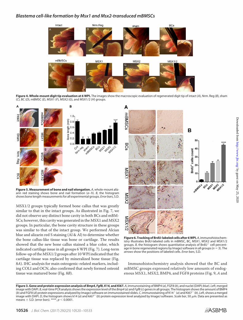
Whole-mount digit tip analysis at 6 weeks after the injection(WPI) showed that the epidermal closure occurred in all exper-imental and control groups. We observed no trace of regener-ation in the Nrm. Reg and Sham groups, which was expected(Fig. 4, B and C). In the BC and mBMSC groups, regeneration ofthe proximal regions occurred in � 10% of the digit tips.Abnormal bone and nail organ formation with no elongationoccurred in both groups (Fig. 4, D and E, and supplementalTable 2). Surprisingly, � 95% of the digit tips showed full digittip regeneration, which included both formation and elonga-tion of the bone and nail in the MSX1/2 group (Fig. 4H andsupplemental Table 2).
In contrast, �5% of the digit tips fully regenerated in theMSX1 and MSX2 groups. We observed new bone and nail for-mation in � 50% of samples from both the MSX1 and MSX2groups (Fig. 4, F and G, and supplemental Table 2).
To quantify the regenerated bone, we performed whole-mount alizarin red staining to visualize the skeletal patterns tomeasure the proximal-distal length of each terminal phalanx(30 digits for each group). As illustrated in Fig. 5A, the Nrm. Regand Sham groups formed no nails or new bone (Fig. 5A, b and c).We observed newly formed bone and nail in the mBMSCs andBCs groups (Fig. 5A, d and e), although they had abnormalmorphologies with thin nails and short bones. On the otherhand, morphological analysis revealed that bone and nail gen-erated up to 50% of the intact group in both the MSX1 andMSX2 groups (Fig. 5, A (f and g) and B). There were no remark-able differences in new bone length between the MSX1 andMSX2 groups. The thickness and length of the new nail remark-ably increased in MSX1/2 because the length of the newlyformed bone was comparable with the intact group (Fig 5, A (h)and B).
Histological analysis of digit tip regeneration
To explore the presence of BlCs in terminal phalange ele-ment regeneration, the labeled cells were first traced betweenthe stump bone and wound epidermis position. Ferridex- andGFP-MSX1-labeled cells comprised 15–20% of total cells indigit tip regenerated regions (supplemental Fig. 3). Similarly,BrdU labeling showed the presence of injected cells at the site ofinjury 6 WPI in all experimental groups (Fig. 6).
Histological analysis showed that epidermal closure oc-curred in all groups, and the thickness of the epidermis anddermis layer had no significant differences among the groups(Fig. 7). H&E and Masson’s trichrome staining results indicatedthat tissue atrophy occurred in the connective tissue (cartilage,
Blastema cell-like formation by Msx1 and Msx2-transduced mBMSCs
J. Biol. Chem. (2017) 292(25) 10520 –10533 10523
by guest on May 25, 2020
http://ww
w.jbc.org/
Dow
nloaded from

nail, and bone stump) in the Nrm. Reg and Sham groups. How-ever, we observed no apoptotic cells or tissue in these groups(Fig. 7, D and H). In the cell-injected groups, the distal end ofthe bone stump was elongated and formed a bony cap similar toa bone callus (Fig. 6, J and N). Specifically, the bone stump
showed slight elongation in BCs and mBMSCs, whereas thebone callus was significantly larger in the BlC groups (Fig. 7,Q–U, W, and X). The formed bone callus was spongy in bothBCs and mBMSCs. This structure was going to be replaced by astructure similar to compact bone in BlCs. Interestingly, the
Figure 2. Osteogenic activity. The data represent osteogenic differentiation of BlCs, BCs, and mBMSCs after 7, 14, and 21 days. A, nodule-like aggregationsincreased over time during 3 weeks and showed strong mineralization in all groups. B, the histogram shows ALP activity in the BC, mBMSC, MSX1, MSX2, andMSX1/2 groups. C, the histogram represents the calcium content in BC, mBMSC, MSX1, MSX2, and MSX1/2 groups. D, real-time PCR analysis for ColI (a), Runx2(b), and Ocn (c) genes showed enhanced expression levels in the MSX groups compared with mBMSCs and BCs. E, the histogram shows quantitative analysisof mineralized nodules in the BC, mBMSC, MSX1, MSX2, and MSX1/2 groups. Data are presented as means � S.D. (error bars) (n � 3). ****, p � 0.0001.
Blastema cell-like formation by Msx1 and Msx2-transduced mBMSCs
10524 J. Biol. Chem. (2017) 292(25) 10520 –10533
by guest on May 25, 2020
http://ww
w.jbc.org/
Dow
nloaded from

Blastema cell-like formation by Msx1 and Msx2-transduced mBMSCs
J. Biol. Chem. (2017) 292(25) 10520 –10533 10525
by guest on May 25, 2020
http://ww
w.jbc.org/
Dow
nloaded from
MSX1/2 groups typically formed bone callus that was greatlysimilar to that in the intact groups. As illustrated in Fig. 7, wedid not observe any distinct bone cavity in both BCs and mBM-SCs; however, this cavity was generated in the MSX1 and MSX2groups. In particular, the bone cavity structure in these groupswas similar to that of the intact group. We performed Alcianblue and alizarin red S staining (Al & Al) to determine whetherthe bone callus-like tissue was bone or cartilage. The resultsshowed that the new bone callus stained a blue color, whichindicated cartilage issue in all groups 6 WPI (Fig. 7). Long-termfollow-up of the MSX1/2 groups after 10 WPI indicated that thecartilage tissue was replaced by mineralized bone tissue (Fig.8A). IHC analysis for main osteogenic-related markers, includ-ing COLI and OCN, also confirmed that newly formed osteoidtissue was matured bone (Fig. 8B).
Immunohistochemistry analysis showed that the BC andmBMSC groups expressed relatively low amounts of endog-enous MSX1, MSX2, BMP4, and FGF8 proteins (Fig. 9, A and
Figure 3. Gene and protein expression analysis of Bmp4, Fgf8, K14, and Ki67. A, immunostaining of BMP4 (a), FGF8 (b), and nuclei (DAPI; blue). Left, mergedimage with DAPI. B, real-time PCR analysis shows the expression level of the Bmp4 (a) and Fgf8 (c) genes in all groups. The histogram shows the amount of BMP4(b) and FGF8 (d) protein expressions analyzed by ImageJ software on immunostained slides. C, immunostaining of K14� (a) and Ki67� (b). Left, shows a mergedimage with DAPI. D, the histogram shows k14 (a) and ki67� (b) protein expression level analyzed by ImageJ software. Scale bar, 50 �m. Data are presented asmeans � S.D. (error bars). ****, p � 0.0001.
Figure 4. Whole-mount digit tip evaluation at 6 WPI. The images show the macroscopic evaluation of regenerated digit tip of intact (A), Nrm. Reg (B), sham(C), BC (D), mBMSC (E), MSX1 (F), MSX2 (G), and MSX1/2 (H) groups.
Figure 5. Measurement of bone and nail elongation. A, whole-mount aliz-arin red staining shows bone and nail formation (a– h). B, the histogramshows bone length measurements for all experimental groups. Error bars, S.D.
Figure 6. Tracking of BrdU-labeled cells after 6 WPI. A, immunohistochem-istry illustrates BrdU-labeled cells in mBMSC, BC, MSX1, MSX2 and MSX1/2groups. B, the histogram shows quantitative analysis of BrdU� cell percent-age in bone regenerated regions by ImageJ software in all groups (n � 3). Thearrows show the positions of labeled cells. Error bars, S.D.
Blastema cell-like formation by Msx1 and Msx2-transduced mBMSCs
10526 J. Biol. Chem. (2017) 292(25) 10520 –10533
by guest on May 25, 2020
http://ww
w.jbc.org/
Dow
nloaded from

B). In contrast, the expression levels of the same markersdramatically increased in the BlC groups, particularly MSX1/2(Fig. 9, A and B). These results agreed with the expressionprofiles of Msx1, Msx2, Fgf8, and Bmp4 under in vitroconditions.
Discussion
Digit tip regeneration in adult mice is restricted to the distalhalf of the terminal phalangeal element (P3), whereas regener-ation fails following amputation through the proximal third ofP3 (7) due to lack of BCs and Msx genes. Msx genes regulatecellular behavior like proliferation and differentiation as well asdigit patterning during limb development and regeneration(31). The unavailability of BCs is a major challenge regardingBC therapy. On the other hand, lack of positional informationin other cell sources, an exclusive ability of BCs, has hampered
the cell-based therapeutic approaches. Therefore, in this study,we have developed a new cell source by overexpression of theMsx1 and Msx2 genes in mBMSCs, which created BlCs, afterwhich we explored the regenerative potential these BlCs inproximally amputated digit tips (Fig. 10).
Our ICC and qRT-PCR results showed that Msx1 and Msx2genes were accurately transduced and up-regulated in mBM-SCs. Additionally, transduction of exogenous Msx genes hasresulted in up-regulation of endogenous Msx2 and Msx1 inMSX1 and MSX2 cells, respectively. The Msx2 expressiondomain has been shown to expand into the Msx1 domain, andthey overlap (7). Therefore, up-regulation of endogenous geneswould be expected. Also, overexpression of one of the Msxgenes may affect expression of the other related genes. A com-parison of BlCs and original BCs has confirmed the higherexpression levels of the Bmp4, Fgf8, and Ki67 genes in BlCsaccording to ICC results. To determine whether BlCs have thesimilar characteristics as BCs, we assessed the expression levelof specific blastema markers, CD31, Vim, and Sca1, in allgroups. Surprisingly, the flow cytometry results confirmed thatspecific blastema cell-surface markers were significantlyexpressed in BlCs compared with mBMSCs. These significantdifferences among mBMSCs, BCs, and BlCs might be attributedto overexpression of Msx, which led to a higher proliferationrate and osteogenic activity in the transduced cells. Bmp4 isconsidered to be a crucial osteogenic marker (32). Its up-regu-lation in BlCs enhanced osteogenic differentiation capacitycompared with mBMSCs and BCs. Likewise, the higher prolif-eration rate of BlCs occurred because of Fgf8 and Ki67 geneup-regulation as proliferation markers. The results of cell-cycleanalysis confirmed that S phase in BlCs was longer than mBM-SCs and BCs. Of note, the decreased activity of BCs relative toBlCs was most likely related to cellular expansion followingseveral passages in vitro.
Figure 7. Histological analysis. H&E, Masson’s trichrome, Alcian blue, and alizarin red S staining in the intact (A–C), Nrm. Reg (D–F), sham (G–I), BC (J–L),mBMSC (M–O), MSX1 (P–R), MSX2 (S–U), and MSX1/2 (V–Y) groups. The H&E results showed that no apoptotic cells or tissues were found in any of the groups.Alcian blue-alizarin red S staining indicated that the region containing new bone callus was a blue color; we assumed the presence of cartilage tissues that werestained by Alcian blue.
Figure 8. Bone regeneration analysis in MSX1/2 after 10 WPI. A, Al & Alstaining of MSX1/2 group (a, low magnification; b, high magnification) 10 WPIthat shows the endochondral bone was replaced by mineralized bone. B, IHCanalysis shows the expression of COLI (a, low magnification; b, high magnifi-cation) and OCN (c, low magnification; d, high magnification) in mature bonegenerated in MSX1/2 group after 10 weeks.
Blastema cell-like formation by Msx1 and Msx2-transduced mBMSCs
J. Biol. Chem. (2017) 292(25) 10520 –10533 10527
by guest on May 25, 2020
http://ww
w.jbc.org/
Dow
nloaded from

We explored the regenerative capacity of BlCs followingtransplantation into non-regenerative digit tips of an adultmouse model. In this model, the cartilage tissue of the joint,stump bone, and nail of P3 were preserved, whereas weremoved the bone and nail within the bone marrow cavity. Ourwhole-mount results combined with histological analysisshowed a lack of new bone and nail formation as well as tissueatrophy in the non-cell-injected groups (Nrm. Reg and Sham)after 6 WPI, which confirmed that the proximally amputatedmodel was non-regenerative. We observed a significant regen-erative response in BlCs, particularly MSX1/2, as indicated bycomplete nail and bone formation, which was like that of theintact group. We observed abnormal bone and thin nail forma-tion in both the mBMSC and BC groups. These findingsshowed that the new cell fate of mBMSCs by overexpression ofthe Msx genes efficiently improved regrowth of the amputateddigit.
To precisely address how the process of bone formation pro-gresses, we performed Al & Al staining. According to the data,
the cell-injected groups induced osteogenesis via an endochon-dral ossification manner as in a previous study that demon-strated endochondral bone formation in mouse digit tip regen-eration (33). The transient cartilage was suggested to bereplaced by newly mineralized bone. However, visible changesoccurred in the generated bone tissue in terms of volume andhistology among the BlCs, mBMSCs, and BCs groups. Asexpected, injection of BlCs led to regrowth of bone and nailorgans, although full regeneration that contained normal bonecavity and compact bone-like was only observed in the MSX1/2groups after 6 WPI. Long-term follow-up confirmed comple-tion of ossification after 10 WPI in the MSX1/2 group. Thepresence of very large blood vessels containing blood cells wasclearly detected within the regenerated region. Our results areconsistent with previous studies that have showed that cellsexpressing the endothelial markers CD31 and stem cell antigen1 (Sca1) in the blastema region could differentiate into endo-thelial progenitor cells and form blood vessels (4). Regeneratedbone marrow cavity along with blood vessel formation in BlC
Figure 9. Immunohistochemistry analysis of the MSX1, MSX2, BMP4, and FGF8. A, IHC staining shows that MSX1, MSX2, BMP4, and FGF8 were expressedin the BC, mBMSC, MSX1, MSX2, and MSX1/2 groups. B, histograms show the percentage of MSX1, MSX2, BMP4, and FGF8 protein expressions in the BCs,mBMSCs, MSX1, MSX2, and MSX1/2 groups (a– d). Error bars, S.D.
Blastema cell-like formation by Msx1 and Msx2-transduced mBMSCs
10528 J. Biol. Chem. (2017) 292(25) 10520 –10533
by guest on May 25, 2020
http://ww
w.jbc.org/
Dow
nloaded from

groups, particularly MSX1/2, compared with BC and mBMSCgroups, was most probably related to the increased expressionlevel of CD31 and Sca1 markers after Msx transduction. Toallocate the improved regeneration to our Msx-transducedcells, Ferridex- and BrdU-labeled cells were tracked in theregenerated regions 6 WPI. The presence of labeled cells at theinjury site along with expression of Msx genes as evidenced byGFP� cells demonstrated the contribution of BlCs to digitregeneration.
The expression of Msx and its related genes was also shownby IHC results. Interestingly, excellent regeneration occurredin the MSX1, MSX2, and particularly MSX1/2 groups thatexpressed the highest levels of Msx1 and Msx2. Msx1 and Msx2are greatly expressed in mesenchymal and epithelial cells,respectively (9). Thus, their combination in MSX1/2 causedregeneration of both mesenchymal tissue, such as bone, andepithelial tissue like nail.
It should be taken into consideration that we could not dis-tinguish the expression level of endogenous and exogenousgenes due to the lack of a distinct marker in vivo. However, thediscrepancy of expressed genes between various groups isrelated to the exogenous gene transduction, which contributesto acceleration of digit regeneration.
The Msx genes directly regulate Bmp4 expression as anessential signaling molecule for bone formation and digit devel-opment (13, 34). Runx2 is also controlled indirectly by Msxgenes through up-regulation of Bmp4. Therefore, enhancedexpression level of the Bmp4 gene in the BlC group comparedwith mBMSCs and BCs resulted in remarkable bone formation.According to the literature, although essential for bone forma-tion, Bmp4 expression is not sufficient for full digit tip regener-ation. Yu et al. (34) have reported improved regeneration but
not complete restoration of an injury within the terminal pha-langeal element after BMP4 treatment. Exogenous BMP4 par-tially rescued a digit defect in an Msx1 mutant in culturedexplanted autopods of embryonic day 14.5 neonatal mice. Thiseffect was dose-dependent (7). Therefore, full digit tip regener-ation in our study might be related to an enhanced expressionlevel of Fgf8, which recovered the ecto-mesodermal interac-tions as vital events in limb regeneration and development. Msxgenes control the FGF8 expression indirectly through either theBMP or sonic hedgehog (SHH) signaling pathway (35). Thecrucial role of Fgf8 in cell proliferation, as assessed by BrdUlabeling, showed significant outgrowth of the regenerating digitin the MSX1/2 group compared with the mBMSC and BCgroups. Recently, Satoh et al. (36) observed the cooperativeinputs of Fgf and Bmp signaling for full-competence limbregeneration in axolotl. Therefore, normal digit regeneration inMSX1/2 might be attributed to direct contribution of BlCs aswell as paracrine activity of BlCs through co-overexpressionof Fgf8 and Bmp4 genes caused by Msx1 and Msx2 genetransduction.
We sought to explain why normal nail formation occurred inthe BlC groups, whereas we did not observe nail formation inthe mBMSC and BC groups. The essential nail-related marker,K14, is a well-established marker expressed in mitotically activekeratinocytes in the basal epidermis during nail developmentand regeneration (37). Elevated expression level of K14 in theBlC groups was believed to be the rationale behind the nail plateformation.
More importantly, Msx genes exert their role through thepositional information properties on limb patterning (38).Therefore, lack of Hox genes (i.e. Msx1 and Msx2) in mBMSCsled to bone formation but without patterning.
Figure 10. Schematic representation of study design and related mechanism. A, mBMSCs are transduced by Msx1 and Msx2 genes (A). A proximalnon-regenerative digit tip model is created (B), followed by BlC injection 4 DPA into the models (C). Digit tip regeneration has occurred 6 WPI (D). B, schematicrepresentation of the proposed mechanism by Msx-transduced cells on limb regeneration. In this model, Msx genes indirectly regulate Runx2 throughup-regulation of Bmp4, and Runx2 in turn triggers an osteogenic pathway via Ocn and ColI expression. Msx genes also control the expression of Fgf8 indirectlyeither by Bmp4 or by SHH, which has caused cell proliferation and bone elongation. The up-regulation of K14 as a consequence of Msx genes led to nailformation. D, dermis; M, mesoderm; E, ectoderm; N, nail; P2, phalanx 2; P3, phalanx 3; BM, bone marrow.
Blastema cell-like formation by Msx1 and Msx2-transduced mBMSCs
J. Biol. Chem. (2017) 292(25) 10520 –10533 10529
by guest on May 25, 2020
http://ww
w.jbc.org/
Dow
nloaded from

Our results demonstrated that overexpression of Msx1 andMsx2 in mBMSCs fully regenerated proximally amputated digittips in adult mice. This study showed that Msx-transduced cellsexhibited up-regulation of Bmp4 and Fgf8 as well as K14 com-pared with BCs and mBMSCs. This finding has suggested thatectopic MSCs, which contain exogenous essential genes,activate endogenous signaling pathways and effectivelyaccelerate the regeneration process (Fig. 10B). Further ex-periments are necessary to clearly elucidate the molecularmechanism implicated in up-regulation of the K14 and Fgf8genes related to Msx.
Experimental procedures
Isolation and expansion of mBMSCs and BCs
MSCs were isolated from bone marrow of inbred C57LB/6strain as described previously (39). Isolated cells were culturedin DMEM (Gibco) supplemented with 15% FBS (Gibco), 2mM L-glutamine (Sigma-Aldrich), and 1% penicillin/strepto-mycin (Gibco) with medium changes every 3 days. Non-ad-herent cells were removed by changing the medium, allow-ing MSCs to proliferate. mBMSCs at passage 3 were used forfurther experiments.
As we previously reported (30), BCs were isolated from digittips of 3-day-old newborn C57BL/6 mice, followed by digestionwith 0.2% collagenase type I (Gibco) and 0.5% dispase (Sigma-Aldrich). Collected cells were cultured in DMEM supplementedwith 15% FBS, 2 mM L-glutamine, and 1% penicillin/streptomycin.Adherent cells were used for subsequent experiments.
Plasmid constructions, virus production, and Msx1 and Msx2gene transduction
Lentiviral plasmid and virus production was performedaccording to a protocol described previously (40, 41). Briefly,the cDNAs of Msx1 and Msx2 were amplified by PCR andcloned into third generation lentiviral expression vectors. Msx1and Msx2 were cloned into IRES-GFP and IRES-tdTomato viralplasmids, respectively, to form Msx1-IRES-GFP and Msx2-IRES-tdTomato vectors. The plasmids had similar constitutivepromoters, but they differed in co-overexpression markergenes.
To produce lentiviral particles, the HEK293 cells were cul-tured in fibroblast medium that included G418 (200 �g/ml;Sigma) and nonessential amino acids (10 �l/ml; Life Technol-ogies, Inc.).
Next, 5 � 106 cells were transfected by using Lipofectamine3000 (Life Technologies) along with the vectors and lentiviralpackaging vectors: pMDL, pRev, and pVSVG. Transfectionmedium was renewed after several hours. The filtered viralsupernatant was used for mBMSC transduction along withPolybrene (6 �g/ml; Sigma). Thereafter, transduced cells wereexpanded for several passages in culture medium. The GFP�
and tdTomato� cells were sorted using FACS (BD FACSAriaTM II cell sorter). Sorted cells were expanded for severalpassages and used for subsequent experiments.
Flow cytometry
To analyze the expression of cell-surface markers, cell-sur-face antigens were detected using a flow-cytometric technique.
Passage-3 BCs and BlCs (mBMSCs as a control group) werefirst trypsinized, washed, and suspended in PBS. Next, theywere incubated with phycoerythrin-conjugated anti-mousesca1, CD31 (Abcam), and FITC-conjugated anti-mouse Vim(Sigma). As isotype controls, murine FITC-conjugated IgG1and phycoerythrin-conjugated IgG2b (eBioscience) were sub-stituted for primary antibodies. Data from all samples were col-lected using a FACScan flow cytometer (BD FACSCaliber, BDBiosciences) and analyzed by Flowing Software version 2.5.
Cell proliferation assay
The MTT assay was performed to evaluate the proliferationof mBMSCs, BCs, and BlCs. Cells were seeded at a density of5 � 104 cells/ml in triplicate in 96-well tissue culture plates. Weadded the MTT solution (5 mg/ml) to each well after 1, 3, and 7days, and the cells were incubated for 3 h at 37 °C. Formazancrystals were dissolved in DMSO, and we measured the inten-sity of the MTT product at 570 nm using a Thermo ScientificMultiskanTM GO microplate spectrophotometer (ThermoScientific).
CFU-F assay
We evaluated the colony-forming efficiency of the isolatedand transduced cells by the CFU-F assay. Approximately 1000cells were plated in 60-mm dishes and maintained for 7 days.The colonies were subsequently fixed and stained by crystalviolet for 10 min, after which they were counted under aninverted phase-contrast microscope (Olympus, Tokyo, Japan).
Cell-cycle analysis
Cell-cycle analysis was used to quantitatively evaluate cellproliferation. About 1 � 106 cells of mBMSCs, BCs, MSX1, andMSX2 were cultured in the same conditions. After 24 –36 h,cells were fixed by 70% ethanol and incubated with RNase for 15min at 37 °C. PI was used to label DNA content, and the cell-cycle stage of each population was determined by FACSanalysis.
Real-time PCR analysis
The gene expression levels of osteogenic-related markers(ColI, Runx2, and Ocn) were examined by qRT-PCR. Briefly,total RNA from cells was extracted using TRI Reagent� (Sigma-Aldrich, T9424). cDNA was produced by the RevertAid FirstStrand cDNA synthesis kit (Fermentas, K1632) according to themanufacturer’s instructions. The expression levels of the targetgenes were normalized to GAPDH as a reference gene. Analysiswas performed by the comparative CT method. Supplemen-tal Table 1 lists the primers used in this study.
ALP activity
The osteogenic differentiation of BlCs, BCs, and mBMSCswas examined as a function of ALP activity at days 7, 14, and 21.ALP activity was determined with respect to the release of p-ni-trophenol from p-nitrophenyl phosphate substrate using analkaline phosphatase assay kit (colorimetric; ab83369; Abcam,Cambridge, MA) according to the manufacturer’s protocol.Briefly, cells were cultured on 24-well plates at a density of 5 �104 cells/well. The medium was replaced after 48 h with osteo-
Blastema cell-like formation by Msx1 and Msx2-transduced mBMSCs
10530 J. Biol. Chem. (2017) 292(25) 10520 –10533
by guest on May 25, 2020
http://ww
w.jbc.org/
Dow
nloaded from

genic medium that contained 0.2 mM ascorbic acid, 10 mM
�-glycerophosphate, and 1 nM dexamethasone. For each of thetime points, we used lysis buffer to scrape the cell layer from thesurface, followed by sonication and centrifugation to collectthe cell lysis solution. We added p-nitrophenyl phosphate tothe cell lysis solution to begin the reaction, which was haltedafter 60 min by the addition of a stop solution. Optical densitywas analyzed at 405 nm with a Thermo Scientific MultiskanTM
GO microplate spectrophotometer (Thermo Scientific). ALPactivity values were normalized with respect to the total proteincontent obtained from the same cell lysate and expressed asunits/�g of total protein. Total protein content was measuredusing the BCA protein assay kit (EMD Millipore Co., Darm-stadt, Germany). The absorbance of the reaction product wasmeasured at 562 nm. The protein concentration was calculatedfrom a standard curve.
Calcium content
Calcium content was measured to evaluate the matrix min-eralization ability of BlCs at days 7, 14, and 21 post-induction.The calcium concentration was determined using a calciumcolorimetric assay kit (Biovision, Inc.) based on the formationof stable purple-colored complexes with free calcium. Thecolor intensity was detected at 575 nm by a Thermo ScientificMultiskanTM GO microplate spectrophotometer (Thermo Sci-entific). Color intensity is directly proportional to the calciumconcentration of the samples.
ICC and protein expression analysis
We used the immunofluorescent technique to assess thepresence of Msx1 and Msx2 as main markers of blastema, aswell as Bmp4, Fgf8, Ki67, and K14 as bone differentiation, pro-liferation, and nail formation markers. Cells were fixed in 4%paraformaldehyde for 20 min and permeabilized with 1% Tri-ton X-100. The fixed cells were blocked with 1% BSA in PBS for30 min at room temperature and then incubated overnight at4 °C with primary antibodies that included rat polyclonal anti-mouse BMP4, FGF8, MSX1, MSX2, (Invitrogen), Ki67 (SantaCruz), and K14 (Biorbyt). Cells were finally incubated with goatanti-rat Alexa Fluor� 488 secondary antibody (1:500; Invitro-gen) and goat anti-rat Alexa Fluor� 568 secondary antibody(1:500; Invitrogen) for 60 min at room temperature. Nucleiwere counterstained with DAPI (Invitrogen) and analyzedusing a fluorescence microscope (Olympus BX51).
Digit tip amputation
C57B/L6 female mice were used in all experiments. Adultmice were anesthetized by ketamine (80 mg/kg body weight)and xylazine (8 mg/kg body weight). Amputations were per-formed on digits 2, 3, and 4 of the forelimb, at the proximalnon-regenerating level. Procedures for care and use of micewere performed in accordance with the standard operatingprocedures approved by the institutional animal care and ethicsCommittee of Royan Institute.
Cell labeling by Feridex and BrdU
We used two cell-labeling methods to track cells followingtransplantation. In the first method, cells were labeled with
superparamagnetic iron oxide nanoparticles known as FeridexIV (Sigma) before transplantation. A mixture of Feridex IV (100�g/ml) and protamine sulfate (45 �g/ml) prepared in serum-free culture medium was directly applied to the attached cells,after which they were incubated for 2 h at 37 °C. Subsequently,10% FBS, 1% L-glutamine, and 1% penicillin/streptomycin wereadded to the cells and incubated for 48 h. Cell labeling wasperformed by incorporation of BrdU using BrdU Detection KitII (Roche Applied Science) according to the manufacturer’sinstructions. The labeled cells in the histological sections weretracked using an IX70 inverted microscope (Olympus).
Cell preparation and transplantation
We injected a solution of fibrin glue and medium that con-tained labeled cells (at a 1:1 ratio) with a final cell concentrationof 1 � 105 cells/�l (�5–7 � 105 cells/digit) into the amputationwound 4 DPA. Injection was performed using a Hamilton nee-dle under a Stereo Loop (Olympus) between the wound epider-mis and the stump of the amputated bone and allowed tohydrate in situ before needle removal. The experimental groupsconsisted of cell-injected groups that included mBMSCs, BCs,MSX1, MSX2, and the combination of MSX1 and MSX2(MSX1/2, 1:1 ratio). Non-cell-injected groups included anintact digit group (control), Nrm. Reg, and sham (fibrin glue �medium, 1:1 ratio) groups.
Digit harvest and bone length analysis
Mice were euthanized by CO2 euthanasia chambers, andtheir digit tips were harvested from both the left and right fore-limbs at 6 and 10 WPI. The right digit tip was used for the intactdigit group (control). To observe the entire mount digit regen-eration, we fixed a random number of digits in 4% PFA over-night at 4 °C. After washing in 1% KOH in H2O, the digits wereincubated serially in 20% glycerol, 1% KOH for 3– 6 h; 50%glycerol, 1% KOH for 4 –16 h; and 100% glycerol for 2–3 h atroom temperature.
To quantify newly formed bone, we measured the proximal-distal length of each terminal phalanx (about 30 random digitsfor each group). Before staining, the skin was thoroughlyremoved, and the limbs were fixed in 100% ethanol for 4 – 6 h.Fixed limbs were then stained with 60% ethanol, 5% acetic acid,0.015% Alcian blue, 0.005% alizarin red at room temperaturefor 24 h, followed by washing in 1% KOH, and then transferredinto 50% glycerol and stored in 100% glycerol.
Histological analysis
Digits from all experimental groups as well as cell trackingsamples were fixed in 4% paraformaldehyde for 24 h at 4 °C,followed by decalcification in Morse’s solution (20% formic acidand 10% sodium citrate, pH 7.2) with constant agitation atroom temperature for 4 days. Tissues were then embedded inparaffin and cut into 6-�m thickness sections for histologicalstaining.
Prussian blue staining
Ferridex-labeled cells were tracked using Prussian blue stain-ing. Tissue sections from the cell tracking groups were rehy-drated and incubated in 2% potassium ferrocyanide (Ajax
Blastema cell-like formation by Msx1 and Msx2-transduced mBMSCs
J. Biol. Chem. (2017) 292(25) 10520 –10533 10531
by guest on May 25, 2020
http://ww
w.jbc.org/
Dow
nloaded from

Chemical) in 6% hydrochloric acid for 30 min. After washing,nuclear staining was carried out with hematoxylin.
H&E, Masson’s trichrome, and Al & Al staining
Samples were first deparaffinized and dehydrated accordingto standard protocols. Subsequently, sections were stained withMasson’s trichrome according to the manufacturer’s instruc-tions (Gomori, procedure HT10, Sigma-Aldrich). H&E stainsused Gill’s hematoxylin to stain nuclei and acidified eosin tocounterstain the cytoplasms. To observe the skeletal elements,we stained the samples with alizarin red S and Alcian blue (Al &Al) to detect bone and cartilage, respectively.
Immunohistochemistry
Immunostaining was performed using the following primaryantibodies: MSX1 (Abcam), MSX2 (Abcam), BMP4 (eBiosci-ence), and FGF8 (eBioscience). Briefly, the slides were blockedfor 30 min in 10% BSA with 2% goat serum, followed by anovernight incubation with primary antibodies at 4 °C. HRP wasthe secondary antibody at a 1:5000 concentration for 1 h (Invit-rogen). The results were visualized by a light microscope(Olympus).
Statistical analysis
Statistical analyses were carried out on data sets that con-sisted of at least three independent experiments, using anunpaired Student’s t test that compared two groups: one-wayanalysis of variance with Tukey’s multiple-comparison test,which compared more than two groups, or two-way analysis ofvariance with Tukey’s multiple-comparison test for nonpara-metric results with GraphPad Prism software (GraphPad, LaJolla, CA). All data are expressed as the mean � S.D. *, p � 0.05;**, p � 0.01; ***, p � 0.001; ****, p � 0.0001.
Author contributions—L. T. designed, performed, and analyzed theexperiments shown in all figures and wrote the paper. M. H. designedand constructed vectors for transduction and expression of Msxgenes. F. A. S. provided the quantitative PCR technical assay andcontributed to the data analysis of quantitative PCR. L. S. providedtechnical assistance and cell injection. S. H. edited the paper andcontributed to the preparation of the figures. N. A. coordinated thestudy. M. B. E. conceived and coordinated the study, provided tech-nical assistance, and wrote the paper. All authors reviewed theresults and approved the final version of the manuscript.
References1. Suzuki, M., Yakushiji, N., Nakada, Y., Satoh, A., Ide, H., and Tamura, K.
(2006) Limb regeneration in Xenopus laevis froglet. ScientificWorld-Journal 6, 26 –37
2. Muller, T. L., Ngo-Muller, V., Reginelli, A., Taylor, G., Anderson, R., andMuneoka, K. (1999) Regeneration in higher vertebrates: limb buds anddigit tips. Semin. Cell Dev. Biol. 10, 405– 413
3. Kragl, M., Knapp, D., Nacu, E., Khattak, S., Maden, M., Epperlein, H. H.,and Tanaka, E. M. (2009) Cells keep a memory of their tissue origin duringaxolotl limb regeneration. Nature 460, 60 – 65
4. Fernando, W. A., Leininger, E., Simkin, J., Li, N., Malcom, C. A., Sathy-amoorthi, S., Han, M., and Muneoka, K. (2011) Wound healing and blas-tema formation in regenerating digit tips of adult mice. Dev. Biol. 350,301–310
5. Allan, C. H., Fleckman, P., Fernandes, R. J., Hager, B., James, J., Wisecarver,Z., Satterstrom, F. K., Gutierrez, A., Norman, A., Pirrone, A., Underwood,R. A., Rubin, B. P., Zhang, M., Ramay, H. R., and Clark, J. M. (2006) Tissueresponse and Msx1 expression after human fetal digit tip amputation invitro. Wound Repair Regen. 14, 398 – 404
6. Carlson, M. R., Bryant, S. V., and Gardiner, D. M. (1998) Expression ofMsx-2 during development, regeneration, and wound healing in axolotllimbs. J. Exp. Zool. 282, 715–723
7. Han, M., Yang, X., Farrington, J. E., and Muneoka, K. (2003) Digit regen-eration is regulated by Msx1 and BMP4 in fetal mice. Development 130,5123–5132
8. Wang, Y., and Sassoon, D. (1995) Ectoderm-mesenchyme and mesen-chyme-mesenchyme interactions regulate Msx-1 expression and cellulardifferentiation in the murine limb bud. Dev. Biol. 168, 374 –382
9. Bensoussan-Trigano, V., Lallemand, Y., Saint Cloment, C., and Robert, B.(2011) Msx1 and Msx2 in limb mesenchyme modulate digit number andidentity. Dev. Dyn. 240, 1190 –1202
10. Barker, D. M., and Beck, C. W. (2009) Overexpression of the transcriptionfactor Msx1 is insufficient to drive complete regeneration of refractorystage Xenopus laevis hindlimbs. Dev. Dyn. 238, 1366 –1378
11. Han, M. J., An, J. Y., and Kim, W. S. (2001) Expression patterns of Fgf-8during development and limb regeneration of the axolotl. Dev. Dyn. 220,40 – 48
12. Ide, H. (2012) Bone pattern formation in mouse limbs after amputation atthe forearm level. Dev. Dyn. 241, 435– 441
13. Yu, L., Han, M., Yan, M., Lee, J., and Muneoka, K. (2012) BMP2 inducessegment-specific skeletal regeneration from digit and limb amputations byestablishing a new endochondral ossification center. Dev. Biol. 372, 263–273
14. Simon, H. G., and Tabin, C. J. (1993) Analysis of Hox-4.5 and Hox-3.6expression during newt limb regeneration: differential regulation ofparalogous Hox genes suggest different roles for members of different Hoxclusters. Development 117, 1397–1407
15. Rao, N., Song, F., Jhamb, D., Wang, M., Milner, D. J., Price, N. M., Belecky-Adams, T. L., Palakal, M. J., Cameron, J. A., Li, B., Chen, X., and Stocum,D. L. (2014) Proteomic analysis of fibroblastema formation in regeneratinghind limbs of Xenopus laevis froglets and comparison to axolotl. BMC Dev.Biol. 14, 32 25063185
16. Stoltz, J. F., de Isla, N., Li, Y. P., Bensoussan, D., Zhang, L., Huselstein, C.,Chen, Y., Decot, V., Magdalou, J., Li, N., Reppel, L., and He, Y. (2015) Stemcells and regenerative medicine: myth or reality of the 21th century. StemCells Int. 2015, 734731
17. Vojnits, K., Pan, H., Mu, X., and Li, Y. (2015) Characterization of an injuryinduced population of muscle-derived stem cell-like cells. Sci. Rep. 5,17355
18. Bajek, A., Gurtowska, N., Olkowska, J., Kazmierski, L., Maj, M., andDrewa, T. (2016) Adipose-derived stem cells as a tool in cell-based thera-pies. Arch. Immunol. Ther. Exp. 64, 443– 454
19. Wu, X., Wang, W., Meng, C., Yang, S., Duan, D., Xu, W., Liu, X., Tang, M.,and Wang, H. (2013) Regulation of differentiation in trabecular bone-derived mesenchymal stem cells by T cell activation and inflammation.Oncol. Rep. 30, 2211–2219
20. Cai, T. Y., Zhu, W., Chen, X. S., Zhou, S. Y., Jia, L. S., and Sun, Y. Q. (2013)Fibroblast growth factor 2 induces mesenchymal stem cells to differentiateinto tenocytes through the MAPK pathway. Mol. Med. Rep. 8, 1323–1328
21. Toma, C., Pittenger, M. F., Cahill, K. S., Byrne, B. J., and Kessler, P. D.(2002) Human mesenchymal stem cells differentiate to a cardiomyocytephenotype in the adult murine heart. Circulation 105, 93–98
22. Maleki, M., Ghanbarvand, F., Reza Behvarz, M., Ejtemaei, M., and Ghad-irkhomi, E. (2014) Comparison of mesenchymal stem cell markers in mul-tiple human adult stem cells. Int. J. Stem. Cells 7, 118 –126
23. Ono, I., Yamashita, T., Hida, T., Jin, H. Y., Ito, Y., Hamada, H., Akasaka, Y.,Ishii, T., and Jimbow, K. (2004) Combined administration of basic fibro-blast growth factor protein and the hepatocyte growth factor gene en-hances the regeneration of dermis in acute incisional wounds. WoundRepair Regen. 12, 67–79
24. Shah, M., Foreman, D. M., and Ferguson, M. W. (1995) Neutralisation ofTGF-�1 and TGF-�2 or exogenous addition of TGF-�3 to cutaneous ratwounds reduces scarring. J. Cell Sci. 108, 985–1002
Blastema cell-like formation by Msx1 and Msx2-transduced mBMSCs
10532 J. Biol. Chem. (2017) 292(25) 10520 –10533
by guest on May 25, 2020
http://ww
w.jbc.org/
Dow
nloaded from

25. Muller, I., Kordowich, S., Holzwarth, C., Isensee, G., Lang, P., Neun-hoeffer, F., Dominici, M., Greil, J., and Handgretinger, R. (2008) Appli-cation of multipotent mesenchymal stromal cells in pediatric patientsfollowing allogeneic stem cell transplantation. Blood Cells Mol. Dis. 40,25–32
26. Klinker, M. W., and Wei, C. H. (2015) Mesenchymal stem cells in thetreatment of inflammatory and autoimmune diseases in experimental an-imal models. World J. Stem Cells 7, 556 –567
27. Penfornis, P., and Pochampally, R. (2011) Isolation and expansion of mes-enchymal stem cells/multipotential stromal cells from human bone mar-row. Methods Mol. Biol. 698, 11–21
28. Masaki, H., and Ide, H. (2007) Regeneration potency of mouse limbs. Dev.Growth Differ. 49, 89 –98
29. Rinkevich, Y., Lindau, P., Ueno, H., Longaker, M. T., and Weissman, I. L.(2011) Germ-layer and lineage-restricted stem/progenitors regenerate themouse digit tip. Nature 476, 409 – 413
30. Taghiyar, L., Hosseini, S., Hesaraki, M., Sayahpour, F. A., Aghdami, N.,and Baghaban Eslaminejad, M. (2017) Isolation, characterization andosteogenic potential of mouse digit tip blastema cells in comparisonwith bone marrow-derived mesenchymal stem cells in vitro. Cell(Yakhteh), in press
31. Hu, G., Lee, H., Price, S. M., Shen, M. M., and Abate-Shen, C. (2001) Msxhomeobox genes inhibit differentiation through upregulation of cyclinD1. Development 128, 2373–2384
32. Bandyopadhyay, A., Tsuji, K., Cox, K., Harfe, B. D., Rosen, V., and Tabin,C. J. (2006) Genetic analysis of the roles of BMP2, BMP4, and BMP7 inlimb patterning and skeletogenesis. PLoS Genet. 2, e216
33. Lehoczky, J. A., Robert, B., and Tabin, C. J. (2011) Mouse digit tip regen-eration is mediated by fate-restricted progenitor cells. Proc. Natl. Acad.Sci. U.S.A. 108, 20609 –20614
34. Yu, L., Han, M., Yan, M., Lee, E. C., Lee, J., and Muneoka, K. (2010) BMPsignaling induces digit regeneration in neonatal mice. Development 137,551–559
35. Nacu, E., Gromberg, E., Oliveira, C. R., Drechsel, D., and Tanaka, E. M.(2016) FGF8 and SHH substitute for anterior-posterior tissue interactionsto induce limb regeneration. Nature 533, 407–510
36. Satoh, A., Makanae, A., Nishimoto, Y., and Mitogawa, K. (2016) FGF andBMP derived from dorsal root ganglia regulate blastema induction in limbregeneration in Ambystoma mexicanum. Dev. Biol. 417, 114 –125
37. Coulombe, P. A., Kopan, R., and Fuchs, E. (1989) Expression of keratin K14in the epidermis and hair follicle: insights into complex programs of dif-ferentiation. J. Cell Biol. 109, 2295–2312
38. Pizette, S., Abate-Shen, C., and Niswander, L. (2001) BMP controls proxim-odistal outgrowth, via induction of the apical ectodermal ridge, and dorsoven-tral patterning in the vertebrate limb. Development 128, 4463–4474
39. Eslaminejad, M. B., Nikmahzar, A., Taghiyar, L., Nadri, S., and Massumi,M. (2006) Murine mesenchymal stem cells isolated by low density primaryculture system. Dev. Growth Differ. 48, 361–370
40. Dehghan, S., Hesaraki, M., Soleimani, M., Mirnajafi-Zadeh, J., Fathollahi,Y., and Javan, M. (2016) Oct4 transcription factor in conjunction withvalproic acid accelerates myelin repair in demyelinated optic chiasm inmice. Neuroscience 318, 178 –189
41. Reiser, J. (2000) Production and concentration of pseudotyped HIV-1-based gene transfer vectors. Gene. Ther. 7, 910 –913
Blastema cell-like formation by Msx1 and Msx2-transduced mBMSCs
J. Biol. Chem. (2017) 292(25) 10520 –10533 10533
by guest on May 25, 2020
http://ww
w.jbc.org/
Dow
nloaded from
Hosseini, Naser Aghdami and Mohamadreza Baghaban EslaminejadLeila Taghiyar, Mahdi Hesaraki, Forough Azam Sayahpour, Leila Satarian, Samaneh
mesenchymal stem cells resemble blastema cells and enhance regeneration in mice-overexpressing bone marrow-derivedMsx2)- and Msx1Msh homeobox 1 (
doi: 10.1074/jbc.M116.774265 originally published online May 1, 20172017, 292:10520-10533.J. Biol. Chem.
10.1074/jbc.M116.774265Access the most updated version of this article at doi:
Alerts:
When a correction for this article is posted•
When this article is cited•
to choose from all of JBC's e-mail alertsClick here
Supplemental material:
http://www.jbc.org/content/suppl/2017/05/01/M116.774265.DC1
http://www.jbc.org/content/292/25/10520.full.html#ref-list-1
This article cites 40 references, 9 of which can be accessed free at
by guest on May 25, 2020
http://ww
w.jbc.org/
Dow
nloaded from


![KM C554e-20171228175357 · 2015 2015 2016 [Zit] 2015 105.02 35.42 32.83 109.46 22.05 761.29 2.98 2.46 1503.38 274.62 74.90 661.86 31.60 2017 412 h 2017 2016 10 h 2016 2014](https://static.fdocuments.in/doc/165x107/5f08ae157e708231d423332d/km-c554e-2015-2015-2016-zit-2015-10502-3542-3283-10946-2205-76129-298.jpg)