MRI
4
MRI OVERVIEW: In this activity, students will explore the technology used in having an MRI. Students will visit a medical facility observing an MRI and interviewing a radiologist about the purpose, benefits, and possible risks of having a MRI. CONCEPTS: National Science Foundation Standards: Standard E: Science and Technology (Understandings about Science and Technology) • Limitations and tradeoffs in technological solutions. • Improving safety and reducing risks. • Intended and unintended benefits of technological solutions. • Predictable and unpredictable consequences. Benchmark 3: The Nature of Technology C: Issues in Technology • The human ability to shape the future comes from a capacity for generating knowledge and developing new technologies – and for communication ideas to others. • New technologies increase some risks and decrease other. Some of the same technologies that have improved the length and quality of life for many people have also brought new risks. OBJECTIVES: Students will: • Identify the benefits of having an MRI • Identify possible side effects of having an MRI. • Interview a radiologist for the purpose of understanding the functions of a MRI PROCEDURES: • Allow 1 hour to present the background information and to complete the activity. • Present the background information. • Use the computer or a light box to show a few MRI images. • Complete the activity (Part A.) • Follow up activity with discussion questions (see Part B.) These questions may be used for assessment purposes. MATERIALS: • Background information • Transparency: Schematic Diagram of MRI Unit • MRI imager • Computers • Light Box • Steel Wool Wrapped in Duct Tape 126
description
Transcript of MRI
MRI
OVERVIEW: In this activity, students will explore the technology used in having an MRI. Students will visit a medical facility observing an MRI and interviewing a radiologist about the purpose, benefits, and possible risks of having a MRI. CONCEPTS: National Science Foundation Standards: Standard E: Science and Technology (Understandings about Science and Technology)
• Limitations and tradeoffs in technological solutions. • Improving safety and reducing risks. • Intended and unintended benefits of technological solutions. • Predictable and unpredictable consequences.
Benchmark 3: The Nature of Technology C: Issues in Technology
• The human ability to shape the future comes from a capacity for generating knowledge and developing new technologies – and for communication ideas to others.
• New technologies increase some risks and decrease other. Some of the same technologies that have improved the length and quality of life for many people have also brought new risks.
OBJECTIVES: Students will:
• Identify the benefits of having an MRI • Identify possible side effects of having an MRI. • Interview a radiologist for the purpose of understanding the functions of a MRI
PROCEDURES:
• Allow 1 hour to present the background information and to complete the activity. • Present the background information. • Use the computer or a light box to show a few MRI images. • Complete the activity (Part A.) • Follow up activity with discussion questions (see Part B.) These questions may be used
for assessment purposes. MATERIALS:
• Background information • Transparency: Schematic Diagram of MRI Unit • MRI imager • Computers • Light Box • Steel Wool Wrapped in Duct Tape
126
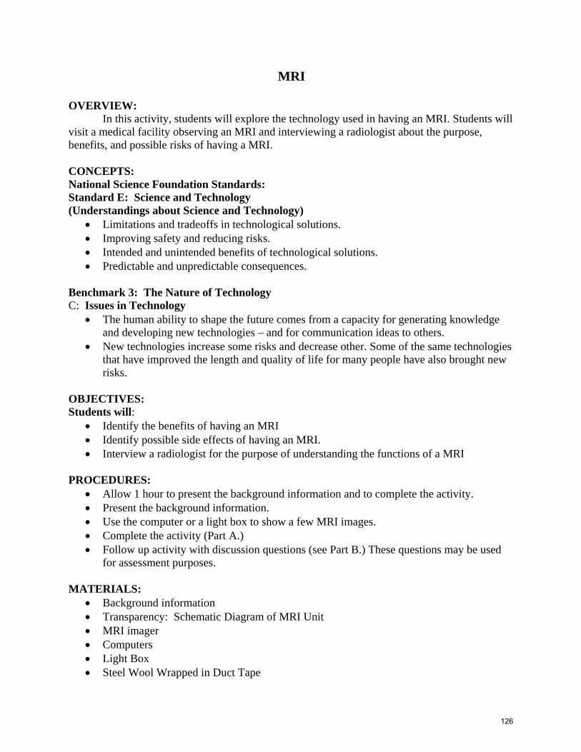
BACKGROUND: We have often heard the term MRI used especially in hospitals. The letters MRI stand for Magnetic Resonance Imaging. MRI is a technique in radiology which uses magnetism, radio waves and a computer to produce images of body structures. MRI scans are used as an extremely accurate method of detecting disease throughout the body. Head trauma, Brain Aneurysms, Stroke, Tumors of the brain, and Spine tumors, are all abnormalities that are detected by a MRI scan. The Imager is a long tubular hole which is surrounded by a giant circular magnet. To get an idea on this, here is a sketch:
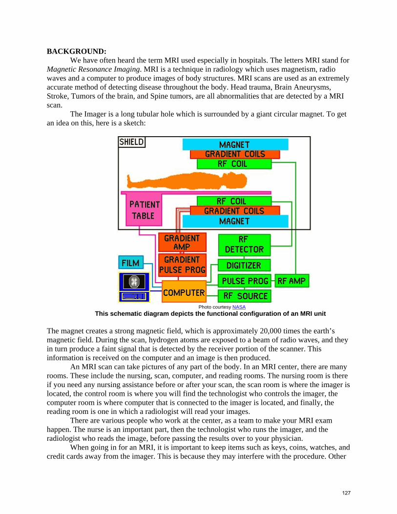
Photo courtesy NASA
This schematic diagram depicts the functional configuration of an MRI unit The magnet creates a strong magnetic field, which is approximately 20,000 times the earth’s magnetic field. During the scan, hydrogen atoms are exposed to a beam of radio waves, and they in turn produce a faint signal that is detected by the receiver portion of the scanner. This information is received on the computer and an image is then produced. An MRI scan can take pictures of any part of the body. In an MRI center, there are many rooms. These include the nursing, scan, computer, and reading rooms. The nursing room is there if you need any nursing assistance before or after your scan, the scan room is where the imager is located, the control room is where you will find the technologist who controls the imager, the computer room is where computer that is connected to the imager is located, and finally, the reading room is one in which a radiologist will read your images. There are various people who work at the center, as a team to make your MRI exam happen. The nurse is an important part, then the technologist who runs the imager, and the radiologist who reads the image, before passing the results over to your physician. When going in for an MRI, it is important to keep items such as keys, coins, watches, and credit cards away from the imager. This is because they may interfere with the procedure. Other
127

interferences may be caused by any objects placed inside your body, like metal chips, or certain medical conditions. People with pacemakers cannot go through with an MRI scan, because the pacemaker may cease functioning due to the magnetic field, in the process of the scan. An MRI exam ranges from half an hour to one and a half hours to be completed. Once in the imager, one is expected to lie still for periods of 3 to 10 minutes at a time. In cases of claustrophobia, a mild sedative can be given to help alleviate the feeling. The physician requesting the scan and the radiology staff should be notified in such cases. It is also recommended that one wear ear protections because of the sound produced by the imager. MRI scans are painless and avoid x-ray radiation exposure. There are no known side effects and the benefits relate to an MRI’s precise accuracy in detecting structural abnormalities of the body. To get into an MRI career you will need an Associate degree (2 years) or a Bachelor’s Degree (4 years). People in the professions make amounts ranging from $20,000 to $90,000 year, depending on your area of specialty. References: Hornak, Joseph P.; The Basics of MRI, Chapter 13: Your MRI Exam. Copyright 1996-2002. Retrieved from: http://www.cis.rit.edu/htbooks/mri/author.htm, October 6, 2004. http://www.medicinenet.com/script/main/art.asp?articlekey=421&pf=3&track=qpa421 ACTIVITY: Part A:
• Tour a hospital (or other facility) which uses MRI technology. • Arrange for a radiologist to talk about the purpose and risks of having an MRI. • Take the students back to the Image room. • Ask the students to predict what will happen when the imager is turned on and someone
enters holding the steel wool wrapped in duct tape. • Turn on the imager and have them test the magnetic field by holding the steel wool
wrapped in duct tape by the entrance of the imager. Part B: Ask the following questions and allow time for discussion. Share: For what reasons would an individual need to have a MRI scan taken? Process: What role does magnetism play in this type of imaging? Generalize: Why is it important to let the technician doing the MRI know your medical history prior to having the scan taken? Apply: Could an individual with a heart pacemaker or an artificial limb have an MRI? Explain
128

This schematic diagram depicts the functional configuration of an MRI unit
129