MRI preprocessing and segmentation. Bias References.
39
MRI preprocessing and segmentation
Transcript of MRI preprocessing and segmentation. Bias References.
MRI preprocessing and
segmentation

Bias References
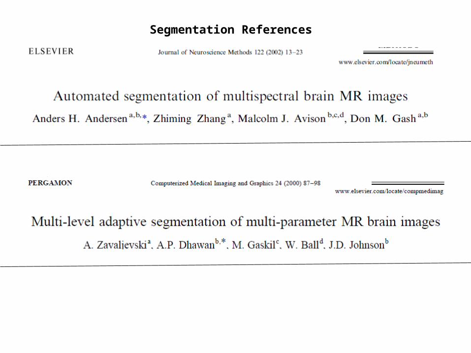
Segmentation References
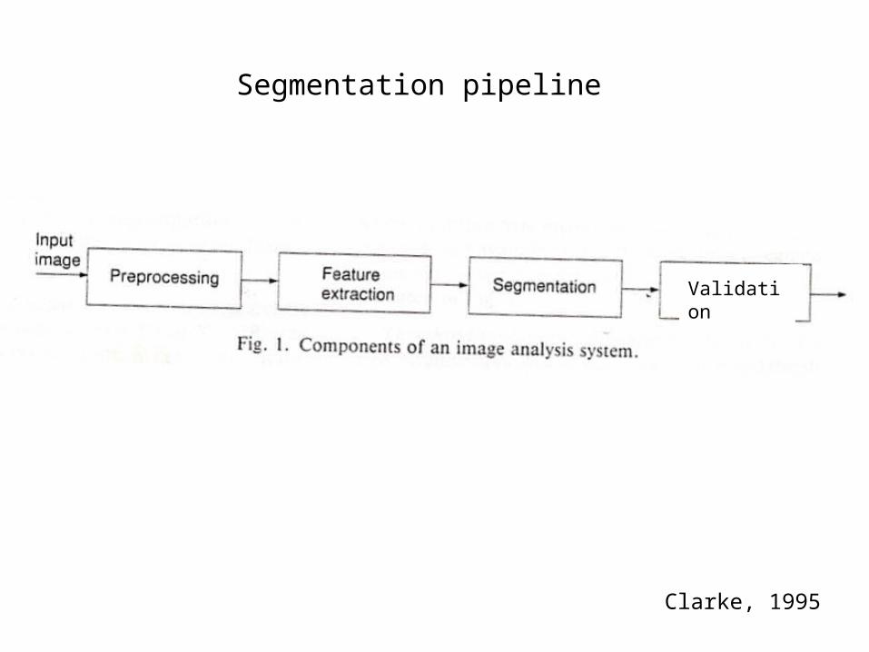
Segmentation pipeline
Clarke, 1995
Validation
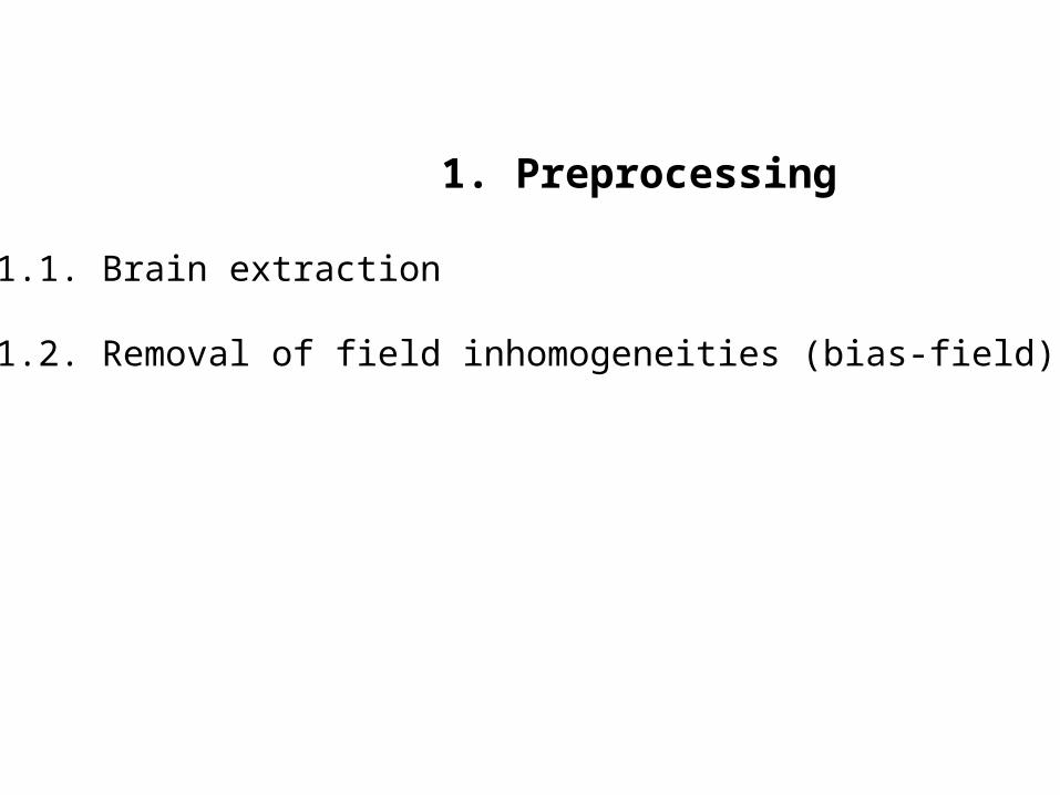
1. Preprocessing
1.1. Brain extraction
1.2. Removal of field inhomogeneities (bias-field)

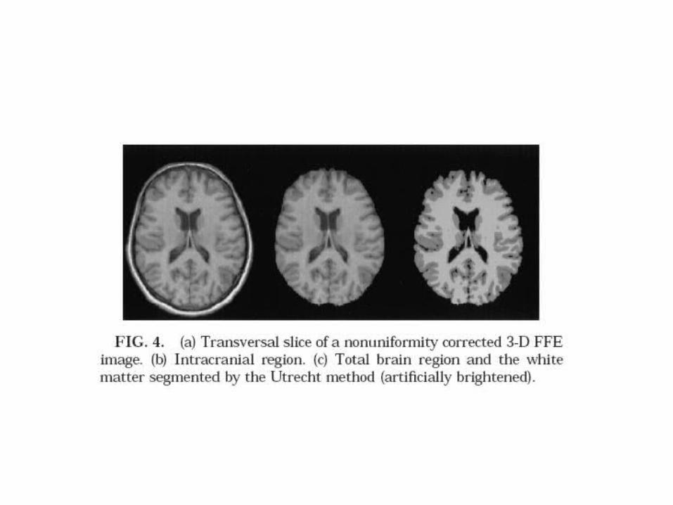
1.1. Brain extraction
MRI of head Intracranial volume Extracted brain

1.1. Brain extraction
FSL: Initiate a mesh inside the skull and expand-wrap onto brain surface
Huh, 2002 method: go to mid sagittal, find brain, copy mask on adjacent slicescorrect the copied mask

1.1. Brain extraction
initial mask adjacent slice j mask of slice j
challengeHuh, 2002
1.1. Brain extraction
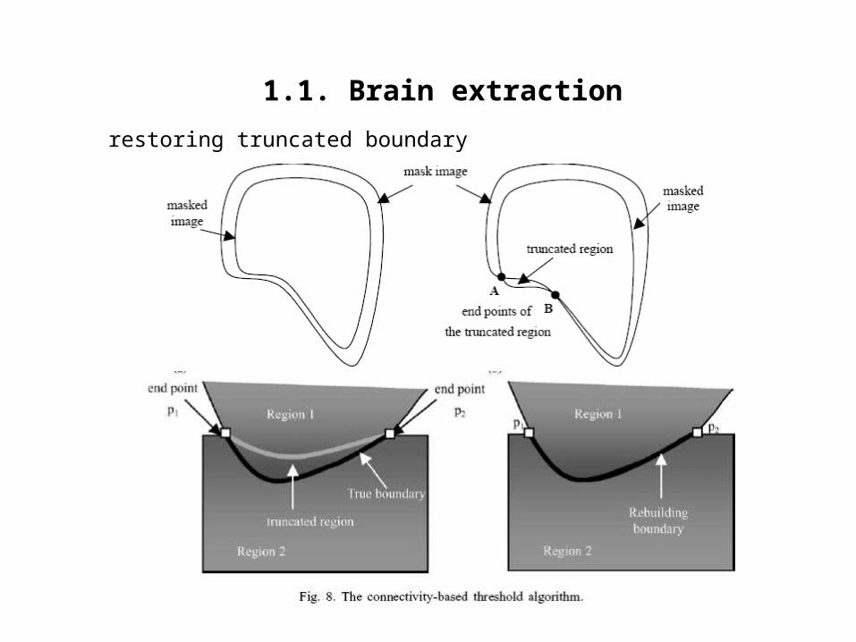
restoring truncated boundary
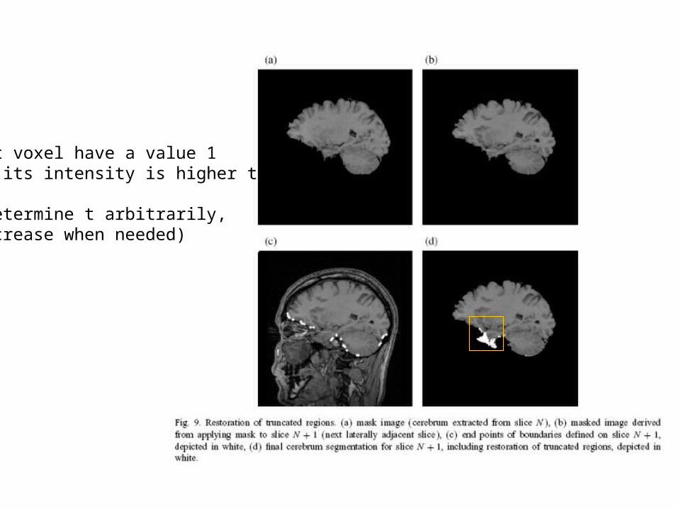
Let voxel have a value 1 if its intensity is higher than t
(determine t arbitrarily,increase when needed)
1.2. Removal of field inhomogeneities
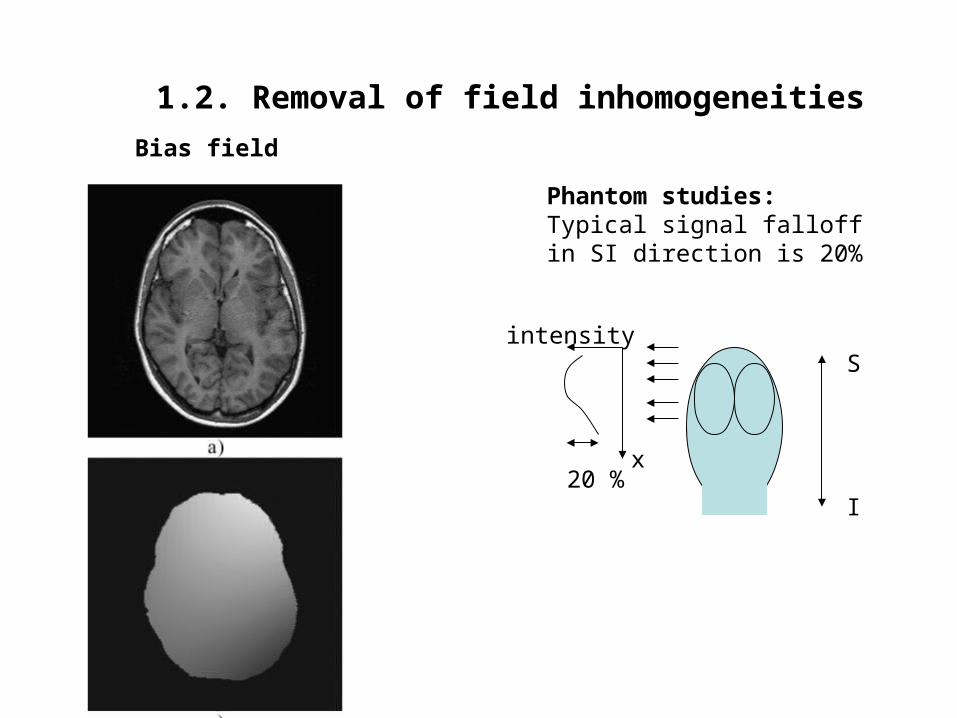
Bias field
Phantom studies:Typical signal falloff in SI direction is 20%
S
I20 %
x
intensity

1.2. Removal of field inhomogeneities
Statistical methods: probabilistic, gaussian and mixture models of bias-field
Polynomial methods: smooth polynomial fit to bias-field
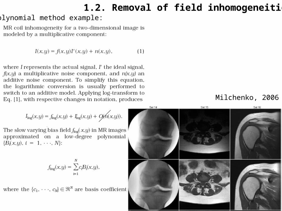
1.2. Removal of field inhomogeneitiesPolynomial method example:
Milchenko, 2006
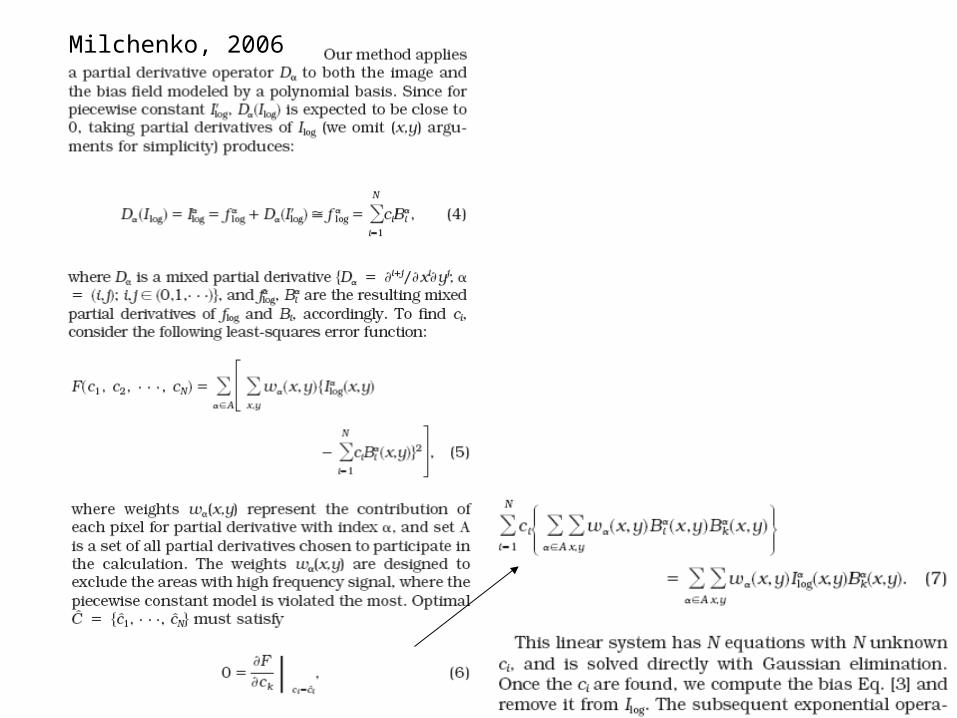
Milchenko, 2006
1.2. Removal of field inhomogeneities
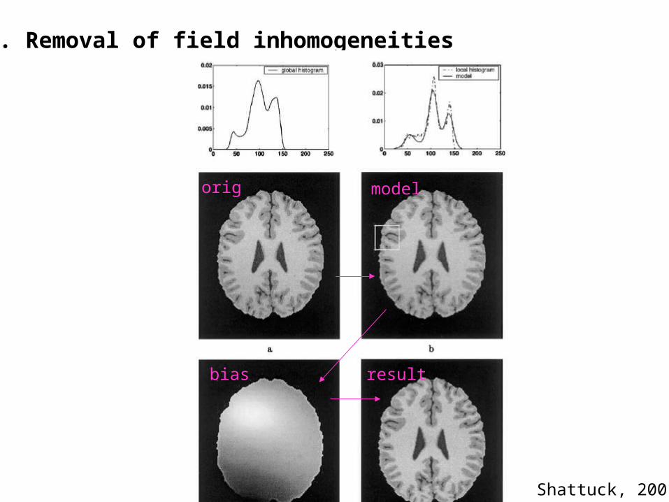
Shattuck, 2001
orig model
bias result
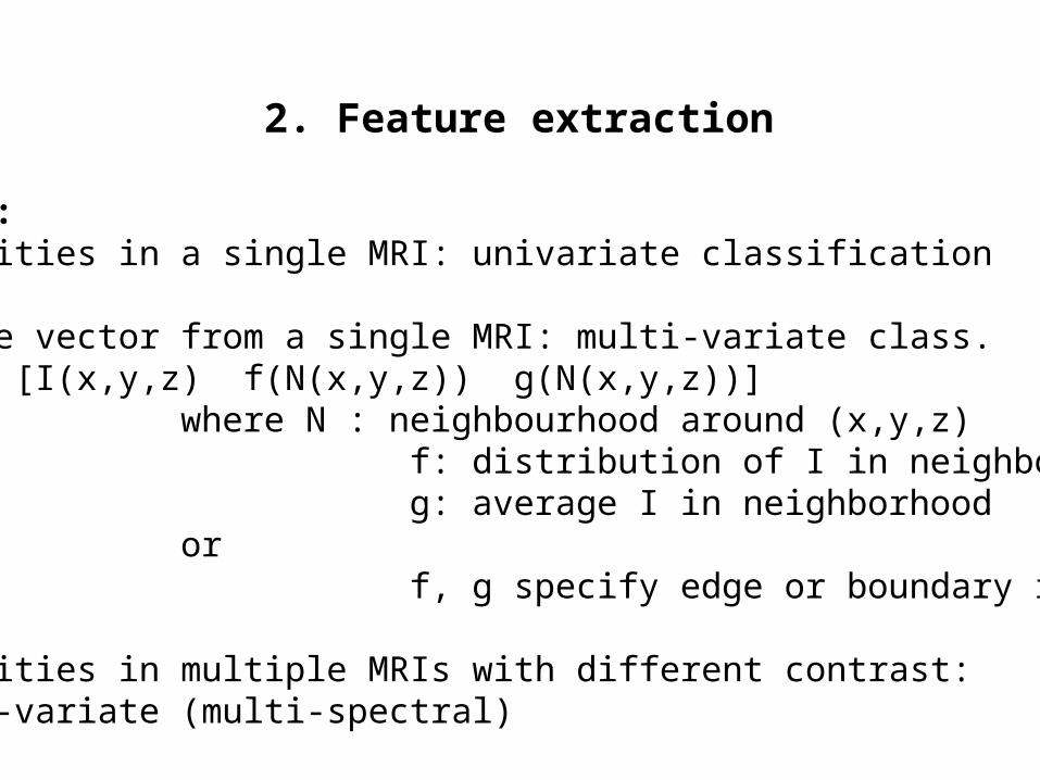
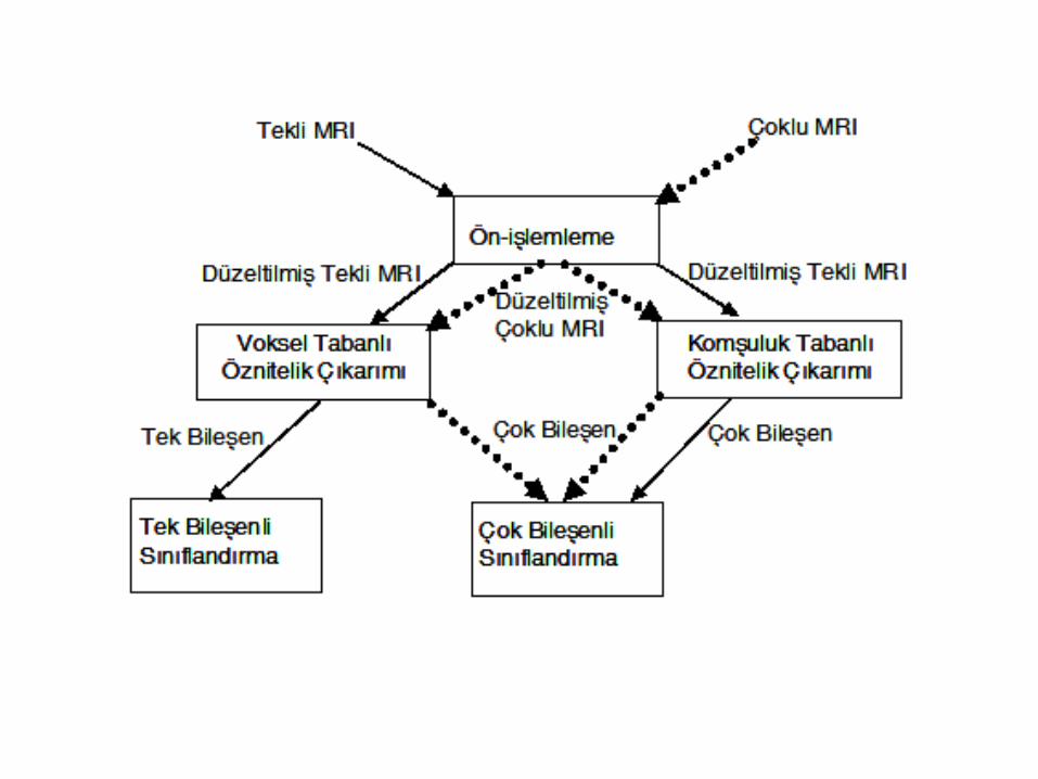
2. Feature extraction
Features:- Intensities in a single MRI: univariate classification
- Feature vector from a single MRI: multi-variate class.ex: [I(x,y,z) f(N(x,y,z)) g(N(x,y,z))]
where N : neighbourhood around (x,y,z) f: distribution of I in neighborhood (entropy) g: average I in neighborhood or f, g specify edge or boundary information
- Intensities in multiple MRIs with different contrast: multi-variate (multi-spectral)
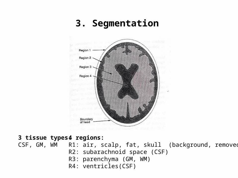
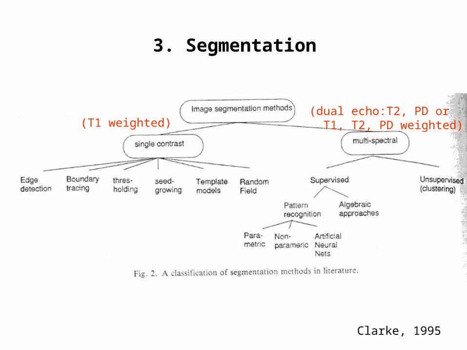
3. Segmentation
4 regions:R1: air, scalp, fat, skull (background, removed)R2: subarachnoid space (CSF)R3: parenchyma (GM, WM)R4: ventricles(CSF)
3 tissue types:CSF, GM, WM
3. Segmentation
Clarke, 1995
(T1 weighted)(dual echo:T2, PD or T1, T2, PD weighted)
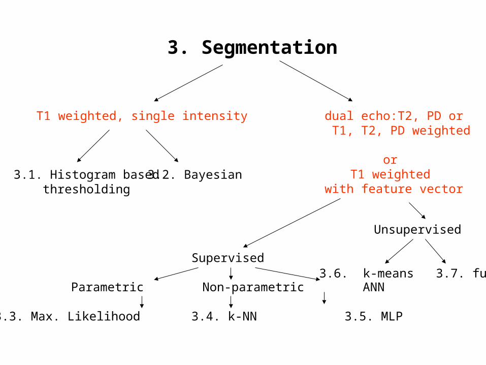
3. Segmentation
T1 weighted, single intensity dual echo:T2, PD or T1, T2, PD weighted
or T1 weighted
with feature vector3.1. Histogram based
thresholding
Unsupervised
3.6. k-means 3.7. fuzzy cmeans
Supervised
Parametric Non-parametric ANN
3.3. Max. Likelihood 3.4. k-NN 3.5. MLP
3.2. Bayesian
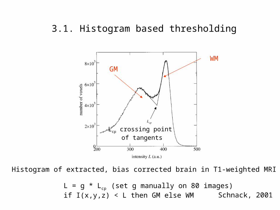
3.1. Histogram based thresholding
Schnack, 2001
WM
GM
Histogram of extracted, bias corrected brain in T1-weighted MRI
Lcp crossing point of tangents
L = g * Lcp (set g manually on 80 images)if I(x,y,z) < L then GM else WM
Population1
Population2
Population3
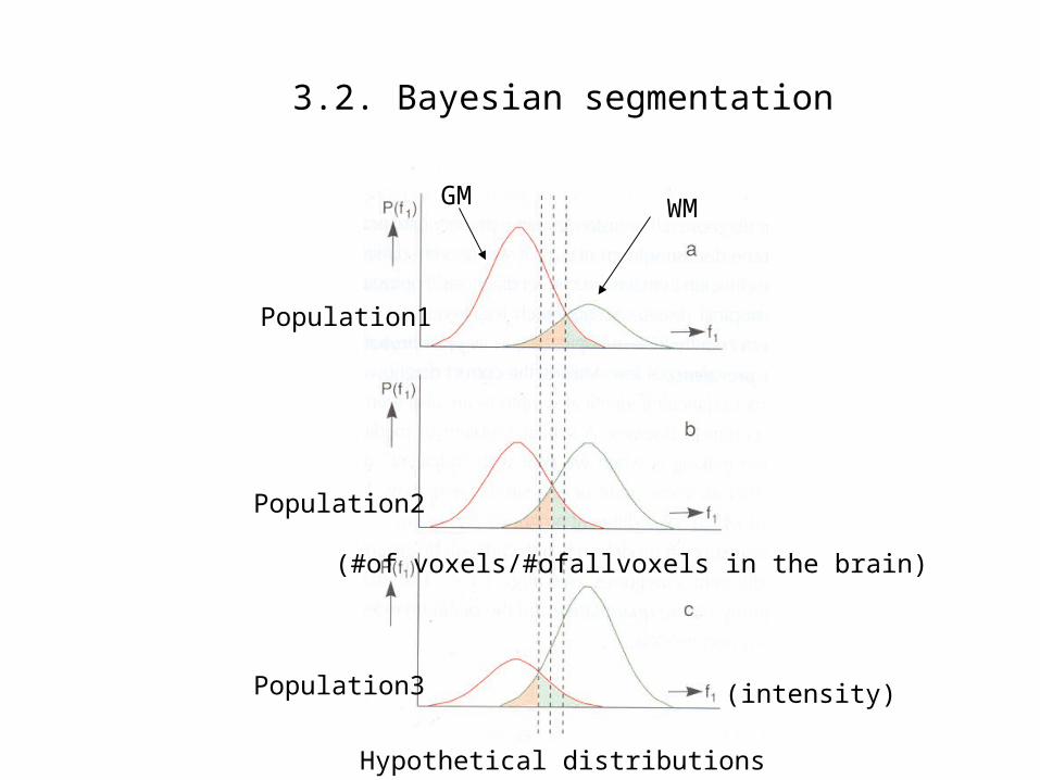
3.2. Bayesian segmentation
WMGM
Hypothetical distributions
(intensity)
(#of voxels/#ofallvoxels in the brain)

3.2. Bayes’ classifier
For each voxel, x,y,z:Assume K tissue types (for eg. T1, T2, ..., Tk) possible, for 1 observed intensity, I:
P(Tj ! I) = P(I ! Tj) . P(Tj)
Ξ P(I ! Tk). P(Tk) k
GM, WM, CSF ratiosfrom volumetric studies
setup graphs above from regional data
Decide on tissue type m if: P(Tm ! I) > P(Tj ! I) for all j
Kovacevic, 2002
J,k=1,2,3:1: CSF, 2: GM, 3:WM

Methods based on feature vector or multi-spectral data
Supervised vs unsupervised Methods
Supervised: - Color indicates known classes - Separation contour is to be found during training phase- Separation contour is used for classification during recall phase
Unsupervised: - No color, classes unknown- Clusters are found during training phase- Association with clusters are made during recall phase
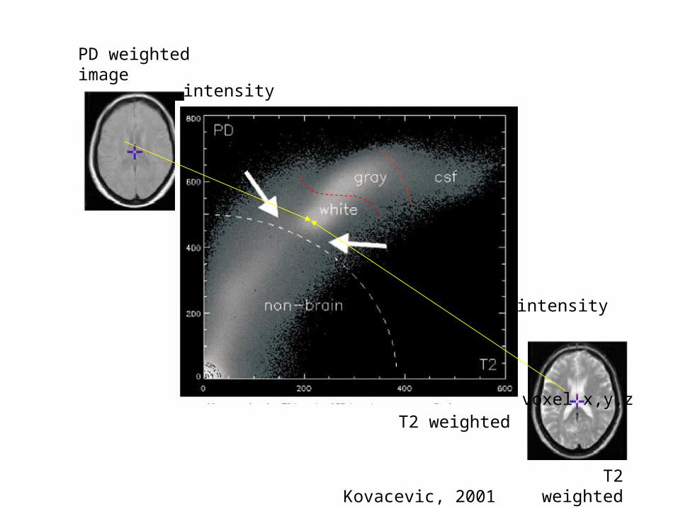
Kovacevic, 2001
T2 weightedvoxel x,y,z
PD weightedimage
T2 weightedimage
intensity
intensity

Suckling, 1999

3.3. Maximum likelihood classifier
- Assume the distribution P(I ! Tj) in Bayes can be obtained by a mixture of Gaussian or Normal distribution- Estimate means and co-variance matrix- For better results use Hidden Markov fields within neighborhoods
Zavaljevski, 2000
15 classes
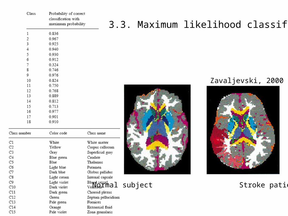
3.3. Maximum likelihood classifier
Zavaljevski, 2000
Normal subject Stroke patient

3.4. K-NN, K-Nearest neighbor classifier
T1 intensity
T2 intensity
Hypothetical distribution
- k is always odd, 1<k<15 (as k increases comput time increases)- given a point p find k closest samples known from before- decide on class m where m is the highest number of classes among these k samples
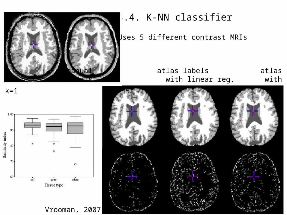
3.4. K-NN classifier
k=1 k=45
manual atlas labels atlas labels labels with linear reg. with non-lin reg.
Vrooman, 2007
Uses 5 different contrast MRIs
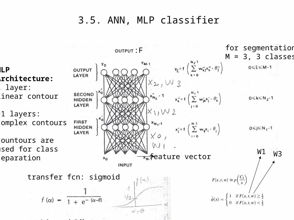
MLPArchitecture:1 layer: linear contour
>1 layers: complex contours
countours areused for classseparation
transfer fcn: sigmoid
W1 W3
:F
3.5. ANN, MLP classifier
for segmentation,M = 3, 3 classes
feature vector
3.5. ANN, MLP classifier
Results
This page is empty on purpose

3.6. k-means classifier
Algorithm:- k is equal to number of classes- choose k arbitrary initial seed points (*)- assume seed points are class centroids1 for each sample point j, find distance to all k centroids Let j belong to class m if j is closest to centroid m2 for each class k, recalculate centroids
repeat steps 1 and 2 above until no change in centroids
Note how class assignments changeat each iteration
Minimized measure:
This classifier is not used much in segmentation, but explained here as an introduction to fuzzy c-means
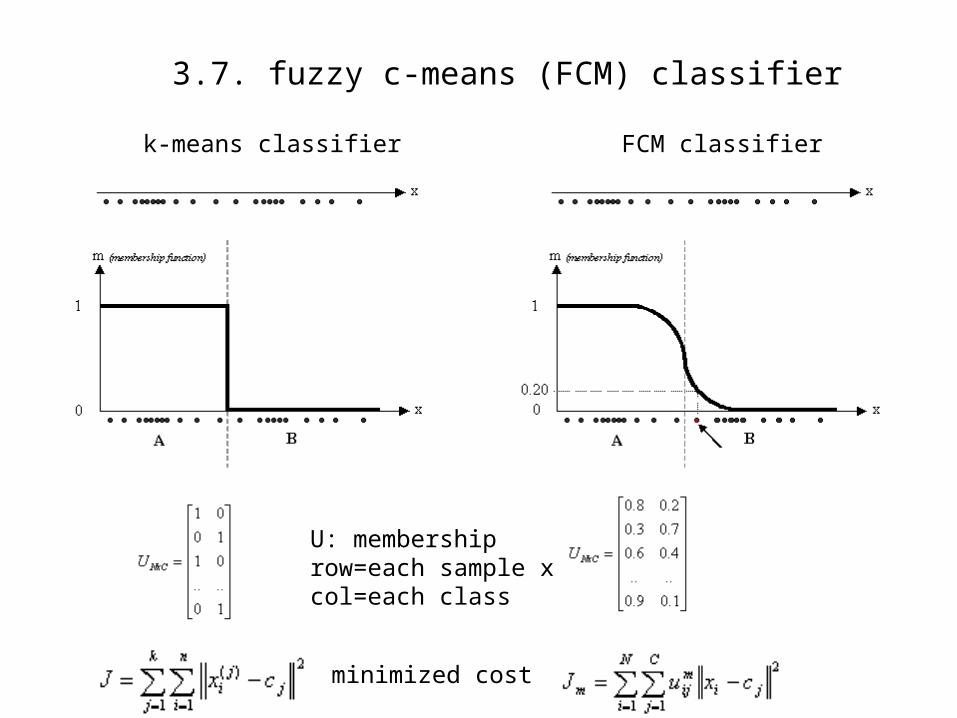
3.7. fuzzy c-means (FCM) classifier
k-means classifier FCM classifier
U: membership row=each sample xcol=each class
minimized cost
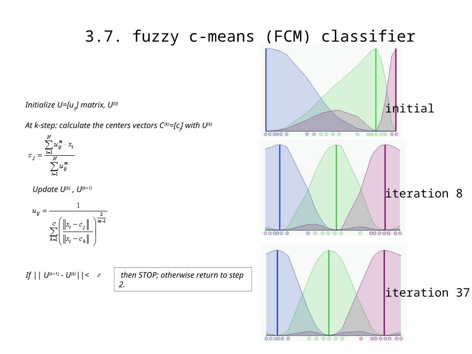
3.7. fuzzy c-means (FCM) classifier
initial
iteration 8
iteration 37
Initialize U=[uij] matrix, U(0)
At k-step: calculate the centers vectors C(k)=[cj] with U(k)
Update U(k) , U(k+1)
If || U(k+1) - U(k)||< then STOP; otherwise return to step 2.

3.7. fuzzy c-means classifier
Results
4. Validation
Important issues:
- Partial volume effect, visualization
- Validation in manually segmented image
- Performance comparison with other methods on simulated image: Ex: Brainweb from Mcgill
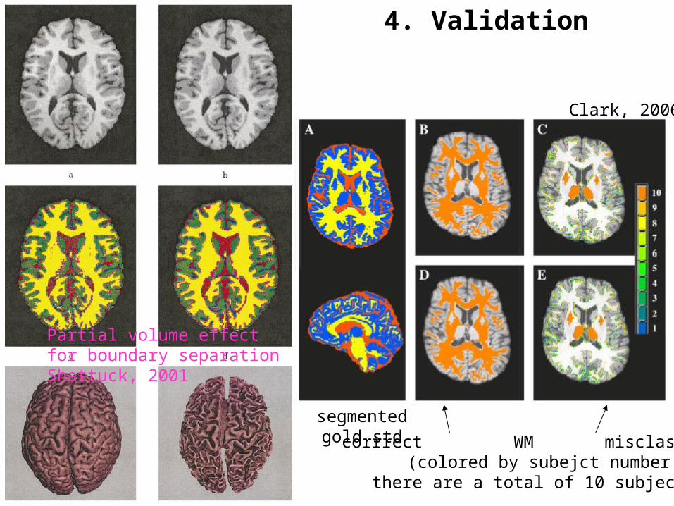
4. Validation
Partial volume effectfor boundary separationShattuck, 2001
corrrect WM misclassified(colored by subejct number
there are a total of 10 subjects)
segmentedgold std
Clark, 2006