MRI Interpretation for Dementia -ANATOMY
52
Sharon Best, PA-C, MHS MRI Interpretation for Dementia - ANATOMY APP2APP Virtual Lectures, Inc https://app2app.org/
Transcript of MRI Interpretation for Dementia -ANATOMY

Sharon Best, PA-C, MHS
MRI Interpretation for Dementia - ANATOMY
APP2APP Virtual Lectures, Inc
https://app2app.org/

Objectives
1. Imaging Planes 2. T1 vs T2 vs FLAIR3. MRI Anatomy Pertinent for Dementia
• Medial Temporal Lobe structures and atrophy• Parietal atrophy - Koedam scale • Frontotemporal atrophy • Small Vessel Ischemic Disease - Fazekas scale• Microangiopathy: microhemorrhages and superficial siderosis
APP2APP Virtual Lectures, Inc

Imaging Planes
APP2APP Virtual Lectures, Inc
Imaging Planes
image ref: http://www.radtechonduty.com/2017/03/radiography-imaging-planes.html
APP2APP Virtual Lectures, Inc

Imaging Planes
https://www.webmd.com/brain/picture-of-the-brain#1
https://online.king.edu/infogra
phics/parts-of-the-brain/
Note: Sylvian fissureCentral sulcusPons and Poles
https://www.imaios.com/en/e-Anatomy/Head-and-Neck/Brain-MRI-3D
IAMOS practice

Imaging Planes
Sagittal Axial Coronal
Cingulate gyrus

T1 vs T2 vs FLAIR
APP2APP Virtual Lectures, Inc

Gray Matter (peripheral cortex and deep brain nuclei) vs White Matter (myelinated axons- subcortical regions)
https://antranik.org/cerebral-white-matter-and-gray-matter-and-basal-ganglia/ https://www.78stepshealth.us/human-physiology/descending-tracts.html
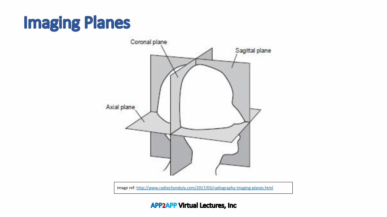
T1 vs T2
T1 Imaging: • Gray matter is dark• White matter is lighter• CSF is darkest
T2 Imaging: • Gray matter is light• White matter is darker• CSF is lightest
T1 MRI T2 MRI
Image REF: http://casemed.case.edu/clerkships/neurology/web%20neurorad/mri%20basics.htm
APP2APP Virtual Lectures, Inc
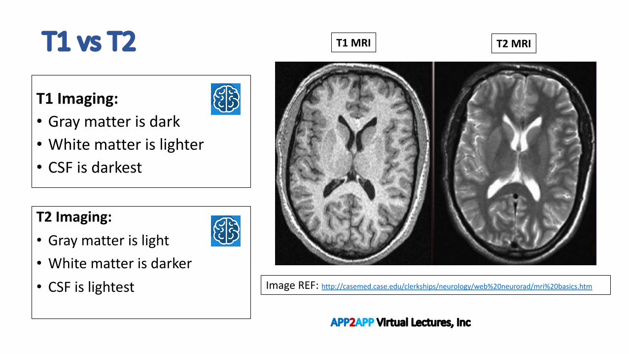
FLAIR- Fluid Attenuated Inversion Recovery Sequences
• A T2 image with “fluid attenuated” (ie: the fluid signal is suppressed) • Very sensitive to pathology• Think! Patients’ permanent record • RECALL: T2 gray matter is light, white matter
is darker, CSF is lightest. • HOWEVER, in a FLAIR image: • The CSF is attenuated (suppressed) • Therefore, gray matter is light, white matter is
darker, CSF if black.
https://www.imaios.com/en/e-Anatomy/Head-and-Neck/Brain-MRI-3D
MRI Anatomy Pertinent for Dementia
APP2APP Virtual Lectures, Inc

https://www.imaios.com/en/e-Anatomy/Head-and-Neck/Brain-MRI-3D
Important Gyri and Sulci and Poles of the Cortex
Postcenteral gyrus
Temporal pole
Occipital pole(Sylvian Fissure)
Inferior orbital frontal gyri

Important Motor and Sensory Regions of the Cortex
https://owlcation.com/stem/Exploring-the-Brain-Three-Regions-Named-after-Scientists
APP2APP Virtual Lectures, Inc

Brain Stem- Midline- Sagittal View
https://radiopaedia.org/cases/normal-brain-mri-5
Brain Stem:
A. Medulla oblongata
B. Pons
C. midbrain (cerebral crus)
https://www.pinterest.com/pin/744149538403526897/

Brain Stem Axial ViewBrain Stem (inferior à superior) A. Medulla oblongataB. PonsC. Midbrain (note: cerebral crus)CerebellumTemporal lobesParietal lobes:
- At the level of the medulla, look at the cerebellum.
- At the level of the Pons, look at the Poles (temporal and occipital).
- At the level of the midbrain, look at hippocampi and the temporal horns.
B
C
A
A
B
C
Image Ref:1. https://testmyprep.com/subject/economy/new-and-emerging-theories-of-
international-trade2. 2. http://w-radiology.com/atlas_brain_mri.php
A medulla
B pons
C midbrain
BG and R/L Thalami
*always scroll from base of the brain superiorly

Brain Stem – Coronal View A
B
C
Cerebral Peduncle: the most anterior portion of the midbrain; contains large ascending and descending white matter tracts.
Middle Cerebellar Peduncles: connects the cerebellum to the pons; comprised of whitematter tracks arising from the pontine nuclei and projecting to the contralateral cerebellar cortex.
Inferior Olives: the largest paired nuclei (gray matter) in the olivary bodies found in the medulla oblongata. The primary source of motor nuclei projecting to the cerebellum.
Pyramids: paired white matter structures in the medulla oblongata, contain groups of descending motor fibers collectively known as pyramidal tracts. The fibers cross at the lower level of the medulla. Image Ref: https://www.imaios.com/en/e-Anatomy/Head-and-Neck/Brain-MRI-3D
IAMOS practice

Thalami and Basal Ganglia • R/L Thalamus- chiefly gray matter structures that will
radiate to much of the cortex and basal ganglia. • Basal ganglia: deep brain nuclei; intimately connected
w/ brain stem, thalamus, and cerebral cortex. We will identify three BG on MRI: • 1) Globus pallidus• 2) Putamen• 3) Caudate nucleus
Image Ref:1. https://en.wikipedia.org/wiki/Basal_ganglia2. https://www.imaios.com/en/e-Anatomy/Head-and-Neck/Brain-MRI-3D
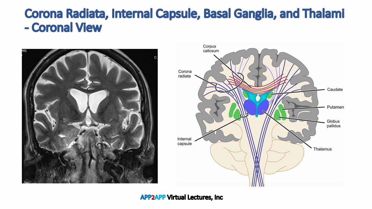
Corona Radiata, Internal Capsule, Basal Ganglia, and Thalami- Coronal View
APP2APP Virtual Lectures, Inc
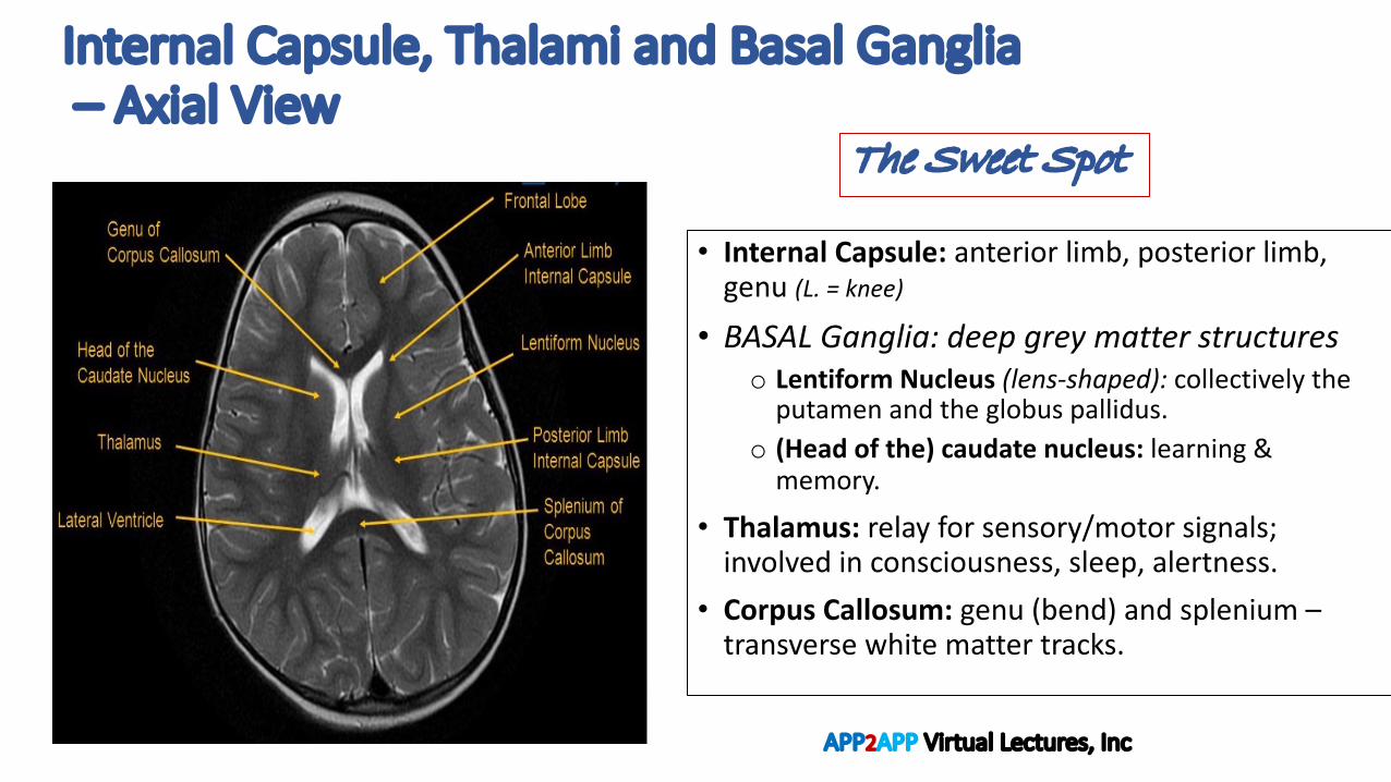
Internal Capsule, Thalami and Basal Ganglia – Axial View
• Internal Capsule: anterior limb, posterior limb, genu (L. = knee)
• BASAL Ganglia: deep grey matter structures o Lentiform Nucleus (lens-shaped): collectively the
putamen and the globus pallidus.o (Head of the) caudate nucleus: learning &
memory.
• Thalamus: relay for sensory/motor signals; involved in consciousness, sleep, alertness.
• Corpus Callosum: genu (bend) and splenium –transverse white matter tracks.
The Sweet Spot
APP2APP Virtual Lectures, Inc
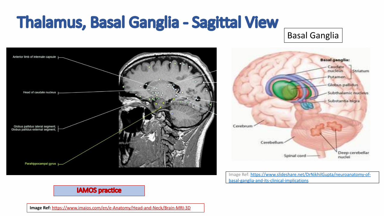
Thalamus, Basal Ganglia - Sagittal View
Image Ref: https://www.slideshare.net/DrNikhilGupta/neuroanatomy-of-basal-ganglia-and-its-clinical-implications
Basal Ganglia
Image Ref: https://www.imaios.com/en/e-Anatomy/Head-and-Neck/Brain-MRI-3D
IAMOS practice
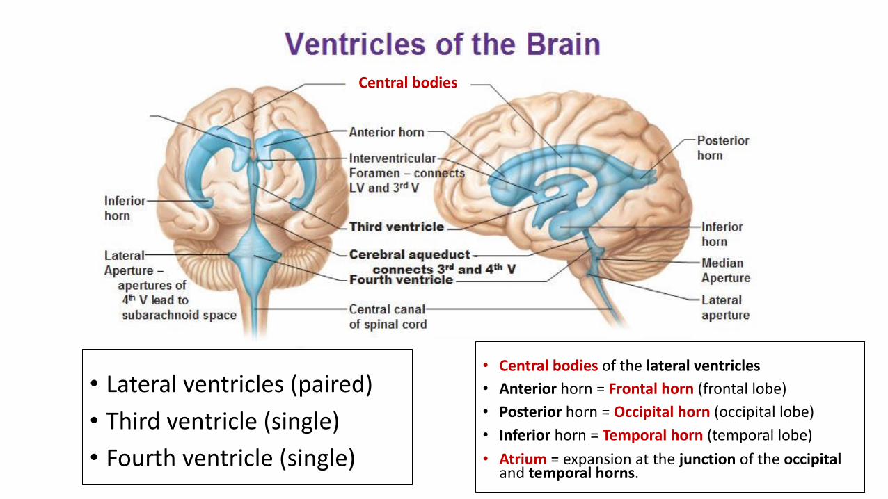
• Central bodies of the lateral ventricles • Anterior horn = Frontal horn (frontal lobe) • Posterior horn = Occipital horn (occipital lobe) • Inferior horn = Temporal horn (temporal lobe) • Atrium = expansion at the junction of the occipital
and temporal horns.
Central bodies
• Lateral ventricles (paired)• Third ventricle (single)• Fourth ventricle (single)

Choroid Plexus & the Choroid Fissure
Image Ref: https://www.researchgate.net/figure/A-The-scheme-of-the-cerebrospinal-fluid-system-with-location-of-the-choroid-plexuses_fig8_316520924
The choroid plexus is a network of specialized cells located in the cerebral ventricles that produces the CSF.
The choroid fissure: a cleft that forms in the medial wall of the lateral ventricle
APP2APP Virtual Lectures, Inc

Fourth Ventricle
Tegmentum of pons (dorsal) Basilar pons- most
anterior portion (ventral)
Basilar pons – most anterior portion (ventral)
Tegmentum of pons (dorsal)
Fourth ventricle
Fourth ventricle

Hippocampus, Temporal horn, Thalamus, BG, Collateral sulcus, and Calcarine fissure
Image REF: https://www.imaios.com/en/e-Anatomy/Head-and-Neck/Brain-MRI-3D
Temporal horn of the lateral ventricles Globus pallidus (BG) superimposed over Thalamus (superior to midbrain)
Collateral sulcus
Calcarine fissure
Orbital frontal gyri

Third Ventricle
APP2APP Virtual Lectures, Inc

Image REF: https://www.imaios.com/en/e-Anatomy/Head-and-Neck/Brain-MRI-3D
Central sulcus
Lateral Ventricles- frontal horn- central body- note: choroid plexus within Frontal Pole - Most anterior portion of frontal lobeInsular sulci - separates insular lobe from temporal lobeCentral Sulcus - separates frontal lobe from parietal lobeLateral Sulcus (ie: Sylvian Fissure)- separates frontal and parietal lobes from
temporal lobe Cingulate gyrus: - The posterior portion is labeled, which is typically atrophied in Alzheimer’s Disease (A.D.) but preserved in DLB (ie: “Cingulate Island Sign”). Intraparietal sulcus- sulci of parietal lobe- atrophied in A.D.Parietooccipital sulcus - separates parietal lobe and occipital lobe
Frontal pole
Insular sulcus
Intraparietal sulci
Cingulate gyrus
Lateral Ventricles, Sulci, and Lobes
IAMOS practice
Parietal Atrophy
APP2APP Virtual Lectures, Inc

Parietal Atrophy
The Koedam scale • Used to evaluate parietal atrophy.• Widening of the parieto-occipital
and posterior cingulate sulci.• The precuneus is a portion of the
superior parietal lobule on the medial surface of the brain.
• MCI pts may have parietal atrophy prior to MTA (particularly in the precuneus).
REF: http://www.radiologyassistant.nl/en/p43dbf6d16f98d/dementia-role-of-mri.html
Parenchyma Sulci
Grade 0 No parietal atrophy
Closed sulci of parietal lobes and precuneus
Grade 1 Mild parietal atrophy
Mild widening of posterior cingulate and parieto-occipital sulci
Grade 2 Substantial parietal atrophy
Substantial widening of posterior cingulate and parieto-occipital sulci
Grade 3 End-stage “knife-blade” atrophy
Extreme widening of the posterior cingulate and parieto-occipital sulci
Parieto-occipital sulcusPosterior cingulate sulcus Parieto-occipital sulcus
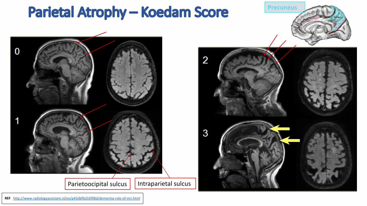
Parietal Atrophy – Koedam Score
REF: http://www.radiologyassistant.nl/en/p43dbf6d16f98d/dementia-role-of-mri.html
Intraparietal sulcusParietoocipital sulcus
Precuneus
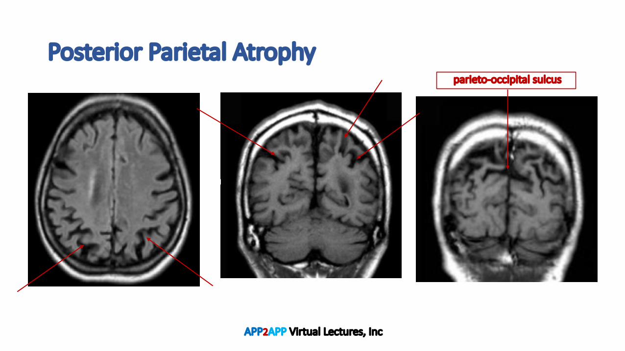
Posterior Parietal Atrophy
APP2APP Virtual Lectures, Inc
parieto-occipital sulcus

Medical Temporal Lobe
1.Cingulate gyrus2.Hippocampal formation
CA1-CA4, dentate gyrus, and subiculum
3.Parahippocampal gyrus
APP2APP Virtual Lectures, Inc
Cingulate gyrus

MRI Hippocampus - Axial View
Hippocampus:1 = head is located anterior to the mesencephalon (mid-brain)2 = body is at the level of the mesencephalon 3 = tail is posterior to the mesencephalon4 = midbrain (note the cerebral crus)
Image Ref: Dekeyzer, et al. 2017
Image: :https://qbi.uq.edu.au/brain-basics/memory/where-are-memories-stored
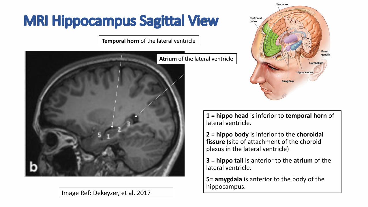
MRI Hippocampus Sagittal View
1 = hippo head is inferior to temporal horn of lateral ventricle.
2 = hippo body is inferior to the choroidal fissure (site of attachment of the choroid plexus in the lateral ventricle)
3 = hippo tail Is anterior to the atrium of the lateral ventricle.
5= amygdala is anterior to the body of the hippocampus.
Image Ref: Dekeyzer, et al. 2017
Temporal horn of the lateral ventricle
Atrium of the lateral ventricle
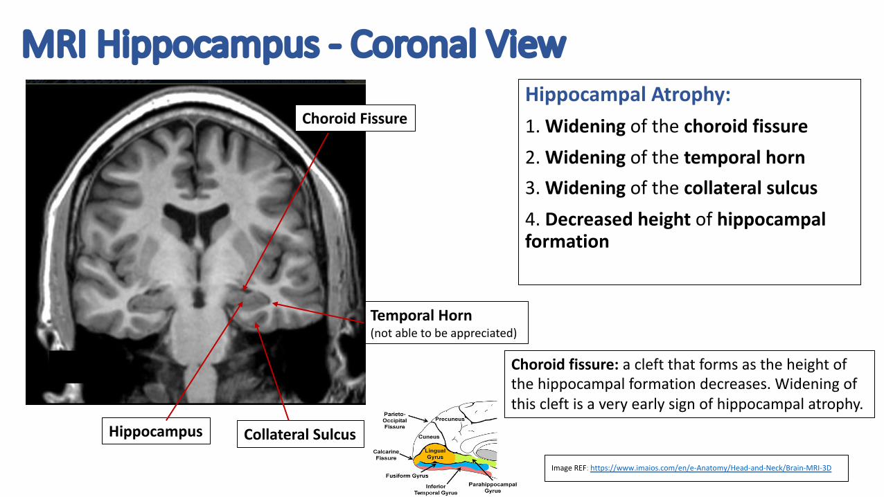
MRI Hippocampus - Coronal View
Hippocampus Collateral Sulcus
Temporal Horn (not able to be appreciated)
Hippocampal Atrophy: 1. Widening of the choroid fissure2. Widening of the temporal horn3. Widening of the collateral sulcus 4. Decreased height of hippocampal formation
Choroid Fissure
Choroid fissure: a cleft that forms as the height of the hippocampal formation decreases. Widening of this cleft is a very early sign of hippocampal atrophy.
Image REF: https://www.imaios.com/en/e-Anatomy/Head-and-Neck/Brain-MRI-3D

Medial Temporal Lobe Atrophy
APP2APP Virtual Lectures, Inc

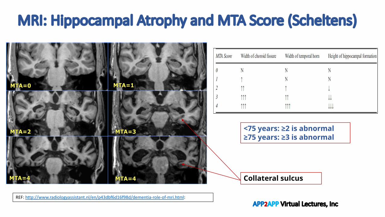
Hippocampal Atrophy and MTA-Score (Scheltens)
MTA-score will be best assessed via Coronal view1. Width of choroid fissure2. Width of temporal horn3. Height of hippocampal formation
Image REF: https://www.imaios.com/en/e-Cases/Channels/Radiology/Radiological-classifications-commonly-used-in-medical-imaging/MTA-scale-for-Medial-Temporal-lobe-Atrophy-Scheltens
MRI: Hippocampal Atrophy and MTA Score (Scheltens)
<75 years: ≥2 is abnormal≥75 years: ≥3 is abnormal
REF: http://www.radiologyassistant.nl/en/p43dbf6d16f98d/dementia-role-of-mri.html:
Collateral sulcus
APP2APP Virtual Lectures, Inc
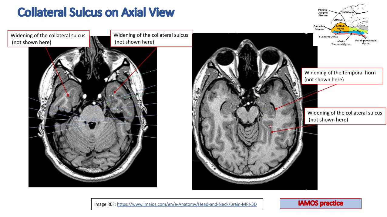
Collateral Sulcus on Axial View
Image REF: https://www.imaios.com/en/e-Anatomy/Head-and-Neck/Brain-MRI-3D
Widening of the collateral sulcus(not shown here)
Widening of the temporal horn (not shown here)
Widening of the collateral sulcus(not shown here)
Widening of the collateral sulcus(not shown here)
IAMOS practice

Frontotemporal Atrophy
APP2APP Virtual Lectures, Inc

Frontotemporal Atrophy
Ref: https://radiopaedia.org/cases/behavioral-variant-frontotemporal-dementia-1?lang=us
Image A/B:• Axial T2/Axial FLAIR with R frontal lobe atrophy.Image C: • Coronal T2 also showing the R frontal lobe
atrophy.Image D: • Coronal T2 showing the R frontotemporal
atrophy. • Note the widening of the R Sylvian fissure.• Note the widening of the sulci b/t the
superior/middle and inferior temporal gyri. • Note hippocampal atrophy R>L.
Frontotemporal Atrophy
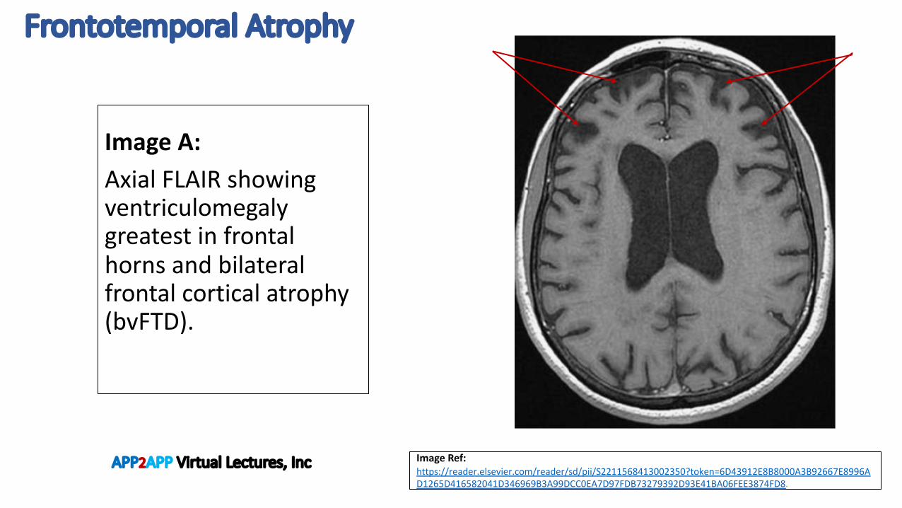
Image Ref: https://reader.elsevier.com/reader/sd/pii/S2211568413002350?token=6D43912E8B8000A3B92667E8996AD1265D416582041D346969B3A99DCC0EA7D97FDB73279392D93E41BA06FEE3874FD8.
Image A: Axial FLAIR showing ventriculomegaly greatest in frontal horns and bilateral frontal cortical atrophy (bvFTD).
APP2APP Virtual Lectures, Inc
Small Vessel Ischemic Disease
APP2APP Virtual Lectures, Inc

Small Vessel Ischemic Disease
Ref: https://www.imaios.com/en/e-Cases/Channels/Radiology/Radiological-classifications-commonly-used-in-medical-imaging/Fazekas-scale-in-ARWMC-scale-on-MRI
Fazekas 1 = normal in elderly pts.Fazekas 2/3 = is considered pathological. May be seen in cognitively normal individuals, but they are considered high risk for cognitive decline.
Periventricular lesions
0 No lesions
1 Caps or thin lines
2 Smooth halos
3 Extends into white matter
White matter lesions
0 No lesions
1 Punctate foci
2 Confluence beginning
3 Large confluent areas
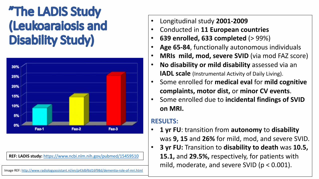
”The LADIS Study (Leukoaraiosis and Disability Study)
Image REF: http://www.radiologyassistant.nl/en/p43dbf6d16f98d/dementia-role-of-mri.html
REF: LADIS study: https://www.ncbi.nlm.nih.gov/pubmed/15459510
• Longitudinal study 2001-2009• Conducted in 11 European countries• 639 enrolled, 633 completed (> 99%)• Age 65-84, functionally autonomous individuals• MRIs mild, mod, severe SVID (via mod FAZ score)• No disability or mild disability assessed via an
IADL scale (Instrumental Activity of Daily Living).• Some enrolled for medical eval for mild cognitive
complaints, motor dist, or minor CV events. • Some enrolled due to incidental findings of SVID
on MRI.
RESULTS: • 1 yr FU: transition from autonomy to disability
was 9, 15 and 26% for mild, mod, and severe SVID.• 3 yr FU: Transition to disability to death was 10.5,
15.1, and 29.5%, respectively, for patients with mild, moderate, and severe SVID (p < 0.001).

Microangiopathy
APP2APP Virtual Lectures, Inc

Microhemorrhages Two types of MR Imaging: 1. SWI: Susceptibility Weighted Imaging2. GRE: Gradient (Recalled) Echo
ImagingImages A, B, C:
• Cerebral amyloid angiopathy (CAA):microhemorrhages in the peripheral cortical distribution, associated with Alzheimer’s disease.
Image D:
• Hypertensive microangiopathy: microhemorrhages in the basal ganglia, pons and cerebellar hemispheres, associated with chronic HTN.
A
DC
B
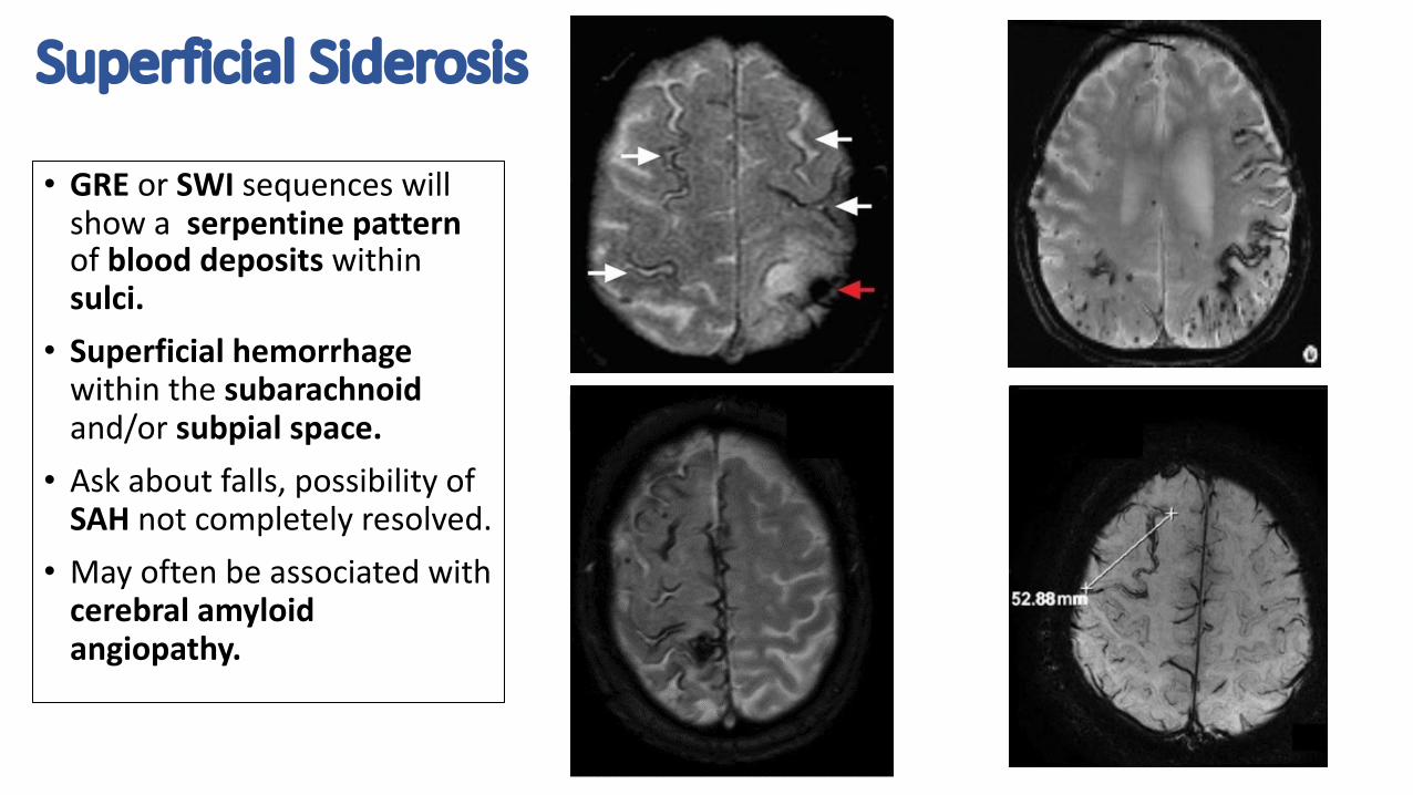
Superficial Siderosis
• GRE or SWI sequences will show a serpentine pattern of blood deposits withinsulci. • Superficial hemorrhage
within the subarachnoidand/or subpial space. • Ask about falls, possibility of
SAH not completely resolved. • May often be associated with
cerebral amyloid angiopathy.
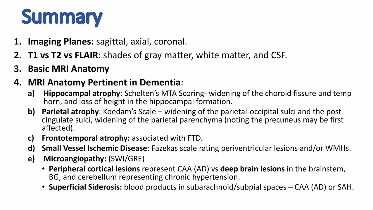
Summary1. Imaging Planes: sagittal, axial, coronal.2. T1 vs T2 vs FLAIR: shades of gray matter, white matter, and CSF. 3. Basic MRI Anatomy4. MRI Anatomy Pertinent in Dementia:
a) Hippocampal atrophy: Schelten’s MTA Scoring- widening of the choroid fissure and temp horn, and loss of height in the hippocampal formation.
b) Parietal atrophy: Koedam’s Scale – widening of the parietal-occipital sulci and the post cingulate sulci, widening of the parietal parenchyma (noting the precuneus may be first affected).
c) Frontotemporal atrophy: associated with FTD. d) Small Vessel Ischemic Disease: Fazekas scale rating periventricular lesions and/or WMHs. e) Microangiopathy: (SWI/GRE)
• Peripheral cortical lesions represent CAA (AD) vs deep brain lesions in the brainstem, BG, and cerebellum representing chronic hypertension.
• Superficial Siderosis: blood products in subarachnoid/subpial spaces – CAA (AD) or SAH.

References1. Dekeyzer, DeKock, et al. “Unforgettable” – a pictorial essay on anatomy and pathology of the
hippocampus. Insights Imaging (2017) 8:122-212.
2. Imaios: Online Radilogy Reference https://www.imaios.com/en/e-Anatomy/Head-and-Neck/Brain-MRI-3D
3. Inzitari, D., et al. LADIS Study Group. “Risk of rapid global functional decline in elderly patients with severe cerebral age-related white matter changes: the LADIS study.” Arch Intern Medicine (2007) Jan 8;167(1):81-8.
4. Korhonen, V. E., Soje, E., et al. “Frontotemporal dementia as a comorbidity to idiopathic normal pressure hydrocephalus (iNPH): a short review of literature and an unusual case.” Fluids and Barriers of the CNS 2017, volume 14, Article number: 10.
5. LADIS Study Group: 2001–2011: “A Decade of the LADIS (Leukoaraiosis And DISability) Study: What Have We Learned about White Matter Changes and Small-Vessel Disease?” CerebrovascDis 2011;32:577–588
6. Quach, C. Hommet, C., et al. “Early-onset dementias: Specific etiologies and contribution of MRI. Diagnostic and Interventional Imaging (2014) 95, 377-398.
7. Radiology Assistant: free radiology reference: http://www.radiologyassistant.nl/en/p43dbf6d16f98d/dementia-role-of-mri.html
8. Radiopaedia: Online Radiology Reference: https://radiopaedia.org/?lang=us

Four Functional Cognitive Systems and their correlation with MRI and
FDG PET Imaging
Imaging for Dementia with Clinical Cases
APP2APP Virtual Lectures, Inc
APP2APP Virtual Lectures, Inc

Four Functional Cognitive Systems & MRI Cortical Atrophy Patterns1. Medial Temporo-Limbic Network: memory and learning
ü Alzheimer’s Disease and MCI with AD etiology• MRI: predominant MTL, posterior parietal atrophy. SWI-cortical microbleeds/superficial siderosis. PET same.
2. Occipito-Temporal /Occipito-Parietal Network: vision or object recognitionü Posterior Cortical Atrophy (PCA)
ü MRI: Predominant occipito-parietal or occipito-temporal atrophy (posterior cingulate gyrus involved on PET. üDementia of Lewy Bodies (DLB)
ü MRI: often normal. May have occipito-parietal atrophy (posterior cingulate gyrus spared on PET.
3. Perisylvian Language Network: PPAs- languageü Primary Progressive Aphasia- Logopenic (lvPPA)
• MRI: atrophy in the temporo-parietal junction L>R and posterior parietal cortex. PET same. ü Primary Progressive Aphasias- Semantic (svPPA)
• MRI: atrophy in the anterior temporal pole, L>R . PET same. ü Primary Progressive Aphasia- Agrammatic/Non-fluent (nfvPPA)
• MRI: atrophy in ventro-lateral portion of inferior frontal gyrus (Broca's area) and premotor cortex. PET same
4. Fronto-Temporal Network: executive, attention, behavior ü Behavioral Variant of Frontotemporal Dementia (bvFTD). MRI: frontal and anterior temporal atrophy. PET same.