MRI in Thalassemiathalassemia.org/boduw/wp-content/uploads/2016/08/MRI-in-Thalassemia.pdf · MRI in...
20
MRI in Thalassemia John C. Wood, MD, PhD Children’s Hospital Los Angeles [email protected] Cooley’s Anemia Foundation Patient-Family Conference San Diego, California, July 9 th , 2016
Transcript of MRI in Thalassemiathalassemia.org/boduw/wp-content/uploads/2016/08/MRI-in-Thalassemia.pdf · MRI in...

MRI in Thalassemia John C. Wood, MD, PhD
Children’s Hospital Los Angeles [email protected]
Cooley’s Anemia Foundation Patient-Family Conference San Diego, California, July 9th, 2016

How Does Iron Overload Occur ?
� Patients with thalassemia require regular transfusions
� Blood contains iron which cannot be removed because it carries oxygen to the tissues.
� Each blood transfusions contains as much iron as a person would absorb in one year.
� The body can only eliminate one transfusion per year.
l Without iron chelation, dangerous levels of iron can build up in 1 year of transfusions.

Organs Affected by Iron
� Liver
� Glands � Pituitary
� Pancreas � Thyroid/Parathyroid � Adrenal
� Ovary/Testis
� Heart & blood vessels
http://2.bp.blogspot.com/_LFCjWQlobaM/TD-9EEsMSmI/AAAAAAAAABk/PWAZ-TMcjcQ/s1600/sickle+cell+6.jpg

Why measure in each organ ?
� Iron loads and unloads at different rates in the different organs.
� While high ferritin and poor chelator compliance are always bad, iron can build up in gland and heart tissue even if ferritin values are low.
� Not all organs are equally sensitive to iron.
� Iron can accumulate silently for many years.

How does MRI measure iron?
� Iron in a magnet becomes a magnet.
� Iron darkens images.
� Rate of darkening is proportional to iron.
� We use computer programs to measure the darkening rate and relate it to iron.

How is it done ? � Patients lie quietly on a table for approximately
an hour. They may be asked to hold their breath for 10-15 seconds. Scan of heart and liver is one hour.
� No IV’s or medications are necessary.
� Porta-Caths and surgical clips are not a problem but patients with pacemakers can’t be scanned

Does MRI measure iron ?
� We compare MRI results to direct iron measurements.
0
50
100
150
200
250
300
0 10 20 30 40
HepatitisHemochromatosisβ-thalassemia/ Hb Eβ-thalassemia
Mea
n tr
ansv
erse
rela
xatio
n ra
te <
R2>
(s-1
)
Biopsy iron concentration (mg.g-1 dry tissue)
p<0.0001r=0.98 n=105 20
40
60
0.5 2

What organs can iron be measured?

Liver Iron � Liver iron levels are the best
single marker of iron balance in the body. � Serum ferritin is a helpful number
but liver iron is more accurate. � Liver iron levels help adjust
chelation.
� High liver iron causes liver scarring and liver cancer.
� High liver iron also raises the risk that iron will build up elsewhere.
Iron In Iron Out

Levels of liver iron ? � In the past, we thought that liver iron levels could be
divided into low, intermediate and high risk, with < 7 mg/g being “safe” and > 15 mg/g being dangerous.
� Patients with liver iron levels greater than 15-20 mg/g dry weight still appear to be at much higher risk for liver, endocrine, and heart complications.
� However, there is really no “safe” level. � Missed chelator doses put the heart and glands at risk.
� We are now striving to normalize liver iron, with liver iron concentrations between 1 & 2 mg/g and ferritin values 200-400.

Heart Iron
� Heart iron is measured by a MRI value known as T2*.
� Low T2* means high iron and higher chance of heart problems.
� A heart T2* > 20 ms is normal
� A heart T2* between 10 & 20 ms is caution zone.
� A heart T2* < 10 ms represents higher danger.

Meaning of heart T2*
� Patients should try to get screened every 1 – 2 years, starting at around 8 years of age.
� Intensive chelation can prevent heart failure, even if the T2* is < 10 ms. But time is crucial.
Kirk P, et al, Circulation, 2009 From Anderson et al, Eur. J. Cardiology, 2001
Pum
pin
g Fr
acti
on (
%)
Heart T2* (ms)

Pancreas Iron The pancreas makes insulin, a hormone that regulates sugar levels. 3 levels of pancreas problems
- Increased fasting level(100-126)
- Impaired response to sugar. - Diabetes.
Older, no iron Older, iron, no diabetes Older, iron, diabetes
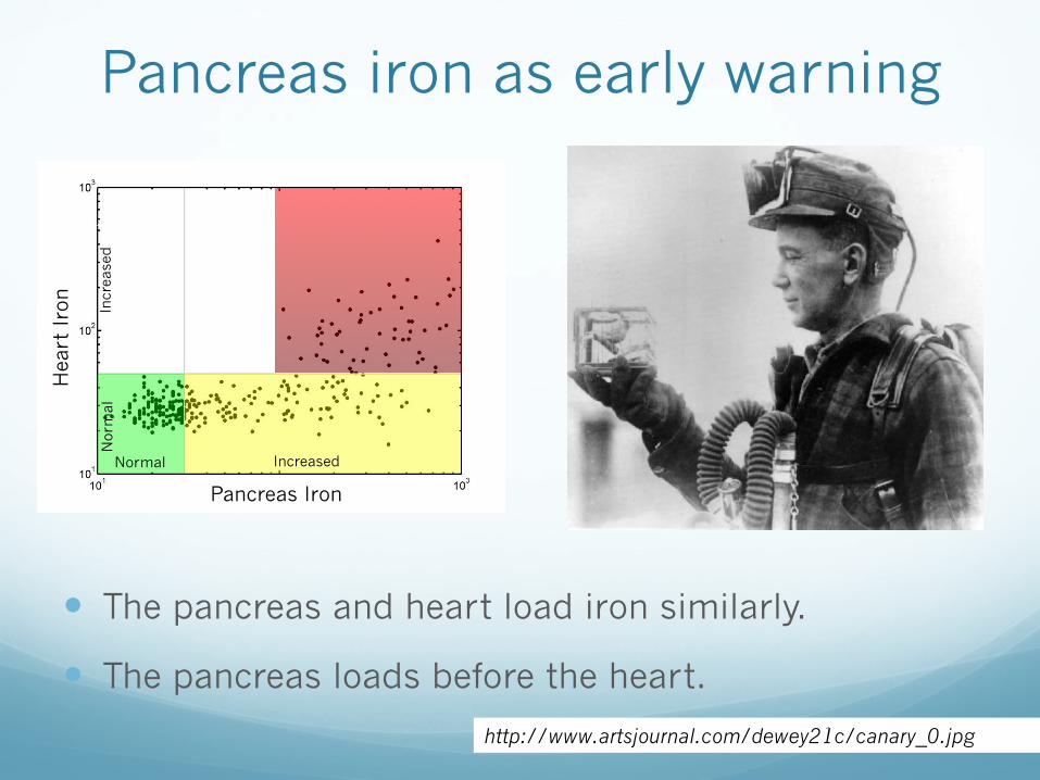
Pancreas iron as early warning
� The pancreas and heart load iron similarly.
� The pancreas loads before the heart.
http://www.artsjournal.com/dewey21c/canary_0.jpg
+ Pancreas + Heart
- Pancreas + Heart
Pancreas Iron
Hea
rt I
ron
Normal
Nor
mal
Increased
Incr
ease
d

Timing of Gland Problems
Growth Failure
Pubertal Delay
Diabetes
Hypothyroidism
Heart Failure
Birth 5 yr 10 yr 15 yr 20 yr 25 yr 30 yr 35 yr
Silent Symptoms
Silent
Silent
Symptoms
Silent Symptoms
Silent Symptoms

Pituitary Gland
� So-called “master” gland.
� Makes hormones that control other glands. � Sex hormones � Growth
� Stress response � Thyroid function
� Pituitary iron causes hypogonadatropic hypogonadism (HH)

How do these organs relate ?
� The organs load and get rid of iron at different speeds.
� Chelation has to be taken nearly every day to protect the pituitary, pancreas, and heart.
Fast Slow
Heart Liver Pituitary Pancreas

Who can make the measurements ?
� Only Thalassemia centers can do the iron MRI’s.
� Experience matters. Most radiologists don’t know how to make these measurements properly.
� Liver and heart represent standard of care.
� Pancreas is routine at CHLA but not yet at many other centers.
� Not as easy as the liver/heart.
� Pituitary is performed at CHLA.

How can you help ?
� Ask your doctors to learn how to use MRI in their clinical practice if they are unfamiliar with it.
� Press your insurers to cover MRI for iron overload for routine clinical care if they do not.
� Participate in research validating MRI techniques.

Summary
� MRI measures the iron stored in the organs.
� Iron damage depends on how much and how long.
� MRI allows doctors to see the iron before damage occurs.
� Different organs load/unload at different rates.
� Regular MRI measurements of heart & liver are the standard of care.
� MRI of the pituitary and pancreas may help reduce iron toxicity in the future.