MRI conditional devices. A luxury or a real clinical...
29
MRI conditional devices. A luxury or a real clinical need? Sophie Mavrogeni, MD, FESC Onassis Cardiac Surgery Center, Athens, Greece
Transcript of MRI conditional devices. A luxury or a real clinical...
MRI conditional devices. A luxury or a real clinical need?
Sophie Mavrogeni, MD, FESC Onassis Cardiac Surgery Center, Athens,
Greece

Increase of MRI scans in recent decades
• In the USA, the number of procedures rose from 7.7 million in 1993 to nearly 22 million in 2002.
• More recent data indicate that, in 2011, 32 million scans were performed in that country.
• Worldwide, it is estimated that approximately 60 million scans are performed each year.
• MRI will continue to grow due to a combination of aging population, expanding indications and increasing accessibility to this technique.

Increase of implantable devices in recent decades
• Between 1993 and 2009, 2.9 million patients received permanent pacemakers in the USA and overall use increased by more than 50%.
• A worldwide survey showed that, in 2009 alone, more than one million pacemakers were implanted, and virtually all countries reported increases in implant numbers.
• The combination of these two growing phenomena results in an estimated 50%–75% probability of a patient needing an MRI over the lifetime of the device.
• These patients will generally see their MRI studies denied due to safety concerns.

Compelling reasons for choosing an MRI-compatible pacemaker
• Previous or planned MRI • Known comorbidities • – Present or previous malignancy • – Neurological disorder (eg, stroke, multiple sclerosis,
seizures) • – Musculoskeletal disorder (eg, back pain, arthritis) • – Congenital heart disease • – Cardiomyopathy • Contraindication for iodinated contrast (known allergy, renal
failure) • Planned surgery

MRI versus CT MRI HAS ADVANTAGES IN DIAGNOSING FOR SOME CONDITIONS
• The images below show two different CT scans and the following MRI for the same patient
▪ Where the CT scan was not conclusive, the MRI provided diagnostic information that enabled the treating physician to choose the correct therapy
CT MRIMRICT
Source: courtesy of Torsten Sommer, Director, Department of Radiology, German Red Cross Hospital Bonn, Academic Institution of the University of Bonn, Marktstr, Neuwied, GermanySource: Brain images courtesy knol.com.
Ischemic Brain Injury Tumor
5

6
Historically, pacemaker patientsWERE DENIED ACCESS TO MRI
Source: Data from 2010 MarketScan Commercial and Medical databases from Truven Health Analytics, Inc.
MRI0.32%
MRI15%
Pacemaker Cohort n = 27,580
Non-Pacemaker Cohort n = 27,580
15%of patients without pacemakers are, annually, receiving an MRI and only 0.32% of patients with traditional pacemakers are receiving them.
Data from 2010 were used to characterize non-pacemaker and pacemaker cohorts and utilization of radiology services. Cohorts were matched based on age, gender and comorbidities.

1 Nazarian S, Reynolds MR, et al. Utilization and likelihood of radiologic diagnostic imaging in patients with implantable cardiac defibrillators. J Magn Reson Imaging. Published online June 27, 2015.2 Medtronic data on file 2015: Data from MarketScan® 2012 Commercial and Medicare Database, Truven Health Analytics.
Data from 2012 were used to project MRI utilization in the ICD patient cohort over 4 years; whereas the actual MRI utilization rate over 4 years was measured in the non-ICD cohort.
Historically, ICD patientsWERE DENIED ACCESS TO MRI
36%of ICD patients are likely to have an MRI ordered over 4 years and only 1.4% of patients are receiving them annually1
ICD Patient Cohort1
n = 9 385Non-ICD Patient Cohort2
n = 9 385
7

Footer Text 8
Need for 3T MRI
3.0T1.5T
Europe
Cerebral familial cavernomatosis.
Many of the small cavernomas visible on 3 T are not detected on 1.5 T.
Source :3 T MRI: Advances in brain imaging. Juan Alvarez-Linera. European Journal of Radiology 67 (2008) 415–426
20%
80%
1.5T3.0T
2014

Footer Text 9
Why 3T?
24%76%
1.5T3.0T
Japan 20%
3yr CAGR2014
27%73%
1.5T3.0T
US 6%
2014 3yr CAGR
% OF MACHINES THAT ARE 3T
3.0T1.5T
Europe
20%
80%
1.5T3.0T
~5x 1.5T
Growth2014
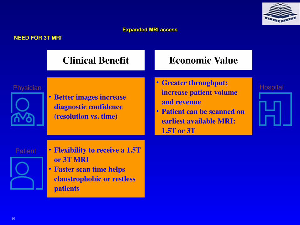
• Better images increase diagnostic confidence (resolution vs. time)
• Flexibility to receive a 1.5T or 3T MRI
• Faster scan time helps claustrophobic or restless patients
• Greater throughput; increase patient volume and revenue
• Patient can be scanned on earliest available MRI: 1.5T or 3T
Clinical Benefit Economic Value
Expanded MRI accessNEED FOR 3T MRI
Physician
Patient
Hospital
10

Unanswered questions and future directions
• Cost-effectiveness versus conventional pacemakers unknown.
• Other economic issues such as reimbursement.
• Legal implications especially in cases where, despite the availability of MRI-conditional pacemakers, a conventional device is implanted and the patient subsequently needs to undergo MRI.
• The impact on image quality especially in thoracic imaging.
• As 3.0 T imaging becomes more prevalent, it will be important to have devices tested and approved for scanning at 3T
• The medical community should try to reach consensus and establish clear criteria for the implantation of these devices.
• Large multicenter studies and education on this subject.

Potential risks of MRI in patients with conventional pacemakers.
Guidelines about devices in MRI
• The AHA guidelines discourage MRI in non-pacemaker-dependent patients, except in cases with a strong clinical indication.
• Among pacemaker-dependent patients, MRI should not be performed unless there are highly compelling circumstances.
• The ESC guidelines strongly recommend against MRI in non-pacemaker-dependent patients, except in the presence of a documented, extremely serious, life-threatening, or severely “quality of life-limiting” condition.
• In pacemaker-dependent patients, the indication for MRI should be seriously reconsidered if the underlying rhythm is too slow, and the threshold for imaging and safety requirements are higher
• Restriction of MRI in patients with conventional pacemakers to specialized centers with specific expertise and equipment for close monitoring.
• In 2013, the American College of Radiology issued a Guidance Document on Safe MR Practices, where a case-by-case and site-by-site appraisal is recommended before scanning patients with non-MRI-conditional devices.

Design modifications made to the pacemakersystem to improve MRI compatibility
• The leads modified to reduce RF lead tip heating.
• Internal circuits changed to reduce potential cardiac stimulation.
• The amount of ferromagnetic materials was limited.
• Internal circuit protection improved to prevent disruption of internal power supply.
• The reed switch was replaced by a Hall sensor (predictable behavior).
• A dedicated programming care pathway developed to facilitate the choice between asynchronous vs nonstimulation modes and restoration of prescan program.

Pacing system integrity checks
• Pacemaker and both leads implanted 6 weeks • Pectoral implantation • No other active pacing or implantable
cardioverterdefibrillator devices or leads • No abandoned leads, lead extenders, or adapters • Leads electrically intact, with stable, normal function • Lead impedance between 200 and 1,500 • Capture threshold

RF-induced heating
• Amount of transmitted RF energy (quantified by SAR)
• Patient size and anatomy
• Patient position within the scanner bore
• Position of the pacemaker lead within the RF field
• Specific lead design and lead length

RF-induced heating
• Substantial increases in CPK and serum troponin can be observed after MRI at 1.5 T in patients implanted with conventional pacemakers.
• CPK changes have been attributed to RF-induced heating of the cardiac tissue in proximity to the pacemaker lead tip, resulting in thermal injury.

Unintended cardiac stimulation
• Gradient magnetic and RF electromagnetic fields produced by MRI induce pulsed voltages in pacemaker leads that are conducted to the heart
• If MRI-induced voltage pulses are large enough, they could directly stimulate the heart, known as unintended cardiac stimulation (UCS).
• UCS includes palpitations or hemodynamic collapse and potentially fatal sustained VT.

Interference with pacemaker function
• If the pacemaker stays in synchronous mode during an MRI examination, the gradient field can mimic intrinsic cardiac electrical activity and thus inhibit pacemaker output, which may be fatal for a pacemaker-dependent patient.
• Fixed cardiac pacing in an asynchronous mode due to MRI-related closure of the reed switch may lead to competitive rhythms, creating a risk of inducing possibly fatal tachyarrhythmias.
• Use of a Hall sensor mitigates this risk because of its predictable behavior in MRI and its specific MRI operation.
• The Hall sensor is suspended and not influenced by the static magnetic field.

Electrical reset
• Electrical reset is an emergency safety feature that guarantees minimal pacemaker functionality in case of battery depletion.
• An electrical reset causes a change in the programmed parameters to basic settings and to an inhibited pacing mode (VVI) with a manufacturer-determined stimulation rate.
• Electrical reset has been reported upon exposure to the MRI field in 6% of pacemaker patients undergoing MRI.
• An electrical reset has important safety implications: • (1) when the reed switch remains open, pacemaker inhibition
potentially leads to bradycardia/asystole; • (2) when the reed switch is closed by the MRI, competitive rhythms
(fixed pacemaker stimulation in asynchronous mode and intrinsic rhythm) may occur, with the risk of inducing fatal tachyarrhythmias.
• The risk of electrical reset is minimized by reducing MRI-induced voltage on the leads and improving internal circuit protection.

Magnetic resonance imaging in patients with a pacemaker system designed for MRI environment.
• Patients (n = 464) were randomized to undergo an MRI scan between 9 and 12 weeks postimplant (MRI group, n = 258) or not to undergo MRI (control group, n = 206) after successful implantation of the specially designed dual-chamber pacemaker and leads.
• No MRI-related complications occurred during or after MRI, including sustained VT, pacemaker inhibition or output failures, electrical resets, or other pacemaker malfunctions.
• Pacing capture threshold and sensed electrogram amplitude changes were minimal and similar between study groups.
Wilcoff BLet al. Heart rhythm 2011

MRI-conditional pacemakers: “safe by design”(1)
• Minimization of ferromagnetic content (use of nonferromagnetic materials, which have to be appropriately conductive, durable, and biocompatible).
• Replacement of reed switches (Reed switch behavior in the MRI environment may vary with the strength of the magnetic field and the orientation of the reed switch, and is essentially unpredictable).
• To overcome this problem, reed switches have been replaced by solid-state Hall sensors, which have more predictable behavior when exposed to magnetic fields

MRI-conditional pacemakers: “safe by design”(2)
• Lead design to minimize heat and electrical currents induced on the leads by MRI.
• To achieve this, the resonant frequency should be avoided to prevent the lead from acting as a receiver of electro-magnetic impulses and can be accomplished by modifications in lead geometry.
• Lead tip coating and bipolar lead designs were also used to minimize electromagnetic interference.
• It should be emphasized that MRI-conditional pacemakers are designed to be used only with compatible MRI-conditional leads in accordance with manufacturer.

MRI-conditional pacemakers: “safe by design”(3)
• Special pacemaker circuitry
• MRI-conditional pacemakers have been equipped with special filters that limit the transfer of certain frequencies and dissipate energy, reducing the risk of damage to the internal power supply.
• Generator shielding has been used to minimize electromagnetic energy transfer
• Dedicated pacemaker programming
• An “MRI mode” must be switched on before entering the scanner, and switched off immediately afterwards.
• Specific programming was developed to assist in the choice between asynchronous vs nonstimulation modes.
• Programming also includes mandatory system-integrity checks, increased pacing output during MRI scanning, and effortless restoration of prescan program.

Balanced steady-state free precession (b-SSFP) cine images of the heart in a patient with an implanted
magnetic resonance imaging-conditional pacemaker.

BRADY Devices MRI condition of industry

ICD – CRT Devices MRI condition of industry

MRI condition FDA approved
Medtronic Biotronik Boston Scientific
St. Jude
Pacemakers 1.5T/3T full body 1.5T full body 1.5T full body No
ICDs 1.5T/3T full body 1.5T full body No (in clinical) No (VR in clinical)
CRT-D 1.5T/3T full body 1.5T full body (bipolar/no quad)
No (in clinical) No
Pacing leads Active/passive Active Active Active (in clinical)
ICD leads IS-1/DF4 IS-1/DF-4 DF-4 (in clinical) DF-4 (in clinical)
LV leads IS-1/IS-4 IS-1 only IS-4 (in clinical) No

Conclusions
• MRI of pacemaker patients is contraindicated due to documented potential risks from hazardous interactions between the MRI and pacemaker.
• 5 million patients worldwide currently are implanted with a pacemaker or implantable cardioverter-defibrillator.
• At least 50% of these patients are expected to be indicated to undergo clinical MRI over the lifetime of their device.
• MRI conditional devices are not a luxury. • They are a real clinical need in the era of increased
MRI applications.

“Transform CRISIS into INSPIRATION” THANK YOU!