MRI characteristics of brain edema in preeclampsia ...
8
Mai et al. BMC Pregnancy Childbirth (2021) 21:669 https://doi.org/10.1186/s12884-021-04145-1 RESEARCH ARTICLE MRI characteristics of brain edema in preeclampsia/eclampsia patients with posterior reversible encephalopathy syndrome Hui Mai 1† , Zhiyu Liang 1,2† , Zhanhang Chen 1 , Zhaoran Liu 1 , Yaxi Xu 1 , Xuting Chen 1 , Xiujian Du 1 , Yuling Peng 1 , Yonglu Chen 1 and Tianfa Dong 1* Abstract Background: The neuroimaging manifestations of eclampsia and preeclampsia often overlap, mainly presenting as posterior reversible encephalopathy syndrome (PRES). The purpose of this retrospective study was to compare the extent and nature of brain edema in eclampsia and preeclampsia patients with PRES based on MRI characteristics. Methods: One hundred fifty women diagnosed with preeclampsia-eclampsia and undergoing cranial MRI were enrolled; 24 of these were diagnosed as having eclampsia. According to clinicoradiologic diagnosis of PRES, eligible patients were classified as having eclampsia with PRES (group E-PRES) and preeclampsia with PRES (group P-PRES). A scale on T2W FLAIR-SPIR images was established to evaluate the extent of brain edema, and the score of brain edema (SBE) of both groups was compared. In patients of the two groups who also underwent DWI sequence, the presence or absence of hyperintensity on DWI and hypointensity on ADC maps were determined to compare the nature of brain edema. Furthermore, clinical and biochemical data of the two groups were compared. Results: The incidence of PRES in eclampsia patients was significantly higher than that in preeclampsia patients (87.50% vs. 46.03%, P<0.001). The SBE of all regions and typical regions in group E-PRES patients were significantly higher than those in group P-PRES patients (15.88±8.72 vs. 10.90±10.21, P=0.021; 8.52±3.87 vs. 5.01±4.19, P=0.002; respectively). The presence of hyperintensity on DWI was determined more frequently in group E-PRES patients than group P-PRES patients (71.43% vs. 32.00%, P=0.024). Age, systolic blood pressure, white blood cell count, neutrophil count and percentage of neutrophils were significantly different between the two groups (P<0.05). Conclusions: Certain MRI characteristics that reflect the extent and nature of brain edema were different between eclampsia and preeclampsia patients with PRES. Additional prospective studies are still required to explore whether these MRI characteristics of brain edema may further become a potential predictor for eclamptic seizures in preec- lampsia patients with PRES. © The Author(s) 2021. Open Access This article is licensed under a Creative Commons Attribution 4.0 International License, which permits use, sharing, adaptation, distribution and reproduction in any medium or format, as long as you give appropriate credit to the original author(s) and the source, provide a link to the Creative Commons licence, and indicate if changes were made. The images or other third party material in this article are included in the article’s Creative Commons licence, unless indicated otherwise in a credit line to the material. If material is not included in the article’s Creative Commons licence and your intended use is not permitted by statutory regulation or exceeds the permitted use, you will need to obtain permission directly from the copyright holder. To view a copy of this licence, visit http://creativecommons.org/licenses/by/4.0/. The Creative Commons Public Domain Dedication waiver (http://creativeco mmons.org/publicdomain/zero/1.0/) applies to the data made available in this article, unless otherwise stated in a credit line to the data. Open Access *Correspondence: [email protected] † Hui Mai and Zhiyu Liang have contributed equally to this work as co-first authors. 1 Department of Radiology, The Third Affiliated Hospital of Guangzhou Medical University, No. 63 Duobao Road, Guangzhou 510150, China Full list of author information is available at the end of the article
Transcript of MRI characteristics of brain edema in preeclampsia ...

Mai et al. BMC Pregnancy Childbirth (2021) 21:669 https://doi.org/10.1186/s12884-021-04145-1
RESEARCH ARTICLE
MRI characteristics of brain edema in preeclampsia/eclampsia patients with posterior reversible encephalopathy syndromeHui Mai1†, Zhiyu Liang1,2†, Zhanhang Chen1, Zhaoran Liu1, Yaxi Xu1, Xuting Chen1, Xiujian Du1, Yuling Peng1, Yonglu Chen1 and Tianfa Dong1*
Abstract
Background: The neuroimaging manifestations of eclampsia and preeclampsia often overlap, mainly presenting as posterior reversible encephalopathy syndrome (PRES). The purpose of this retrospective study was to compare the extent and nature of brain edema in eclampsia and preeclampsia patients with PRES based on MRI characteristics.
Methods: One hundred fifty women diagnosed with preeclampsia-eclampsia and undergoing cranial MRI were enrolled; 24 of these were diagnosed as having eclampsia. According to clinicoradiologic diagnosis of PRES, eligible patients were classified as having eclampsia with PRES (group E-PRES) and preeclampsia with PRES (group P-PRES). A scale on T2W FLAIR-SPIR images was established to evaluate the extent of brain edema, and the score of brain edema (SBE) of both groups was compared. In patients of the two groups who also underwent DWI sequence, the presence or absence of hyperintensity on DWI and hypointensity on ADC maps were determined to compare the nature of brain edema. Furthermore, clinical and biochemical data of the two groups were compared.
Results: The incidence of PRES in eclampsia patients was significantly higher than that in preeclampsia patients (87.50% vs. 46.03%, P<0.001). The SBE of all regions and typical regions in group E-PRES patients were significantly higher than those in group P-PRES patients (15.88±8.72 vs. 10.90±10.21, P=0.021; 8.52±3.87 vs. 5.01±4.19, P=0.002; respectively). The presence of hyperintensity on DWI was determined more frequently in group E-PRES patients than group P-PRES patients (71.43% vs. 32.00%, P=0.024). Age, systolic blood pressure, white blood cell count, neutrophil count and percentage of neutrophils were significantly different between the two groups (P<0.05).
Conclusions: Certain MRI characteristics that reflect the extent and nature of brain edema were different between eclampsia and preeclampsia patients with PRES. Additional prospective studies are still required to explore whether these MRI characteristics of brain edema may further become a potential predictor for eclamptic seizures in preec-lampsia patients with PRES.
© The Author(s) 2021. Open Access This article is licensed under a Creative Commons Attribution 4.0 International License, which permits use, sharing, adaptation, distribution and reproduction in any medium or format, as long as you give appropriate credit to the original author(s) and the source, provide a link to the Creative Commons licence, and indicate if changes were made. The images or other third party material in this article are included in the article’s Creative Commons licence, unless indicated otherwise in a credit line to the material. If material is not included in the article’s Creative Commons licence and your intended use is not permitted by statutory regulation or exceeds the permitted use, you will need to obtain permission directly from the copyright holder. To view a copy of this licence, visit http:// creat iveco mmons. org/ licen ses/ by/4. 0/. The Creative Commons Public Domain Dedication waiver (http:// creat iveco mmons. org/ publi cdoma in/ zero/1. 0/) applies to the data made available in this article, unless otherwise stated in a credit line to the data.
Open Access
*Correspondence: [email protected]†Hui Mai and Zhiyu Liang have contributed equally to this work as co-first authors.1 Department of Radiology, The Third Affiliated Hospital of Guangzhou Medical University, No. 63 Duobao Road, Guangzhou 510150, ChinaFull list of author information is available at the end of the article

Page 2 of 8Mai et al. BMC Pregnancy Childbirth (2021) 21:669
BackgroundEclampsia is the most severe stage of the preeclamp-sia–eclampsia spectrum, with an incidence of approx-imately 3/1000 to 9/1000 in pregnant or recently delivered women [1, 2]. Despite significant improve-ment in medical care and the popularity of prenatal diagnosis in recent years, the worldwide incidence and mortality of eclampsia remain high, seriously threaten-ing maternal and fetal lives. Therefore, the early pre-diction of eclampsia has always been one of the severe challenges in obstetrics [3].
Neuroimaging manifestations of eclampsia and preec-lampsia often overlap, mainly presenting as posterior reversible encephalopathy syndrome (PRES) [4, 5]. PRES is a distinctive clinicoradiologic syndrome character-ized by headaches, visual disturbances, and seizures with predominantly parieto-occipital vasogenic edema, occa-sionally with cytotoxic edema [4]. Thus far, the exact pathophysiological mechanism of preeclampsia-eclamp-sia with PRES has not been clarified and is still controver-sial [6–8]. Not much is known about whether differences exist in the extent and nature of brain edema between eclampsia and preeclampsia patients with PRES; moreo-ver, a “threshold” trigger in the occurrence of eclampsia has not yet been verified [9, 10].
Cranial conventional magnetic resonance imaging (MRI) is the preferred imaging modality for preec-lampsia-eclampsia patients with PRES [4]. Gao et al. used conventional MRI sequences to score the extent of brain edema in PRES patients and found that the score was significantly correlated with serum levels of lactate dehydrogenase [6]. Diffusion-weighted imaging (DWI) is another commonly performed MRI sequence and is highly sensitive for distinguishing between cytotoxic and vasogenic edema [4, 11]. Recent MRI studies have shown that DWI can reflect the changes of pathophysi-ology regarding PRES patients in the ictal or peri-ictal phase of epilepsy [9]. The purpose of this retrospective research was to compare the extent and nature of brain edema in eclampsia and preeclampsia patients with PRES based on the MRI characteristics.
MethodsPatientsThe study protocol was approved by the institutional review board, and written informed consent was waived given the retrospective nature of the study.
In all, 150 women diagnosed with preeclampsia-eclampsia and undergoing cranial MRI from Septem-ber 2012 to March 2020 were enrolled; 24 of these were diagnosed as having eclampsia. The inclusion criteria was as follows: (i) preeclampsia was diagnosed accord-ing to the diagnostic criteria established by the Ameri-can College of Obstetricians and Gynecologists [12], and eclampsia was defined by new-onset seizures that could not be attributed to other causative conditions in these women; (ii) women with preeclampsia under-went cranial MRI within 3 days before or after the onset of symptoms such as headache, vomiting, visual disturbance, mental status changes, and consciousness impairment, while those with eclampsia underwent cranial MRI within 3 days before or after the onset of seizures; and (iii) patients without neurological dis-eases such as epilepsy, brain tumors, and congenital brain malformations, which were unrelated to preec-lampsia-eclampsia. The exclusion criterion was as fol-lows: (i) patients that lacked clinicoradiologic findings of PRES [4]; and (ii) patients with abnormal neuroimag-ing features unrelated to PRES [7]. Finally, women who met these criteria were classified as having eclamp-sia with PRES (group E-PRES) and preeclampsia with PRES (group P-PRES).
MRI assessmentMRI were performed on a 3.0T scanner (Achieva, Philips Healthcare, Best, The Netherlands) with an 8-channel head coil.
Conventional MRI was performed for all study patients. The conventional MRI scan sequences and parameters were as follows: (i) axial T1-weighted (T1W) spin echo sequence (repetition time: 500 ms, echo time: 7.5 ms, number of signal averaged: 2, slice thickness: 6 mm, and matrix size: 328×196); (ii) axial T2-weighted (T2W) turbo spin echo sequence (repetition time: 5000 ms, echo time: 80 ms, number of signal averaged: 2, slice thickness: 6 mm, and matrix size: 328×196); and (iii) axial T2W fluid attenuated inversion recovery - spectral presaturation inversion recovery (FLAIR-SPIR) sequence (repetition time / inversion time: 7000/2200 ms, echo time: 156 ms, number of signal averaged: 2, slice thick-ness: 6 mm, and matrix size: 328×196). The imaging diagnosis of PRES was based on the MRI finding of brain edema, which presented as hypointense or isointense signals on T1W and as hyperintense signal on T2W and T2W FLAIR-SPIR [4]. The incidence of PRES in patients
Keywords: eclampsia, preeclampsia, posterior reversible encephalopathy syndrome, brain edema, magnetic resonance imaging

Page 3 of 8Mai et al. BMC Pregnancy Childbirth (2021) 21:669
with eclampsia and preeclampsia was calculated and compared.
Amongst these conventional MRI sequences, T2W FLAIR-SPIR is considered to be the most sensitive scan technique for detecting brain edema lesions of PRES [4]; therefore, we established a scale for T2W FLAIR-SPIR images to score and evaluate the extent (distribution and degree) of brain edema. The distribution of brain edema was divided into 15 regions according to anatomy: (1, 2) bilateral occipital lobes; (3, 4) bilateral parietal lobes; (5, 6) bilateral frontal lobes; (7, 8) bilateral temporal lobes; (9, 10) bilateral insular lobes; (11, 12) bilateral basal gan-glia, thalamus, internal capsule, and external capsule; (13) cerebellum; (14) brain stem; (15) corpus callosum. The degree of brain edema in each region was divided into 0–III grades: grade 0, normal signal, score: 0 point; grade I, single fleck, patch, or nodule of abnormal signal, score: 1 point; grade II, multiple flecks, patches, or nod-ules of abnormal signals without fusion, score: 2 points; grade III, large area fusion of abnormal signals, score: 3 points. The sum of scores in each region was the score of brain edema (SBE) of all regions. In addition, we defined bilateral occipital and parietal lobes as typical regions of PRES and the rest as atypical regions of PRES. We then evaluated the SBE of both groups for typical and atypical regions.
Furthermore, MRI DWI sequence images of the two groups were collected. Axial DWI with b values of 0 and 1000 s/mm2 utilized a single-shot echo planar imag-ing (repetition time: 2258 ms, echo time: 86 ms, num-ber of signal averaged: 2, slice thickness: 6 mm, and matrix size: 152×122). An axial apparent diffusion coeffi-cient (ADC) map was generated automatically on a voxel-by-voxel basis from the two b values. The presence or absence of hyperintensity on DWI with b value of 1000 s/mm2 and hypointensity on the ADC maps were deter-mined to evaluate the nature of brain edema.
MRI assessment for each patient enrolled in this study was independently evaluated by two radiologists with 13 and 5 years of experience, respectively, blinded to the clinical and biochemical data. For quantitative variables, the average was used; for the cases with discrepancies in the assessment of qualitative variables, a consensus inter-pretation was reached by joint review.
Clinical and biochemical data assessmentClinical data of the two groups was collected, including age, primiparity, gravidity, parity, length of hospital stay, and blood pressure. The biochemical data of both groups were collected, including blood cell count and indicators of liver and renal function. Biochemical indicators and blood pressure were obtained immediately at the occur-rence of seizures in group E-PRES patients and at the occurrence of symptoms in group P-PRES patients.
Statistical analysisDescriptive statistics for continuous variables were pre-sented as the mean±SD, while categorical variables were presented as frequencies and percentages. We used inde-pendent sample t-test (normally distributed variables) or Mann-Whitney U-test (non-normally distributed variables) to compare the continuous variables of the two groups, and chi-square or Fisher’s exact test to com-pare the categorical variables. The intraclass correlation efficient (ICC) was used to evaluate the inter-observer repeatability of SBE measurement. An ICC value>0.75 indicates that the repeatability is excellent. All statistical analyses were performed using SPSS version 26.0 (IBM Corporation, Armonk, NY, USA). P<0.05 was considered to indicate statistical significance.
ResultsIn our study, PRES occurred in 87.50% eclampsia patients (21/24) and 46.03% preeclampsia patients (58/126). The incidence of PRES in eclampsia patients was significantly higher than that in preeclampsia patients (P<0.001). The inter-observer repeatability of SBE measurement indi-cated excellent with ICC>0.75. The SBE of all regions and typical regions in group E-PRES patients were sig-nificantly higher than those in group P-PRES patients (15.88±8.72 vs. 10.90±10.21, P=0.021; 8.52±3.87 vs. 5.01±4.19, P=0.002; respectively). The SBE of atypi-cal regions in group E-PRES patients was slightly higher than that in group P-PRES patients, but this difference was not statistically significant (7.36±5.62 vs. 5.91±6.79, P=0.097). Table 1 shows the SBE of the two groups, and examples are shown in Figs. 1 and 2.
14 group E-PRES and 25 group P-PRES patients were imaged using the MRI DWI sequence. The majority of
Table 1 SBE in eclampsia patients with PRES (group E-PRES) and preeclampsia patients with PRES (group P-PRES)
SBE Score of brain edema, PRES Posterior reversible encephalopathy syndrome
Parameter Group E-PRES (n=21) Group P-PRES (n=58) T value P
SBE of all regions 15.88±8.72 10.90±10.21 402.00 0.021
SBE of typical regions 8.52±3.87 5.01±4.19 377.50 0.002
SBE of atypical regions 7.36±5.62 5.91±6.79 460.00 0.097

Page 4 of 8Mai et al. BMC Pregnancy Childbirth (2021) 21:669
brain edema lesions appeared as hyperintense or isoin-tense signals on DWI and as hyperintense signals on the ADC map. Furthermore, coexistence of different signals on DWI and ADC maps of the affected areas was some-times observed. The presence of hyperintensity on DWI was determined in 71.43% group E-PRES patients (10/14) and 32.00% group P-PRES patients (8/25), with statisti-cally significant differences (P=0.024). The hypointense signal on ADC map presented in group E-PRES patients (4/14, 28.57%) was slightly more frequent than that pre-sented in group P-PRES patients (2/25, 8.00%), but this difference was not statistically significant (P=0.163). Table 2 shows the features of DWI and ADC map on brain edema lesions in the two groups, and examples are shown in Figs 1 and 2.
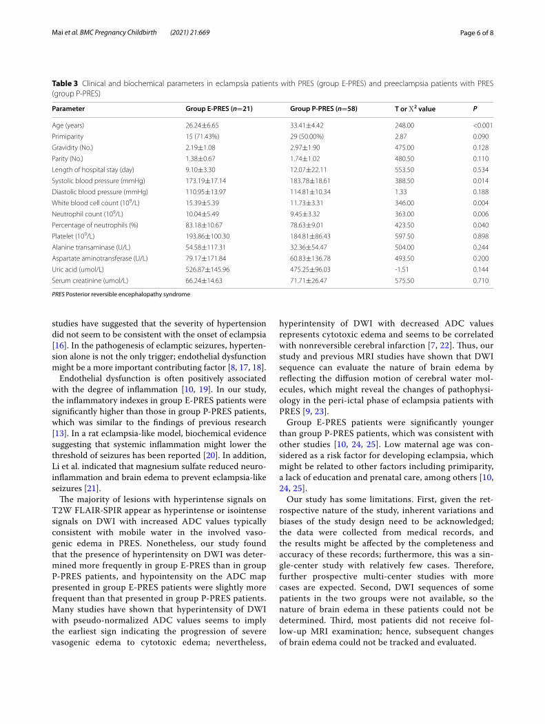
Group E-PRES patients were significantly younger than group P-PRES patients (26.24±6.65 vs. 33.41±4.42 years old, P<0.001). Systolic blood pressure, white blood cell count, neutrophil count, and percentage of neutrophils in group E-PRES patients were significantly higher than those in group P-PRES patients (P<0.05). Other clinical and biochemical parameters collected in this study did not significantly differ between the two groups. Table 3
shows the clinical and biochemical parameters of the two study groups.
DiscussionPrevious studies have shown that abnormal imaging find-ings of PRES typically associated with eclampsia were also observed frequently in preeclampsia patients with-out seizures [5, 8, 13]. In this study, although the inci-dence of PRES in eclampsia patients was significantly higher than that in preeclampsia patients, nearly half the preeclampsia patients with symptoms also experienced PRES. Fang et al. further reported that there were no sig-nificant differences between preeclampsia and eclampsia patients in the distribution score or degree score of brain edema on MRI [10]. The sample size of our study was similar to that of Fang et al’s study. However, our study found that the SBE of typical regions, atypical regions, and all regions in group E-PRES patients were all higher than those in group P-PRES patients; furthermore, the SBE of typical regions and all regions were significantly different between the two groups. The distribution score and degree score of brain edema were combined into the measure of SBE in our study rather than counted
Fig. 1 Cranial MRI of a 31-year-old woman with eclampsia with PRES: a axial T2W FLAIR-SPIR, b axial DWI with b value of 1000 s/mm2, c axial ADC map. SBE was evaluated according to the distribution and the degree of abnormal edema signal, which presented as heterogeneous hyperintense signal (arrow) on T2W FLAIR-SPIR (a). Large area fusions of abnormal edema signals were shown in corpus callosum, bilateral occipital, parietal, frontal, and temporal lobes; and multiple flecks, patches or nodules of abnormal edema signals without fusion were seen in the cerebellum and right basal ganglia. The SBE of typical regions, atypical regions, and all regions in this patient were 12, 19, and 31, respectively. Some of the abnormal edema signals in bilateral frontal lobes, left parietal and temporal lobes presented as hyperintense signals (arrow) on DWI (b), with the lesion in left temporal lobe locally appearing as hypointense signal (arrow) on ADC map (c)

Page 5 of 8Mai et al. BMC Pregnancy Childbirth (2021) 21:669
independently as in Fang et al’s study, which may be a more comprehensive scale for assessing the extent of brain edema. This scoring method for brain edema has been rarely mentioned in the literature except for in a previous study, which found that this scoring method for brain edema showed positive correlation with the ratio of soluble endoglin to placental growth factor that reflected the degree of vascular endothelial injury in preeclampsia patients with PRES [14].
The results showed that the difference in SBE of all regions between the two study groups was mainly due
to the difference in the edema score of typical regions. The preferential distribution of posterior parietal and occipital lobe regions in PRES is not well understood. It is generally accepted that posterior circulation arteries have sparser mural sympathetic innervation and are thin-ner than other cerebral arteries, which make them more vulnerable to endothelial dysfunction and more prone to autoregulation breakthrough in acute hypertension [7, 15]. Interestingly, in our study, the systolic blood pressure of group E-PRES patients was not higher, rather signifi-cantly lower than that of group P-PRES patients. Previous
Fig. 2 Cranial MRI of a 31-year-old woman with preeclampsia with PRES: a axial T2W FLAIR-SPIR, b axial DWI with b value of 1000 s/mm2, and c axial ADC map. SBE was evaluated according to the distribution and degree of abnormal edema signal, which presented as heterogeneous hyperintense signals (arrow) on T2W FLAIR-SPIR (a). Large area fusions of abnormal edema signals were shown in the right occipital and temporal lobes; and multiple flecks, patches, or nodules of abnormal signals without fusion were seen in the left basal ganglia, right parietal lobe, left occipital, temporal and frontal lobes. The SBE of typical regions, atypical regions, and all regions in this patient were 7, 9, and 16, respectively. The abnormal edema signals presented as isointense signals (arrow) on DWI (b) and as hyperintense signals (arrow) on ADC map (c)
Table 2 The features of DWI and ADC map on brain edema lesions in eclampsia patients with PRES (group E-PRES) and preeclampsia patients with PRES (group P-PRES)
DWI Diffusion weighted imaging, ADC Apparent diffusion coefficient, PRES Posterior reversible encephalopathy syndrome
Parameter Group E-PRES (n=14) Group P-PRES (n=25) P
Hyperintensity on DWI 0.024
Present 10 (71.43%) 8 (32.00%)
Absent 4 (28.57%) 17 (68.00%)
Hypointensity on ADC map 0.163
Present 4 (28.57%) 2 (8.00%)
Absent 10 (71.43%) 23 (92.00%)
Page 6 of 8Mai et al. BMC Pregnancy Childbirth (2021) 21:669
studies have suggested that the severity of hypertension did not seem to be consistent with the onset of eclampsia [16]. In the pathogenesis of eclamptic seizures, hyperten-sion alone is not the only trigger; endothelial dysfunction might be a more important contributing factor [8, 17, 18].
Endothelial dysfunction is often positively associated with the degree of inflammation [10, 19]. In our study, the inflammatory indexes in group E-PRES patients were significantly higher than those in group P-PRES patients, which was similar to the findings of previous research [13]. In a rat eclampsia-like model, biochemical evidence suggesting that systemic inflammation might lower the threshold of seizures has been reported [20]. In addition, Li et al. indicated that magnesium sulfate reduced neuro-inflammation and brain edema to prevent eclampsia-like seizures [21].
The majority of lesions with hyperintense signals on T2W FLAIR-SPIR appear as hyperintense or isointense signals on DWI with increased ADC values typically consistent with mobile water in the involved vaso-genic edema in PRES. Nonetheless, our study found that the presence of hyperintensity on DWI was deter-mined more frequently in group E-PRES than in group P-PRES patients, and hypointensity on the ADC map presented in group E-PRES patients were slightly more frequent than that presented in group P-PRES patients. Many studies have shown that hyperintensity of DWI with pseudo-normalized ADC values seems to imply the earliest sign indicating the progression of severe vasogenic edema to cytotoxic edema; nevertheless,
hyperintensity of DWI with decreased ADC values represents cytotoxic edema and seems to be correlated with nonreversible cerebral infarction [7, 22]. Thus, our study and previous MRI studies have shown that DWI sequence can evaluate the nature of brain edema by reflecting the diffusion motion of cerebral water mol-ecules, which might reveal the changes of pathophysi-ology in the peri-ictal phase of eclampsia patients with PRES [9, 23].
Group E-PRES patients were significantly younger than group P-PRES patients, which was consistent with other studies [10, 24, 25]. Low maternal age was con-sidered as a risk factor for developing eclampsia, which might be related to other factors including primiparity, a lack of education and prenatal care, among others [10, 24, 25].
Our study has some limitations. First, given the ret-rospective nature of the study, inherent variations and biases of the study design need to be acknowledged; the data were collected from medical records, and the results might be affected by the completeness and accuracy of these records; furthermore, this was a sin-gle-center study with relatively few cases. Therefore, further prospective multi-center studies with more cases are expected. Second, DWI sequences of some patients in the two groups were not available, so the nature of brain edema in these patients could not be determined. Third, most patients did not receive fol-low-up MRI examination; hence, subsequent changes of brain edema could not be tracked and evaluated.
Table 3 Clinical and biochemical parameters in eclampsia patients with PRES (group E-PRES) and preeclampsia patients with PRES (group P-PRES)
PRES Posterior reversible encephalopathy syndrome
Parameter Group E-PRES (n=21) Group P-PRES (n=58) T or Χ2 value P
Age (years) 26.24±6.65 33.41±4.42 248.00 <0.001
Primiparity 15 (71.43%) 29 (50.00%) 2.87 0.090
Gravidity (No.) 2.19±1.08 2.97±1.90 475.00 0.128
Parity (No.) 1.38±0.67 1.74±1.02 480.50 0.110
Length of hospital stay (day) 9.10±3.30 12.07±22.11 553.50 0.534
Systolic blood pressure (mmHg) 173.19±17.14 183.78±18.61 388.50 0.014
Diastolic blood pressure (mmHg) 110.95±13.97 114.81±10.34 1.33 0.188
White blood cell count (109/L) 15.39±5.39 11.73±3.31 346.00 0.004
Neutrophil count (109/L) 10.04±5.49 9.45±3.32 363.00 0.006
Percentage of neutrophils (%) 83.18±10.67 78.63±9.01 423.50 0.040
Platelet (109/L) 193.86±100.30 184.81±86.43 597.50 0.898
Alanine transaminase (U/L) 54.58±117.31 32.36±54.47 504.00 0.244
Aspartate aminotransferase (U/L) 79.17±171.84 60.83±136.78 493.50 0.200
Uric acid (umol/L) 526.87±145.96 475.25±96.03 -1.51 0.144
Serum creatinine (umol/L) 66.24±14.63 71.71±26.47 575.50 0.710

Page 7 of 8Mai et al. BMC Pregnancy Childbirth (2021) 21:669
ConclusionsWhat triggers the occurrence of eclampsia in preec-lampsia patients with PRES is still unknown. This retrospective study has suggested that certain MRI characteristics that reflect the extent and nature of brain edema are different in eclampsia and preec-lampsia patients with PRES. However, additional pro-spective studies are still required to explore whether these MRI characteristics of brain edema may further become a potential predictor for eclamptic seizures in preeclampsia patients with PRES. Given the high mor-tality related to intracranial complications in eclampsia patients, we recommend early consideration of cranial MRI in the management of preeclampsia patients to evaluate the presence or absence of PRES as well as the extent and nature of brain edema.
AbbreviationsPRES: Posterior reversible encephalopathy syndrome; MRI: Magnetic resonance imaging; DWI: Diffusion-weighted imaging; T1W: T1-weighted; T2W: T2-weighted; FLAIR: Fluid attenuated inversion recovery; SPIR: Spectral presaturation inversion recovery; SBE: Score of brain edema; ADC: Apparent diffusion coefficient; ICC: Intraclass correlation efficient.
AcknowledgementsNot applicable.
Authors’ contributionsHM, ZYL and TFD conceived and designed this study. ZHC, ZRL, YXX, XTC, XJD and YLP conducted the study and collected important background data. YXX, XTC, XJD, YLP and YLC analyzed the medical images. HM, ZYL and TFD drafted the manuscript. All authors read and approved the final manuscript.
FundingThis work was supported by the Guangdong Medical Research Foundation (CN) [grant numbers A2018147, A2015103] and General Guidance Project of Health Science and Technology of Guangzhou (CN) [grant number 2019A010060].
Availability of data and materialsThe datasets used and analyzed during the current study are available from the corresponding author on reasonable request.
Declarations
Ethics approval and consent to participateThis retrospective study was approved by the institutional review board at the third affiliated hospital of Guangzhou Medical University, and the requirement of patients’ informed consent was waived.
Consent for publicationNot applicable.
Competing interestsThe authors declare that they have no competing interests.
Author details1 Department of Radiology, The Third Affiliated Hospital of Guangzhou Medical University, No. 63 Duobao Road, Guangzhou 510150, China. 2 Department of Medical Imaging Center, Nanfang Hospital, Southern Medical University, No. 1838 Guangzhou Avenue North, Guangzhou 510515, China.
Received: 21 March 2021 Accepted: 22 September 2021
References 1. Miguil M, Chekairi A. Eclampsia, study of 342 cases. Hypertens Pregnancy.
2008;27:103–11. 2. Abalos E, Cuesta C, Carroli G, Qureshi Z, Widmer M, Vogel JP, et al. Pre-
eclampsia, eclampsia and adverse maternal and perinatal outcomes: a secondary analysis of the world health organization multicountry survey on maternal and newborn health. BJOG. 2014;121(Suppl 1):14–24.
3. Hastie R, Brownfoot FC, Cluver CA, Walker SP, Hesselman S, Tong S, et al. Predictive value of the signs and symptoms preceding eclampsia: a systematic review. Obstet Gynecol. 2019;134:677–84.
4. Garg RK, Kumar N, Malhotra HS. Posterior reversible encephalopathy syndrome in eclampsia. Neurol India. 2018;66:1316–23.
5. Demirtas O, Gelal F, Vidinli BD, Demirtas LO, Uluc E, Baloglu A. Cranial MR imaging with clinical correlation in preeclampsia and eclampsia. Diagn Interv Radiol. 2005;11:189–94.
6. Gao B, Liu FL, Zhao B. Association of degree and type of edema in poste-rior reversible encephalopathy syndrome with serum lactate dehydroge-nase level: initial experience. Eur J Radiol. 2012;81:2844–7.
7. Ollivier M, Bertrand A, Clarencon F, Gerber S, Deltour S, Domont F, et al. Neuroimaging features in posterior reversible encephalopathy syndrome: a pictorial review. J Neurol Sci. 2017;373:188–200.
8. Mayama M, Uno K, Tano S, Yoshihara M, Ukai M, Kishigami Y, et al. Inci-dence of posterior reversible encephalopathy syndrome in eclamptic and patients with preeclampsia with neurologic symptoms. Am J Obstet Gynecol. 2016;215:239 e1–5.
9. Wakisaka K, Morioka T, Shimogawa T, Murao K, Kanazawa Y, Hagiwara N, et al. Epileptic Ictal Hyperperfusion on Arterial Spin Labeling Perfusion and Diffusion-Weighted Magnetic Resonance Images in Posterior Reversi-ble Encephalopathy Syndrome. J Stroke Cerebrovasc Dis. 2016;25:228–37.
10. Xiaobo F, Yanling L, Dunjin C, Fang H, Jia C, Yuhua Z, et al. Effect of blood pressure on reversible posterior leukoencephalopathy syndrome in pre-eclampsia or eclampsia. Hypertens Res. 2018;41:112–7.
11. Dong XY, Bai CB, Nao JF. Clinical and radiological features of posterior reversible encephalopathy syndrome in patients with pre-eclampsia and eclampsia. Clin Radiol. 2017;72:887–95.
12. Practice Bulletin No ACOG. 202: Gestational Hypertension and Preec-lampsia. Obstet Gynecol. 2019;133:e1–e25.
13. Di X, Mai H, Zheng Z, Guo K, Morse AN, Liu H. Neuroimaging findings in women who develop neurologic symptoms in severe preeclampsia with or without eclampsia. Hypertens Res. 2018;41:598–604.
14. Jiang KM, Zhong X, Tan Y, Liu GC, Mai H, Wu SX. The correlation between cerebral MRI characteristics of posterior reversible encephalopathy syndrome and serum levels of PlGF, sEng in patients with pre-eclampsia. Zhonghua Fu Chan Ke Za Zhi. 2016;51:840–4.
15. Hacein-Bey L, Varelas PN, Ulmer JL, Mark LP, Raghavan K, Provenzale JM. Imaging of Cerebrovascular Disease in Pregnancy and the Puerperium. AJR Am J Roentgenol. 2016;206:26–38.
16. Berhan Y, Berhan A. Should magnesium sulfate be administered to women with mild pre-eclampsia? A systematic review of published reports on eclampsia. J Obstet Gynaecol Res. 2015;41:831–42.
17. McDermott M, Miller EC, Rundek T, Hurn PD, Bushnell CD. Preeclampsia: Association With Posterior Reversible Encephalopathy Syndrome and Stroke. Stroke. 2018;49:524–30.
18. Marra A, Vargas M, Striano P, Del Guercio L, Buonanno P, Servillo G. Pos-terior reversible encephalopathy syndrome: the endothelial hypotheses. Med Hypotheses. 2014;82:619–22.
19. Liu L, Han X, Huang Q, Zhu X, Yang J, Liu H. Increased neuronal seizure activity correlates with excessive systemic inflammation in a rat model of severe preeclampsia. Hypertens Res. 2016;39:701–8.
20. Huang Q, Liu L, Hu B, Di X, Brennecke SP, Liu H. Decreased seizure thresh-old in an eclampsia-like model induced in pregnant rats with lipopolysac-charide and pentylenetetrazol treatments. PLoS One. 2014;9:e89333.
21. Li X, Han X, Yang J, Bao J, Di X, Zhang G, et al. Magnesium Sulfate Provides Neuroprotection in Eclampsia-Like Seizure Model by Ameliorating Neuro-inflammation and Brain Edema. Mol Neurobiol. 2017;54:7938–48.

Page 8 of 8Mai et al. BMC Pregnancy Childbirth (2021) 21:669
• fast, convenient online submission
•
thorough peer review by experienced researchers in your field
• rapid publication on acceptance
• support for research data, including large and complex data types
•
gold Open Access which fosters wider collaboration and increased citations
maximum visibility for your research: over 100M website views per year •
At BMC, research is always in progress.
Learn more biomedcentral.com/submissions
Ready to submit your researchReady to submit your research ? Choose BMC and benefit from: ? Choose BMC and benefit from:
22. Covarrubias DJ, Luetmer PH, Campeau NG. Posterior reversible encepha-lopathy syndrome: prognostic utility of quantitative diffusion-weighted MR images. AJNR Am J Neuroradiol. 2002;23:1038–48.
23. Szabo K, Poepel A, Pohlmann-Eden B, Hirsch J, Back T, Sedlaczek O, et al. Diffusion-weighted and perfusion MRI demonstrates parenchymal changes in complex partial status epilepticus. Brain. 2005;128:1369–76.
24. Mahran A, Fares H, Elkhateeb R, Ibrahim M, Bahaa H, Sanad A, et al. Risk factors and outcome of patients with eclampsia at a tertiary hospital in Egypt. BMC Pregnancy Childbirth. 2017;17:435.
25. Rebahi H, Elizabeth Still M, Faouzi Y. Rhassane El Adib A. Risk factors for eclampsia in pregnant women with preeclampsia and positive neurosen-sory signs. Turk J Obstet Gynecol. 2018;15:227–34.
Publisher’s NoteSpringer Nature remains neutral with regard to jurisdictional claims in pub-lished maps and institutional affiliations.