MRI basic sequences
34
DR MOHIT GOEL JR 1 27 JULY, 2012 MRI PHYSICS ( Basic sequences)
-
Upload
dr-mohit-goel -
Category
Education
-
view
85 -
download
13
Transcript of MRI basic sequences

DR MOHIT GOELJR 1 27 JULY, 2012
MRI PHYSICS ( Basic sequences)


Net magnetization is the macroscopic
measure of many spins
Bo
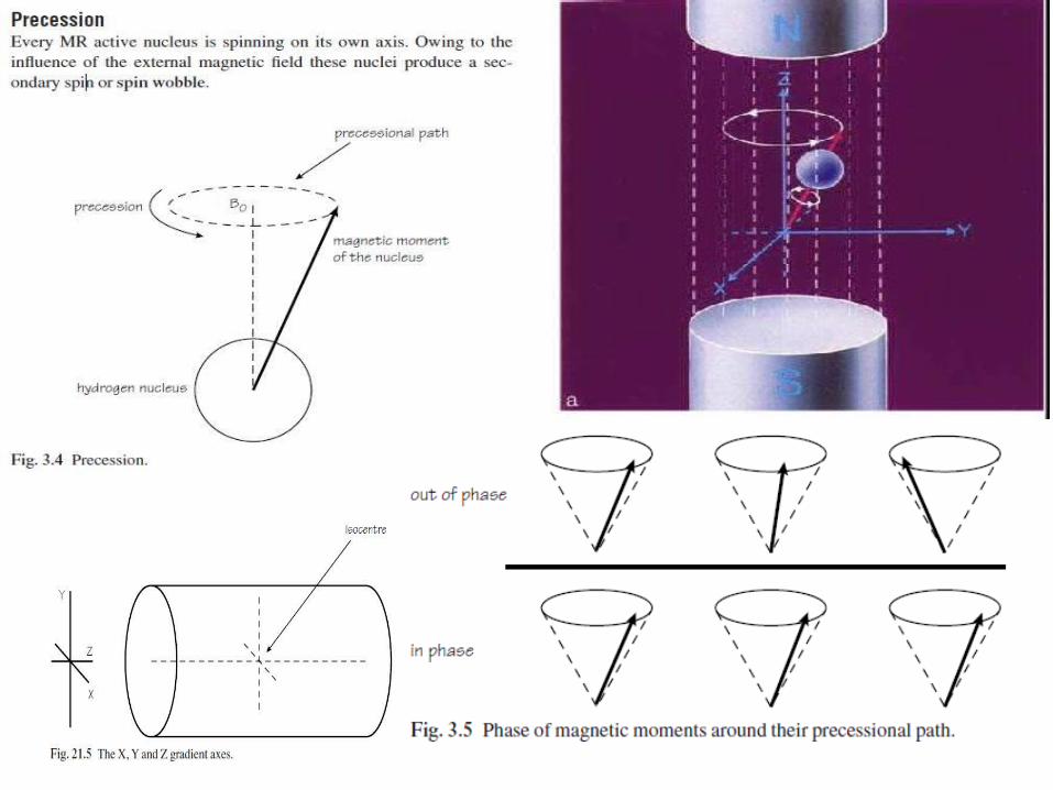

ResonanceResonance is an energy transition that occurs when an object is subjected to a frequency the same as its own. In MR, resonance is induced by applying a radiofrequency (RF) pulse:• at the same frequency as the precessing hydrogen nuclei;• at 90° to B0.


T1 recoveryThe T1 time is defined as the time it takes for 63% of the longitudinal magnetization to recover. The period of time during which this occurs is the time between one excitation pulse and the next or the TR.

T1 recovery in fat• T1 relaxation occurs as a result of nuclei exchanging the energy given to them by the RF pulse to their surrounding environment. The efficiencyof this process determines the T1 time of the tissue in which they are situated.• Owing to the fact that fat is able to absorb energy quickly the T1 time of fat is very short, i.e. nuclei dispose of their energy to the surrounding fat tissue and return to B0 in a short time.T1 recovery in water• Water is very inefficient at receiving energy from nuclei. The T1 time of water is therefore quite long, i.e. nuclei take a lot longer to dispose of their energy to the surrounding water tissue andreturn to B0.

• The TR controls how much of the NMV in fat or water has recovered before the application of the next RF pulse.

T2 decayT2 decay is caused by the exchange of energy from one nucleus to another. It is called spin–spin energy transfer. It occurs as a result of the intrinsic magnetic fields of the nuclei interacting with each other. This energy exchange produces a loss of phase coherence or dephasing, and results
in decay of the NMV in the transverse plane. T2 decay time is the time it takes for63% of the transverse magnetization to be lost due to dephasing, i.e. 37% remains. The period of time over which this occurs is the time between the excitation pulse and the MR signal or the TE. The TE therefore determines how much T2 decay occurs in a particular tissue.

Short TEs do not permit full dephasing in either fat or water so their transverse components are similar. There is little contrast difference between fat and water due to differences in T2 decay using short TEs.
Long TEs allow dephasing of the transverse components in fat and water. There is a contrast difference between fat and water due to differences in T2 decay times when using long TEs.

T1 weightingA TR is selected that is short enough to ensure that the NMV in neither fat nor water has had time to relax back to B0 before the application of the next excitation pulse. If the TR is long, the NMV in both fat and water recovers and their respective T1 relaxation times can no longer be distinguished. A T1 weighted image is an image whose contrast is predominantly due to the differences in T1 recovery times of tissues. In T1 weighted images, tissues with short T1 relaxation times such as fat, are bright (high signal), because they recover most of their longitudinal magnetization during the TR and therefore more magnetization is available to be flipped into the transverse plane by the next RF pulse.Tissues with long T1 relaxation times such as water, are dark (low signal) because they do not recover much of their longitudinal magnetization during the TR and therefore less magnetization is available to be flipped into the transverse plane by the next RF pulse.

T2 weightingA TE is selected that is long enough to ensure that the NMV in both fat and water have time to decay. If the TE is too short, the NMV in neither fat nor water has had time to decay and their respective T2 times cannot be distinguished. A T2 weighted image is an image whose contrast is predominantly due to the differences in T2 recovery times of tissues. For T2 weighting the differences between the T2 times of tissues is exaggerated, therefore the TE must be long. T1 effects are diminished by selecting a long TR.
• Tissues with a short T2 decay time such as fat are dark (low signal) because they lose most of their coherent transverse magnetization during the TE period.
• Tissues with a long T2 decay time such as water are bright (high signal), because they retain most of their transverse coherence during the TE period.• T2 weighted images best demonstrate pathology as most pathology has an increased water content and is therefore bright on T2 weighted images.


Proton density weightingIn a PD weighted image differences in the proton densities (number of hydrogen protons in the tissue) must be demonstrated. To achieve this both T1 and T2 effects are diminished. T1 effects are reduced by selecting a long TR and T2 effects are diminished by selecting a short TE.
• Tissues with a low proton density are dark (low signal) because the low number of protons result in a small component of transverse magnetization.
• Tissues with a high proton density are bright (high signal) because the high number of protons result in a large component of transverse magnetization.

Conventional spin echo pulse sequence (SE)
Conventional spin echo pulse sequences are used to produce T1, T2 or proton density weighted images and are one of the most basic pulse sequences used in MRI. In a spin echo pulse sequence there is a 90° excitation pulse followed by a 180° rephasing pulse followed by anEcho.

T1 weighted T2 weighted
Spin Echo Contrast

Spin Echo Contrast
PD weighted T2 weighted

Fast or turbo spin echo (FSE/ TSE)• FSE employs a train of 180° rephasing pulses, each one producing a spin echo. This train of spin echoes is called an echo train. The number of 180° RF pulses and resultant echoes is called the echo train length (ETL) or turbo factor. The spacing between each echo is called theecho spacing.• After each rephasing, a phase encoding step is performed and data from the resultant echo are stored in K space . Therefore several lines of K space are filled with every TR instead of one line as in conventional spin echo. As K space is filled more rapidly, the scan time decreases.
UsesFSE produces T1, T2 or proton density scans in a fraction of the time of Conventional spin echo. Because the scan times are reduced, matrix size can be increased to improve spatial resolution. FSE is usually used for brains, spines, joints, extremities and the pelvis


Inversion recovery (IR)• Inversion recovery is a spin echo sequence that begins with a 180° inverting pulse. This inverts the NMV through 180°.• When the pulse is removed the NMV begins to relax back to B0. A 90° pulse is then applied at time interval TI (time from inversion) after the 180° inverting pulse.• A further 180° RF pulse is applied which rephases spins in the transverse plane and produces an echo at time TE after the excitation pulse.


STIR (short TI inversion recovery)
It uses short TIs such as 100– 180 ms, depending on field strength . TIs of this magnitudeplace the 90° excitation pulse at the time that NMV of fat is passing exactly through the transverse plane. At this point (called the null point) there is no longitudinal component in fat. Therefore the 90° excitation pulse produces no transverse component in fat and therefore nosignal. In this way a fat suppressed image results .
FLAIR (fluid attenuated inversion recovery)It uses long TIs such as 1700–2200 ms, to null the signal from CSF in exactly the same way as the STIR sequence . Because CSF has a long T1 recovery time, the TI must be longer to correspond with its null point.


Gradient echo pulse sequences ( FFE / GRE /GE)Gradient echo pulse sequences are sequences that use a gradient to reduce magnetic homogeneity effects, as opposed to a 180° RF pulse used in spin echo sequences.
The gradient echo sequence differs from the spin echo sequence in regard to: • the flip angle usually below 90°• the absence of a 180° RF rephasing pulseA flip angle lower than 90° (partial flip angle) decreases the amount of magnetization tipped into the transverse plane. The consequence of a low-flip angle excitation is a faster recovery of longitudinal magnetization that allows shorter TR/TE and decreases scan time.
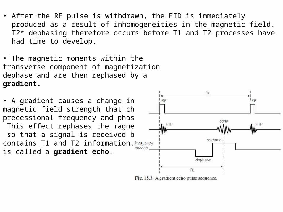
• After the RF pulse is withdrawn, the FID is immediately produced as a result of inhomogeneities in the magnetic field. T2* dephasing therefore occurs before T1 and T2 processes have had time to develop.
• The magnetic moments within the transverse component of magnetization dephase and are then rephased by a gradient.
• A gradient causes a change in the magnetic field strength that changes the precessional frequency and phase of spins. This effect rephases the magnetic moments so that a signal is received by the coil that contains T1 and T2 information. This signal is called a gradient echo.

In a gradient echo pulse sequence ,rephasing is performed by the frequency encoding gradient .• The nuclei are dephased with a negative gradient pulse. The negative gradient slows down the slow nuclei even further and speeds up the fast ones. This accelerates the dephasing process.• The gradient polarity is then reversed to positive. The positive gradient speeds up the slow nuclei and slows down the fast ones. The nuclei rephase and produce a gradient echo.
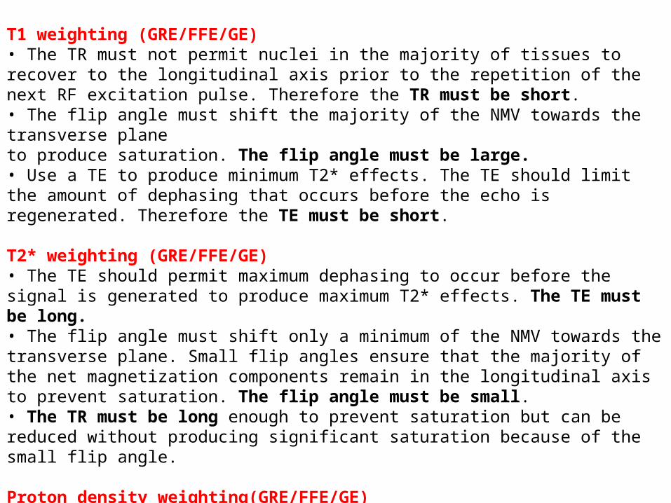
T1 weighting (GRE/FFE/GE)• The TR must not permit nuclei in the majority of tissues to recover to the longitudinal axis prior to the repetition of the next RF excitation pulse. Therefore the TR must be short.• The flip angle must shift the majority of the NMV towards the transverse planeto produce saturation. The flip angle must be large.• Use a TE to produce minimum T2* effects. The TE should limit the amount of dephasing that occurs before the echo is regenerated. Therefore the TE must be short.
T2* weighting (GRE/FFE/GE)• The TE should permit maximum dephasing to occur before the signal is generated to produce maximum T2* effects. The TE must be long.• The flip angle must shift only a minimum of the NMV towards the transverse plane. Small flip angles ensure that the majority of the net magnetization components remain in the longitudinal axis to prevent saturation. The flip angle must be small.• The TR must be long enough to prevent saturation but can be reduced without producing significant saturation because of the small flip angle.
Proton density weighting(GRE/FFE/GE)Select TR and flip angle to produce minimum T1 effects and a TE toproduce minimum T2*/T2 effects. As a result proton density predominates.• The flip angle must be small.• The TR must be long to minimize saturation and T1 effects as well.• The TE must be short to minimize the T2*/T2 effects.

Advantages of Gradient echo (GRE / FFE) sequences
1. To detect haemorrhage, calcification
2. Abdominal breath hold studies3. 3 D imaging4. Time of flight (TOF) or inflow
angiography

Thank you …