MR Kevin Highland MBBS MRCOphth FRCS(Glasgow)
43
MR Kevin Highland MBBS MRCOphth FRCS(Glasgow)
Transcript of MR Kevin Highland MBBS MRCOphth FRCS(Glasgow)

MR Kevin Highland MBBS MRCOphth FRCS(Glasgow)
Dengue chikungunya and zika viruses have emerged as increasingly important causes of human disease
Ocular manifestations of these diseases have become more prevalent over the past few years
This review highlights the current understanding of the ocular findings emphasizing the retinal manifestations related to these diseases
Arbovirus is a term used to refer to any virus that is transmitted by an arthropodvector
Differ in size morphology gene sequence and replication
Arbovirus (ARthropod-
BOrne virus)
Flavivirus
Dengue
Zika
Alphavirus
chikungunya
Vector borne diseases account for over 17 of all infectious diseases causing more than 1 million deaths annually
The illnesses caused by the arboviruses have very similar clinical presentation with prominent fever headache rash myalgia and arthralgia
In fact serologic surveys have demonstrated that outbreaks attributed to DENGUE in the past have actually turned out to be CHIKV or ZIKV infections
Globalization of travel and trade unplanned urbanization and environmental challenges such as climate change are having a significant impact on disease transmission
Vectors Main diseases
Aedes Aegypti Dengue fever
Zika virus
Rift valley fever
Yellow fever
Chikungunya
Anopheles Malaria
Lymphatic filariasis
Culex Japanese encephalitis
Lymphatic filariasis
West Nile fever
DENGUE
Dengue fever (DFV) is the most common mosquito borne viral disease in humans and has become a major health concern due to increased incidence
Aedes aegypti mosquito is the main vector responsible and is predominant in tropical weather regions
The disease has a large clinical spectrum and reports of ocular manifestations have been recently published with complications ranging from mild blurring of vision to significant morbidity with severe visual impairment
Dengue infection is characterized by an acute onset of fever associated with symptoms of malaise sore throat rhinitis and cough headache muscle ache retro-orbital pain joint pain abdominal discomfort and rash
Other clinical manifestations of dengue are related to the bleeding diathesis from thrombocytopenia
Infection is caused by the dengue virus of which there are four closely related but antigenically distinct serotypes that do not confer cross-immunity
As such individuals in endemic countries are not protected from the other serotypes after infection with one serotype
Dengue infection is usually a clinical diagnosis but can be confirmed with lab tests based on the time of presentation
Frequently used tests include PCR and IgM or IgG enzyme immunoassays
During the early phase of the infection when febrile illness is within 5 days dengue PCR is performed
If febrile illness exceeds 5 days the preferred tests are dengue IgM and IgG tests
The precise pathophysiologic mechanism of dengue ophthalmic complications is not well understood however many studies have alluded to the possibility of an immune-mediated process as a likely mechanism
Ocular finding usually present 4-5 days after onset of fever
Blurring of Vision
The most common complaint was blurring of vision
In three case series involving 23 patients all of them complained of blurring of vision with visual acuity decreasing to counting fingers in two of the studies
Ophthalmic Complications of Dengue Fever a Systematic Review
Vivien Cherng-Hui Yip Srinivasan Sanjay and Yan Tong Koh
Scotoma
The next most common symptom was central scotoma
The areas of scotoma generally corresponded to the areas of oedema and haemorrhage in the macula
In the case series by Chan et al (2006) 12 out of 13 patients had central scotoma in association with blurring of vision
Ocular Pain
Usually associated with headache
Location of the pain is typically described as retrobulbar and worse on eye movements
Headache features in patients with dengue virus infection
Domingues RB1 Kuster GW Onuki de Castro FL Souza VA Levi JE Pannuti CS
Subconjunctival Haemorrhage
A study by Kapoor et al accounted for the majority of cases of dengue-related subconjunctival haemorrhage
134 patients hospitalized with a diagnosis of dengue fever during an epidemic were included
50 patients had subconjunctival haemorrhage 42 (84) patients had petechial haemorrhages present in the conjunctivae and eight (16) patients had diffuse haemorrhages noted in one to four quadrants
Of all lab parameters evaluated marked thrombocytopenia emerged to be significantly associated with ocular haemorrhage
Uveitis Patients with dengue infection rarely presented with uveitis
A case series by Gupta et al reported dengue-related uveitis in six patients The pts presented with ocular symptoms 3-5 months after contracting dengue fever without any other attributable cause for uveitis
They were treated with topical steroids cycloplegic and ocular hypotensive medications when required and oral steroid in case of posterior segment involvement
We should be aware of the delayed ophthalmic complications of dengue infection like uveitis which might occur even after complete recovery from the systemic disease
Uveitis following dengue fever
Gupta A Srinivasan R Setia S Soundravally R Pandian DG
Eye (Lond) 2009 Apr23(4)873-6 doi 101038eye2008124 Epub 2008 May 9
CASE STUDY A 32-year old woman was diagnosed with dengue
fever by her GP on day 4 of her febrile illness
Blood investigations showed progressive thrombocytopenia and was positive for dengue virus on PCR analysis
On day 7 after onset of fever she noticed sudden painless bilateral blurring of vision upon waking Platelet count was at its lowest of 26 x 109L
Visual acuity were 20400 and counting fingers right and left eyes respectively
External and slit lamp examinations were unremarkable and intraocular pressures were within normal limits
Dilated fundi examination revealed focal macular chorioretinitis with macular edema associated with vasculitis and flame hemorrhage
Fundus fluorescein angiography revealed extensive periphlebitis in both eyes
httpwwwuveitisorgwp-
contentuploads201705dengue_chorioretinitis_and_dengue_related_ophthalmic_complicationspdf
Dengue infection can affect the retina microcirculation either by direct viral infection or activation of inflammation through an immune-mediated reaction
The pathogenesis of lesions appears to be the same as the clinical disease haemoconcentration vasculitis and coagulation disorders
Central retinal vein occlusion
concomitant with dengue fever Punithamalar VelaithamEmail authorView ORCID ID profile and
Nandini Vijayasingham
International Journal of Retina and Vitreous2016
The majority of ocular changes in DFV are in the vascular system of the posterior segment
Maculopathy is the main retinal manifestation (affecting up to 10 of the cases) and is generally bilateral (73) and characterized by vasculitis and hemorrhages
Arboviruses and the eye
bullGabriel Costa de AndradeEmail author Camila VVenturaPaulo Augusto de Arruda Mello FilhoMauriacutecio Maia Silvana Vianello and
Eduardo Buumlchele Rodrigues
Haemorrhages associated with dengue-related maculopathy are mostly intraretinal and can take the form of dot blot or flame-shaped haemorrhages
Vascular sheathing and vasculitis were often found in association with macular haemorrhage
Dengue-related foveolitis refers to the yellow-orange lesion at the fovea of patients
with dengue maculopathy which corresponds to a disruption of the outer neurosensory retina in optical coherence tomography (OCT)
It was formally described by Loh et al in 10 eyes of 6 patients and the term foveolitis was coined
Retinal manifestations
Optic Neuropathy
Optic neuropathy is relatively uncommon compared to other dengue-related ocular signs This can present with optic disc swelling hyperaemia and disc hemorrhages
Optic neuropathy associated with dengue fever
Sanjay S1 Wagle AM Au Eong KG
Serial photographs of the right
eye in showing the optic disc
1(a) 7(b) and 20(c) days after
onset of dengue fever This case
resolved spontaneously with
supportive care
The main differential diagnoses are herpetic and cytomegalovirus retinitis but both of them are more common in immunodeficient individuals with a more exuberant inflammation
Although Dengue Retinopathy may morphologically mimic herpetic or cytomegalovirus retinitis the history of fever joint pains and skin rash before the onset of visual symptoms is helpful in the diagnosis particularly in the endemic regions
ZIKV is a mosquito-borne infection mainly transmitted in Americas by A aegypti the same vector that transmits DFV and CHIKV
Additionally there are also reports of ZIKV infection following sexual perinatal and blood transfusion
ZIKV can also lead to severe congenital malformations in newborns whose mothers were infected during pregnancy especially in the first trimester of pregnancy
Musso D Roche C Robin E Nhan T Teissier A Cao-Lormeau V et al Potential sexual transmission of
Zika virus Emerg Infect Dis 201521(2)359ndash361 doi 103201eid2102141363
Although microcephaly is the major finding in these newborns recent publications have described other malformations associated with ZIKV congenital infection including hearing loss limb anomalies and ocular findings
Therefore a new terminology has been given to this clinical condition Congenital Zika Syndrome (CZS)
De Paula Freitas B de Oliveira Dias JR Prazeres J et al Ocular findings in infants with microcephaly
associated with presumed Zika virus congenital infection in Salvador Brazil JAMA Ophthalmol 2016
doi101001jamaophthalmol20160267
A cross-sectional study of forty-three infants with congenital Zika syndrome had severe ocular abnormalities and all patients had bilateral involvement
The data revealed that 12 of cases of congenital Zika with microcephaly had anterior segment abnormalities and 88 had important macular and optic nerve abnormalities
The posterior ocular findings were focal pigment mottling chorioretinal atrophy with a predilection for the macular area congenital glaucoma and optic disc abnormalities
Ophthalmic examination is recommended in patients with congenital Zika syndrome
Cynthia A Moore MD PhD1 J Erin Staples MD PhD2 William B Dobyns MD3 et al
Ventura CV Maia M Travassos SB et al Risk factors associated with the
ophthalmoscopic findings identified in infants with presumed Zika virus congenital
infection JAMA Ophthalmol 2016134(8)912-918
Ocular abnormalities were detected in infants clinically diagnosed with ZIKV-related microcephaly
These findings include gross macular pigment mottling macular chorioretinal atrophy optic nerve hypoplasia and increased cup-to-disc ratio
Wide-angle fundus image (Retcamreg) of the right eye of an infant with presumed Zika virus congenital infection showing sharply demarcated chorioretinalscarring with gross pigment mottling on the macula
In particular retinal lesions including
well-defined chorioretinal atrophy and
gross pigmentation generally
affecting the macular region are
unique to ZIKV infection
Only 20 of patients infected with ZIKV are symptomatic The symptoms include fever headache maculopapular rash arthralgia and conjunctivitis which usually lasts for 1 week
Recent reports showed a mild disease in adults with acute infection which can include anterior uveitis and non-purulent conjunctivitis
The treatment of anterior uveitis related to ZIKV evolves topical corticosteroids and has a benign prognosis
Severe disease caused by ZIKV were recently described in patients from Brazil and French Polynesia as the Guillain-Barreacute syndrome and other neurological manifestations in patients infected by the virus
Neurological manifestations of Zika virus infection
Ana-Beleacuten Blaacutezquez and Juan-Carlos Saiz
Chikungunya fever (CHIKV) is an emerging mosquito-borne disease caused by an alphavirus from the Togaviridae family
The vectors are the same species involved in the transmission of DFV and ZIKV
CHIKV has been identified in over 60 countries in Asia Africa Europe and the Americas The virus is transmitted from human to human by the bites of infected female mosquitoes
After the bite of an infected mosquito onset of illness occurs usually between 4 and 8 days but can range from 2 to 12 days
As with several other mosquito-borne alphaviruses CHIKV causes a fever-rash-arthralgiasyndrome in humans
The name ldquoChikungunyardquo derives from the debilitating joint pain noted by local populations during an outbreak in 1952ndash1953 in what is now Tanzania
The local word means ldquothat which bends uprdquo and the name was given as a result of the stooped posture that resulted from the pain of the disease
Acute infection lasts for 1ndash10 days and is characterized by an abrupt onset of fever headache fatigue nausea vomiting rash myalgia and severe arthralgia
Joint pains may persist for months to years in some patients
Diagnosis is made through demonstration of CHIKV IgM antibody in the serum and also by reverse transcriptase-polymerase chain reaction (RT-PCR) from ocular fluids and serum
There is no specific antiviral drug treatment for CHIKV therefore treatment is directed primarily for relieving the symptoms including the joint pain and fever using anti-pyretics optimal analgesics and fluids
Chikungunya virus is known to affect the eye in a myriad of ways ranging from conjunctivitis to retinitis and even optic neuritis
Photophobia and retro orbital pain are often seen in the acute phase without any other signs of ocular involvement
The main ocular manifestation is an anterior uveitis often associated with pigmented keratic precipitates and ocular hypertension
The management of the anterior uveitis evolves topical and systemic corticosteroids to control inflammation
The raised IOP responds well to topical antiglaucoma medications
Posterior segment involvement of CHIKV infection may manifest as choroiditis retinitis neuroretinitis and optic disc neuritis
Chikungunya retinitis (CR) can present at the time of fever or after many weeks or months of the infection
Clinical features include vitritis hyperemic disc retina hemorrhages cotton wool spots and multifocal retinitis
45 year old woman complaining of blurred
vision 6 weeks following the resolution of
chikungunya fever in her left eye
Fundus photograph of the left eye showing
confluent area of retinal whitening suggestive
of retinitis
FFA reveals early hypofluorescence followed by
late hyperfluorescence in the posterior pole
OCT showed fluid-filled spaces in the outer
retina with serous retinal detachment
Fundus photograph and OCT showing resolving
retinitis lesion 2 weeks after initiation of
systemic steroid therapy
OCT after 4 months showing complete
resolution of retinitis with thinning of the inner
retinal layers nasal to the fovea
Chikungunya and the eye a review
Padmamalini Mahendradas 1 Kavitha Avadhani1 and Rohit Shetty
Chikungunya Retinitis
Dengue Zika Chikungunya
type flavivirus flavivirus alphavirus
Duration of disease 4-7 weeks 1-2 weeks 1-2 weeks
Fever gt39 ++ + +++
arthalgia +++ ++ ++++
conjunctivitis + +++ (20 symptomatic) +
Retro orbital pain +++ ++ +
Maculopapular
exanthema
++ +++ ++
uveitis + ++ +++ with High IOP
retina Choroiditis retinitis
macular oedema
neuroretinitis
Macular pigment
mottling macular
chorioretinal atrophy
optic nerve hypoplasia
Choroiditis retinitis
neuroretinitis and optic
disc neuritis
The Vector for Dengue Chikungunya and zika is
A) Anopheles Mosquitoes
B) Culex mosquito
C) Aedes aegypti
D) None of the above
E) All of the above
Confirmatory tests for dengue infection
A) IGG and IGM test should be performed in the first 5 days of presentation
B) PCR testing should be done late after the fever settles
C) Can cross react with testing for chikungunya
D) PCR testing is best done early during the febrile phase
Arboviruses cause the following diseases except
A) Encephalitis
B) Febrile diseases
C) Haemorrhagic fevers
D) Pneumonia
What are symptoms of Zika infection
A) Headache fever rash
B) Red eyes (conjunctivitis)
C) Musclepain joint pain and weakness
D) All of the above
Which of these conditions is least likely to present with conjunctivitis
A) Dengue
B) Zika
C) Chikungunya
What should I do with a patient who is sero-positive for arbovirus infection and is complaining of blurred vision blind spots andor floaters
A) Observe for a few weeks to see if the condition improves
B) Give antibiotic drops and lubricants
C) Refer to an Ophthalmologist for a complete eye exam
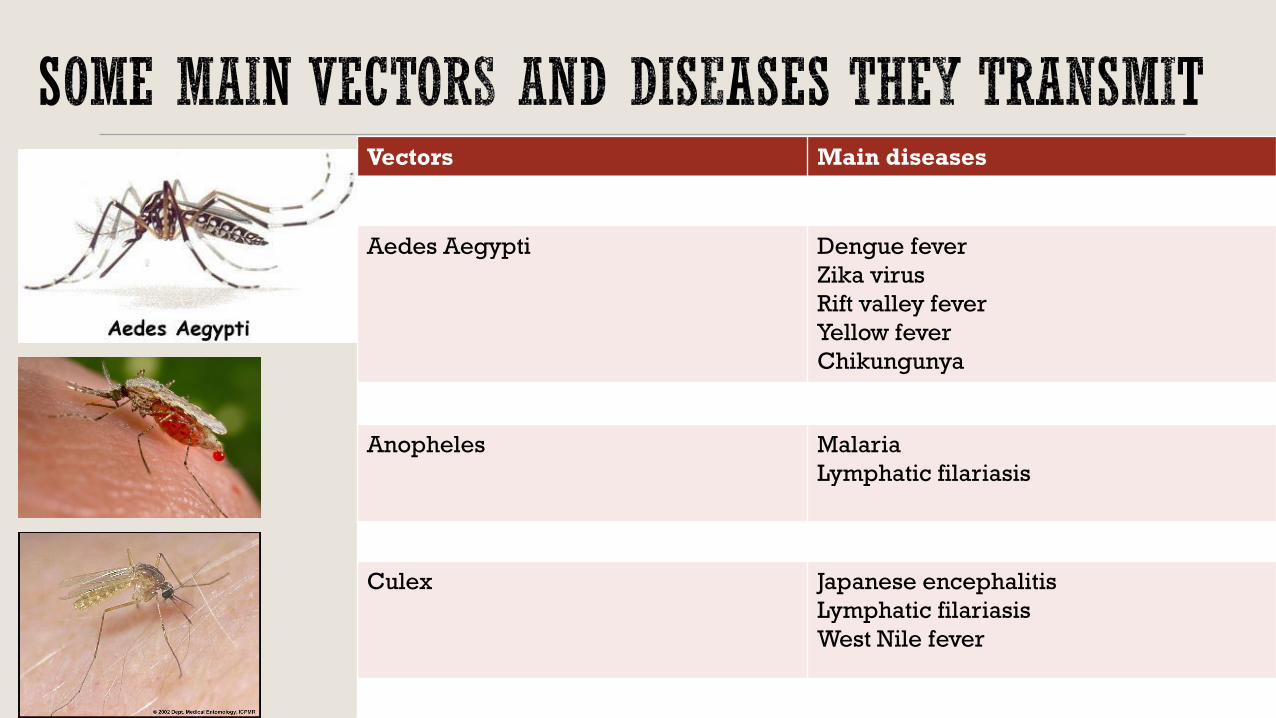
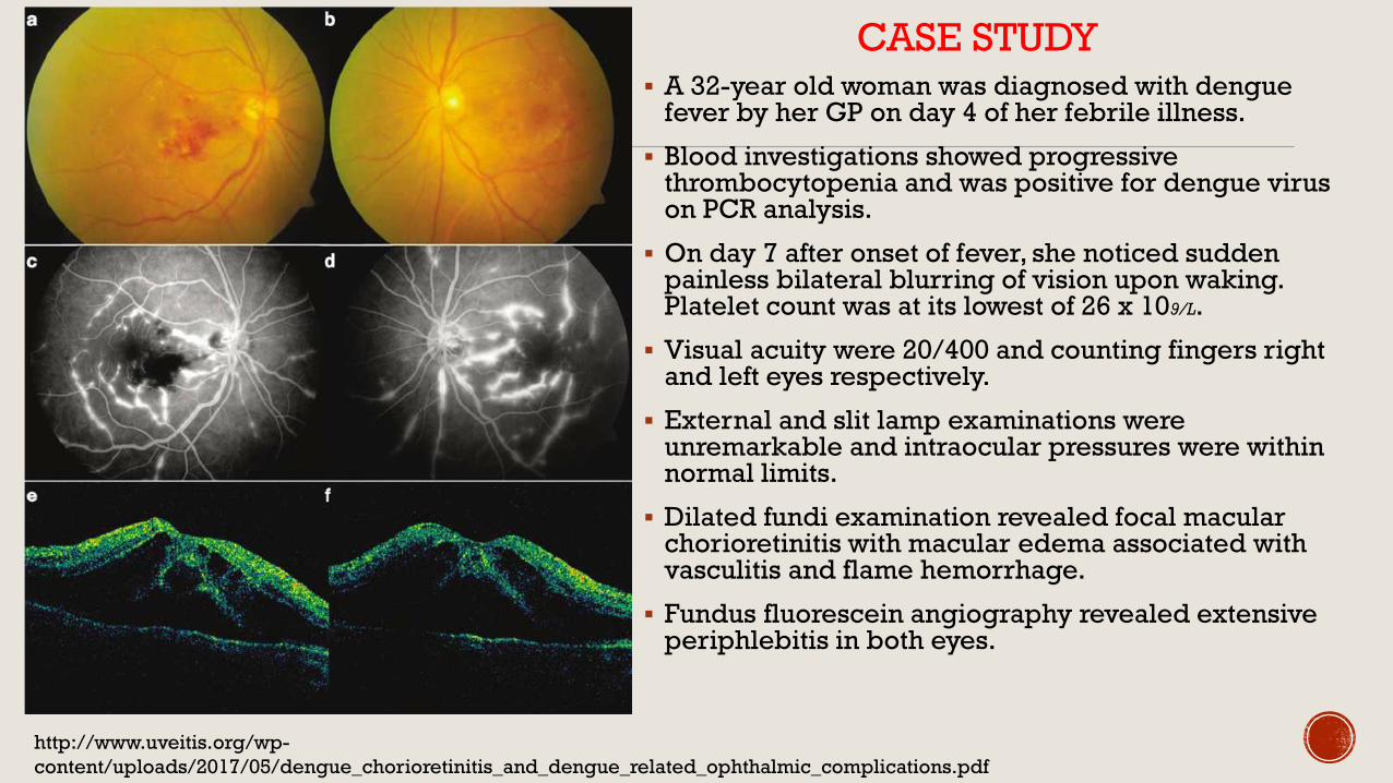
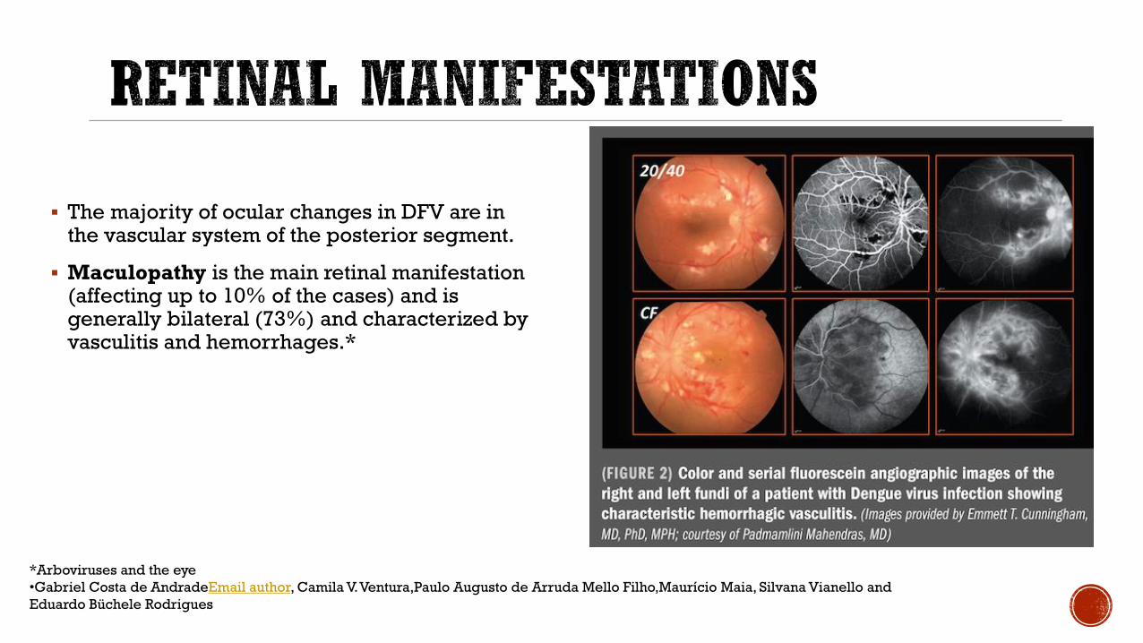
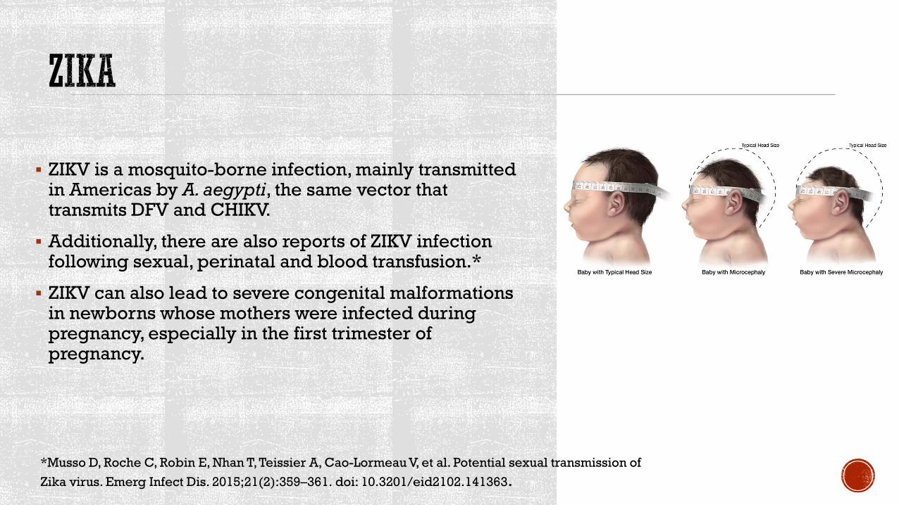
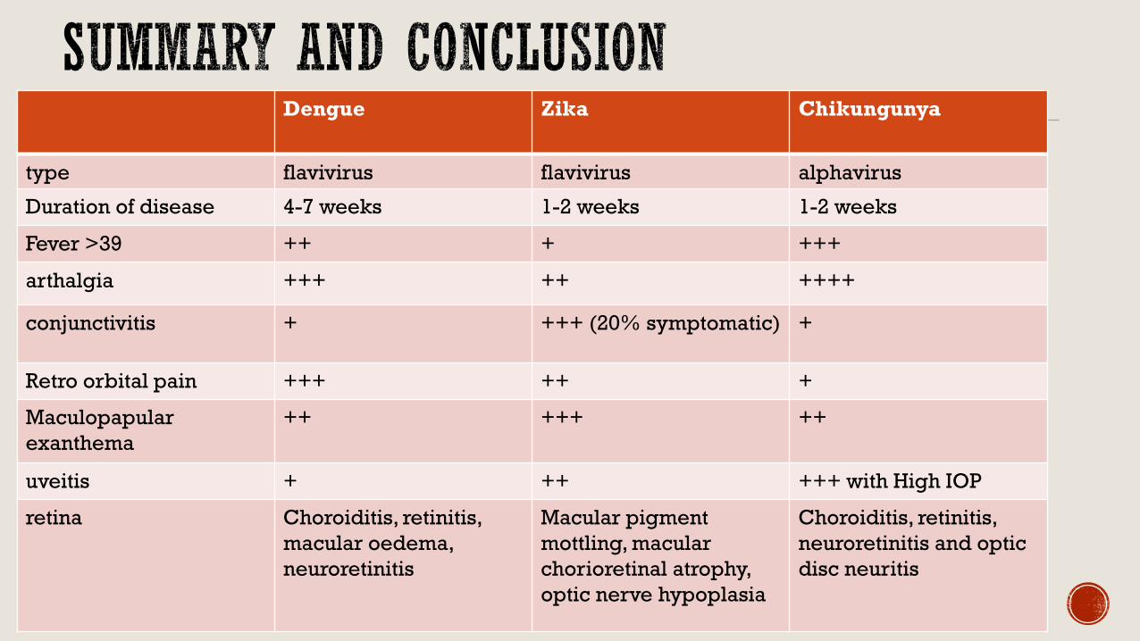

What should I do with a patient who is sero-positive for arbovirus infection and is complaining of blurred vision blind spots andor floaters
A) Observe for a few weeks to see if the condition improves
B) Give antibiotic drops and lubricants
C) Refer to an Ophthalmologist for a complete eye exam