MR Imaging of Solitary Fibrous Tumors in the Head and Neck · MR Imaging of Solitary Fibrous Tumors...
7
136 Korean J Radiol 6(3), September 2005 MR Imaging of Solitary Fibrous Tumors in the Head and Neck Objective: Solitary fibrous tumor (SFT) is a very rare tumor. The purpose of this study is to determine the MR imaging features of SFT in the intracranial and extracranial head and neck regions. Materials and Methods: We retrospectively reviewed six MR images and two CT images of six histologically proven cases of SFT that occurred in four men and two women, and their ages ranged from 46 to 59 years. These imaging find- ings were correlated with the microscopic findings of their surgical specimens. Results: Six SFTs arose in the meninges (the petrous ridge and the pituitary fossa), the parotid gland, the parapharyngeal space, the buccal space and the maxillary sinus. On the MR images, SFTs in the intracranial and extracranial head and neck regions were mostly isointense to the muscle on the T1-weighted images, they were hyperintense on the T2-weighted images and they all had intense enhancement. On the T1- and T2-weighted images, hypointense lines were observed within in five SFTs. On the CT images, the SFTs were hypodense to the muscle on the unenhanced images and they were heterogeneously enhanced on the contrast-enhanced images. An exceptional case of pituitary SFT was hypointense on the T2-weighted images and it was hyperdense on the unenhanced CT images, which correlated with the increased collagenous com- ponent and the cellular compactness. Conclusion: The imaging features of SFT are nonspecific; however, SFT should be included in the differential diagnosis of masses involving the intracra- nial and extracranial head and neck regions. olitary fibrous tumor (SFT) is the current name for a distinctive neoplasm that most frequently occurs in the pleura. Since Klember and Rabin first described SFT as a localized fibrous mesothelioma in 1931 (1), the histogenesis of the solitary fibrous tumor has been debated as to whether it is a tumor of a mesothelial or mesenchymal cell origin, and the latter is now the favored answer (2, 3). Although it is an uncommon tumor, SFT of the pleura is a well-established entity. More recently, attention has been drawn to the presence of the occasional SFTs that arise at extrapleural sites such as the lung, mediastinum, pericardium, peritoneum, meninges, head and neck (4 8, 11 13). Despite its characteristic histologic and immunohistochemical features, SFT of the head and neck remains a diagnostic challenge to both clinicians and radiologists, and it is often poorly recognized and confused with other neoplasms that more commonly occur at the head and neck. A review of the literature reveals sporadic reports on the imaging findings of SFT in the intracranial and extracranial head and neck regions, but really, there is only rather Hyun Jeong Kim, MD 1 Ho Kyu Lee, MD 2 Jeong Jin Seo, MD 3 Hyung Jin Kim, MD 4 Ji Hoon Shin, MD 2 Ae Kyung Jeong, MD 5 Jeong Hyun Lee, MD 2 Kyung Ja Cho, MD 6 Index terms : Magnetic resonance (MR) Head and neck neoplasms Head and neck neoplasms, MR Korean J Radiol 2005 ; 6 : 136-142 Received November 17, 2004; accepted after revision July 7, 2005. 1 Department of Radiology, Daejeon St. Mary’s Hospital, College of Medicine, The Catholic University; 2 Department of Radiology, Asan Medical Center, University of Ulsan College of Medicine; 3 Department of Radiology, Chonnam National University Medical School; 4 Department of Radiology, Samsung Medical Center, Sungkyunkwan University School of Medicine; 5 Department of Radiology, Ulsan University Hospital, University of Ulsan College of Medicine; 6 Department of Diagnostic Pathology, Asan Medical Center, University of Ulsan College of Medicine Address reprint requests to : Ho Kyu Lee, MD, Department of Radiology, Asan Medical Center, University of Ulsan College of Medicine, 388-1 Poongnap-dong, Songpa-gu, Seoul 138-736, Korea. Tel. (822) 3010-4400 Fax. (822) 476-4719 e-mail: [email protected] S
Transcript of MR Imaging of Solitary Fibrous Tumors in the Head and Neck · MR Imaging of Solitary Fibrous Tumors...

136 Korean J Radiol 6(3), September 2005
MR Imaging of Solitary Fibrous Tumorsin the Head and Neck
Objective: Solitary fibrous tumor (SFT) is a very rare tumor. The purpose ofthis study is to determine the MR imaging features of SFT in the intracranial andextracranial head and neck regions.
Materials and Methods: We retrospectively reviewed six MR images and twoCT images of six histologically proven cases of SFT that occurred in four menand two women, and their ages ranged from 46 to 59 years. These imaging find-ings were correlated with the microscopic findings of their surgical specimens.
Results: Six SFTs arose in the meninges (the petrous ridge and the pituitaryfossa), the parotid gland, the parapharyngeal space, the buccal space and themaxillary sinus. On the MR images, SFTs in the intracranial and extracranialhead and neck regions were mostly isointense to the muscle on the T1-weightedimages, they were hyperintense on the T2-weighted images and they all hadintense enhancement. On the T1- and T2-weighted images, hypointense lineswere observed within in five SFTs. On the CT images, the SFTs were hypodenseto the muscle on the unenhanced images and they were heterogeneouslyenhanced on the contrast-enhanced images. An exceptional case of pituitarySFT was hypointense on the T2-weighted images and it was hyperdense on theunenhanced CT images, which correlated with the increased collagenous com-ponent and the cellular compactness.
Conclusion: The imaging features of SFT are nonspecific; however, SFTshould be included in the differential diagnosis of masses involving the intracra-nial and extracranial head and neck regions.
olitary fibrous tumor (SFT) is the current name for a distinctive neoplasmthat most frequently occurs in the pleura. Since Klember and Rabin firstdescribed SFT as a localized fibrous mesothelioma in 1931 (1), the
histogenesis of the solitary fibrous tumor has been debated as to whether it is a tumorof a mesothelial or mesenchymal cell origin, and the latter is now the favored answer(2, 3).
Although it is an uncommon tumor, SFT of the pleura is a well-established entity.More recently, attention has been drawn to the presence of the occasional SFTs thatarise at extrapleural sites such as the lung, mediastinum, pericardium, peritoneum,meninges, head and neck (4 8, 11 13). Despite its characteristic histologic andimmunohistochemical features, SFT of the head and neck remains a diagnosticchallenge to both clinicians and radiologists, and it is often poorly recognized andconfused with other neoplasms that more commonly occur at the head and neck. Areview of the literature reveals sporadic reports on the imaging findings of SFT in theintracranial and extracranial head and neck regions, but really, there is only rather
Hyun Jeong Kim, MD1
Ho Kyu Lee, MD2
Jeong Jin Seo, MD3
Hyung Jin Kim, MD4
Ji Hoon Shin, MD2
Ae Kyung Jeong, MD5
Jeong Hyun Lee, MD2
Kyung Ja Cho, MD6
Index terms:Magnetic resonance (MR)Head and neck neoplasmsHead and neck neoplasms, MR
Korean J Radiol 2005;6:136-142Received November 17, 2004; accepted after revision July 7, 2005.
1Department of Radiology, Daejeon St.Mary’s Hospital, College of Medicine, TheCatholic University; 2Department ofRadiology, Asan Medical Center,University of Ulsan College ofMedicine; 3Department of Radiology,Chonnam National University MedicalSchool; 4Department of Radiology,Samsung Medical Center, SungkyunkwanUniversity School of Medicine;5Department of Radiology, UlsanUniversity Hospital, University of UlsanCollege of Medicine; 6Department ofDiagnostic Pathology, Asan MedicalCenter, University of Ulsan College ofMedicine
Address reprint requests to:Ho Kyu Lee, MD, Department ofRadiology, Asan Medical Center,University of Ulsan College of Medicine,388-1 Poongnap-dong, Songpa-gu, Seoul138-736, Korea. Tel. (822) 3010-4400 Fax. (822) 476-4719 e-mail: [email protected]
S

limited description of the imaging features of this tumor inthe literature. During the past five years, we have encoun-tered six cases of SFT in the intracranial and extracranialhead and neck regions. In this study, we describe the CTand MR imaging features of these six cases of SFT.
MATERIALS AND METHODS
The clinical data and imaging studies of six patients whosuffered with SFT involving the intracranial and extracra-nial head and neck regions were retrospectively reviewed.These patients were diagnosed during the five-year periodfrom January 1998 to December 2002. The study includesfour men and two women, and their mean age was 52years (age range; 46 59 years). All six patients underwentMR imaging and two patients underwent CT examinations.The MR imaging was performed using a 1.5-T MR unit(Signa; GE Medical Systems, Milwaukee, WI, USA)equipped with a head coil. An axial T1-weighted spin echosequence (TR/TE/NEX, 466 516/11 14/2; field of view,18 20 cm; section thickness, 4 5 mm with a 1 mm gap;matrix size, 256 256) and an axial T2-weighted spin echosequence (TR/TE/NEX, 4016 4000/96 104/1; field ofview, 18 20 cm; section thickness, 4 5 mm with a 0.4 1mm gap; matrix size, 256 256) were obtained for theprecontrast MRI. In addition, contrast-enhanced T1-weighted spin echo images were obtained in the axial,sagittal and coronal planes after the intravenous injectionof 0.1 mmol/kg of gadolinium dimeglumine. The CT scanwas obtained using a spiral CT scanner (Hispeed; GEMedical System, Milwaukee, WI, USA). The scanningparameters were 120 kVp, 200 mA and a 0.8-sec scan
time. Both the unenhanced and enhanced scans wereobtained in two patients. One patient underwent onlyunenhanced CT scanning. The enhanced scans wereobtained after the injection of 150 ml of iopromide(Ultravist 300, Schering, Seoul, Korea); 100 ml wasinjected as a bolus before scanning and 50 ml was infusedas a drip during scanning. Contiguous scans with 5-mmcollimation were obtained at 5-mm intervals from theexternal auditory canal to the thoracic inlet for the headand neck imaging, and from the top of the head to the skullbase for the brain imaging with the patient performingquiet respiration. The images were reviewed by two radiol-ogists working in consensus, and they were only aware ofthe histologic diagnosis. Both the CT and MR images wereevaluated for the location, margin, contour, MR signalintensity or CT density, internal architecture, pattern ofenhancement and the change of the adjacent structure. Onthe CT and the T1- and T2-weighted MR images, thedensity and signal intensity abnormalities were categorizedas low, iso- or high density/signal intensity as comparedwith the neck musculature. The margin was graded as welldefined or ill-defined, and the contour was graded assmooth or irregular. The pattern of intratumoral enhance-ment was categorized as homogeneous or heterogeneous,and the degree of enhancement was categorized as strong,moderate or weak. These MR and CT findings werecorrelated with the microscopic findings of the surgicallyobtained specimens. Surgical excision was performed in allpatients. The histologic evaluation and immunohistochemi-cal studies were performed on the excised specimens. Thediagnosis of SFT was established on the basis of thehistopathologic appearance and the immunohistochemical
MR Imaging of Solitary Fibrous Tumors in Head and Neck
Korean J Radiol 6(3), September 2005 137
Table 1. Summary Clinical and Imaging Features in Six Patients
Case Age/ Symptom Location Margin/ Preoperation MR CT
Sex Contour Diagnosis T1 T2 Pattern / Streak Density Pattern/ CommentGrade of En Grade of En
1 56/F headache meninges well/ meningioma iso high homo/ + low hetero/ dural ‘tail’(petrous ridge) smooth strong strong
2 56/M visual meninges well/ pituitary iso low hetero/ high ND dural ‘tail’ disturbance (pituitary fossa) smooth adenoma strong
3 59/M palpable mass deep lobe of well/ pleomorphic iso high hetero/ + ND erosion parotid gland irregular adenoma strong of mandible
4 49/F palpable mass parapharyngeal well/ neurogenic iso high homo/ + ND erosionspace smooth tumor strong of palate
5 46/M palpable mass buccal space well/ pleomorphic iso high homo/ + NDsmooth adenoma strong
6 38/M nasal bleeding maxillary sinus well / angiofibroma iso high homo/ ND erosionsmooth strong + of sinus wall
Note. En = enhancement, hetero = heterogeneously, homo = homogeneously, ill = ill-defined, irregular = irregular-contoured, iso = isointense, ND = notdone, smooth = smooth-contoured, strong = strong-enhanced, weak = weak-enhanced, well = well-defined

pattern of a diffuse and strong positive reaction for CD34.The diagnostic histopathologic findings were the presenceof a haphazard growth pattern of short spindle cells havingscant cytoplasm and a bland cytologic appearance, andthese cells were separated by strands of rope-like collagen(5 8).
RESULTS
The clinical and imaging features of our six patients aregiven in Table 1. There were two intracranial SFTs andfour extracranial SFTs included in this study. The individ-ual locations were the meninges, including the petrousridge and the pituitary fossa, the parotid gland, theparapharyngeal space, the buccal space and the maxillarysinus. The presenting symptoms were headache in a case ofthe meninges location, visual disturbance in a case of the
pituitary fossa location and an indolent palpable mass inthe extracranial located cases. Preoperative diagnosis wasmeningioma for a case of the petrous ridge location,pituitary adenoma for a case of the pituitary fossa location,pleomorphic adenoma for the cases of the parotid glandand buccal space locations, and neurogenic tumor for thecases with parapharyngeal space and paraspinal arealocations. Of the six SFTs, five were well-defined andsmooth-contoured, except for one that had an irregularcontour. On the MR images, all the SFTs were observed ashaving iso-signal intensity on the T1-weighted images(Figs. 1D and 3A) and they had high signal intensity on theT2-weighted images (Figs. 1C and 3B), except for a casehaving a pituitary fossa location, and this tumor had a lowsignal intensity on the T2-weighted images (Fig. 2B). Onthe contrast-enhanced T1-weighted images, homogeneousenhancement was observed in four SFTs (Figs. 1E and 3C)
Kim et al.
138 Korean J Radiol 6(3), September 2005
A B C
Fig. 1. Images of a 56-year-old man present-ing with headache (case 1).A. The axial unenhanced CT image showsan ill-defined low density lesion abutting thetemporal lobe (*). B. The axial contrast-enhanced CT imagedemonstrates the lesion to be a well-defined,heterogeneously well-enhanced, roundedand contoured mass.C. The axial T2-weighted MR image(4000/96 [TR/TE]) shows the lesion to be awell-defined, round mass that is heteroge-neously hyperintense to the muscle andthere are multiple, hypointense streaks(arrows) within the mass.D. The axial T1-weighted MR image(500/11) shows an isointense mass. Thehypointense streaks on the T2-weightedimage are also hypointense. E. The axial contrast-enhanced T1-weighted
MR images (500/11) reveal homogeneous enhancement of the mass that is based on the petrous ridge and it has a dural ‘tail’ (arrow).The streaks are well enhanced, as same as the other portions of the mass.
D E

and heterogeneous enhancement was observed in two. Allsix SFTs were strongly enhanced. On the T1- and T2-weighted images, linear or curvilinear low-signal-intensitylines were observed within five tumors, except a casehaving a pituitary fossa location (Figs. 1C, 1D, 3A, 3B and4A). These hypointese lines were less enhanced than theother portions of the tumor in four cases (Fig. 3C), andthese hypointense lines were enhanced to the same degreeas the other portions of the tumor in one case (Fig. 1E).
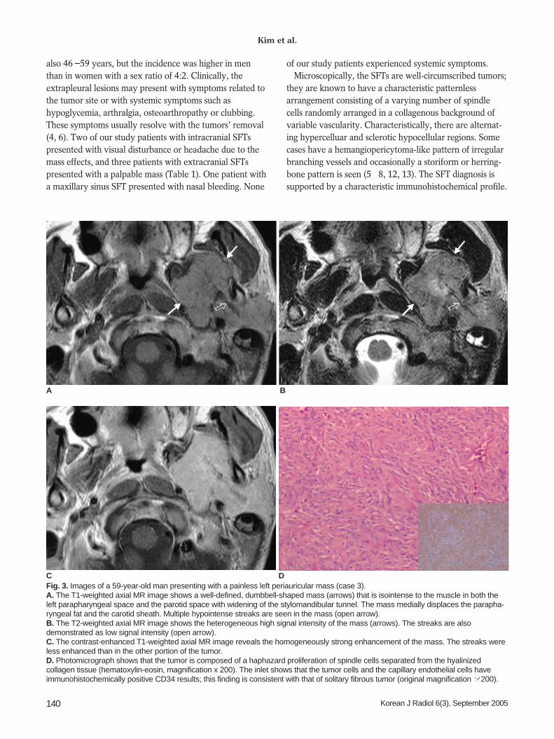
On the unenhanced CT images, the tumors wereobserved as having low density in the case of the petrousridge location (Fig. 1A) and the tumors had a high densityin a case of the pituitary fossa location (Fig. 2A). On thecontrast-enhanced CT images, heterogeneously strongenhancement was observed in a case having a petrousridge location (Fig. 1B). Two tumors in the meningesshowed adjacent meningeal thickening (a dural ‘tail’) (Fig.1E). Adjacent bony erosion was seen in the SFTs havingparotid gland and the parapharyngeal space locations dueto the large size of the tumors. A SFT of the maxillary
sinus expanded into the adjacent nasal cavity and then itwas clinically misdiagnosed as angiofibroma. In a case ofthe tumor with the pituitary fossa location, the tumordisplaced the pituitary gland upward.
Microscopic examination revealed benign SFT for all sixpatients. A more abundant collagenous component andrelative hypocellularity were shown in the pituitary SFT ofa low signal intensity on the T2-weighted images than wasshown for SFTs of a high signal intensity on the T2-weighted images (Figs. 2C and Fig. 3D). The hypocellular,but more collagenous areas in a tumor corresponded to thehypointense lines on the T1- and T2-weighted images.
All patients made uneventful recoveries and they are stillclinically and radiologically free from tumor recurrenceduring the follow-up period that ranged from seven to 27months (average follow-up period; 13 months).
DISCUSSION
Solitary fibrous tumor is an uncommon spindle celltumor that usually occurs in the pleura. The presence ofthis tumor has also been recently reported in a number ofextrapleural sites, including the intracranial and extracra-nial head and neck regions such as the meninges, orbit,nasal cavity, paranasal sinus, soft palate, epiglottis, thyroid,salivary gland, infratemporal fossa, buccal space andparapharyngeal space (4 8, 11 13). In our study, sixSFTs were located, respectively, in the meninges (thepetrous ridge and the pituitary fossa), parotid gland,parapharyngeal space, buccal space and the maxillarysinus. The primary SFT of the pituitary fossa in our study isthe first one to be reported at this site.
Solitary fibrous tumors occur with equal frequency inboth sexes and they usually occur in adults who are agedfrom 30 to 64 years (6). In our study, the age range was
MR Imaging of Solitary Fibrous Tumors in Head and Neck
Korean J Radiol 6(3), September 2005 139
Fig. 2. Images of a 56-year-old manpresenting with visual disturbance (case2).A. The axial unenhanced CT imageshows the lesion to be well-defined andhyperdense. B. The T2-weighted axial MR image(4000/98 [TR/TE]) shows a well-defined‘figure eight’ shaped mass that ishypointense to the muscle in thepituitary fossa and it has a suprasellarextension.C. Photomicrograph shows theprominent collagenous tissue withcellular areas that are composed ofhaphazardly arranged spindle cells.(hematoxylin-eosin stain, originalmagnification (100))A B
C
also 46 59 years, but the incidence was higher in menthan in women with a sex ratio of 4:2. Clinically, theextrapleural lesions may present with symptoms related tothe tumor site or with systemic symptoms such ashypoglycemia, arthralgia, osteoarthropathy or clubbing.These symptoms usually resolve with the tumors’ removal(4, 6). Two of our study patients with intracranial SFTspresented with visual disturbance or headache due to themass effects, and three patients with extracranial SFTspresented with a palpable mass (Table 1). One patient witha maxillary sinus SFT presented with nasal bleeding. None
of our study patients experienced systemic symptoms.Microscopically, the SFTs are well-circumscribed tumors;
they are known to have a characteristic patternlessarrangement consisting of a varying number of spindlecells randomly arranged in a collagenous background ofvariable vascularity. Characteristically, there are alternat-ing hypercelluar and sclerotic hypocellular regions. Somecases have a hemangiopericytoma-like pattern of irregularbranching vessels and occasionally a storiform or herring-bone pattern is seen (5 8, 12, 13). The SFT diagnosis issupported by a characteristic immunohistochemical profile.
Kim et al.
140 Korean J Radiol 6(3), September 2005
A B
Fig. 3. Images of a 59-year-old man presenting with a painless left periauricular mass (case 3).A. The T1-weighted axial MR image shows a well-defined, dumbbell-shaped mass (arrows) that is isointense to the muscle in both theleft parapharyngeal space and the parotid space with widening of the stylomandibular tunnel. The mass medially displaces the parapha-ryngeal fat and the carotid sheath. Multiple hypointense streaks are seen in the mass (open arrow).B. The T2-weighted axial MR image shows the heterogeneous high signal intensity of the mass (arrows). The streaks are alsodemonstrated as low signal intensity (open arrow). C. The contrast-enhanced T1-weighted axial MR image reveals the homogeneously strong enhancement of the mass. The streaks wereless enhanced than in the other portion of the tumor. D. Photomicrograph shows that the tumor is composed of a haphazard proliferation of spindle cells separated from the hyalinizedcollagen tissue (hematoxylin-eosin, magnification x 200). The inlet shows that the tumor cells and the capillary endothelial cells haveimmunohistochemically positive CD34 results; this finding is consistent with that of solitary fibrous tumor (original magnification 200).
C D
SFTs strongly stain for CD34 and the intermediate filamentvimentin, but they are usually negative for epithelial,vascular, neural crest and muscle markers (5 8, 13).
According to the reported MR features of SFTs, thesignal intensity was isointense to muscle on the T1-weighted images, the signal intensity increased withintravenous gadolinium and it was variable on the T2-weighted images. It’s been suggested that this variablesignal intensity depends mainly on the differences in themain components of the tumor, namely, the amount ofcollagen and fibroblasts, and also on the presence ofdegeneration. Intense enhancement of an SFT is generallydue to its high vascularity (8, 15). Several MR studies thatwere done on fibrosis reported that mature fibrous tissueusually has a low signal intensity on the T1-weighted,proton-density-weighted and T2-weighted images, and thisis related to the areas of hypocellularity and the abundantcollagen stroma (9, 10). In our study, the tumor signalintensity on the T2-weighted images decreased as thecollagenous component increased (Figs. 2B, 2C, 3B and3D). We also observed an interesting finding, that is, thelinear or curvilinear hypointense lines within the tumorson the MR images in four cases (Figs. 1C, 1D and 3A).Those lines corresponded to the hypocellular collagenousstroma intervening in the hypercelluar area, and thishypocellular collagenous stroma was composed of ahaphazard proliferation of spindle cells. Even if thesehypointense lines were not specific for SFT, we suggestthat these hypointense lines are helpful for including thisrare tumor in the differential diagnosis of masses involvingthe intracranial and extracranial head and neck regions.
For the CT-pathologic correlation of nine cases having apleural origin, Lee et al. (14) described these tumors ashaving isoattenuation relative to the adjacent musculature,and they had intense enhancement following intravenouscontrast injection. The intratumoral low attenuation areascorrelated with myxoid or cystic degeneration. In ourstudy, an SFT located in the pituitary fossa was hyperdenseon the unenhanced CT images. We presume that thecompactly arranged, abundant, collagenous component ofthis tumor increased the density of the tumor on the CTimages, and this was seen as a decreased signal on the T2-weighted MR images.
Most intracranial SFTs have a dural origin, and althoughthere have been several reports of intracranial and spinalmeningeal SFTs (8, 11), an SFT in the pituitary region hasnever been reported on before. Thickened dural ‘tails’,hyperostosis, skull erosion and capping cysts may all beseen in meningeal SFTs (8). The two meningeal SFTs in ourstudy were also shown to have dural tails.
The radiologic appearance for SFTs of the intracranial
and extracranial head and neck regions is nonspecific, andthis tumor is often misdiagnosed as being another type ofcommon tumor on the initial images. In our case of thepituitary SFT, the tumor was initially considered to be apituitary adenoma with hemorrhage because of its lowsignal intensity on the T2-weighted images. A transsphe-noidal approach for resection of this tumor resulted in itsincomplete excision because of the harder consistency ofthe SFT. This patient had to undergo repeated surgeryusing a transcranial approach in order to achieve completetumor resection. Therefore, any suggestion of the charac-teristics of this hard, fibrous tumor, according to its MRsignal intensity, may be important for the subsequentpatient care.
Most SFTs, especially those in the extrapleural sites, arebenign and they are cured with complete surgical excision.Previous reports have indicated that 13 37% of SFTshave been associated with local recurrence or histologicmalignancy such as an invasive growth pattern, cellularpleomorphism, abundant mitotic activity or metastasis (58, 10, 16). There is no report that a benign SFT has apotential to transform into a malignant SFT.
CONCLUSION
Solitary fibrous tumor is an uncommon tumor occurringin the intracranial and extracranial head and neck regions.On the MR images, this tumor is seen as a well-defined,smooth or irregular, contoured mass. It is isointenserelative to muscle on the T1-weighted images; the intensityincreasing with intravenous gadolinium, and the intensityis variable on the T2-weighted images. Darker signalintensity on the T2-weighted images represents a firmerfibrous tumor composed of abundant collageneous stroma.Solitary fibrous tumor should be included in the differen-tial diagnosis of masses involving the intracranial andextracranial head and neck regions because it has imagingfindings similar to those of the other more common lesionssuch as schwannoma and meningioma.
References1. Klemperer P, Rabin CB. Primary neoplasms of the pleura: a
report of five cases. Arch Pathol 1931;11:385-4122. England DM, Hochholzer L, McCarthy MJ. Localized benign
and malignant tumors of the pleura. A clinicopathologic reviewof 223 cases. Am J Surg Pathol 1989;13:640-658
3. Dervan PA, Tobin B, O’Connor M. Solitary (localized) fibrousmesothelioma: evidence against mesothelial cell origin.Histopathology 1986;10:867-875
4. Goodlad JR, Fletcher CD. Solitary fibrous tumor arising atunusual sites: analysis of a series. Histopathology 1991;19:515-522
5. Shin JH, Sung IY, Suh JH, Yang SO, Jeong YK, Lee JH, et al.
MR Imaging of Solitary Fibrous Tumors in Head and Neck
Korean J Radiol 6(3), September 2005 141

Kim et al.
142 Korean J Radiol 6(3), September 2005
Solitary fibrous tumor in the buccal space: MR Findings withpathologic correlation. AJNR Am J Neuroradiol 2001;22:1890-1892
6. Jeong AK, Lee HK, Kim SY, Cho KJ. Solitary fibrous tumor ofthe parapharyngeal space: MR imaging findings. AJNR Am JNeuroradiol 2002;23:473-475
7. Kim TA, Brunberg JA, Pearson JP, Ross DA. Solitary fibroustumor of the paranasal sinuses: CT and MR appearance. AJNRAm J Neuroradiol 1996;17:1767-1772
8. Martin AJ, Fisher C, Igbaseimokumo U, Jarosz JM, Dean AF.Solitary fibrous tumor of the meninges: case series and literaturereview. J Neurooncol 2001;54:57-69
9. Ferretti GR, Chiles C, Cox JE, Choplin RH, Coulomb M.Localized benign fibrous tumors of the pleura: MR appearance.J Comput Assist Tomogr 1997;21:115-120
10. Kransdorf MJ, Jelinek JS, Moser RP Jr, Utz JA, Hudson TM,Neal J, et al. Magnetic resonance appearance of fibromatosis. Areport of 14 cases and review of the literature. Skeletal Radiol1990;19:495-9
11. Pizzolitto S, Falconieri G, DeMaglio G. Solitary fibrous tumorof the spinal cord: a clinicopathologic study of two cases. Annalsof Diagnostic Pathology 2004;8:268-275
12. Kessler A, Lapinsky J, Berenholz L, Sarfaty S, Segal A. Solitaryfibrous tumor of the nasal cavity. Otolaryngol Head Neck Surg1999;121;826-888
13. Sato J, Asakura K, Yokoyama Y, Satoh M. Solitary fibroustumor of the parotid gland extending to the parapharyngealspace. Eur Arch Otorhinolaryngol 1998;255:18-21
14. Lee KS, Im J-G, Choe KO, Kim CJ, Lee BH. CT findings inbenign fibrous mesothelioma of the pleura: pathologic correla-tion in nine patients. AJR Am J Roentgenol 1992;158:983-986
15. Tateishi U, Nishihara H, Morikawa T, Miyasaka K. Solitaryfibrous tumor of the pleura: MR appearance and enhancementpattern. J Comput Assist Tomogr 2002;26:174-179
16. Sung SH, Chang JW, Kim JG, Lee KS, Han JH, Park SI. Solitaryfibrous tumors of the pleura: surgical outcome and clinicalcourse. Ann Thorac Surg 2005;79:303-7


![Solitary fibrous tumors in abdomen and pelvis: Imaging ......Solitary fibrous tumors (SFTs) were first described by Klemperer and Rabin in 1931 as a localized fibrous me-sothelioma[1].](https://static.fdocuments.in/doc/165x107/6112180e6352b44a0e769a1d/solitary-fibrous-tumors-in-abdomen-and-pelvis-imaging-solitary-fibrous.jpg)


![Solitary fibrous tumor occurring in the parotid gland: a case …...Solitary fibrous tumor (SFT) was described by Klemperer and Rabin in 1931 as a tumor of pleura [1]. Initially, this](https://static.fdocuments.in/doc/165x107/609ae127f5229b054724627b/solitary-fibrous-tumor-occurring-in-the-parotid-gland-a-case-solitary-fibrous.jpg)