Movement System Impairment (MSI) MSI Syndromes - Assumptions · Pelvic Tilt Indicated by Right...
15
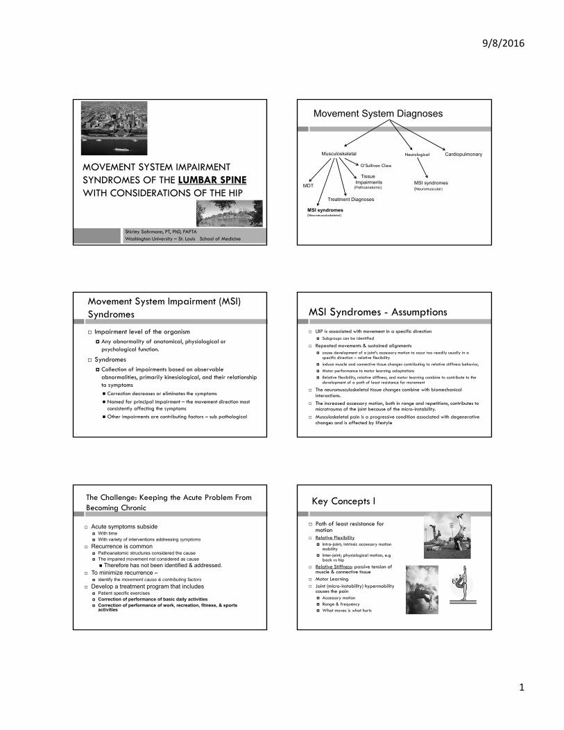
9/8/2016 1 MOVEMENT SYSTEM IMPAIRMENT SYNDROMES OF THE LUMBAR SPINE WITH CONSIDERATIONS OF THE HIP Shirley Sahrmann, PT, PhD, FAPTA Washington University – St. Louis School of Medicine Movement System Diagnoses Musculoskeletal Neurological Cardiopulmonary MSI syndromes (Neuromusculoskeletal) Tissue Impairments (Pathoanatomic) MSI syndromes (Neuromuscular) MDT Treatment Diagnoses O’Sullivan Class Impairment level of the organism Any abnormality of anatomical, physiological or psychological function. Syndromes Collection of impairments based on observable abnormalities, primarily kinesiological, and their relationship to symptoms Correction decreases or eliminates the symptoms Named for principal impairment – the movement direction most consistently affecting the symptoms Other impairments are contributing factors – sub pathological Movement System Impairment (MSI) Syndromes MSI Syndromes - Assumptions LBP is associated with movement in a specific direction Subgroups can be identified Repeated movements & sustained alignments cause development of a joint’s accessory motion to occur too readily usually in a specific direction – relative flexibility induce muscle and connective tissue changes contributing to relative stiffness behavior, Motor performance to motor learning adaptations Relative flexibility, relative stiffness, and motor learning combine to contribute to the development of a path of least resistance for movement The neuromusculoskeletal tissue changes combine with biomechanical interactions. The increased accessory motion, both in range and repetitions, contributes to microtrauma of the joint because of the micro-instability. Musculoskeletal pain is a progressive condition associated with degenerative changes and is affected by lifestyle The Challenge: Keeping the Acute Problem From Becoming Chronic Acute symptoms subside With time With variety of interventions addressing symptoms Recurrence is common Pathoanatomic structures considered the cause The impaired movement not considered as cause Therefore has not been identified & addressed. To minimize recurrence – identify the movement cause & contributing factors Develop a treatment program that includes Patient specific exercises Correction of performance of basic daily activities Correction of performance of work, recreation, fitness, & sports activities Key Concepts l Path of least resistance for motion Relative Flexibility Intra-joint; intrinsic accessory motion mobility Inter-joint; physiological motion, e.g back vs hip Relative Stiffness: passive tension of muscle & connective tissue Motor Learning Joint (micro-instability) hypermobility causes the pain Accessory motion Range & frequency What moves is what hurts
Transcript of Movement System Impairment (MSI) MSI Syndromes - Assumptions · Pelvic Tilt Indicated by Right...
9/8/2016
1
MOVEMENT SYSTEM IMPAIRMENT SYNDROMES OF THE LUMBAR SPINE WITH CONSIDERATIONS OF THE HIP
Shirley Sahrmann, PT, PhD, FAPTAWashington University – St. Louis School of Medicine
Movement System Diagnoses
Musculoskeletal Neurological Cardiopulmonary
MSI syndromes(Neuromusculoskeletal)
TissueImpairments
(Pathoanatomic)MSI syndromes(Neuromuscular)
MDT
Treatment Diagnoses
O’Sullivan Class
Impairment level of the organism Any abnormality of anatomical, physiological or
psychological function.
Syndromes Collection of impairments based on observable
abnormalities, primarily kinesiological, and their relationship to symptoms Correction decreases or eliminates the symptoms
Named for principal impairment – the movement direction most consistently affecting the symptoms
Other impairments are contributing factors – sub pathological
Movement System Impairment (MSI) Syndromes MSI Syndromes - Assumptions
LBP is associated with movement in a specific direction Subgroups can be identified
Repeated movements & sustained alignments cause development of a joint’s accessory motion to occur too readily usually in a
specific direction – relative flexibility
induce muscle and connective tissue changes contributing to relative stiffness behavior,
Motor performance to motor learning adaptations
Relative flexibility, relative stiffness, and motor learning combine to contribute to the development of a path of least resistance for movement
The neuromusculoskeletal tissue changes combine with biomechanical interactions.
The increased accessory motion, both in range and repetitions, contributes to microtrauma of the joint because of the micro-instability.
Musculoskeletal pain is a progressive condition associated with degenerative changes and is affected by lifestyle
The Challenge: Keeping the Acute Problem From Becoming Chronic
Acute symptoms subside With time With variety of interventions addressing symptoms
Recurrence is common Pathoanatomic structures considered the cause The impaired movement not considered as cause
Therefore has not been identified & addressed. To minimize recurrence –
identify the movement cause & contributing factors
Develop a treatment program that includes Patient specific exercises Correction of performance of basic daily activities Correction of performance of work, recreation, fitness, & sports
activities
Key Concepts l
Path of least resistance for motion
Relative Flexibility Intra-joint; intrinsic accessory motion
mobility Inter-joint; physiological motion, e.g
back vs hip
Relative Stiffness: passive tension of muscle & connective tissue
Motor Learning Joint (micro-instability) hypermobility
causes the pain Accessory motion Range & frequency What moves is what hurts

9/8/2016
2
Kinesiopathologic Model of Movement System – A Theoretical Construct
Musculoskeletal Nervous
Biomechanics
Repeated movementsSustained alignmentsINDUCERS Personal Characteristics – intrinsic
Activity Demands - extrinsic
Tissue Adaptations
Joint AccessoryHypermobility
Relative Stiffness of muscle & connective tissue
Relative FlexibilityIntra-jt + Inter-jt
Micro Macro trauma
Path of Least Resistance
Cardio-Pulmon -endocrine
Motor LearningNeural aff/efferent
Key Concepts II
The way everyday activities are performed is the critical issue Repeated movements and
Sustained alignments
Key Concepts III
You get what you train (many strategies to create moments at a joint or within a limb)
Presence of a muscle does not mean appropriate use
No magic in an exercise except if the desired motion is evident Does strengthening the serratus
Improve scapular upward rotation?
How Much Does the Hip Contribute?
Lumbar Rotation hip rotators, abductors
L Flexion Hip extensors
L Extension Hip flexors
Lumbar Rotation Femoral anteversion /
retroversion
L Flexion Cam impingement
Pincer impingement
L Extension
Muscular Structural
Muscular Factors – Affect Low Back
Hip flex/abd flexor Hip Extensors short
The Lumbar Spine Syndromes
Chronic Conditions Extension
Extension-rotation
Rotation Primary and Secondary
Acute Conditions - usually Flexion
Flexion-rotation

9/8/2016
3
Movement System Impairment Syndromes
Identify the cause of the dysfunction Identify the contributing factors
tissue & motor control impairments
Organize specific tissue impairments Minimizing treatment of isolated impairments
Usually limited in effectiveness
Provide a direction for intervention do not require identification of a specific pathoanatomical
structure (source)
Based on anatomy and kinesiology
Young – Tall - Acute
Lumbar Flexion Syndromes
Kendall: Muscles Testing & Function 1983
Tx:ShortenHip flexBack ext
Long psoasLong back extensors
What is his natural unsupported sitting alignment?
short ham-strings
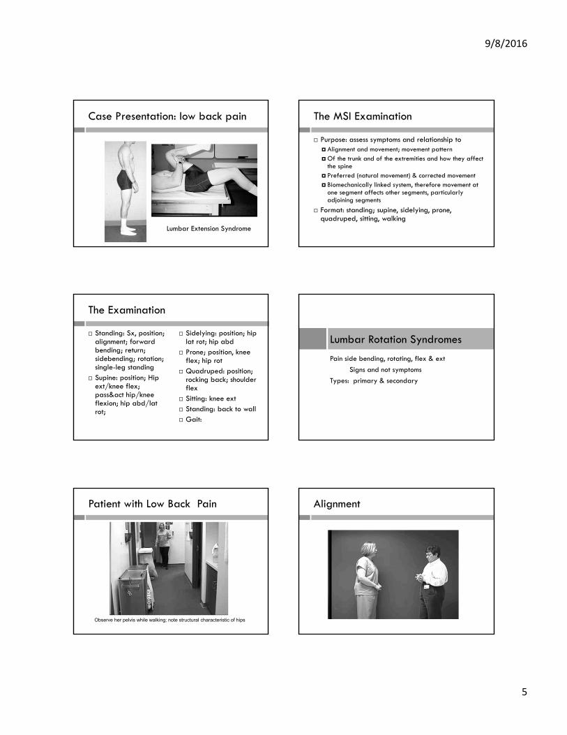
Case Presentation: low back pain - flexion Lumbar Flexion Syndrome
40 yr old ultra-marathonistExecutive
Lordotic?
Excessive flexion
Flexion Moment
Case Presentation: Low Back Pain – flexionYoung – tall – flexible: student/diver
sit-up exercisesHip extensors not short Relative flexibility: abdominals > hip extensors > back ext
Abdominals strongBack extensors more flexible than the hip extensors
Lumbar Flexion Syndrome
64 yo MD – DDD scoliosis
Post
9/8/2016
4
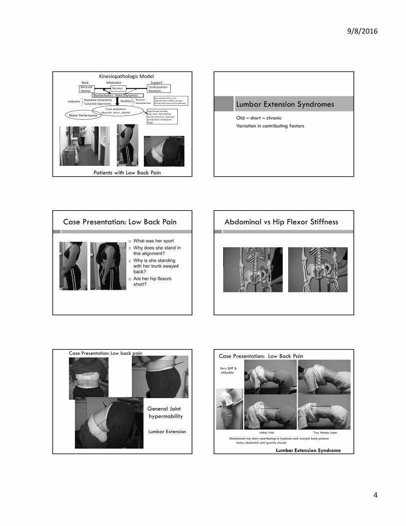
Kinesiopathologic Model Base Modulator SupportMuscularSkeletal
Nervous Cardio/pulmonMetabolic
Biomechanics: Static/Dynamics
Tissue adaptations
Muscular, neuro , skeletalMotor Performance
Inducers ModifiersRepeated movementsSustained alignments
Age, anthopometrics, sexgeneral tissue mobility, genetics,Activity level (excessive/insufficient)
Hypertrophy, atrophy,long, short, stiff, stabilize,recruit, derecruit, cocontractcoordination, boney/jointshape
Personalcharacteristics
Patients with Low Back Pain
Lumbar Extension Syndromes
Old – short – chronic
Variation in contributing factors
Case Presentation: Low Back Pain
What was her sport
Why does she stand in this alignment?
Why is she standing with her trunk swayed back?
Are her hip flexors short?
Abdominal vs Hip Flexor Stiffness
General Jointhypermobility
Lumbar Extension
Case Presentation: Low back pain
Abdominals too short contributing to kyphosis and swayed back posturerectus abdominis anti-gravity muscle
Initial Visit Two Weeks Later
Case Presentation: Low Back Pain
Lumbar Extension Syndrome
Very Stiff &inflexible
9/8/2016
5
Case Presentation: low back pain
Lumbar Extension Syndrome
The MSI Examination
Purpose: assess symptoms and relationship to Alignment and movement; movement pattern Of the trunk and of the extremities and how they affect
the spine Preferred (natural movement) & corrected movement Biomechanically linked system, therefore movement at
one segment affects other segments, particularly adjoining segments
Format: standing; supine, sidelying, prone, quadruped, sitting, walking
The Examination
Standing: Sx, position; alignment; forward bending; return; sidebending; rotation; single-leg standing
Supine: position; Hip ext/knee flex; pass&act hip/knee flexion; hip abd/lat rot;
Sidelying: position; hip lat rot; hip abd
Prone; position, knee flex; hip rot
Quadruped: position; rocking back; shoulder flex
Sitting: knee ext Standing: back to wall Gait:
Pain side bending, rotating, flex & ext
Signs and not symptoms
Types: primary & secondary
Lumbar Rotation Syndromes
Patient with Low Back Pain
Observe her pelvis while walking; note structural characteristic of hips
Alignment

9/8/2016
6
Rotation Side lying Hip Lateral Rotation (L)
Side lying Hip Abduction (L) Prone Knee Flexion
Muscles =Springs in series & inparallel
Passive stretch of stiff & < stiff muscle in series elongation of least stiff muscle
Stiff m< stiff m
Most important:FLEXIBILITY ofjoint of spineNOT stiffnessor shortness ofmusclesRELATIVE
Relative Flexibility/StiffnessProne Hip Rotation

9/8/2016
7
Walking – Corrected Relative flexibility: abdominals vs hip flexorsThumbs Monitoring ASIS
Left – initialposition
Right – followASIS motion
Pelvic Tilt Indicated by Right Thumb Moving as
Hip Extends
Compensatory lumbar extension
Motion before reaching limit of muscle length
Continued Anterior Pelvic Tilt –Thumb Moves Further Distally
Anterior pelvic tilt & lumbar extension (LS seg)
If Pelvis & Spine Were Stable, No Pelvic Tilt, LE Would Remain Suspended
Relative Stiffness/Flexibility
Anterior pelvic tilt, hip is flexed No pelvic tilt, hip is flexed
Lumbar spine more flexible thanHip flexors
Lumbar spine is not flexible

9/8/2016
8
Rotation - Primary
Herniated discscheduled forsurgery
Has pain when rotated
Rotated SpineIncreases when rocking backward
Hip flexion limited – most likely structural
Treatment Effect
Before After quadruped rocking
8/23 8/29
Initial visit 5 days later
Case Presentation: Low backpain with left radiculopathy6 months post-partum - twins
Successive Visits
9/5
8/29
8/23
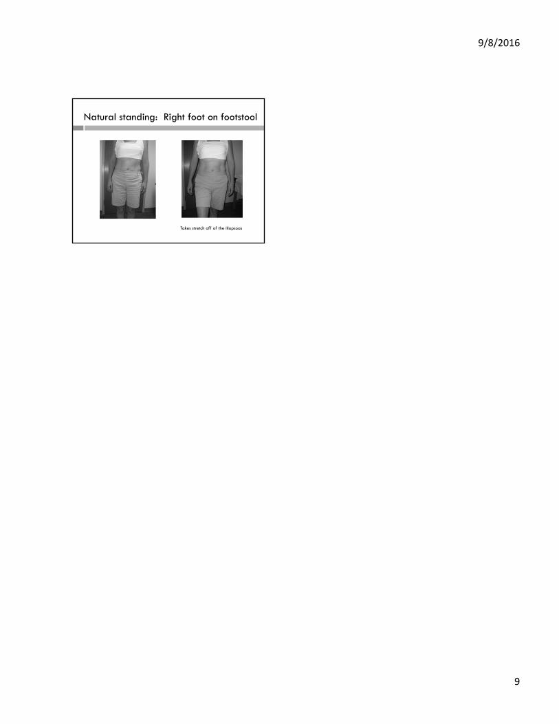
Rotation to left when rocking back
Right iliopsoas pulling> Left iliopsoas
9/8/2016
9
Natural standing: Right foot on footstool
Takes stretch off of the iliopsoas

9/8/2016
1
Movement ExamGeneric
Essential Activities
Standing
• Appearance
• Alignment
• Forward bend & return
• Single Leg standing
• Step up & down
• Walk
• Reach forward – overhead and out to side
• Sit to stand
Recumbent
• Roll to both sides
• Scoot
• Lying to sitting and reverse

9/8/2016
2
Diagnostic Movement Exam
Standing
• Appearance (size, structural proportions, general fitness)• Overweight, athletic, tall, short, long trunk, “apple or pear”
Alignment• Iliac crest height; pelvic rotation, paraspinal asymmetry = rotation• Marked lumbar lordosis, marked anterior pelvic tilt, thoracic kyphosis, swayback = extension
• Flat back, high iliac crest, posterior pelvic tilt = flexion • Symptoms – increased lumbar curve = extension; kyphosis /swayback = extension (compression with well developed abdominal muscles)
• Forward bending: corrected forward bending• Increased lumbar flexion, decreased hip flexion, Sx = flexion• Decreased Sx with hip flexion only = flexion
Standing continued• Return from forward bending: corrected return from forward bending
• lumbar extension & Sx with return = extension• Decreased Sx with hip extension, not lumbar extension = extension
Sidebending; corrected sidebending• Sx, moves at one segment = rotation• Decreased Sx with correction = rotation
• Rotation• Sx, rotates off axis = rotation
• Backbending• Sx = extension
• Single leg stance• Hip drop, lateral trunk flexion = rotation• Lateral pelvic tilt = rotation
9/8/2016
3
Supine
• Hip Extension with knee flexion (flexor length test)• Anterior pelvic tilt 1st 50%, note symmetry, change with abduction = extension/rotation
• Position – Sx = extension
• Log roll test – indication of femoral ante or retroversion, • note resistance & change in ROM with abduction
• Position of hips and knees extended vs. hips and knees flexed• Sx with LEs extended = extension• Sx decreased with hips/knees flexed = extension
• Unilateral hip and knee flexion (passive and active)• Note weight of LE, onset of any stiffness, hip flexion ROM• Sx during passive flexion, if becomes active = extension• At end of ROM, pelvis posterior tilts = flexion• Sx and/or lumbopelvic rotation = rotation• Hip flexion <100 deg = FAI, flexion• Active‐ Sx, lumbopelvic rotation = extension‐rotation• Note the extent of hip extension of contra‐lateral LE.
Sidelying
• Rolling • Asymmetrical timing of upper trunk & lower trunk – pelvis = rotation
• Position• Sx = rotation
• Hip abduction/lateral rotation from flexion• Lumbopelvic rotation in relation to thorax = rotation
• Hip ab & adduction• Pelvic tilt = rotation
Prone• Position
• Sx = extension
• Knee flexion – passive ‐ active• Lumbopelvic rotation, Sx = rotation
• Anterior pelvic tilt, Sx = extension
• Hip lateral rotation – neutral & abducted• Lumbopelvic rotation, Sx = rotation
• Note ROM ? femoral version
• Hip medial rotation – neutral & abducted• Lumbopelvic rotation, Sx = rotation
• Note ROM? Femoral version
9/8/2016
4
Quadruped
• Alignment• Lumbar flexion, <90 deg of hip flexion = flexion
• Paraspinal asymmetry = rotation
• Lumbar extension, Sx = extension
• Rock backward• Lumbar flexion = flexion
• Lumbar rotation = rotation
• Shoulder flexion• Trunk rotation = rotation

LUMBAR MOVEMENT EXAMINATION September 2016
A. STANDING:
1. Appearance (size, structural proportions, general fitness) a. Overweight, athletic, tall, short, long trunk, “apple or pear”
2. Alignment
a. Iliac crest height; pelvic rotation, paraspinal asymmetry = rotation b. Marked lumbar lordosis, marked anterior pelvic tilt, thoracic kyphosis, swayback =
extension c. Flat back, high iliac crest, posterior pelvic tilt = flexion
3. Forward bending: corrected forward bending
a. Increased lumbar flexion, decreased hip flexion, Sx = flexion b. Decreased Sx with hip flexion only = flexion
4. Return from forward bending: corrected return from forward bending a. lumbar extension & Sx with return = extension b. Decreased Sx with hip extension, not lumbar extension = extension
5. Sidebending; corrected sidebending
a. Sx, moves at one segment = rotation b. Decreased Sx with correction = rotation
6. Rotation
a. Sx, rotates off axis = rotation
7. Backbending a. Sx = extension
8. Single leg stance
a. Hip drop, lateral trunk flexion = rotation b. Lateral pelvic tilt = rotation
B. SUPINE: 1. Hip Extension with knee flexion (flexor length test)
a. Anterior pelvic tilt 1st 50%, note symmetry, change with abduction = extension/rotation
2. Log roll test – indication of femoral ante or retroversion, note resistance & change in
ROM with abduction
3. Position of hips and knees extended vs. hips and knees flexed a. Sx with LEs extended = extension b. Sx decreased with hips/knees flexed = extension
4. Unilateral hip and knee flexion (passive and active)
a. Note weight of LE, onset of any stiffness, hip flexion ROM b. Sx during passive flexion, if becomes active = extension c. At end of ROM, pelvis posterior tilts = flexion d. Sx and/or lumbopelvic rotation = rotation e. Hip flexion <100 deg = FAI, flexion f. Active- Sx, lumbopelvic rotation = extension-rotation
a. Note the extent of hip extension of contra-lateral LE.

5. Hip abduction/lateral rotation from flexion
a. Lumbopelvic rotation 1st 60% = rotation 6. Lower abdominal muscle performance inferred by lumbopelvic motion
a. Early & greater than ½” motion = poor performance C. Sidelying
1. Position a. Sx = rolling
2. Hip abduction/lateral rotation from flexion a. Lumbopelvic rotation in relation to thorax = rotation
3. Hip ab & adduction a. Pelvic tilt = rotation
D. Prone 1. Position
a. Sx = extension 2. Knee flexion – passive - active
a. Lumbopelvic rotation, Sx = rotation b. Anterior pelvic tilt, Sx = extension
3. Hip lateral rotation – neutral & abducted a. Lumbopelvic rotation, Sx = rotation b. Note ROM ? femoral version
4. Hip medial rotation – neutral & abducted a. Lumbopelvic rotation, Sx = rotation b. Note ROM? Femoral version
E. Quadruped 1. Alignment
a. Lumbar flexion, <90 deg of hip flexion = flexion b. Paraspinal asymmetry = rotation c. Lumbar extension, Sx = extension
2. Rock backward a. Lumbar flexion = flexion b. Lumbar rotation = rotation
3. Shoulder flexion a. Trunk rotation = rotation
F. Walking 1. Lumbopelvic rotation = rotation