Movement & Muscle
25
1 1 19 th Lecture Fri 27 Feb 2009 Vertebrate Physiology ECOL 437 (MCB/VetSci 437) Univ. of Arizona, spring 2009 Kevin Bonine & Kevin Oh Movement & Muscle Chapter 18 2 Housekeeping, Fri 27 February 2009 Readings Today: Ch 18 Mon: Ch 19 Wed: Ch 19 & 20 LAB Wed 04 Mar: Dickinson et al. 2000 Fri 06 Mar: Ch 21 (gas transport) Lab discussion leaders: 04 Mar 1pm – Deedra, Franz 3pm – AJ, Kareem Lab discussion leaders: 25 Mar 1pm – Sam 3pm – Karri, Jason Fri 13 Feb = Exam 1
Transcript of Movement & Muscle

1
1
19th LectureFri 27 Feb 2009
Vertebrate PhysiologyECOL 437 (MCB/VetSci 437)Univ. of Arizona, spring 2009
Kevin Bonine & Kevin Oh
Movement & MuscleChapter 18
2
Housekeeping, Fri 27 February 2009
ReadingsToday: Ch 18Mon: Ch 19Wed: Ch 19 & 20LAB Wed 04 Mar: Dickinson et al. 2000Fri 06 Mar: Ch 21 (gas transport)
Lab discussion leaders: 04 Mar1pm – Deedra, Franz3pm – AJ, Kareem
Lab discussion leaders: 25 Mar1pm – Sam3pm – Karri, Jason
Fri 13 Feb = Exam 1

2
3
Movement and Behavior
Nervous System and Muscle
integration, control, feedback
4
Dipsosaurusdorsalis
Callisaurusdraconoides
Phrynosomaplatyrhinos
Thanks to Duncan Irschick and Steve Reilly

3
5
~Behavior Initiation
Bring together nervous, endocrine, muscular systems, etc.
Complex
Reflexes / Learned / Plasticity
Respond to situation(s)Parallel Processing Complicated
NeuronalCircuitry
Animal Behavior,Neurobiology
6
Simple Reflexes – basis of neuronal circuitry
Reflex Arc, Stereotypic Behaviore.g., stretch reflex (patellar tendon)
- Tonic tension in muscle- Important for
maintenance of posture via negative feedback
- Only 2 neurons required
- monosynaptic reflex
Stretch receptor activates1a-afferent neuron
Alpha-motor neuron activates quadriceps
Sherwood 1997 (see 11-1 in Eckert)
(ventral)

4
7
Simple ReflexesStretch receptor = muscle spindle organ
- contains intrafusal fibers(as opposed to extrafusal)
- Sensitive to stretch (stretch -> APs)
- Need to be reset for new muscle length- Gamma-efferent neurons innervate
spindle
8
1. 1a-afferent neuron2. Alpha-motor neuron3. Gamma-motor neuron
c. Contracted musclewithout ‘reset’ musclespindle
Sherwood 1997 (see 11-1 in Eckert)

5
9
Simple Reflexes +Other neurons become involved as well:- 1a-afferents inhibit antagonist muscle
(Knee flexor ~hamstring)- Conscious decision to bend leg etc.
- Limb
Fig. 11-2Randall et al. 2002
10
Silverthorn 2001
Stretch Reflex

6
11Hill et al. 2004 Fig 18.7
12Hill et al. 2004 Fig 18.7

7
13
Silverthorn 2001
14
Silverthorn 2001
Golgi Tendon Reflex:

8
15
Law of Nerve-Specific Energy
ventral
dorsalAction Potentials
and Graded Potentials don’t convey specific information.
Rather, the geographic connections, summation of different inputs, and modulation are important for correct response Fig. 11-12
Randall et al. 2002
16
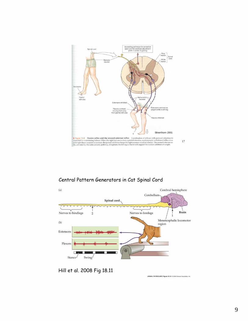
CPG = central pattern generator-neuronal network producing repetitive output
Walking, swimming, flying, breathing
Toad walking with no afferents - awkward- flaccid muscles
Sensory feedbackHigher centers can override
Some patterns at level of spinal cord if stimulateinitially (cats on treadmill)
Peripheral vs. Central Control
9
17
Silverthorn 2001
18Hill et al. 2008 Fig 18.11
Central Pattern Generators in Cat Spinal Cord

10
19
Design of a robot salamander that walks and swims Figure 18.12
20
Activating muscles
NERVOUS SYSTEM CONTROL:
•cerebral cortex•frontal, parietal,temporal, occipital lobes
•Cerebellum•basal ganglia•brain stem•spinal cord•peripheral nerves

11
21
Silverthorn 2001
22
Major Motor Areas,IncludingPRIMARY MOTOR CORTEX
Hill et al. 2004, Fig 18.15&16

12
23
Basal Ganglia(plans&initiates movement)
Cerebellum(fine-tunes execution)
Hill et al. 2004, Fig 18.19
Parkinson’s Disease(akinesia, too much inhibition of motor function, mediated by dopamine)
Huntington’s Disease(chorea, not enough inhibition of motor function)
24
http://salmon.psy.plym.ac.uk/year1/ETHEXPT.HTM#Egg%20retrieval
Herring Gull Egg Retrieval
- prefer the larger of two eggs of the same colour- prefer the speckled egg over an unspeckled egg of the same colour- prefer natural coloured(brown speckled) eggs over brown unspeckled eggs - prefer green speckled eggs over green unspeckled eggs - prefer green eggs over brown eggs (Baerends & Kruijt)
Fixed Action Patterns…
Greater preference

13
25
Research Proposal Tips:
-Physiology and science should be subject, not researchers and experiments-Having interesting question or problem helps give direction and focus-More physiology-Subheadings often helpful
-Synthesize, not serial book reports-Abstract, role is summary of entire paper, not an intro to the intro
-Avoid Pronouns (its, these, this, …which, there are)-Passive voice to be avoided (e.g., Avoid passive voice)-Leading and following zeroes (0.5, .5, .50)
-Page numbers-Citation format (J. of Physiology, instructions to authors, [full journal names])
-Turn in old, graded work with each new version
-Peer editing (read quickly, then read for content and writing, comments helpful)
26
MuscleA. SarcomereB. Cross-bridge cyclingC. Length-tension relationshipD. Excitation-contraction couplingE. Force-Velocity curves, PowerF. Fiber TypesG. Motor Units/RecruitmentH. EnergeticsI. FatigueJ. Repair and Regeneration
Vertebrate Physiology 437
- Smooth and Cardiac introduction

14
27
Uses: - most observable animal behavior - most visceral function- generally act by shortening
Muscle
Classification: - striated
- smoothskeletal or cardiac
All muscle movement based on myofilaments (actin and myosin) sliding past each other…
Utilize: ATP, Ca2+, ~APs
walls of hollow organs
(Myo-, Sarco- = musclerelated)
28
Structure:
Skeletal Muscle
- muscle attached to bone(skeleton) via tendons
- muscle comprises elongate,multinucleate, muscle fibers
- multinucleate muscle fibersderived from combination of manymyoblasts (embryonic muscle cells)
- within each muscle fiber are many parallel myofibrils
- each myofibril contains sarcomeresarranged in series (end-to-end)
- sarcomere is functional unit ofmuscle

15
29
Sarcomere
Z-disk at each end of sarcomere
Sarcomeres in adjacent myofibrils are alignedleading to striated appearance
Actin thin myofilaments attachedto each Z-disk
Myosin thick myofilaments in between actins (6,3)
Actin and Myosin overlap is what allows muscle contraction
(6,3)
30
Sarcomere
Z-disk (actin attaches)
Areas within sarcomere given names:
I-band (actin only) A-band (myosin length)
H-zone (myosin only)
M-line (midpoint of myosin)
During muscle contraction, myosin thick filaments slide past actin thin filaments toward Z-lines

16
31
Which regions change length and which remain the same as the sarcomere shortens?
Z-disk (actin attaches)I-band (actin only) A-band (myosin length)
H-zone (myosin only)
M-line (midpoint of myosin)
During muscle contraction, myosin thick filaments slide past actin thin filaments toward Z-lines
32
Sarcomere Composition
Actin composed of:individual molecules of G-actin (globular)united into chains called F-actin (filamentous) which form a two-stranded helix
In the groove of the two F-actin strands is tropomyosin,which also has globular troponin molecules attached to it

17
33
Sarcomere Composition
Myosin composed of:2 heavy chains with
globular heads2 essential light chains2 regulatory light chains
Myosin molecules spontaneously aggregate into complexes with the heads at the ends and the tails toward the middle
The light chains are involved in the speed of contraction(important for different muscle fiber-types)
34
Sarcomere Function
Cross-bridges form transiently between myosin head and actin filament(actomyosin) Sarcomere shortens
during contraction
Sliding Filament Theory
Actin and Myosin molecules slide past each other, but don’t themselves change length

18
35
Cross Bridges and Force Production
Myosin head binds to actin (actomyosin), then pulls myosin toward z-line thereby shortening sarcomere (= contraction)
Vander et al., 2001
Cross-bridge forces are additive.Same force all along myofibril.
36
Sarcomere Function
Number of Cross-bridges (and therefore contraction magnitude) increased with appropriate overlap of actin with myosin heads

19
37
Length-Tension Relationship
Normal muscle function at or near the plateau (1.8-2.2)
38
Length-Tension Relationship
Why lose force production at short end?
What constrains muscle length in the body?

20
39Hill et al. 2004, Fig 17.12
Length-Tension Relationship
40
Vander et al., 2001
Myosin head has to be able to detach and bind again to actinfurther along in order to continue to generate forceDetachment requires ATP bind to myosin head

21
41
Vander et al., 2001
Cross Bridges and Force ProductionATP required for the (3)dissociation of actin and myosin (else rigor mortis)
Myosin acts as an ATPase, hydrolyzing ATP to ADP + Pi (4) (Energy of ATP hydrolysis “cocks” the myosin head)
Myosin releases ADP and Pi (very slowly (2)unless bound to actin) ATP binds to myosin
Actomyosin complex forms (= crossbridge) (1)
Cross bridge stronger when Pi released, then myosin head rotates
Movement about 10-12 nm
Cycle repeats until Ca++ resequestered or run out of energy
ATP hydrolysis
42
San Diego State University College of Sciences Biology 590 - Human Physiology
Actin Myosin Crossbridge 3D Animation*

22
43
Hill et al. 2004, Fig 17.5
44
Regulation of Contraction
CALCIUM & the cross bridge
Need free Ca2+ in cytosolto get contraction
Calcium binds troponinwhich is attached to tropomyosin on actin
This causes conformational change in tropomyosinexposing actin binding sites for myosin heads (not shown)
Without calcium, contraction is inhibited
Vander et al., 2001

23
45
Excitation-Contraction Coupling, from the beginning…
1. AP from CNS arrives at neuromuscular junction.
2. ACh released into synapse.
3. ACh binds to nicotinic receptors on motor endplate.4. Ion channels for K+ and Na+ open; greater Na+ influx leads to depolarization and AP in muscle plasma membrane
EPP = Endplate Potential(~Excitatory Post-Synaptic Potential or EPSP)
46
Excitation-Contraction Coupling, the middle I…
5. Change in membrane potential (AP) reaches deepinto the muscle cell via transverse tubules (T-tubules; one per Z-disk)

24
47
Excitation-Contraction Coupling, the middle II…
6. T-tubules have voltage sensitive proteins called dihydropyridine receptors
8. Calcium stored in the SR. Released into the cytosol via the ryanodine receptor channel when the RR is mechanically triggered by the voltage sensitive dihydropyridine receptor.
7. Dihydropyridine receptors in the T-tubules are mechanically linkedwith ryanodine receptors (RR) on sarcoplasmic reticulum (SR)
The ends of the SR adjacent to the T-tubule are called terminal cisternae (w/ calsequestrin)
48
Excitation-Contraction Coupling, the last bit…
9. Calcium triggers release of more calcium from some ryanodine receptors that are not linked to dihydropyridine receptors
10. Calcium binds to troponin leading to actomyosin complex…
Called calcium-induced calcium release
11. After repolarization, calcium actively (requires ATP) movedback into SR where much of it is bound to calsequestrin
12. Muscle relaxes as long as ATP is present to allow actomyosin complex to dissociate

25
49
Sherwood, 1997
Review of EC Coupling and Muscle Contraction