Mousa e al, Med Chem (Los Angeles) 2018, 8:8 …...Mousa e al, Med Chem (Los Angeles) 2018, 8:8 M e...
13
Mousavi et al., Med Chem (Los Angeles) 2018, 8:8 DOI: 10.4172/2161-0444.1000515 M e d i c i n a l c h e m i s t r y ISSN: 2161-0444 Medicinal Chemistry t Research Article Open Access Med Chem (Los Angeles), an open access journal ISSN: 2161-0444 Volume 8(8): 205-217 (2018) - 205 Nanosensors for Chemical and Biological and Medical Applications Seyyed Mojtaba Mousavi 1,2 *, Seyyed Alireza Hashemi 1,2 *, Maryam Zarei 3 , Ali Mohammad Amani 1,2 and Aziz Babapoor 4 1 Department of Medical Nanotechnology, School of Advanced Medical Sciences and Technologies, Shiraz University of Medical Sciences, Shiraz, Iran 2 Pharmaceutical Sciences Research Center, Shiraz University of Medical Sciences, Shiraz, Iran 3 Department of Engineering Sciences, Salman Farsi University, Kazerun, Iran 4 Department of Chemical Engineering, University of Mohaghegh Ardabili, Ardabil, Iran Keywords: Nanosensor; Biomedical; Drug carriers; Optical nanosensors; Nanotechnology Introduction A device that can detect changes in the physical stimulus and convert them into signals that can be measured or recorded is called a sensor. e sensor initially acquires this physical value then converts it to the desired signal for processing (for example, optical, electrical, and mechanical). In the scientific term, the prefix "Nano" is considered as one billionth of a single unit. For example, a billionth of a meter is nanometer. On a nanoscale scale, one can use the natural spacing of molecules and interactions between them. In Nanoscience and nanotechnology Nano-scale objects are examined (National Science and Technology Council 1999). One of the major differences in nanoscale with larger scales is the properties and function of objects on this scale. Generally speaking, Nanoscience studies the properties of materials on an atomic, molecular and macromolecular scale, while in nanotechnology, scientists design, construct, and use devices and systems by controlling their shape and size on a nanometer scale (Royal Society and Royal Academy of Engineering 2004). Nanomedicine is the result of advances in Nanoscience and nanotechnology and is generally referred to as biotechnology applications in Nanoscience and nanotechnology. Nanomedicine can be considered as a boundary between medical, physics, chemistry and biological sciences. Also, the Nanomedicine development of fields of analysis and treatment in biological systems at the cellular and sub-cellular levels has provided revolutionary methods for the detection and prevention of certain fatal diseases. For example, the National Cancer Institute of America expects nanotechnology and Nanoscience to be used to control cancer and thus reduce mortality. Because of the proximity of Nanoscience to fertility, the US Department of the Food and Drug Administration has been exploring the complex issues associated with nanoscale testing, nanoparticles, and Nanosystems to improve human life. Nanoscale materials can be described as a range of atoms up to 100 nanometers, and virtually a nanoscale material is defined as a material with the smallest dimensions of less than 100 nanometers (Figure 1). Nanomaterials have unique properties compared to larger ones, and sometimes the nanoscale range can increase to 1,000 nm. Since a number of biological *Corresponding author: Mousavi SM, Department of Medical Nanotechnology, School of Advanced Medical Sciences and Technologies, Shiraz University of Medical Sciences, Shiraz, Iran, Tel: +987132305410; E-mail: [email protected] Received August 07, 2018; Accepted August 10, 2018; Published August 17, 2018 Citation: Mousavi SM, Hashemi SA, Zarei M, Amani AM, Babapoor A (2018) Nanosensors for Chemical and Biological and Medical Applications. Med Chem (Los Angeles) 8: 205-217. doi: 10.4172/2161-0444.1000515 Copyright: © 2018 Mousavi SM, et al. This is an open-access article distributed under the terms of the Creative Commons Attribution License, which permits unrestricted use, distribution, and reproduction in any medium, provided the original author and source are credited. organisms, such as proteins, antibodies, viruses, and bacteria, are located within the Nano range, they are oſten referred to as Nano-biological materials. e high tendency to design non-biological Nano-materials is due to their specific functions and the properties of nanoscale biological materials. At the same time, nanoscale materials can be used to access or manipulate non-biological Nanomaterials because they have the right size [1]. Nanomaterials that are less than 50 nm in size do not have the problem of entering many cells, while Nanomaterials which are smaller than 20 nm can move beyond the blood vessels and move around the body's circulatory system. erefore, aſter special treatments, Nanomaterials are widely used as drug delivery systems whose role is to transfer chemotherapeutic materials or therapeutic genes to malignant cells, while protecting healthy cells and store them. As a momentous note it can be said that, in many practical sections of literature, nanomaterials are commonly called as non-biological nanomaterials, however, in the preparation and fabrication of non- biological nanomaterials, organisms and biological methods have been remarkably used [2]. One of the reasons for the emergence of nanotechnology is the extraordinary knowledge-based and technical development in diverse areas, such as pharmacy and biomedicine. One nanometer can be described as one billionth of a meter and three times smaller than micron [3]. Over the past few years, nanotechnology has been instrumental in the field of medicine and its development. Studies show that a wide range of Nanomaterials has emerged that have biomedical Abstract Nanotechnology is one of the areas that have attracted much attention in all areas of scientific research. Nanotechnology has inherent potential in the production of unique materials, new equipment and systems with molecular level control and the usage of material features in nanometer scales. Therefore, the use of Nano-sized objects such as a variety of natural nanoparticles as drug carriers and/or tumor detection agent can be considered as a major achievement. The recent increase in the number of articles and publications in this field is due to the same. Due to the complexity of this field, collaborative research teams with expertise such as medicine, engineering, physics, chemistry, and biotechnology are necessary. This study focuses on the elements such as elementary concepts, the application of nanotechnology in medicine, the structure, and form of operating nanosensors and their use in detecting microorganisms and cancer. In other sections, various types of nanosensors such as nano-chemical sensors, electrochemical nanosensors, and biosensors. As well as carbon-based materials in nanosensors are described, and finally, it discusses the advancement and development of nanotechnology since its inception and emergence until now, with emphasis on sensor technology and their application in biomedical systems.
Transcript of Mousa e al, Med Chem (Los Angeles) 2018, 8:8 …...Mousa e al, Med Chem (Los Angeles) 2018, 8:8 M e...

Mousavi et al., Med Chem (Los Angeles) 2018, 8:8DOI: 10.4172/2161-0444.1000515Med
icinal chemistry
ISSN: 2161-0444
Medicinal Chemistryt Research Article Open Access
Med Chem (Los Angeles), an open access journalISSN: 2161-0444
Volume 8(8): 205-217 (2018) - 205
Nanosensors for Chemical and Biological and Medical ApplicationsSeyyed Mojtaba Mousavi1,2*, Seyyed Alireza Hashemi1,2*, Maryam Zarei3, Ali Mohammad Amani1,2 and Aziz Babapoor4
1Department of Medical Nanotechnology, School of Advanced Medical Sciences and Technologies, Shiraz University of Medical Sciences, Shiraz, Iran2Pharmaceutical Sciences Research Center, Shiraz University of Medical Sciences, Shiraz, Iran3Department of Engineering Sciences, Salman Farsi University, Kazerun, Iran4Department of Chemical Engineering, University of Mohaghegh Ardabili, Ardabil, Iran
Keywords: Nanosensor; Biomedical; Drug carriers; Optical nanosensors; Nanotechnology
IntroductionA device that can detect changes in the physical stimulus and
convert them into signals that can be measured or recorded is called a sensor. The sensor initially acquires this physical value then converts it to the desired signal for processing (for example, optical, electrical, and mechanical). In the scientific term, the prefix "Nano" is considered as one billionth of a single unit. For example, a billionth of a meter is nanometer. On a nanoscale scale, one can use the natural spacing of molecules and interactions between them. In Nanoscience and nanotechnology Nano-scale objects are examined (National Science and Technology Council 1999). One of the major differences in nanoscale with larger scales is the properties and function of objects on this scale. Generally speaking, Nanoscience studies the properties of materials on an atomic, molecular and macromolecular scale, while in nanotechnology, scientists design, construct, and use devices and systems by controlling their shape and size on a nanometer scale (Royal Society and Royal Academy of Engineering 2004). Nanomedicine is the result of advances in Nanoscience and nanotechnology and is generally referred to as biotechnology applications in Nanoscience and nanotechnology. Nanomedicine can be considered as a boundary between medical, physics, chemistry and biological sciences. Also, the Nanomedicine development of fields of analysis and treatment in biological systems at the cellular and sub-cellular levels has provided revolutionary methods for the detection and prevention of certain fatal diseases. For example, the National Cancer Institute of America expects nanotechnology and Nanoscience to be used to control cancer and thus reduce mortality. Because of the proximity of Nanoscience to fertility, the US Department of the Food and Drug Administration has been exploring the complex issues associated with nanoscale testing, nanoparticles, and Nanosystems to improve human life. Nanoscale materials can be described as a range of atoms up to 100 nanometers, and virtually a nanoscale material is defined as a material with the smallest dimensions of less than 100 nanometers (Figure 1). Nanomaterials have unique properties compared to larger ones, and sometimes the nanoscale range can increase to 1,000 nm. Since a number of biological
*Corresponding author: Mousavi SM, Department of Medical Nanotechnology, School of Advanced Medical Sciences and Technologies, Shiraz University of Medical Sciences, Shiraz, Iran, Tel: +987132305410; E-mail: [email protected]
Received August 07, 2018; Accepted August 10, 2018; Published August 17, 2018
Citation: Mousavi SM, Hashemi SA, Zarei M, Amani AM, Babapoor A (2018) Nanosensors for Chemical and Biological and Medical Applications. Med Chem (Los Angeles) 8: 205-217. doi: 10.4172/2161-0444.1000515
Copyright: © 2018 Mousavi SM, et al. This is an open-access article distributed under the terms of the Creative Commons Attribution License, which permits unrestricted use, distribution, and reproduction in any medium, provided the original author and source are credited.
organisms, such as proteins, antibodies, viruses, and bacteria, are located within the Nano range, they are often referred to as Nano-biological materials. The high tendency to design non-biological Nano-materials is due to their specific functions and the properties of nanoscale biological materials. At the same time, nanoscale materials can be used to access or manipulate non-biological Nanomaterials because they have the right size [1]. Nanomaterials that are less than 50 nm in size do not have the problem of entering many cells, while Nanomaterials which are smaller than 20 nm can move beyond the blood vessels and move around the body's circulatory system. Therefore, after special treatments, Nanomaterials are widely used as drug delivery systems whose role is to transfer chemotherapeutic materials or therapeutic genes to malignant cells, while protecting healthy cells and store them. As a momentous note it can be said that, in many practical sections of literature, nanomaterials are commonly called as non-biological nanomaterials, however, in the preparation and fabrication of non-biological nanomaterials, organisms and biological methods have been remarkably used [2].
One of the reasons for the emergence of nanotechnology is the extraordinary knowledge-based and technical development in diverse areas, such as pharmacy and biomedicine. One nanometer can be described as one billionth of a meter and three times smaller than micron [3]. Over the past few years, nanotechnology has been instrumental in the field of medicine and its development. Studies show that a wide range of Nanomaterials has emerged that have biomedical
AbstractNanotechnology is one of the areas that have attracted much attention in all areas of scientific research.
Nanotechnology has inherent potential in the production of unique materials, new equipment and systems with molecular level control and the usage of material features in nanometer scales. Therefore, the use of Nano-sized objects such as a variety of natural nanoparticles as drug carriers and/or tumor detection agent can be considered as a major achievement. The recent increase in the number of articles and publications in this field is due to the same. Due to the complexity of this field, collaborative research teams with expertise such as medicine, engineering, physics, chemistry, and biotechnology are necessary. This study focuses on the elements such as elementary concepts, the application of nanotechnology in medicine, the structure, and form of operating nanosensors and their use in detecting microorganisms and cancer. In other sections, various types of nanosensors such as nano-chemical sensors, electrochemical nanosensors, and biosensors. As well as carbon-based materials in nanosensors are described, and finally, it discusses the advancement and development of nanotechnology since its inception and emergence until now, with emphasis on sensor technology and their application in biomedical systems.

Citation: Mousavi SM, Hashemi SA, Zarei M, Amani AM, Babapoor A (2018) Nanosensors for Chemical and Biological and Medical Applications. Med Chem (Los Angeles) 8: 205-217. doi: 10.4172/2161-0444.1000515
Med Chem (Los Angeles), an open access journalISSN: 2161-0444
Volume 8(8): 205-217 (2018) - 206
is considered as a technology for designing, building, and using nanomaterials and nanostructures. Nanotechnology covers the fundamental understanding of physical properties and nanomaterial and nanostructure phenomena and includes the fundamental study of the relationship between physical properties and the size of materials in nanometer scale. In order to propose a comprehensive definition, nanotechnology is related to materials and systems whose structure and components are new and their biological, chemical and physical properties are significantly developed. This can be assigning to nanoparticle size. Although the term “nanotechnology” is novel, the study of nanometer scale is not something new. It is accepted that, the research of biological systems and engineering of many materials such as colloidal dispersion metal quantum dots and catalysts have existed for many years. What is quite new about nanotechnology can be considered as a combination of our ability to observe and manipulate the material in nanoscale. Invention and development of devices such as transmission electron microscopy (TEM), scanning tunneling microscopy (STM), and scanning probe microscope (SPM) have been used for developing new facilities for specifying, measuring and manipulating nanomaterials and nanostructures. So, it can be stated that by using these tools, it is possible to study and manipulate nanomaterials and nanostructures at atom level. Iron-reducing bacteria (IRB) can be introduced as one of the most beneficial micro-organisms that present in different industrial and environmental
applications such as skin ulcer healing, bone tissue engineering, and smart drug delivery systems [4-6]. Smart drug delivery systems relate to successful and comprehensive examples of Nanomaterials with high potential for imaging, sensitivities, or therapies. Over the past decade, nanoscale materials have been reported with a good drug delivery capability and cancer treatment ability [7].
Nanotechnologies in medicineMolecular nanotechnology is formed on the measurement scale
in which it operates [8-11]. It indicates hundreds or thousands of nanometers (in fact molecule size). It can be considered that 3 or 4 atoms are present in a nanometer. In nanotechnology, the size of viruses can be assumed as 100 nanometers (Figure 2). Also, the diameter of human hair is considered as almost 200 µm. Atom can be considered as the main element of molecular nanotechnology structure. The characteristics of the product result such as tension, conductance or power are improved by groups of atoms. Conversion of graphite into the strong and hard structure of diamond is a clear example of this process. Also, reorganization and improvement of sand atoms can lead to the formation of a medium silicone plate for building semiconductor devices. Therefore, physicochemical conditions and biotic elements of these nanostructures can be useful for improvement of biomedical applications [12].
If comprehensive efforts are made for developing nanotechnology and its completion, it is possible to make the world a safer place for living. So, the sensors available in the industry are not excluded from this rule. In the past decade, nanosensors have been used for conducting researches. A nanosensor can be defined as a sensor that is built on an atomic scale which is based on nanometer measurements. Advance and evaluation of nanosensors can be observed in applications such as medicine, native safety, aerospace, combined circuit, etc. In addition to the variety of distinct usage for nanosensors, there are different kinds of nanosensors and different methods for preparing and producing them. There are challenges for construction of these nanosensors. However, whenever they get advanced and improved for regular use, their advantages are increased and their role in daily routine in enhances. The purpose of nanosensors can be mainly considered as achieving information on a nuclear scale and transferring them to the information which can be simply inspected. Using nanocomponents, usually in microscopic or submicroscopic scales, such sensors can be known as “physical or chemical sensors”. One of the characteristics of these particles is their powerful sensitivity. So, they have the capacity of identifying subatomic of viruses or indeed very low concentrations of the material which is naturally harmful [13,14]. Nanotechnology usually has the ability to design and controlling a nano-size object. However, according to the researchers’ opinions, it can be said that they have different ideas about nanotechnology, and it seems that definitions of nanotechnology are also varied as its applications. Some people have focused on the study of microstructure materials by an electron microscope and determining narrow sheets as nanotechnology. Some others have investigated the synthesis method and building materials as nanotechnology. Protein system, which is a nanotechnology, is defined as a set for protein delivery to a specific position in the body. The above definitions have meanings in specific research areas, but none of them cover the whole range of nanotechnology. Since nanotechnology covers a wide range of research areas and requires interdisciplinary and multidiscipline efforts, different definitions have been proposed for nanotechnology. According to the concepts and definitions of nanotechnology, it can be inferred that the only common feature of nanotechnology is its small size (Royal Society and Royal Academy of Engineering 2004). In general, nanotechnology
Figure 1: Nanoscale and typical materials whose dimension ranges are comparable to nanoscale [1].
Figure 2: Objects with dimensions from units to thousands of nm (see online version for colours) [13].
Citation: Mousavi SM, Hashemi SA, Zarei M, Amani AM, Babapoor A (2018) Nanosensors for Chemical and Biological and Medical Applications. Med Chem (Los Angeles) 8: 205-217. doi: 10.4172/2161-0444.1000515
Med Chem (Los Angeles), an open access journalISSN: 2161-0444
Volume 8(8): 205-217 (2018) - 207
activities. The ability of IRB to build iron nanostructures has made the bacteria interesting in nanobiology technology [15]. According to recent studies, it can be stated that these materials can also be used as nanosensors, many application can be considered for them.
Mechanism and structure of actionOver the past decade, nanotechnology has dramatically increased
the development of medicine, grounding, and shaping Nanoscience. Today, many of these improvements have entered the clinical field. The improvement and development of composite Nanosystems are of great importance to diagnosis and treatment of diseases. Such systems are divided into parts such as 1) a carrier platform 2) disposable for sensor imaging or therapeutic 3) optional target ligands. Exceeding such basic systems, the breadth of design allows the development of complex Nanosystems that can be very useful in medicine. After the advent of Nano-based platforms of different basic materials, they were tested for biological applications such as delivery of medication and cancer treatment. Nanoparticles (NPs) are Nano-sized materials that can carry various loads, such as imaging agents, proteins, small molecular drugs, nucleic acids or other materials [16-19]. The goal of Nanocarrier design is the promotion of efficiency of safety for the delivery of drugs in general, and for non-viral drug delivery purposes [20,21]. The design of such nanoscale materials requires the ability to control particle size, ensure biological compatibility and hidden properties, optimize and achieve distribution and controlled performance [22,23]. As Nano-carrier platforms, liposomes, dendrimers, polymers, Carbon nanotubes, metal NPs, organic NPs, quantum dots, Nano gels, and nanoparticles have been used (Figure 3).
A Nano-sensor mainly consists of a biologically sensitive layer that can contain biomedical diagnostic elements transmitted to the converter. For creating physicochemical changes, the interaction between the target analysis and the biological receptor has been made, which can become a computable factor, for instance an electrical signal [24]. Biological receptors are important and have been able to provide properties for bio-sensor technologies. They provide the substrate for attaching the ideal analyzer to the sensing element in order to quantify with the lowest level of intervention from other pieces in the sampling mix. Biologically sensitive components act as either a biotic molecular genre (such as an antibody, an enzyme, a protein, or a Nucleic acid) or a living biotic organization (such as cells, tissues, or entire organisms) that is used to identify a biochemical procedure. Different electronic, electrochemical or sensitive light detection methods employ in order to discover biosensors. The group of receptor molecules from most of the Nanosensors includes catalyzed and conjunction Nanosensors. In order to bind desirable, non-reversible and non-catalytic molecules, conjunction-based Nano-sensors can be used. For example, antibodies, nucleic acids, and hormone receptors can be considered. On the other side, catalytic-based sensing elements for instance microbiological cells and enzymes can identify and couple the molecule, and subsequently the catalytic chemical transformation of that molecule into a manufactured which was subsequently identified [25]. This is done by transferring the charges between the molecules and the sensitive materials, and at the end, it can be understood that the visual and/or electrical signal is depended on the kind and quantity of molecules [26].
NanosensorsThe sensing tools, that at least one of the dimensions of their
measurement are not more than about 100 nm are called nanosensors. As a matter of fact, within the scope of nanoscience, nanosensors are devices that can (a) administrate chemical and physical elements in areas difficult to control and reach, (b) diagnosis biochemicals in
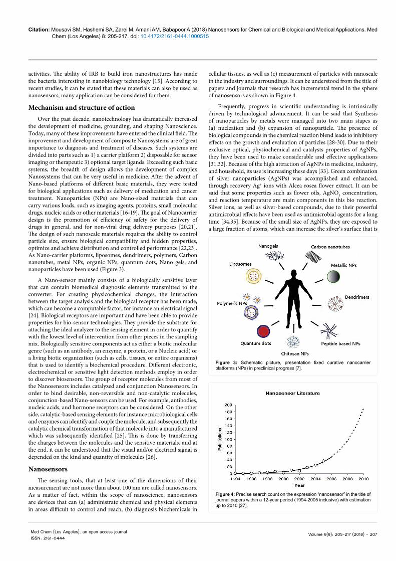
cellular tissues, as well as (c) measurement of particles with nanoscale in the industry and surroundings. It can be understood from the title of papers and journals that research has incremental trend in the sphere of nanosensors as shown in Figure 4.
Frequently, progress in scientific understanding is intrinsically driven by technological advancement. It can be said that Synthesis of nanoparticles by metals were managed into two main stapes as (a) nucleation and (b) expansion of nanoparticle. The presence of biological compounds in the chemical reaction blend leads to inhibitory effects on the growth and evaluation of particles [28-30]. Due to their exclusive optical, physiochemical and catalysts properties of AgNPs, they have been used to make considerable and effective applications [31,32]. Because of the high attraction of AgNPs in medicine, industry, and household, its use is increasing these days [33]. Green combination of silver nanoparticles (AgNPs) was accomplished and enhanced, through recovery Ag+ ions with Alcea rosea flower extract. It can be said that some properties such as flower oils, AgNO3 concentration, and reaction temperature are main components in this bio reaction. Silver ions, as well as silver-based compounds, due to their powerful antimicrobial effects have been used as antimicrobial agents for a long time [34,35]. Because of the small size of AgNPs, they are exposed to a large fraction of atoms, which can increase the silver‘s surface that is
Figure 3: Schematic picture, presentation fixed curative nanocarrier platforms (NPs) in preclinical progress [7].
Figure 4: Precise search count on the expression “nanosensor” in the title of journal papers within a 12-year period (1994-2005 inclusive) with estimation up to 2010 [27].

Citation: Mousavi SM, Hashemi SA, Zarei M, Amani AM, Babapoor A (2018) Nanosensors for Chemical and Biological and Medical Applications. Med Chem (Los Angeles) 8: 205-217. doi: 10.4172/2161-0444.1000515
Med Chem (Los Angeles), an open access journalISSN: 2161-0444
Volume 8(8): 205-217 (2018) - 208
in connect with microorganisms. This point can be reminded that the germicide characteristics of AgNPs result from the oxidation of silver molecules and Ag+ ions release from the AgNPs surface [33]. There are bioactive compounds in Alcea rosea flower which can influence bioreduction of Ag+ ions. So, the AgNPs manufactured are covered by hydrophilic biochemical compounds which have made them colloidal stable. As a matter of fact the main factors in preparing quality-based AgNPs include silver concentration, flower extract amount and reaction temperature. Thus for making uniform particles these factors must be manipulated and checked [36]. So, in order to diminution of Ag+ ions to AgNPs, an optimum quantity of the green source oil is necessitating. For the time of AgNPs biogenesis, a useful diminishing and covering factor was natural carbohydrates existing in average Ephedra [36]. It can be said that absorbed nanoparticles to the outer surface of an organism have the ability to damage cell walls or membranes or release of ions that affect the performance of the organism. As shown in Previous studies, the main toxicity of silver nanoparticles in the environment is related to the free ion in the aqueous phase [37,38]. The bacterial activity decreases, as the concentration of AgNPs increase [39,40]. AgNPs were synthesized using bark extracts of Ficus benghalensis and Azadirachta indica without the use of any external reducing or capping agent Ficus benghalensis (family Moraceae) commonly known as ‘banyan’ is an evergreen tree found all over India. It can be said that, for the silver nanoparticles synthesized by bark extracts of F. benghalensis and A. indica, the average size of particles was approx. 29 nm and 39 nm [41]. one of the most challenging and important issues in polymer nanocomposites is the formation of bubble and cavities resulting from it, which in practice can affect their electrical and mechanical properties [42-47]. Therefore, in recent years, different methods have been proposed to reduce the number of bubble cavities by the researchers, which consist of molecular dispersion of bubbles in the polymer matrix [44], simultaneous usage of vacuum and vibration, and vacuum shock technique [42]. So as previously mentioned, some researchers used vacuum shock technique in order to bubble reduction. They combining DC electric field and improved vacuum shock technique, so they generated electrified (highly aligned SWCNT) and non-electrified (randomly aligned SWCNT) epoxy-based nanocomposites with low void content, and their electrical conductivity and EMI shielding were investigated as is shown in Figure 5, the detail of the nanocomposite preparation explained.
From the electrical characteristics, one can find that the non-electrified nanocomposites have lower electrical conductivity and electromagnetic interference shielding than the electrified nanocomposites. It shows that electrification can develop the level of conductive network formation in the SWCNT/epoxy nanocomposites [48]. Before strengthened with silica nanoparticles and sasobit at various filler charges by multistage fabricating process, first nanocomposites including phenol novolac epoxy resin (PNER) were altered through unsaturated polyester resin (UPR). In the next step, after recognizing the effective factors on the mechanical and thermal features of nanocomposites, the effect of silica and sasobit loads on their morphology was also analyzed. It can be said that with increasing the loading of silica nanoparticles, the thermic and mechanical features are increasing. However if this growth in load of silica exceeds 3%, it can extend to a reduction in mechanical properties [49]. Moreover, When some pure and non-synthetic fillers including nanobioceramic (the egg shell nanoparticle) and also unmixed polymers (starch and glycerin) added to PNER / UPR, then this action can cause a notable diminishing below the soil and in order to progress absorption of water, presence of egg shell nanoparticles in the treated PNER / UPR is required [50]. Furthermore, the addition of montmorillonite nanoparticles to the
ER not only can improve the general mechanical features also this can cause development the thermal properties of nanocomposites [51]. In fact some factors for instance the radiation of electron rays to direct low-density-polyethylene (LLDPE), ethylene-co-vinyl acetate (EVA) and clay nanoparticles can also affect the mechanical or thermal properties of manufactured nanocomposites [52]. What can extremely cause diminishing the general mechanical and electrical properties is cumulus of fillers all over the matrix as well as formation of bubble based holes in the matrix, thus in order to reach an appropriate composite structure some agents such as elimination of bubbles before finishing the treatment mechanism and also uniform diffusion of fillers over the matrix are required [42]. Nanoparticles generated by microbiological methods showed the slower rate of forming than when plant extracts are used so nanosensors can be used. Although the sensors have been widely used for many years, but the subject of nanosensors is relatively new. Different nanosensors can be easily divided into three categories: 1) optical nanosensors 2) electromagnetic nanosensors and 3) mechanical and/or vibrational nanosensors.
Optical Nanosensors
The first nanosensor used to measure PH was fluorescein-based nanosensor which was trapped by polyacrylamide nanoparticle [53]. According to basic concepts, it can be said that fluorescent sensors are particles including leastwise one substratum binding component(s) and photoactive units(s) [54,55]. In fact, the luminescence phenomena is a process performed by a fluorophore that takes light of a particular wavelength due to the emission of a quantum of light with the energy associated with the conflict between the earth and the stimulated states [56,57]. As shown in Figure 6, a conceptual and comprehensible design for a conventional luminescent sensor is provided, where the receiver’s connection to the analyte is accompanied by impressive change in color. As a point, it can be said that the change in the vibrational properties of the photo depends on the meaning of the measurement.
An intracellular molecular colored arm [58] is considered as the main kind of nano-optical sensor that can load fluorescent cells. Unlike the fiber-optic arm, in this initial method, the physical impairment of the cell is minimized which can be considered an important advantage. On the other hand, such matters as toxicity, cellular degradation and chemical interference are known as defects in free color method. The specified nanoparticles, which include receptor molecules attached to
Figure 5: Manufacturing method for production of electrified and non-electrified nanocomposites [48].

Citation: Mousavi SM, Hashemi SA, Zarei M, Amani AM, Babapoor A (2018) Nanosensors for Chemical and Biological and Medical Applications. Med Chem (Los Angeles) 8: 205-217. doi: 10.4172/2161-0444.1000515
Med Chem (Los Angeles), an open access journalISSN: 2161-0444
Volume 8(8): 205-217 (2018) - 209
the outer surface of nanoparticles, have a remarkable deviation from the free color method [59,60]. The major difference between the specified nanoparticles in comparison with the free color method of the receiver molecules and labeled nanoparticles that move freely are respectively the solid state and nature of the primary and secondary fluids. Optical measurement includes absorption/ fluorescence/ phosphorescence/ Raman/ dispersion /refraction/ interference spectroscopy. Indeed, amplitude, energy, polarization time or phase of failure are the hallmarks of measurement [61,62].
Fiber optic nanosensors
Before acting the test, typical approaches for intracellular examination require “fixing” of cell samples. It can be said that, in addition to being able to destroy the cell’s bioavailability, it may also be able to alter intracellular structure in large amounts. One of the characteristics of optical fiber nanosensors is that it can examine and test the essential cellular processes in the laboratory. If biology processes can be controlled substantially at the molecular level, then a better understanding of dynamic cellular activity can be obtained. In order to create a physiochemical disorder that can change into electrical or other measurable signals, a relation between the target cell and the receptor cell is presented [63-67]:
R+A→ RA+ measurable signal
In the next step, the main signal is raised via the optical arm and then conveyed into the database. Since the optical fiber arm can be spaced between the surrounding and sensitive area, it may be stated that the disadvantages associated with the chemical interference of the color cell in the color free method are more that its advantages. Another positive point of the optical nanosensors is that the invasion of this method has reached its minimum level in comparison to typical arm-wire methods.
Electromagnetic nanosensors
In order to divide nanosensors based on their physical mechanisms there are two types of nanosensors:
(a) Finding via electricity measurement
(b) Finding by magnetism measurement.
First of all, we can review the class of electricity measurement for two samples: finding by current enhancement and finding by current inhibition. The label-free methodology throughout the usage of colors is a distinguished positive point of this method. Geng et al. [68] worked on analyzing fundamental interaction between hydrogen sulfide and gold nanoparticles and finally found that hydrogen sulfide atoms have high adsorption. By using the current and voltage across chromium and gold electrodes in the existence of an applied electrical field, the hopping of electrons was determined (Figure 7). Table 1 showed a short summary of the different types of nanosensors with their applications.
Biosensors
One of the most important and useful types of nanosensors is biosensor, because of its high ability to cancer diagnosis and even severe disease. Also, it can be said that the biosensors can be utilized for identification particular sorts of DNA. Dendrimers are the sensors made layer-by-layer onto areas with a diameter of fewer than five nanometers and produced by combinated polymers. Due to tiny dimension of these sensors, it is allowed to organize them via epidermis. It can be said that a nano-biosensor can be employed to uncovering asthma up to several weeks.
Drug DiscoveryThe molecules that can hold together particularly to proteins
are organic molecules which are able to utilize for development and detection of pharmaceuticals and they are also very important. An appropriate example of this topic can be the diagnosis of molecular inhibitors to tyrosine kinases. Proteins that intervene signal conveyance in mammalian cells via phosphorylation of a tyrosine residue from a substratum protein through adenonsine triphosphate (ATP) are called Tyrosine Kinases. Many illnesses for instance cancer are caused by deregulation of the phosphorylation procedure. It can be said that, the kinase Abl adsorbed on the exterior part of Si nanosensors and tested the bonding of ATP as well as keen prohibition of ATP bonding with biological atoms, such as the drug Gleevec in order to manage nanosensor tools in order to control and fix small-molecule inhibitors to tyrosine kinases. As a rise or drop in the performance of the p-type nanosensor tool, inhibition or binding to the bonding of the negatively charged ATP to Abl was done. The bonding of negative charged ATP to Abl was highly compatible with increased conductivity which could be an interesting point for testing. Significantly it can be said that conductivity decrease in low molecular concentrations, which also shows a strong dependence of degree of inhibition on the molecular structure [69,70]. What can use as the major means to treat diabetes and a key peptide hormone that conjointly with its receptor regulates blood glucose levels is Insulin. Factors such as various chemical changes during industrial processing, pharmaceutical formulation, and endogenous storage in the pancreatic β-cells can affect this therapeutic hormone, highly sensitive to the environmental stresses and easily
Figure 6: Conceptual schematics of a luminescent dye for intracellular sensing [27].
Figure 7: Discovering of analytes by inhibition of electron hopping (a) before bonding and (b) after bonding [27].

Citation: Mousavi SM, Hashemi SA, Zarei M, Amani AM, Babapoor A (2018) Nanosensors for Chemical and Biological and Medical Applications. Med Chem (Los Angeles) 8: 205-217. doi: 10.4172/2161-0444.1000515
Med Chem (Los Angeles), an open access journalISSN: 2161-0444
Volume 8(8): 205-217 (2018) - 210
undergoes structural changes are related to Insulin, and also it has the ability to unfold and aggregate. It can be mention that insulin may have an important impact on the biological efficacy in relation to the physiological and pharmacological activities of this hormone only by small changes altering the structural integrity of insulin. In order to improve its pharmaceutical properties, there must be a random or planned chemical change. In a conceptual definition, gene therapy can be considered as the delivery of DNA or RNA to cells, which can be used to cure or obstruct genetic untidiness. Recently, many approaches and gene-transporter have been advanced to getting better gene transfer ability. In order to provide a novel approach to cure illnesses and also merging gene therapy and chemotherapy, the usage of Polyethylenimine (PEI) based transfer material is a smart choice [71]. It can be said that, when ketones is exist, improvement methods for the chemoselective reduction of aldehydes are very important and gained notable consideration or even via usage of some additives materials such as thermoplastic thermoplastic, (PET, ABS, SAN) [49,51,72] nano tube [42,48] resins [73,74] graphene oxide in polymer composite For X-ray radiation shielding [75] and features of nanocomposites, linear low density polyethylene, ethylene-co-vinyl acetate and nano-clay particles through electron rays [52]. Due to the features mentioned, nanosensors can also be used to do this [76].
Cancer diagnosis
Raman spectroscopy (RS) is a powerful analytical technique that can be utilized in the identification of biological specimens, even an individual cell [77] Surface enhanced Raman spectroscopy (SERS) is an up to date approach that can increase the accuracy of identification and is very useful. The most famous particles are gold and silver nanoparticles which can be utilized in SERS, various nanostructures, combination and synthesize with other nanoparticles, covering substances, biomolecules in order to detect tumors (Figure 8) [78]. Raman can be exploited in various experiments, not only as a probe instrument in the laboratory, but also for histological and pathological diagnosis in clinical affairs RS and SERS in particular, is a powerful tool which can be utilized for identification and tracking biological targets which are cell and biochemical compounds [75]. So some of these features can be attributed to nano sensors.
QD bioconjugates which are very luminescent and firm came from recent progresses. It can be said that these bioconjugates have the ability to provide novel facilities for tissue specimens, gene therapy, proteins and drug aims in even single cells, and it is able to identification of cancer cells in organisms. For investigating protein analysts, one method has been improved which works based on magnetic microparticle probes with antibodies that can particularly match an aim of interest such as prostate-specific antigen (PSA) in case of prostate cancer. In comparison with other fluorescent markers, the labeling method has properties that are much better and more secure In order to come behind cells at high lever plan in organisms, present significant advantages throughout organic fluorophores for this target, QDs are capable to merge with fluorescence microscopy [79]. A major burden in today’s life and one of the leading causes of death in industrialised countries is cancer. There are various ways in order to the diagnosis of cancer, which are comtaining histology, ELISA and PCR. As a result, researchers concentrated on nanomechanical biosensors taken by atomic force microscopy cantilevers. This new technology can be used for many usage, and in some ways, it can be said that it has surpassed exiting methods for instance microarray, quartz crystal microbalance and surface plasmon resonance. It can be mentioned that label-free biomarker detection, such as BRAF mutation test in virulent melanoma can be provided by this technology while
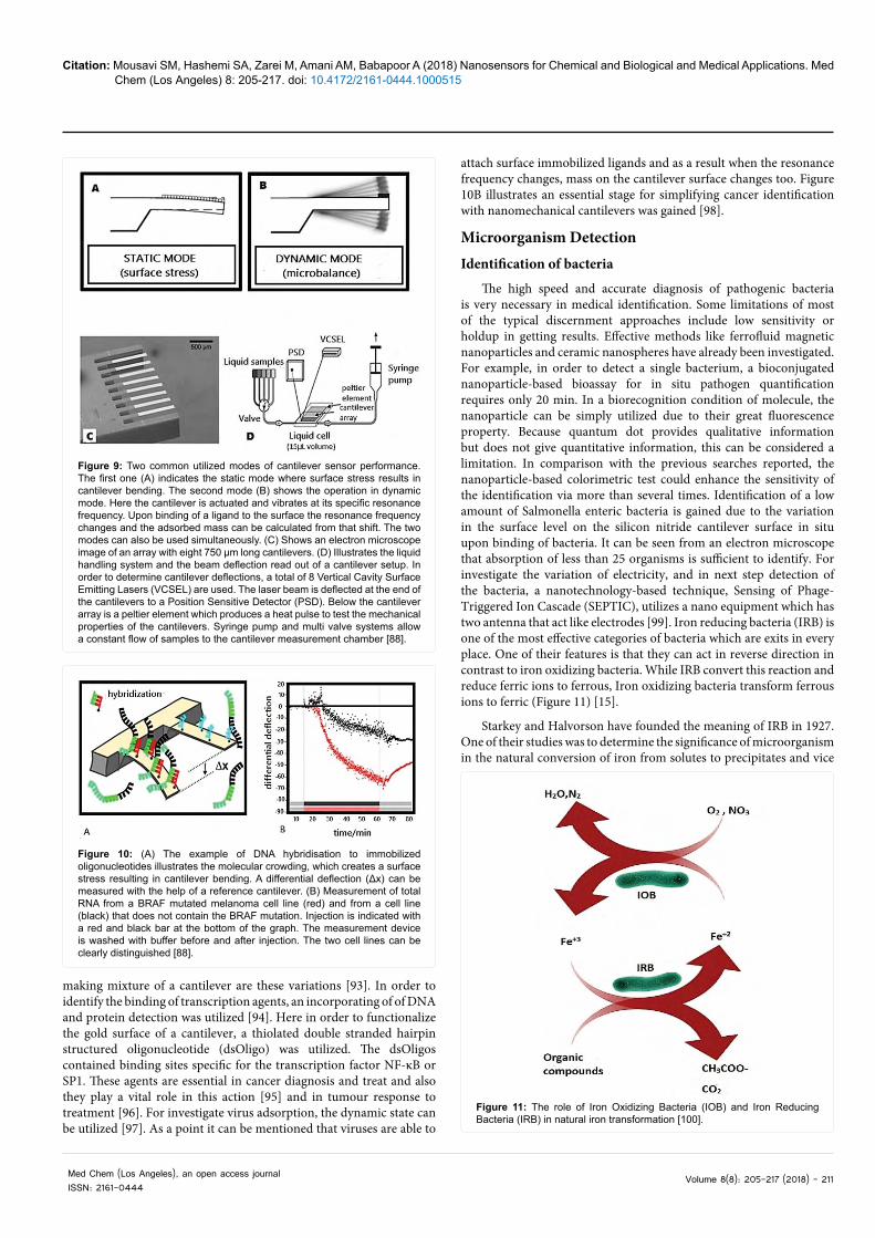
it does not require strengthening the aim in a cellular context. The search of basic dynamics of membrane proteins intercommunicate to surface level alterations is an exclusive usage of the cantilever array style. For example, it can be said that the properties of exhaled breath which allows assessment of a patient's condition in a non-invasive manner is another development. What is refers to the development of the atomic force microscope (AFM) [80] is the application of nanomechanical cantilevers, where in order to image surfaces on the molecular and atomic scale, a cantilever with a sharp tip at one end is utilized. From recent research and development it can be found that, high rate AFM has come into the sphere of time at the nanoscale and millisecond resolution in chemical procedure for instance imagination of the motion of myosin on an actin filament in real time [81]. Figure 9 provides a clear illustration of biomolecular interaction on the cantilever surface. some bonding methods are investigated either via cantilever bending down to the nanoscale due to variations in surface level based on molecular interactions on the cantilever surface (static mode, Figure 9A) [82], or extracting variations in resonance frequencies of a vibrating cantilever due to growth in mass upon binding of biomolecules (dynamic mode, Figure 9B) [83]. The dynamic mode is similar to the operation principle of a quartz crystal microbalance [84]. Recently, arrays of cantilevers manufactured from silicon or silicon nitride have been batch-fabricated (Figure 9C). Different approaches to measure cantilever bending or vibration have been progressed, among them laser rays deflection (Figure 9D) [85], interferometry [86] and the usage of piezo resistive cantilevers [87]. Functionalization by gold-thiol chemistry allows the formation of a self. As shown in (Figure 10A), the deflexion of the cantilever is because of adsorption and cellular relations between the SAM and supplementary molecules. For the first time this action was demonstrated in DNA hybridisation tests [89,90], where the binding of a 12 base long oligonucleotide at a concentration of 80 nM to its complement immobilised on the cantilever surface was studied with single base-pair specificity [86]. More researches concentrated on antibody antigen activitis on cantilevers. These factors illustrated sensitivity in the higher nanomolar to micromolar limited area, when using whole antibodies (Ab) functionalised on the surface [91]. Through oriented individual chain antibody fragments of the variable limitation (scFv), more progress in antigen identification sensitivity was gained that because of their tiny dimension and unmixed direction on the cantilever surface, which expanded the sensitivity about 500 times to low nanomolar concentrations [92]. Many applications rely on the field of structural variations in proteins. It can be said that this field and subject especially is appropriate for static measurements. Constructional variations for protein mechanisms are usually required for instance perform certain functions like enzymatic transformation or binding of ligands. What can raise the surface level in a monolayer
Figure 8: A typical Raman spectrum of a chemical compound and related peaks [78].
Citation: Mousavi SM, Hashemi SA, Zarei M, Amani AM, Babapoor A (2018) Nanosensors for Chemical and Biological and Medical Applications. Med Chem (Los Angeles) 8: 205-217. doi: 10.4172/2161-0444.1000515
Med Chem (Los Angeles), an open access journalISSN: 2161-0444
Volume 8(8): 205-217 (2018) - 211
making mixture of a cantilever are these variations [93]. In order to identify the binding of transcription agents, an incorporating of of DNA and protein detection was utilized [94]. Here in order to functionalize the gold surface of a cantilever, a thiolated double stranded hairpin structured oligonucleotide (dsOligo) was utilized. The dsOligos contained binding sites specific for the transcription factor NF-κB or SP1. These agents are essential in cancer diagnosis and treat and also they play a vital role in this action [95] and in tumour response to treatment [96]. For investigate virus adsorption, the dynamic state can be utilized [97]. As a point it can be mentioned that viruses are able to
Figure 9: Two common utilized modes of cantilever sensor performance. The first one (A) indicates the static mode where surface stress results in cantilever bending. The second mode (B) shows the operation in dynamic mode. Here the cantilever is actuated and vibrates at its specific resonance frequency. Upon binding of a ligand to the surface the resonance frequency changes and the adsorbed mass can be calculated from that shift. The two modes can also be used simultaneously. (C) Shows an electron microscope image of an array with eight 750 μm long cantilevers. (D) Illustrates the liquid handling system and the beam deflection read out of a cantilever setup. In order to determine cantilever deflections, a total of 8 Vertical Cavity Surface Emitting Lasers (VCSEL) are used. The laser beam is deflected at the end of the cantilevers to a Position Sensitive Detector (PSD). Below the cantilever array is a peltier element which produces a heat pulse to test the mechanical properties of the cantilevers. Syringe pump and multi valve systems allow a constant flow of samples to the cantilever measurement chamber [88].
Figure 10: (A) The example of DNA hybridisation to immobilized oligonucleotides illustrates the molecular crowding, which creates a surface stress resulting in cantilever bending. A differential deflection (Δx) can be measured with the help of a reference cantilever. (B) Measurement of total RNA from a BRAF mutated melanoma cell line (red) and from a cell line (black) that does not contain the BRAF mutation. Injection is indicated with a red and black bar at the bottom of the graph. The measurement device is washed with buffer before and after injection. The two cell lines can be clearly distinguished [88].
attach surface immobilized ligands and as a result when the resonance frequency changes, mass on the cantilever surface changes too. Figure 10B illustrates an essential stage for simplifying cancer identification with nanomechanical cantilevers was gained [98].
Microorganism DetectionIdentification of bacteria
The high speed and accurate diagnosis of pathogenic bacteria is very necessary in medical identification. Some limitations of most of the typical discernment approaches include low sensitivity or holdup in getting results. Effective methods like ferrofluid magnetic nanoparticles and ceramic nanospheres have already been investigated. For example, in order to detect a single bacterium, a bioconjugated nanoparticle-based bioassay for in situ pathogen quantification requires only 20 min. In a biorecognition condition of molecule, the nanoparticle can be simply utilized due to their great fluorescence property. Because quantum dot provides qualitative information but does not give quantitative information, this can be considered a limitation. In comparison with the previous searches reported, the nanoparticle-based colorimetric test could enhance the sensitivity of the identification via more than several times. Identification of a low amount of Salmonella enteric bacteria is gained due to the variation in the surface level on the silicon nitride cantilever surface in situ upon binding of bacteria. It can be seen from an electron microscope that absorption of less than 25 organisms is sufficient to identify. For investigate the variation of electricity, and in next step detection of the bacteria, a nanotechnology-based technique, Sensing of Phage-Triggered Ion Cascade (SEPTIC), utilizes a nano equipment which has two antenna that act like electrodes [99]. Iron reducing bacteria (IRB) is one of the most effective categories of bacteria which are exits in every place. One of their features is that they can act in reverse direction in contrast to iron oxidizing bacteria. While IRB convert this reaction and reduce ferric ions to ferrous, Iron oxidizing bacteria transform ferrous ions to ferric (Figure 11) [15].
Starkey and Halvorson have founded the meaning of IRB in 1927. One of their studies was to determine the significance of microorganism in the natural conversion of iron from solutes to precipitates and vice
Figure 11: The role of Iron Oxidizing Bacteria (IOB) and Iron Reducing Bacteria (IRB) in natural iron transformation [100].

Citation: Mousavi SM, Hashemi SA, Zarei M, Amani AM, Babapoor A (2018) Nanosensors for Chemical and Biological and Medical Applications. Med Chem (Los Angeles) 8: 205-217. doi: 10.4172/2161-0444.1000515
Med Chem (Los Angeles), an open access journalISSN: 2161-0444
Volume 8(8): 205-217 (2018) - 212
versa [100,101] In two environments such as aerobic and anaerobic can oxidize iron microbials. On the other hand the iron-reducing metabolic rout is done under anaerobic and or microaerobic situations [100,102] Because IRB is capable to combine iron nanostructures, it can be very much considered in nanotechnology. It can be said that a significant and effective portion of iron minerals in the geological subsurface is supposed to be presented as nano-sized colloids including iron oxides (hematite, magnetite) iron oxyhydr oxides (goethite, akaganeite, lepidocrocite, feroxyhyte) and iron hydrous oxides (ferrihydrite, hydro hematite maghemite). Iron nanoparticles play an essential role in biochemical mechanisms, due to their extensive event, inclination to nucleated and provide the surfaces of other phases, essential redox potential and also high reactivity [103]. Furthermore iron nanostructures have been widely utilized by many applications especially biomedical sciences [14]. Figure 12 is showing the intense activity of AgNPs against bacterial growth and Figure 13 indicates antimicrobial agents of AgNPs against E. coli and S. aureus.
Detection of viruses
Factors such as the effective and sensitive selection of viruses for implementing the effective reponse are very is very important and vital. Several methods like plaque and immunological experiments, transmission electron microscopy, and PCR- based testing of viruses are presented for more essential search. These methods generally require a high level of control, which is not suitable for infectious
agents, nor is detected quickly. Real-time electrical diagnosis has been gained by usage of nanowire field effect transistors single virus particles [104]. Electrical and optical measurements by fluorescently labelled influenza at the same time were utilized to illustrate undeniably which the conductance variations related to single viruses at the level of nanowire tools. In a past few years, magnetic nanoparticles act as a new antimicrobial agent and material. Effective agents of listeriosis are Listeria monocytogenes and play an essential role in public health fields. One of the main features of this bacterium is that, it is capable to fix to the host cell membrane and stimulate molecular [105].
Carbon based materials in nanosensors
Because of its magnificent applications in many areas, carbon is called as a unique element. This black substance has some characteristics such as low water solubility and weak fluorescence. It can be found in many forms including graphite, diamond, fullerenes, and graphene so it can be said that carbon is an astonishing element [106-114]. The carbon that acquires various properties can be used for different purposes, based on how the atoms are organized. Figure 14 illustrates the timeline of carbon-based materials started with the discoveries of fullerene [106,107], carbon nanotubes [108-110] and then graphene [111,113].
Carbon can forms many different chains with different length, and electronic configuration as stated in Figure 15. This hybridization property gives the possibility carbon to form more than hundred million compounds with different properties. Carbon based nanomaterials are common forms with hollow spheres, ellipsoids, or tubes. Spherical and ellipsoidal carbon nanomaterials are referred to as fullerenes and the cylindrical ones are called as nanotubes.
Nanowires are wires with a very small diameter; sometimes their size can be equal to 1 nanometer. Carbon nanowires are single nanoparticles or highly ordered arrays of nanoparticles that are being used independently as the electrode. The structure of a nanowire is so simple that there’s no room for defects, and electrons pass through without any obstacle [117-120]. In the last years, carbon nanotubes have overshadowed nanowires (Figure 16).
Recent applications on pharmaceutical analyses using carbon based nanosensors
The most destructive factors during the manufacture of composite materials are bubbles and cavities derived from them. Finally, because of an increase in gas concentrations of bubbles can cause a significant reduction in some properties and variations in the anatomy of the models. As a result, nanosensors may be used to reduce bubbles and improve properties and structure. The presence or absence of voids represents smooth and non-smooth surfaces in the composite samples as shown in SEM images. It can be said that in Figure 17 part A indicates the surface of sample B and the upper part of bubble based voids.
Figure 12: Antibacterial agent of AgNPs against E. coli and S. aureus [36].
Figure 13: Antimicrobial effects of AgNPs against E. coli and S. aureus [124].
Figure 14: Timeline of major milestones in the field of carbon nanomaterial based biomedical imaging and therapy. Reprinted with permission [115].

Citation: Mousavi SM, Hashemi SA, Zarei M, Amani AM, Babapoor A (2018) Nanosensors for Chemical and Biological and Medical Applications. Med Chem (Los Angeles) 8: 205-217. doi: 10.4172/2161-0444.1000515
Med Chem (Los Angeles), an open access journalISSN: 2161-0444
Volume 8(8): 205-217 (2018) - 213
It can be found from the results that, if there is a bubble in the composite, it can affect some properties such as electrical conductivity and Electromagnetic waves absorption. Moreover, it is accepted that bubbles have a great desire to combine with each other that can cause the emergence of huge size bubbles, by optical examination. Due to the growth in their internal gas concentration and dimension,
enormous size bubbles can make serious destruction in the composite construction and also diminish the overall features of composite models. Furthermore, bubbles attend to move from high concentration areas to lower places which can lead failure in the surface of composite models. In addition, Bubble based voids can lead to expansion of fracture in their direction that can cause the reduction in the mechanical features of composite models as is shown in SEM images [40]. In recent years carbon-based nanosensors have great use in pharmaceutical analyses application, further for real sample applications like tablets, human serum etc. In their work, Cheemalapati et al. developed an electroanalytical resolution of anxiolytic Buspirone hydrochloride using multi-walled carbon nanotubes on glassy carbon electrode was achieved. They used MWCNTs with the lengths of 0.1–10 _m which was prepared in dimethylformamide. Zhai et al. developed an electrochemical nanosensor using sulfonated graphene sheets/oxygen-functionalized multi-walled carbon nanotubes for the determination of β2-adrenergic agonists, Clenbuterol. After characterization of sulfonated graphene sheets/oxygen-functionalized multi-walled carbon nanotubes, Clenbuterol was detected with the differential pulse voltammetric method, with a detection limit of 0.0046 μM between 0.01-5.0 μM [121]. Fakhari et al. suggested reduced graphene oxide modified screen-printed electrode for the determination of NMDA receptor antagonist Dextromethorphan in urine and plasma. Using differential pulse voltammetric technique Dextromethorphan was detected with the limit of detection 1.5 ng mL-1 [122]. For the determination of Rivastigmine, Kalambate et al. developed the adsorptive stripping differential pulse voltammetric method using graphene nanosheet-gold nanoparticle/carbon paste nanosensor. The preparation of the nanosensor is shown in Figure 18. In the range of 2.0 × 10⁻⁷-6.0 × 10-
4M with a detection limit of 5.3 × 10⁻⁸M Rivastigmine was well detected with the suggested [124].
ConclusionRecently, nanosensors have taken a lot of applications in the fields
of pharmacy, medicine, industry and etc. Nano sensors can be utilized to solve many human problems and treat disease as they can easily be adapted to the environment. In this review article, the effects of nanotechnology on everyday life and its application in pharmacy and medical sciences were investigated. In addition sorts of nanosensors
Figure 15: Carbon nanomaterials such as the fullerene C60, CNT, graphene and nanodiamond for advanced technological applications Reprinted with permission [116].
Figure 16: (a) Low resolution and (b) high resolution SEM images of carbon nanowires. (c) TEM image of carbon nanowires, and (d) HRTEM image of carbon nanowires synthesized at the current of ∼10 A. Reprinted with permission [120].
Figure 17: SEM images from surface of composite samples, (A) sample-B, (B) sample-A (For interpretation of the references to colour in this figure legend, the reader is referred to the web version of this article) [42].

Citation: Mousavi SM, Hashemi SA, Zarei M, Amani AM, Babapoor A (2018) Nanosensors for Chemical and Biological and Medical Applications. Med Chem (Los Angeles) 8: 205-217. doi: 10.4172/2161-0444.1000515
Med Chem (Los Angeles), an open access journalISSN: 2161-0444
Volume 8(8): 205-217 (2018) - 214
has been introduced and categorized by their working mechanism. In general, the prominent properties of nanosensors were studied and it was also accepted that their electromagnetic species are used for chemical evaluation and also optical nanosensors are very useful for detection of intracellular chemicals. Finally, in this review article, the specificity of the diagnosis of cancer and microorganisms was also addressed.
References
1. Yih WY, Kratochvil FJ, Stewart JC (2005) Intraoral minor salivary gland neoplasms: review of 213 cases. Journal of Oral and Maxillofacial Surgery 63: 805-810.
2. Varadan VK, Chen L, Xie J (2008) Nanomedicine: design and applications of magnetic nanomaterials, nanosensors and nanosystems. John Wiley & Sons.
3. Datta SP (2016) Future Healthcare: Bioinformatics, Nano-Sensors, and Emerging Innovations. Nanosensors: Theory and Applications in Industry, Healthcare and Defense, p: 247.
4. Poinern GE, Fawcett D, Ng YJ, Ali N, Brundavanam RK, et al. (2010) Nanoengineering a biocompatible inorganic scaffold for skin wound healing. Journal of Biomedical Nanotechnology 6: 497.
5. Reis RL, Neves NM, Mano JF, Gomes ME, Marques AP, et al. (2008) Natural-based polymers for biomedical applications. Elsevier.
6. Broz P, Driamov S, Ziegler J, Ben-Haim N, Marsch S, et al. (2006) Toward intelligent nanosize bioreactors: a pH-switchable, channel-equipped, functional polymer nanocontainer. Nano Letters 6: 2349-235.
Figure 18: Illustration of the preparation procedure for GNS-AuNP nanocomposites. Reprinted with permission [123].
Nanosensor kind Subset Measured samples or Physical properties
OpticalFiber optic Benzopyrene tetrol, benzo[a]-pyrene, caspase-9 (an apoptosis protein), cytochrome c (a protein involved in
producing cellular energy), pH, K+, Ca2+, NO, NO2-, Cl-, Na+
PEBBLE (direct) H+, Ca2+, Mg2+, Zn2+, glucose and dissolved O2
PEBBLE (ion-correlation) K+, Na+, Cl-
Electromagnetic Current measurement H2S, GOx, lactase oxidase, dehydrogenase, peroxidase, hydrogen peroxide, catalase, organophosphorus pesticides, organophosphorus substrates of organophosphorushydrolase, DNA, ATP
Magnetism measurement Molecular interactions, oligonucleotide sequences, enzymatic activity, viral particles, magnetic field, speed, position sensing
Mechanical Vibrational Resonance frequency, spring constant Inertial Pressure, acceleration, yaw rate
Table 1: An abstract on different kinds of nanosensors and their usage [27].
7. Lehner R, Wang X, Marsch S, Hunziker P (2013) Intelligent nanomaterials for medicine: carrier platforms and targeting strategies in the context of clinical application. Nanomedicine: Nanotechnology, Biology and Medicine 9: 742-757.
8. Giljohann DA, Mirkin CA (2009) Drivers of biodiagnostic development. Nature 462: 461.
9. Rosi NL, Giljohann DA, Thaxton CS, Lytton-Jean AK, Han MS (2006) Oligonucleotide-modified gold nanoparticles for intracellular gene regulation. Science 312: 1027-1030.
10. Mirkin CA, Taton TA (2000) Materials chemistry: Semiconductors meet biology. Nature 405: 626.
11. Mirkin CA, Letsinger RL, Mucic RC, Storhoff JJ (1996) A DNA-based method for rationally assembling nanoparticles into macroscopic materials. Nature 382: 607.
12. Kianpour S, Ebrahiminezhad A, Mohkam M, Tamaddon AM, Dehshahri A (2017) Physicochemical and biological characteristics of the nanostructured polysaccharide iron hydrogel produced by microorganism Klebsiella oxytoca. Journal of Basic Microbiology 57: 132-140.
13. Chomoucka J, Drbohlavova J, Masarik M, Ryvolova M, Huska D, et al. (2012) Nanotechnologies for society. New designs and applications of nanosensors and nanobiosensors in medicine and environmental analysis. International Journal of Nanotechnology 9: 746-783.
14. Riu J, Maroto A, Rius FX (2006) Nanosensors in environmental analysis. Talanta 69: 288-301.
15. Ebrahiminezhad A, Manafi Z, Berenjian A, Kianpour S, Ghasemi Y (2017)

Citation: Mousavi SM, Hashemi SA, Zarei M, Amani AM, Babapoor A (2018) Nanosensors for Chemical and Biological and Medical Applications. Med Chem (Los Angeles) 8: 205-217. doi: 10.4172/2161-0444.1000515
Med Chem (Los Angeles), an open access journalISSN: 2161-0444
Volume 8(8): 205-217 (2018) - 215
Iron-Reducing Bacteria and Iron Nanostructures. Journal of Advanced Medical Sciences and Applied Technologies 3: 9-16.
16. Xu S, Luo Y, Graeser R, Warnecke A, Kratz F, et al. (2009) Development of pH-responsive core–shell nanocarriers for delivery of therapeutic and diagnostic agents. Bioorganic & Medicinal Chemistry Letters 19: 1030-1034.
17. Peer D, Karp JM, Hong S, Farokhzad OC, Margalit R, et al. (2007) Nanocarriers as an emerging platform for cancer therapy. Nature Nanotechnology 2: 751.
18. Itaka K, Chung UI, Kataoka K (2006) Supramolecular nanocarrier for gene and siRNA delivery. Nihon rinsho, Japanese Journal of Clinical Medicine 64: 253-257.
19. White RR, Sullenger BA, Rusconi CP (2000) Developing aptamers into therapeutics. The Journal of clinical investigation 106: 929-934.
20. Parveen S, Sahoo SK (2008) Polymeric nanoparticles for cancer therapy. Journal of drug targeting 16: 108-123.
21. Park TG, Jeong JH, Kim SW (2006) Current status of polymeric gene delivery systems. Advanced drug delivery reviews 58: 467-486.
22. Petros RA, DeSimone JM (2010) Strategies in the design of nanoparticles for therapeutic applications. Nature reviews Drug discovery 9: 615.
23. Torchilin VP (2006) Multifunctional nanocarriers. Advanced drug delivery reviews 58: 1532-1555.
24. Colombo M, Ronchi S, Monti D, Corsi F, Trabucchi E, et al. (2009) Femtomolar detection of autoantibodies by magnetic relaxation nanosensors. Analytical biochemistry 392: 96-102.
25. Devreese JT (2007) Importance of nanosensors: Feynman's vision and the birth of nanotechnology. Mrs Bulletin 32: 718-725.
26. Yonzon CR, Stuart DA, Zhang X, McFarland AD, Haynes CL, et al. (2005) Towards advanced chemical and biological nanosensors-an overview. Talanta 67: 438-448.
27. Lim TC, Ramakrishna S (2006) A conceptual review of nanosensors. Zeitschrift für Naturforschung A 61: 402-412.
28. Gholami A, Rasoul-amini S, Ebrahiminezhad A, Seradj SH, Ghasemi Y (2015) Lipoamino acid coated superparamagnetic iron oxide nanoparticles concentration and time dependently enhanced growth of human hepatocarcinoma cell line (Hep-G2). Journal of Nanomaterials 16: 150.
29. Ebrahiminezhad A, Ghasemi Y, Rasoul-Amini S, Barar J, Davaran S (2013) Preparation of novel magnetic fluorescent nanoparticles using amino acids. Colloids and Surfaces B: Biointerfaces 102: 534-539.
30. Ebrahiminezhad A, Ghasemi Y, Rasoul-Amini S, Barar J, Davaran S (2012) Impact of amino-acid coating on the synthesis and characteristics of iron-oxide nanoparticles (IONs). Bulletin of the Korean Chemical Society 33: 3957-3962.
31. Jiang ZJ, Liu CY, Sun LW (2005) Catalytic properties of silver nanoparticles supported on silica spheres. The Journal of Physical Chemistry B 109: 1730-1735.
32. McFarland AD, Van Duyne RP (2003) Single silver nanoparticles as real-time optical sensors with zeptomole sensitivity. Nano letters 3: 1057-1062.
33. Haider A, Kang IK (2015) Preparation of silver nanoparticles and their industrial and biomedical applications: a comprehensive review. Advances in materials science and engineering.
34. Xiu ZM, Zhang QB, Puppala HL, Colvin VL, Alvarez PJ (2012) Negligible particle-specific antibacterial activity of silver nanoparticles. Nano letters 12: 4271-4275.
35. Xiu ZM, Ma J, Alvarez PJ (2011) Differential effect of common ligands and molecular oxygen on antimicrobial activity of silver nanoparticles versus silver ions. Environmental science & technology 45: 9003-9008.
36. Ebrahiminezhad A, Barzegar Y, Ghasemi Y, Berenjian A (2016) Green synthesis and characterization of silver nanoparticles using Alcea rosea flower extract as a new generation of antimicrobials. Chemical Industry and Chemical Engineering Quarterly pp: 2.
37. Ebrahiminezhad A, Taghizadeh S, Berenjiand A, Rahi A, Ghasemi Y (2016) Synthesis and characterization of silver nanoparticles with natural carbohydrate capping using Zataria multiflora. Advanced Materials Letters 7: 939-44.
38. Faramarzi MA, Yazdi MT, Ghostinroudi H, Amini M, Ghasemi Y, et al (2006) Nostoc muscorum: a regioselective biocatalyst for 17-carbonyl reduction
of androst-4-en-3, 17-dione and androst-1, 4-dien-3, 17-dione. Annals of microbiology 56: 253.
39. Ashrafi H, Amini M, Mohammadi-Samani S, Ghasemi Y, Azadi A, et al. (2013) Nanostructure L-asparaginase-fatty acid bioconjugate: synthesis, preformulation study and biological assessment. International journal of biological macromolecules 62: 180-187.
40. Mitra RN, Das PK (2008) In situ preparation of gold nanoparticles of varying shape in molecular hydrogel of peptide amphiphiles. The Journal of Physical Chemistry C 112: 8159-8166.
41. Zomorodian K, Moein M, Lori ZG, Ghasemi Y, Rahimi MJ, et al. (2013) Chemical composition and antimicrobial activities of the essential oil from Myrtus communis leaves. Journal of Essential Oil Bearing Plants. 16: 76-84.
42. Hashemi SA, Mousavi SM (2016) Effect of bubble based degradation on the physical properties of Single Wall Carbon Nanotube/Epoxy Resin composite and new approach in bubbles reduction. Composites Part A: Applied Science and Manufacturing 90: 457-469.
43. Bodaghi M, Cristóvão C, Gomes R, Correia NC (2016) Experimental characterization of voids in high fibre volume fraction composites processed by high injection pressure RTM. Composites Part A: Applied Science and Manufacturing 82: 88-99.
44. Li Y, Li Q, Ma H (2015) The voids formation mechanisms and their effects on the mechanical properties of flax fiber reinforced epoxy composites. Composites Part A: Applied Science and Manufacturing 72: 40-48.
45. Hernández S, Sket F, Molina-Aldareguı JM, González C, LLorca J (2011) Effect of curing cycle on void distribution and interlaminar shear strength in polymer-matrix composites. Composites science and technology 71: 1331-1341.
46. Liu L, Zhang BM, Wang DF, Wu ZJ (2006) Effects of cure cycles on void content and mechanical properties of composite laminates. Composite structures 73: 303-309.
47. Park J, Taweeplengsangsuke J, Theis C, Osenbach J (2001) Epoxy adhesive used in optical fiber/passive component: kinetics, voids and reliability. In Electronic Components and Technology Conference, 2001, Proceedings 51: 637-644.
48. Hashemi SA, Mousavi SM, Arjmand M, Yan N, Sundararaj U (2018) Electrified single‐walled carbon nanotube/epoxy nanocomposite via vacuum shock technique: Effect of alignment on electrical conductivity and electromagnetic interference shielding. Polymer Composites 39: E1139-E1148.
49. Mousavi SM, Hashemi SA, Jahandideh S, Baseri S, Zarei M (2017) Modification of Phenol Novolac Epoxy Resin and Unsaturated Polyester Using Sasobit and Silica Nanoparticles. Polymers from Renewable Resources 8: 3.
50. Mousavi SM, Hashemi SA, Jahandideh S, Baseri S, Zarei M (2015) Biodegradation study of nanocomposites of phenol novolac epoxy/unsaturated polyester resin/egg shell nanoparticles using natural polymers. Journal of Materials 8.
51. Mousavi SM, Arjmand O, Hashemi SA, Banaei N (2016) Modification of the Epoxy Resin Mechanical and Thermal Properties with Silicon Acrylate and Montmorillonite Nanoparticles. Polymers from Renewable Resources 7.
52. Mousavi SM, Aghili A, Hashemi SA, Goudarzian N, Bakhoda Z, et al. (2016) Improved Morphology and Properties of Nanocomposites, Linear Low Density Polyethylene, Ethylene-co-vinyl Acetate and Nano Clay Particles by Electron Beam. Polymers from Renewable Resources 7: 135.
53. Sasaki K, Shi ZY, Kopelman R, Masuhara H (1996) Three-dimensional pH microprobing with an optically-manipulated fluorescent particle," Chemistry Letters 25: 141-142.
54. De Silva AP, Gunaratne HN, Gunnlaugsson T, Huxley AJ, McCoy CP, et al. (1997) Signaling recognition events with fluorescent sensors and switches. Chemical reviews 97: 1515-1566.
55. Czarnik AW (1993) Fluorescent chemosensors for ion and molecule recognition. American Chemical Society.
56. Kulmala S, Suomi J (2003) Current status of modern analytical luminescence methods. Analytica Chimica Acta 500: 21-69.
57. Lakowicz JR (1999) Fluorophores. In Principles of fluorescence spectroscopy: Springer pp: 63-93.
58. Haugland RP (1996) Handbook of fluorescent probes and research chemicals. Molecular Probes, Eugene 8.
Citation: Mousavi SM, Hashemi SA, Zarei M, Amani AM, Babapoor A (2018) Nanosensors for Chemical and Biological and Medical Applications. Med Chem (Los Angeles) 8: 205-217. doi: 10.4172/2161-0444.1000515
Med Chem (Los Angeles), an open access journalISSN: 2161-0444
Volume 8(8): 205-217 (2018) - 216
59. Ji J, Rosenzweig N, Jones I, Rosenzweig Z (2001) Molecular oxygen-sensitive fluorescent lipobeads for intracellular oxygen measurements in murine macrophages. Analytical chemistry 73: 3521-3527.
60. Ji J, Rosenzweig N, Griffin C, Rosenzweig Z (2000) Synthesis and application of submicrometer fluorescence sensing particles for lysosomal pH measurements in murine macrophages. Analytical chemistry 72: 3497-3503.
61. Kim YP, Daniel WL, Xia Z, Xie H, Mirkin CA, et al (2010) Bioluminescent nanosensors for protease detection based upon gold nanoparticle–luciferase conjugates. Chemical communications 46: 76-78.
62. Fehr M, Frommer WB, Lalonde S (2002) Visualization of maltose uptake in living yeast cells by fluorescent nanosensors. Proceedings of the National Academy of Sciences 99: 9846-9851.
63. Kasili PM, Cullum BM, Griffin GD, Vo-Dinh T, et al. (2002) Nanosensor for in vivo measurement of the carcinogen benzo [a] pyrene in a single cell. Journal of nanoscience and nanotechnology 2: 653-658.
64. Vo-Dinh T, Griffin GD, Alarie JP, Cullum B, Sumpter B, et al. (2000) Development of nanosensors and bioprobes. Journal of Nanoparticle Research 2: 17-27.
65. Vo-Dinh T, Cullum B (2000) Biosensors and biochips: advances in biological and medical diagnostics. Fresenius' journal of analytical chemistry 366: 540-551.
66. Vo-Dinh T, Alarie JP, Cullum BM, Griffin GD (2000) Antibody-based nanoprobe for measurement of a fluorescent analyte in a single cell. Nature biotechnology 18: 764.
67. Cullum BM, Griffin GD, Miller GH, Vo-Dinh T (2000) Intracellular measurements in mammary carcinoma cells using fiber-optic nanosensors. Analytical Biochemistry 277: 25-32.
68. Geng J, Thomas MD, Shephard DS, Johnson BF (2005) Suppressed electron hopping in a Au nanoparticle/H 2 S system: development towards a H 2 S nanosensor. Chemical Communications 1895-1897.
69. Akyildiz IF, Jornet JM (2010) Electromagnetic wireless nanosensor networks. Nano Communication Networks vol. 1: 3-19.
70. Schellenberger E (2009) Bioresponsive nanosensors in medical imaging. Journal of the royal society Interface.
71. Zakeri A, Kouhbanani MA, Beheshtkhoo N, Beigi V, Mousavi SM, et al. (2018) Polyethylenimine-based nanocarriers in co-delivery of drug and gene: a developing horizon. Nano Reviews & Experiments 9: 1488497.
72. Amani AM, Hashemi SA, Mousavi SM, Pouya H, Arash V. Electric Field Induced Alignment of Carbon Nanotubes: Methodology and Outcomes.
73. Mousavi SM, Hashemi SA, Amani AM, Saed H, Jahandideh S, et al. (2017) Polyethylene Terephthalate/Acryl Butadiene Styrene Copolymer Incorporated with Oak Shell, Potassium Sorbate and Egg Shell Nanoparticles for Food Packaging Applications: Control of Bacteria Growth, Physical and Mechanical Properties. Polymers from Renewable Resources 8.
74. Goudarzian N, Hashemi SA, Mirjalili M (2016) Unsaturated Polyester Resins Modified With Cresol Novolac Epoxy and Silica Nanoparticles: Processing and Mechanical Properties. International Journal of Chemical and Petroleum Sciences 5: 13-26.
75. Hashemi SA, Mousavi SM, Faghihi R, Arjmand M, Sina S, et al. (2018) Lead oxide-decorated graphene oxide/epoxy composite towards X-Ray radiation shielding. Radiation Physics and Chemistry 146: 77-85.
76. Akbarian M, Ghasemi Y, Uversky VN, Yousefi R (2018) Chemical modifications of insulin: Finding a compromise between stability and pharmaceutical performance. International journal of pharmaceutics.
77. Ravanshad R, Karimi Zadeh A, Amani AM, Mousavi SM, Hashemi SA, et al (2018) Application of nanoparticles in cancer detection by Raman scattering based techniques. Nano Reviews & Experiments 9: 1373551.
78. Kiefer J (2015) Recent advances in the characterization of gaseous and liquid fuels by vibrational spectroscopy. Energies 8: 3165-3197.
79. Nagel DJ, Smith S (2003) Nanotechnology-Enabled Sensors: Possibilities, Realities, and Applications-Operating on the scale of atoms and molecules, emerging nanotechnologies promise dramatic changes in sensor designs and. Sensors-the Journal of Applied Sensing Technology 20: 22-28.
80. Binnig G, Quate CF, Gerber C (1986) Atomic force microscope. Physical review letters 56: 930.
81. Kodera N, Yamamoto D, Ishikawa R, Ando T (2010) Video imaging of walking myosin V by high-speed atomic force microscopy. Nature 468: 72.
82. Thundat T, Warmack RJ, Chen GY, Allison DP (1994) Thermal and ambient‐induced deflections of scanning force microscope cantilevers. Applied Physics Letters 64: 2894-2896.
83. Thundat T, Chen GY, Warmack RJ, Allison DP, Wachter EA (1995) Vapor detection using resonating microcantilevers. Analytical Chemistry 67: 519-521.
84. Marx KA (2003) Quartz crystal microbalance: a useful tool for studying thin polymer films and complex biomolecular systems at the solution- surface interface. Biomacromolecules 4: 1099-1120.
85. Meyer G, Amer NM (1988) Erratum: Novel optical approach to atomic force microscopy [Appl. Phys. Lett. 5 3, 1045 (1988)]. Applied physics letters 53: 2400-2402.
86. Helm M, Servant JJ, Saurenbach F, Berger R (2005) Read-out of micromechanical cantilever sensors by phase shifting interferometry. Applied Physics Letters 87: 064101.
87. Datskos PG, Oden PI, Thundat T, Wachter EA, Warmack RJ, et al. (1996) Remote infrared radiation detection using piezoresistive microcantilevers. Applied Physics Letters 69: 2986-2988.
88. Huber F, Lang HP, Zhang J, Rimoldi D, Gerber C (2015) Nanosensors for cancer detection. Swiss medical weekly 145: w14092.
89. Fritz J, Baller MK, Lang HP, Rothuizen H, Vettiger P, et al. (2000) Translating biomolecular recognition into nanomechanics. Science 288: 316-318.
90. McKendry R, Zhang J, Arntz Y, Strunz T, Hegner M, et al. (2002) Multiple label-free biodetection and quantitative DNA-binding assays on a nanomechanical cantilever array. Proceedings of the National Academy of Sciences 99: 9783-9788.
91. Arntz Y, Seelig JD, Lang HP, Zhang J, Hunziker P, et al. (2002) Label-free protein assay based on a nanomechanical cantilever array. Nanotechnology 14: 86.
92. Backmann N, Zahnd C, Huber F, Bietsch A, Plückthun A, et al. (2005) A label-free immunosensor array using single-chain antibody fragments. Proceedings of the National Academy of Sciences 102: 14587-14592.
93. Braun T, Backmann N, Vögtli M, Bietsch A, Engel A, et al. (2006) Conformational change of bacteriorhodopsin quantitatively monitored by microcantilever sensors. Biophysical Journal 90: 2970-2977.
94. Huber F, Hegner M, Gerber C, Güntherodt HJ, Lang HP (2006) Label free analysis of transcription factors using microcantilever arrays. Biosensors and Bioelectronics 21: 1599-1605.
95. Xie K, Wei D, Huang S (2006) Transcriptional anti-angiogenesis therapy of human pancreatic cancer. Cytokine & growth factor reviews 17: 147-156.
96. Criswell T, Leskov K, Miyamoto S, Luo G, Boothman DA (2003) Transcription factors activated in mammalian cells after clinically relevant doses of ionizing radiation. Oncogene 22: 5813.
97. Braun T, Ghatkesar MK, Backmann N, Grange W, Boulanger P, et al. (2009) Quantitative time-resolved measurement of membrane protein–ligand interactions using microcantilever array sensors. Nature nanotechnology 4: 179.
98. Huber F, Lang HP, Backmann N, Rimoldi D, Gerber C (2013) Direct detection of a BRAF mutation in total RNA from melanoma cells using cantilever arrays. Nature nanotechnology 8: 125.
99. Kaittanis C, Santra S, Perez JM (2010) Emerging nanotechnology-based strategies for the identification of microbial pathogenesis. Advanced drug delivery reviews 62: 408-423.
100. Erbs M, Spain J (2002) Microbial iron metabolism in natural environments. Microbial Diversity.
101. Starkey RL, Halvorson HO (1927) Studies on the transformations of iron in nature. II. Concerning the importance of microörganisms in the solution and precipitation of iron. Soil Science 24: 381-402.
102. Hedrich S, Schlömann M, Johnson DB (2011) The iron-oxidizing proteobacteria. Microbiology 157: 1551-1564.
103. Bosch J, Heister K, Hofmann T, Meckenstock RU (2010) Nanosized iron oxide colloids strongly enhance microbial iron reduction. Applied and environmental microbiology 76: 184-189.

Citation: Mousavi SM, Hashemi SA, Zarei M, Amani AM, Babapoor A (2018) Nanosensors for Chemical and Biological and Medical Applications. Med Chem (Los Angeles) 8: 205-217. doi: 10.4172/2161-0444.1000515
Med Chem (Los Angeles), an open access journalISSN: 2161-0444
Volume 8(8): 205-217 (2018) - 217
104. Jain KK (2005) The role of nanobiotechnology in drug discovery. Drug discovery today. 10: 1435-1442.
105. Ebrahiminezhad A, Rasoul-Amini S, Davaran S, Barar J, Ghasemi Y (2014) Impacts of iron oxide nanoparticles on the invasion power of Listeria monocytogenes. Current Nanoscience 10: 382-388.
106. Kroto HW, Heath JR, O'Brien SC, Curl RF, Smalley RE (1985) C60: Buckminsterfullerene. Nature 318: 162.
107. Kroto HW, McKay K (1988) The formation of quasi-icosahedral spiral shell carbon particles. Nature 331: 328.
108. Iijima S, Ichihashi T (1993) Single-shell carbon nanotubes of 1-nm diameter. Nature 363: 603.
109. Iijima S, Yudasaka M, Yamada R, Bandow S, Suenaga K, et al. (1999) Nano-aggregates of single-walled graphitic carbon nano-horns. Chemical Physics Letters 309: 165-170.
110. Iijima S (2002) Carbon nanotubes: past, present, and future. Physica B: Condensed Matter 323: 1-5.
111. Novoselov KS, Geim AK, Morozov SV, Jiang DA, Zhang Y (2004) Electric field effect in atomically thin carbon films. science 306: 666-669.
112. Barsan MM, Carvalho RC, Zhong Y, Sun X, Brett CM (2012) Carbon nanotube modified carbon cloth electrodes: Characterisation and application as biosensors. Electrochimica Acta 85: 203-209.
113. Novoselov KS, Fal VI, Colombo L, Gellert PR, Schwab MG, et al. (2012) A roadmap for grapheme. Nature 490: 192.
114. Barsan MM, Ghica ME, Brett CM (2015) Electrochemical sensors and biosensors based on redox polymer/carbon nanotube modified electrodes: a review. Analytica chimica acta 881: 1-23.
115. Hong G, Diao S, Antaris AL, Dai H (2015) Carbon nanomaterials for biological imaging and nanomedicinal therapy. Chemical reviews 115: 10816-10906.
116. Zhai W, Srikanth N, Kong LB, Zhou K (2017) Carbon nanomaterials in tribology. Carbon 119: 150-171.
117. Tang YH, Wang N, Zhang YF, Lee CS, Bello I, et al. (1999) Synthesis and characterization of amorphous carbon nanowires. Applied physics letters 75: 2921-2923.
118. Zhao X, Ando Y, Liu Y, Jinno M, Suzuki T (2003) Carbon nanowire made of a long linear carbon chain inserted inside a multiwalled carbon nanotube. Physical review letters 90: 187401.
119. Chang J, Min BK, Kim J, Lee SJ, Lin L (2009) Electrostatically actuated carbon nanowire nanotweezers. Smart Materials and Structures 18: 065017.
120. Qin Z, Li ZJ, Yang BC (2012) Synthesis of carbon nanowires as electrochemical electrode materials. Materials Letters 69: 55-58.
121. Zhai H, Liu Z, Chen Z, Liang Z, Su Z, et al. (2015) A sensitive electrochemical sensor with sulfonated graphene sheets/oxygen-functionalized multi-walled carbon nanotubes modified electrode for the detection of clenbuterol. Sensors and Actuators B: Chemical 210: 483-490.
122. Fakhari AR, Koruni MH, Ahmar H, Shahsavani A, Movahed SK (2014) Electrochemical Determination of Dextromethorphan on Reduced Graphene Oxide Modified Screen Printed Electrode after Electromembrane Extraction. Electroanalysis 26: 521-529.
123. Kalambate PK, Biradar MR, Karna SP, Srivastava AK (2015) Adsorptive stripping differential pulse voltammetry determination of rivastigmine at graphene nanosheet-gold nanoparticle/carbon paste electrode. Journal of Electroanalytical Chemistry 757: 150-158.
124. Ebrahiminezhad A, Bagheri M, Taghizadeh SM, Berenjian A, Ghasemi Y (2016) Biomimetic synthesis of silver nanoparticles using microalgal secretory carbohydrates as a novel anticancer and antimicrobial. Advances in Natural Sciences: Nanoscience and Nanotechnology 7: 015018.