Mounted CP Stent - numedforchildren.com · Mounted CP Stent ™ COARCTATION OF ... (800) 443-8362...
24
Mounted CP Stent COARCTATION OF THE AORTA INSTRUCTIONS FOR USE General Instructions………..…………Page 2 Stent Sizing Charts and Information………………..Page 21 CAUTION: FEDERAL (USA) LAW RESTRICTS THIS DEVICE TO SALE BY OR ON THE ORDER OF A PHYSICIAN. Read all instructions prior to use. Distributed by: B. Braun Interventional Systems Inc. 824 Twelfth Avenue Bethlehem, PA 18018 Customer Service: TEL: (877) 836-2228 FAX: (610) 849-1334 Technical Support TEL: (800) 443-8362 Made in U.S.A. Manufactured By: NuMED, Inc. 2880 Main Street Hopkinton, New York USA 12965 Telephone: (315) 328-4491 Facsimile: (315) 328-4941 Email: [email protected] Internet: www.numedforchildren.com
Transcript of Mounted CP Stent - numedforchildren.com · Mounted CP Stent ™ COARCTATION OF ... (800) 443-8362...
Mounted CP Stent
COARCTATION OF THE AORTA
INSTRUCTIONS FOR USE
General Instructions………..…………Page 2
Stent Sizing Charts and Information………………..Page 21
CAUTION: FEDERAL (USA) LAW RESTRICTS THIS DEVICE TO
SALE BY OR ON THE ORDER OF A PHYSICIAN.
Read all instructions prior to use.
Distributed by: B. Braun Interventional Systems Inc.
824 Twelfth Avenue
Bethlehem, PA 18018
Customer Service:
TEL: (877) 836-2228
FAX: (610) 849-1334
Technical Support
TEL: (800) 443-8362
Made in U.S.A.
Manufactured By:
NuMED, Inc.
2880 Main Street
Hopkinton, New York
USA 12965
Telephone: (315) 328-4491
Facsimile: (315) 328-4941
Email: [email protected]
Internet: www.numedforchildren.com
2
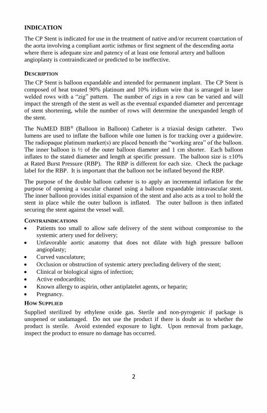
INDICATION
The CP Stent is indicated for use in the treatment of native and/or recurrent coarctation of
the aorta involving a compliant aortic isthmus or first segment of the descending aorta
where there is adequate size and patency of at least one femoral artery and balloon
angioplasty is contraindicated or predicted to be ineffective.
DESCRIPTION
The CP Stent is balloon expandable and intended for permanent implant. The CP Stent is
composed of heat treated 90% platinum and 10% iridium wire that is arranged in laser
welded rows with a “zig” pattern. The number of zigs in a row can be varied and will
impact the strength of the stent as well as the eventual expanded diameter and percentage
of stent shortening, while the number of rows will determine the unexpanded length of
the stent.
The NuMED BIB® (Balloon in Balloon) Catheter is a triaxial design catheter. Two
lumens are used to inflate the balloon while one lumen is for tracking over a guidewire.
The radiopaque platinum marker(s) are placed beneath the “working area” of the balloon.
The inner balloon is ½ of the outer balloon diameter and 1 cm shorter. Each balloon
inflates to the stated diameter and length at specific pressure. The balloon size is ±10%
at Rated Burst Pressure (RBP). The RBP is different for each size. Check the package
label for the RBP. It is important that the balloon not be inflated beyond the RBP.
The purpose of the double balloon catheter is to apply an incremental inflation for the
purpose of opening a vascular channel using a balloon expandable intravascular stent.
The inner balloon provides initial expansion of the stent and also acts as a tool to hold the
stent in place while the outer balloon is inflated. The outer balloon is then inflated
securing the stent against the vessel wall.
CONTRAINDICATIONS
• Patients too small to allow safe delivery of the stent without compromise to the
systemic artery used for delivery;
• Unfavorable aortic anatomy that does not dilate with high pressure balloon
angioplasty;
• Curved vasculature;
• Occlusion or obstruction of systemic artery precluding delivery of the stent;
• Clinical or biological signs of infection;
• Active endocarditis;
• Known allergy to aspirin, other antiplatelet agents, or heparin;
• Pregnancy.
HOW SUPPLIED
Supplied sterilized by ethylene oxide gas. Sterile and non-pyrogenic if package is
unopened or undamaged. Do not use the product if there is doubt as to whether the
product is sterile. Avoid extended exposure to light. Upon removal from package,
inspect the product to ensure no damage has occurred.
3
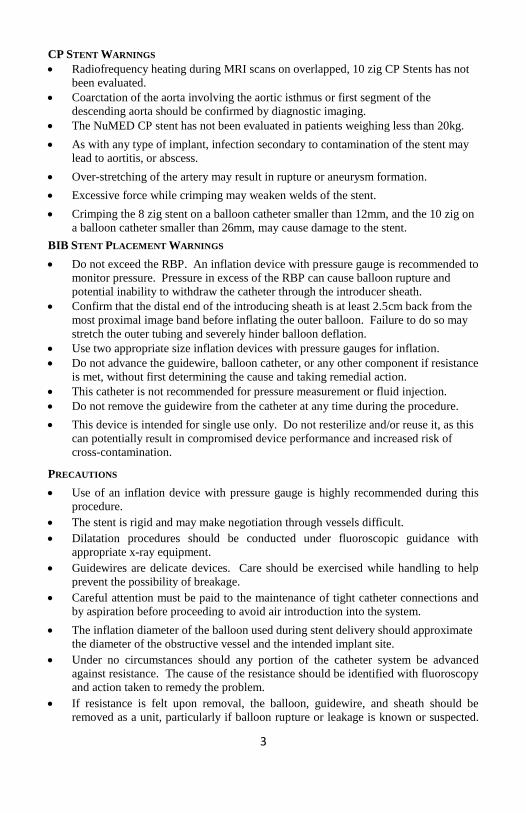
CP STENT WARNINGS
• Radiofrequency heating during MRI scans on overlapped, 10 zig CP Stents has not
been evaluated.
• Coarctation of the aorta involving the aortic isthmus or first segment of the
descending aorta should be confirmed by diagnostic imaging.
• The NuMED CP stent has not been evaluated in patients weighing less than 20kg.
• As with any type of implant, infection secondary to contamination of the stent may
lead to aortitis, or abscess.
• Over-stretching of the artery may result in rupture or aneurysm formation.
• Excessive force while crimping may weaken welds of the stent.
• Crimping the 8 zig stent on a balloon catheter smaller than 12mm, and the 10 zig on
a balloon catheter smaller than 26mm, may cause damage to the stent.
BIB STENT PLACEMENT WARNINGS
• Do not exceed the RBP. An inflation device with pressure gauge is recommended to
monitor pressure. Pressure in excess of the RBP can cause balloon rupture and
potential inability to withdraw the catheter through the introducer sheath.
• Confirm that the distal end of the introducing sheath is at least 2.5cm back from the
most proximal image band before inflating the outer balloon. Failure to do so may
stretch the outer tubing and severely hinder balloon deflation.
• Use two appropriate size inflation devices with pressure gauges for inflation.
• Do not advance the guidewire, balloon catheter, or any other component if resistance
is met, without first determining the cause and taking remedial action.
• This catheter is not recommended for pressure measurement or fluid injection.
• Do not remove the guidewire from the catheter at any time during the procedure.
• This device is intended for single use only. Do not resterilize and/or reuse it, as this
can potentially result in compromised device performance and increased risk of
cross-contamination.
PRECAUTIONS
• Use of an inflation device with pressure gauge is highly recommended during this
procedure.
• The stent is rigid and may make negotiation through vessels difficult.
• Dilatation procedures should be conducted under fluoroscopic guidance with
appropriate x-ray equipment.
• Guidewires are delicate devices. Care should be exercised while handling to help
prevent the possibility of breakage.
• Careful attention must be paid to the maintenance of tight catheter connections and
by aspiration before proceeding to avoid air introduction into the system.
• The inflation diameter of the balloon used during stent delivery should approximate
the diameter of the obstructive vessel and the intended implant site.
• Under no circumstances should any portion of the catheter system be advanced
against resistance. The cause of the resistance should be identified with fluoroscopy
and action taken to remedy the problem.
• If resistance is felt upon removal, the balloon, guidewire, and sheath should be
removed as a unit, particularly if balloon rupture or leakage is known or suspected.
4
This may be accomplished by firmly grasping the balloon catheter and sheath as a
unit and withdrawing both together, using a gentle twisting motion combined with
traction.
• Proper functioning of the catheter depends on its integrity. Care should be used
when handling the catheter. Damage may result from kinking, stretching, or
forceful wiping of the catheter.
POTENTIAL COMPLICATIONS / ADVERSE EFFECTS
NOTE: Circumferential tear of the delivery balloon catheter prior to complete expansion
of the stent may cause the balloon to become tethered to the stent, requiring surgical
removal. In case of rupture of an adequately sized balloon after stent expansion, it can be
withdrawn and a new balloon catheter exchanged over a guidewire to complete expansion
of the stent.
Cardiac catheterization carries certain risks. In addition, potential complications, and
related adverse effects associated with implants include, but are not limited to:
• Femoral artery injury, thrombosis or pseudoaneurysm
• Stent Migration • Stent Stenosis
• Stent Fracture • Aortic Aneurysm/Pseudoaneurysm
• Aortic Rupture/Tear • Stent Malposition
• Hematoma • Sepsis/infection
• Thrombosis/Thromboembolism • AV fistula formation
• Death • Transitory arrhythmia
• Endocarditis • Bleeding
• Cell necrosis at the site of implant
• Cerebrovascular Incident
MRI SAFETY INFORMATION
Nonclinical testing and modeling has demonstrated that the CP Stent is MR Conditional.
A patient with this device can be safely scanned in an MR system meeting the following
conditions:
• Static magnetic field of 1.5 T and 3 T
• Maximum spatial gradient magnetic field of 2500 gauss/cm (25 T/m)
• Maximum MR system reported, whole body averaged specific absorption rate (SAR) of
2.0 W/kg for 15 minutes of scanning (Normal Operating Mode)
Based on nonclinical testing and modeling, under the scan conditions defined above, the
CP Stent is expected to produce a maximum in vivo temperature rise of less than 2°C
after 15 minutes of continuous scanning.
MR image quality may be compromised if the area of interest is in the same area, or
relatively close to the position of the device. In nonclinical testing, the image artifact
caused by the device extends approximately 3 mm from the CP Stent when imaged with a
spin echo pulse sequence and 6 mm when imaged with a gradient echo pulse sequence
and a 3 T MRI System. The lumen of the device was obscured.
5
The presence of other implants or medical circumstances of the patient may require lower
limits on some or all of the above parameters.
Warning: Radiofrequency heating during MRI scans on overlapped, 10 zig CP Stents has
not been evaluated.
INSTRUCTIONS FOR USE:
Select Stent Size
1. Measure the length of the target stricture to determine the length of stent required.
Size the stent length to extend slightly proximal and distal to the stricture.
2. The appropriate stent length should be selected based on covering the entire
obstructed segment with a single stent.
Note: Should more than one stent be required, place the stent most distal from the
puncture site first, followed by placement of the proximal stent in tandem.
3. Measure the diameter of the reference stricture and vessel proximal and distal to the
target lesion to determine the appropriate size stent and delivery system.
Preparation of Stent Delivery System
• Visually inspect the balloon/stent assembly to assure proper placement of the stent.
Stent Deployment
1. Use of the tools supplied with the stent is necessary to defeat the hemostasis
valve without damaging the stent or covering. Once the stent is past the
hemostasis valve, the tool must be pulled out of the valve.
2. The system is advanced through the long delivery sheath and over the stiff
guidewire into the desired location for implant.
3. After correct positioning of the stent, pull back on the sheath to expose the stent.
Confirm proper stent position by a small injection of contrast through the sidearm of
sheath or through a second catheter.
4. Expand the stent initially by inflating the inner balloon until it is fully expanded.
One may “reposition” the stent at this point by moving the BIB catheter. The
unexpanded outer balloon and expanded inner balloon hold the stent tightly against
the BIB catheter. DO NOT deflate the inner balloon before expansion of the outer
balloon. This could cause the stent to slip off the balloon catheter.
5. Confirm positioning and inflate the outer balloon to rated diameter. Do not exceed
the rated burst pressure.
Delivery System Withdrawal
1. Once the stent is expanded, deflate both balloons completely and rotate to ensure the
stent is free and properly deployed. If there is a residual waist in the stent, expand
only the outer balloon again, making sure not to exceed the rated burst pressure.
2. Remove the balloon catheter and confirm the result with angiography.
NOTE: Diameter of the stent may be increased after placement by expanding with a
larger diameter balloon. Do not exceed the maximum recommended expanded stent
diameter of 24mm for the 8 zig stents, and 30mm for the 10 zig stents.
6
CLINICAL STUDY INFORMATION
The COAST clinical study was performed to establish a reasonable assurance of safety
and effectiveness of implantation of the Bare CP Stent in the native and/or recurrent
coarctation of the aorta in the US. The study was a prospective, multi-center, single-arm
clinical study comparing stent treatment of native or recurrent aortic coarctation to a
performance goal (PG) derived from surgical treatment. Surgical PGs are derived from
retrospective data collection at selected participating centers and from the literature.
1. Clinical Inclusion and Exclusion Criteria
Enrollment in the COAST study was limited to patients who met the following
inclusion criteria:
Pre-catheterization Inclusion Criteria:
a. Native or recurrent aortic coarctation
b. Weight 35 kg
c. Noninvasive, arm-leg cuff systolic blood pressure* difference or
catheter measured systolic coarctation gradient 20 mmHg
*Patients receiving antihypertensive therapy can be included in the
study. The type and dose of the medication will be recorded and used
for comparisons with follow up evaluations.
Catheterization Inclusion Criteria:
a. Coarctation of the aorta, either native or recurrent that is
demonstrated, angiographically to involve the aortic isthmus or first
segment of the descending aorta
b. Coarctation of the aorta found to be compliant on pre-stent balloon
dilation
c. Patency of at least one femoral artery
Patients were not permitted to enroll in the COAST study if they met any of the
following exclusion criteria:
Pre-catheterization Exclusion Criteria:
a. Age > 60 years
b. Connective tissue disorders, including Marfan syndrome and other
genetic syndromes such as Turner syndrome and Noonan syndrome
c. Inflammatory aortitis
d. Bloodstream infection, including endocarditis
e. Pregnancy
f. Aortic aneurysm
g. Prior stent placement
h. Adults lacking capacity to consent
i. Foster children and/or wards of the court
Catheterization Exclusion Criteria
a. Angiography that demonstrates aortic coarctation involving a
“curved” region of the aorta, the transverse aortic arch, carotid

7
arterial branches or obstruction extending into or beyond the mid-
thoracic descending aorta
b. Complete aortic atresia demonstrated angiographically
c. Anatomic location of coarctation judged by operator to preclude safe
placement of a stent
d. Coarctation of the aorta found to be non-compliant on pre-stent
balloon dilation
2. Follow-up Schedule
All patients were scheduled to return for follow-up examinations at 1 month, 6
months, 12 months, 24 months, and annually to 5 years. Adverse events and
complications were recorded at all visits.
3. Clinical Endpoints
With regards to safety, the following criteria were evaluated:
Primary Safety Endpoint #1: Occurrence of any serious or somewhat serious
adverse event attributed to the stent or implantation procedure within 30 days of the
catheterization procedure.
The following hypothesis was tested using a one-sample, one-sided test of
proportions conducted at the 0.05 level of significance:
H0: p ≥ 0.18 vs. HA: p < 0.18
Primary Safety Endpoint #2: Occurrence of post-procedure paradoxical
hypertension.
The following hypothesis was tested using a two-sided, one-sample test of
proportions conducted at the 0.05 level of significance:
H0: p = 0.84 vs. HA: p ≠ 0.84
With regards to effectiveness, the following criteria were evaluated.
Primary Effectiveness Endpoint #1: Reduction in arm-leg systolic blood pressure
gradient from pre-dilation to the 12 month post-dilation follow-up.
Assuming that μ represents the true mean gradient reduction among stent patients,
the following hypothesis was tested using a one-sample, one-sided t test conducted
at the 0.05 level of significance:
H0: μ ≤ 31 vs HA: μ > 31
Primary Effectiveness Endpoint #2: Length of stay in the hospital, measured in days
The following hypothesis was evaluated using a two-sided, one-sample t test
conducted at the 0.05 level of significance:
H0: μ = 3.5 vs. HA: μ ≠ 3.5

8
Of the 167 enrolled patients, 102 met the study eligibility criteria and were treated with
the Bare CP Stent. 112 patients were evaluated for safety, of which 5 patients crossed-
over to the Covered CP Stent therapy. Approximately 107 stents were implanted in 105
patients and these patients were included in the evaluation of effectiveness. Study
accountability is detailed in Table 1.
Table 1. COAST Accountability
Possible N
(100%)
1 Month
Visit
n (%)
12 Month
Visit
n (%)
24 Month
Visit
n (%)
3
years
n (%)
4
years
n (%)
5
years
n (%)
Safety Cohort 112 102
(91%)
94
(84%)
90
(80%)
80
(71%)
76
(68%)
73
(65%)
Effectiveness
Cohort
105* 100
(95%)
92
(88%)
87
(83%)
77
(73%)
69
(66%)
56
(53%) *112 patients underwent catheterization and pre-stenting balloon angioplasty, five then received Covered CP
Stents and were entered into the COAST II trial where they have been followed since. Two patients did not
receive study stents and are followed for safety outcomes only.
Subject Demographics
Table 2 presents subject demographics and baseline characteristics analyzed for the
enrolled subjects.
Table 2. COAST Patient Characteristics
Assessment Number (Percent) or
Median (Range)
Safety Cohort
(n=112)
Efficacy Cohort
(n=105)
Gender
Male 77 (69%) 73 (70%)
Female 35 (31%) 32 (30%)
Age, years 16 (8 to 52) 16 (8 to 52)
NYHA Classification
I 88 (79%) 82 (78%)
II 22 (20%) 21 (20%)
III 1 (1%) 1 (1%)
IV 1 (1%) 1 (1%)
Primary Indication
Native Coarctation 65 (58%) 60 (57%)
Recurrent Coarctation 47 (42%) 45 (43%)
The analysis of safety was based on the implanted cohort of 112 COAST patients. The
primary safety outcomes are presented in Table 3.

9
Table 3. Summary of COAST Outcomes and Pre-Specified Safety Endpoints
COAST Safety Endpoint Event
Rate
P Value
(CI)
Primary
Serious or Somewhat Serious Adverse event attributed to
the Stent or Implantation procedure within 30 days of the
procedure
8.9% 0.006
(4.9%,
14.7%)*
Post-procedure paradoxical hypertension
7.5%
<0.001
(3.3%,
14.2%)+
*90% Confidence interval + 95% Confidence Interval # confidence interval provided to illustrate the variability only and should not be used to draw any
statistical conclusion.
The COAST primary safety endpoints were met with the occurrence of any serious or
somewhat serious adverse events within 30 days post procedure being less than the
predefined 18%. Post procedural paradoxical hypertension was observed in 7.5% of
patients in the COAST Trial. The COAST primary safety endpoint for the incidence of
paradoxical hypertension (< 84%) was met.
The overall incidence and types of adverse events were within expected ranges. Aortic
wall injuries were rare and treated appropriately without the need for emergency surgery.
There were no late complications related to device fracture noted, though incidence of
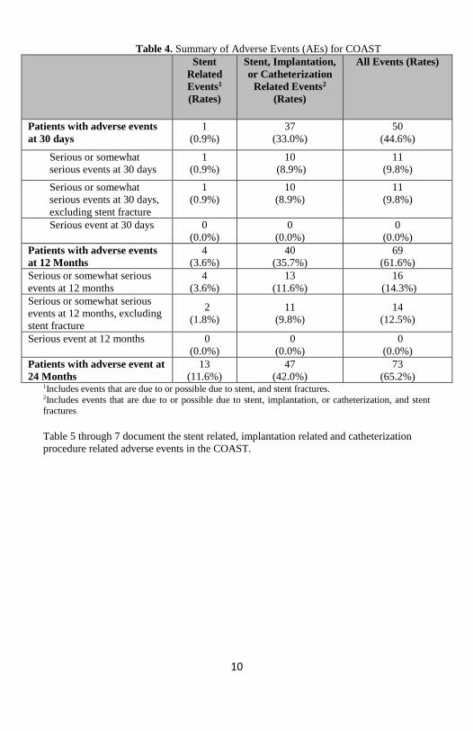
stent fracture increased with time for COAST patients. Table 4 provides a summary of
the adverse events reported under COAST.
10
Table 4. Summary of Adverse Events (AEs) for COAST
Stent
Related
Events1
(Rates)
Stent, Implantation,
or Catheterization
Related Events2
(Rates)
All Events (Rates)
Patients with adverse events
at 30 days
1
(0.9%)
37
(33.0%)
50
(44.6%)
Serious or somewhat
serious events at 30 days
1
(0.9%)
10
(8.9%)
11
(9.8%)
Serious or somewhat
serious events at 30 days,
excluding stent fracture
1
(0.9%)
10
(8.9%)
11
(9.8%)
Serious event at 30 days 0
(0.0%)
0
(0.0%)
0
(0.0%)
Patients with adverse events
at 12 Months
4
(3.6%)
40
(35.7%)
69
(61.6%)
Serious or somewhat serious
events at 12 months
4
(3.6%)
13
(11.6%)
16
(14.3%)
Serious or somewhat serious
events at 12 months, excluding
stent fracture
2
(1.8%)
11
(9.8%)
14
(12.5%)
Serious event at 12 months 0
(0.0%)
0
(0.0%)
0
(0.0%)
Patients with adverse event at
24 Months
13
(11.6%)
47
(42.0%)
73
(65.2%) 1Includes events that are due to or possible due to stent, and stent fractures.
2Includes events that are due to or possible due to stent, implantation, or catheterization, and stent fractures
Table 5 through 7 document the stent related, implantation related and catheterization
procedure related adverse events in the COAST.
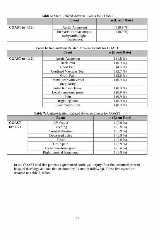
11
Table 5: Stent Related Adverse Events for COAST
Event n (Event Rate)
COAST (n=112) Aortic Aneurysm 1 (0.9 %)
Increased cardiac output,
tachycardia/light-
headedness
1 (0.9 %)
Table 6: Implantation Related Adverse Events for COAST
Event n (Event Rate)
COAST (n=112) Aortic Aneurysm 2 (1.8 %)
Back Pain 1 (0.9 %)
Chest Pain 5 (4.5 %)
Confined Vascular Tear 3 (2.7 %)
Groin Pain 4 (3.6 %)
Intimal tear with vessel
irregularity
1 (0.9 %)
Jailed left subclavian 1 (0.9 %)
Local hematoma groin 1 (0.9 %)
Pain 1 (0.9 %)
Right leg pain 1 (0.9 %)
Stent malposition 1 (0.9 %)
Table 7: Catheterization Related Adverse Events for COAST
Event n (Event Rate)
COAST
(n=112)
AV fistula 1 (0.9 %)
Bleeding 1 (0.9 %)
Corneal abrasion 1 (0.9 %)
Decreased pulse 1 (0.9 %)
Fever 1 (0.9 %)
Groin pain 1 (0.9 %)
Local hematoma groin 4 (3.6 %)
Right inguinal hematoma 1 (0.9 %)
In the COAST trial five patients experienced aortic wall injury, four that occurred prior to
hospital discharge and one that occurred by 24 month follow-up. These five events are
detailed in Table 8, below.

12
Table 8: COAST Aortic Wall Injuries by 24 Month Follow-up
Patient Bare Metal CP Stent
Implanted
Type of Injury Outcome of Event
1 No Small aneurysm after
dilation
Cross-over to Covered CP
Stent
2 Yes Therapeutic and localized
tear
Resolved after implantation
of Bare Metal CP Stent; tear
no longer visible and not
noted on further imaging
3 Yes Contained rupture Possible minimal aneurysm
with no progression during
admission; not noted on
further imaging
4 Yes Aneurysm Implantation of Covered CP
Stent
5 No Acute, rapidly expanding
aneurysm after dilation
Cross-over to Covered CP
Stent
In COAST, four patients with events underwent catheter reinterventions, representing
3.7% of patients with an event. No surgical interventions were performed. Table 9
provides a summary of these four interventions.
Table 9: COAST Coarctation-Related Reintervention by 24 Months
Approximate Time to
Intervention Post-
procedure (Months)
Indication for Reintervention Procedure Performed
11 Planned reintervention to fully
expand stent
Redilation of stent,
implantation of non-
study stent
20 Planned re-expansion of stent Redilation of stent
14 Aneurysm detected at 12m visit Therapy for new aortic
wall injury - implantation
of Covered CP Stent
14 Planned re-expansion of stent,
signs of restenosis at coarctation
site
Redilation of stent
In COAST, two patients experienced non-coarctation related reinterventions that were
documented by the 24 month follow-up. These patients underwent surgical interventions.
The time to intervention for one patient was 20 months, at which time the patient had an
aortic valve replacement to address progressive and severe left ventricular enlargement
and progression of exercise intolerance. The time to intervention for the second patient
was approximately two months, when the patient received a mitral valve replacement to
address mitral regurgitation.
Stent fracture was increasingly common during follow-up ranging from 12% at 24
months to 36% at 60 months in patients treated with bare metal stents (COAST trial).
13
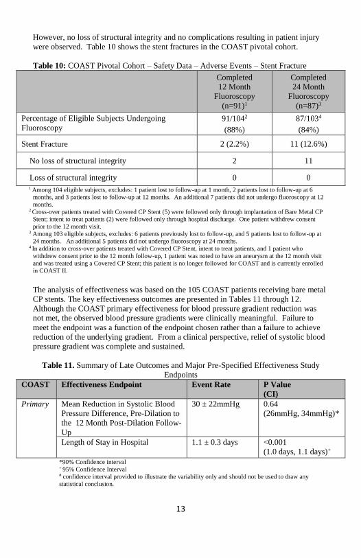
However, no loss of structural integrity and no complications resulting in patient injury
were observed. Table 10 shows the stent fractures in the COAST pivotal cohort.
Table 10: COAST Pivotal Cohort – Safety Data – Adverse Events – Stent Fracture
Completed
12 Month
Fluoroscopy
(n=91)1
Completed
24 Month
Fluoroscopy
(n=87)3
Percentage of Eligible Subjects Undergoing
Fluoroscopy
91/1042
(88%)
87/1034
(84%)
Stent Fracture 2 (2.2%) 11 (12.6%)
No loss of structural integrity 2 11
Loss of structural integrity 0 0
1 Among 104 eligible subjects, excludes: 1 patient lost to follow-up at 1 month, 2 patients lost to follow-up at 6
months, and 3 patients lost to follow-up at 12 months. An additional 7 patients did not undergo fluoroscopy at 12
months. 2 Cross-over patients treated with Covered CP Stent (5) were followed only through implantation of Bare Metal CP
Stent; intent to treat patients (2) were followed only through hospital discharge. One patient withdrew consent
prior to the 12 month visit. 3 Among 103 eligible subjects, excludes: 6 patients previously lost to follow-up, and 5 patients lost to follow-up at
24 months. An additional 5 patients did not undergo fluoroscopy at 24 months. 4 In addition to cross-over patients treated with Covered CP Stent, intent to treat patients, and 1 patient who
withdrew consent prior to the 12 month follow-up, 1 patient was noted to have an aneurysm at the 12 month visit
and was treated using a Covered CP Stent; this patient is no longer followed for COAST and is currently enrolled
in COAST II.
The analysis of effectiveness was based on the 105 COAST patients receiving bare metal
CP stents. The key effectiveness outcomes are presented in Tables 11 through 12.
Although the COAST primary effectiveness for blood pressure gradient reduction was
not met, the observed blood pressure gradients were clinically meaningful. Failure to
meet the endpoint was a function of the endpoint chosen rather than a failure to achieve
reduction of the underlying gradient. From a clinical perspective, relief of systolic blood
pressure gradient was complete and sustained.
Table 11. Summary of Late Outcomes and Major Pre-Specified Effectiveness Study
Endpoints
COAST Effectiveness Endpoint Event Rate P Value
(CI)
Primary Mean Reduction in Systolic Blood
Pressure Difference, Pre-Dilation to
the 12 Month Post-Dilation Follow-
Up
30 ± 22mmHg 0.64
(26mmHg, 34mmHg)*
Length of Stay in Hospital 1.1 ± 0.3 days <0.001
(1.0 days, 1.1 days)+
*90% Confidence interval + 95% Confidence Interval # confidence interval provided to illustrate the variability only and should not be used to draw any
statistical conclusion.
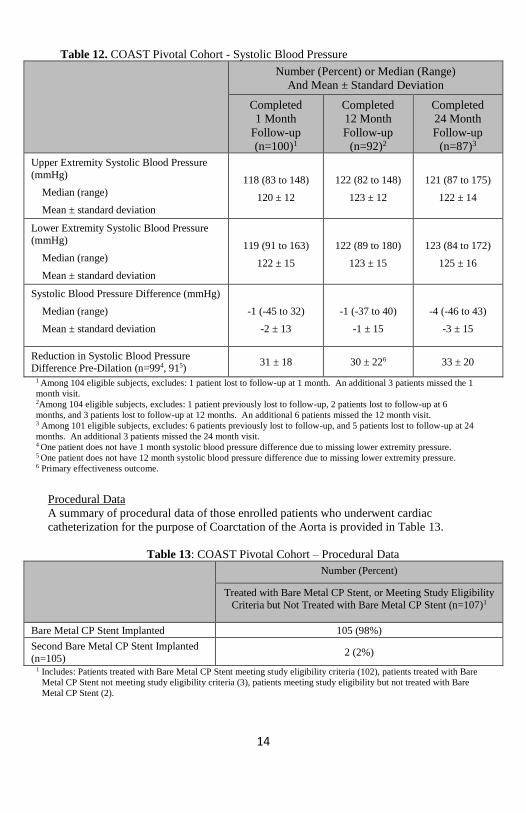
14
Table 12. COAST Pivotal Cohort - Systolic Blood Pressure
Number (Percent) or Median (Range)
And Mean ± Standard Deviation
Completed
1 Month
Follow-up
(n=100)1
Completed
12 Month
Follow-up
(n=92)2
Completed
24 Month
Follow-up
(n=87)3
Upper Extremity Systolic Blood Pressure
(mmHg)
Median (range)
Mean ± standard deviation
118 (83 to 148)
120 ± 12
122 (82 to 148)
123 ± 12
121 (87 to 175)
122 ± 14
Lower Extremity Systolic Blood Pressure (mmHg)
Median (range)
Mean ± standard deviation
119 (91 to 163)
122 ± 15
122 (89 to 180)
123 ± 15
123 (84 to 172)
125 ± 16
Systolic Blood Pressure Difference (mmHg)
Median (range)
Mean ± standard deviation
-1 (-45 to 32)
-2 ± 13
-1 (-37 to 40)
-1 ± 15
-4 (-46 to 43)
-3 ± 15
Reduction in Systolic Blood Pressure Difference Pre-Dilation (n=994, 915)
31 ± 18 30 ± 226 33 ± 20
1 Among 104 eligible subjects, excludes: 1 patient lost to follow-up at 1 month. An additional 3 patients missed the 1
month visit. 2Among 104 eligible subjects, excludes: 1 patient previously lost to follow-up, 2 patients lost to follow-up at 6
months, and 3 patients lost to follow-up at 12 months. An additional 6 patients missed the 12 month visit. 3 Among 101 eligible subjects, excludes: 6 patients previously lost to follow-up, and 5 patients lost to follow-up at 24
months. An additional 3 patients missed the 24 month visit. 4 One patient does not have 1 month systolic blood pressure difference due to missing lower extremity pressure. 5 One patient does not have 12 month systolic blood pressure difference due to missing lower extremity pressure. 6 Primary effectiveness outcome.
Procedural Data
A summary of procedural data of those enrolled patients who underwent cardiac
catheterization for the purpose of Coarctation of the Aorta is provided in Table 13.
Table 13: COAST Pivotal Cohort – Procedural Data
Number (Percent)
Treated with Bare Metal CP Stent, or Meeting Study Eligibility
Criteria but Not Treated with Bare Metal CP Stent (n=107)1
Bare Metal CP Stent Implanted 105 (98%)
Second Bare Metal CP Stent Implanted
(n=105) 2 (2%)
1 Includes: Patients treated with Bare Metal CP Stent meeting study eligibility criteria (102), patients treated with Bare
Metal CP Stent not meeting study eligibility criteria (3), patients meeting study eligibility but not treated with Bare
Metal CP Stent (2).

15
COAST/COAST II Follow-up Data
A.1. Percent of Patients with New Stent Fracture
% Stent Fracture (new at each interval)
Interval COAST I COAST II
12 months 4/121 (3.3%)
1/127 (0.8%)
24 months 12/121
(9.9%)
5/127
(3.9%)
36 months 2/121 (1.6%)
1/127 (0.8%)
48 months 10/121
(8.2%)
4/127
(3.1%)
60 months 9/121 (7.4%)
2/127 (1.6%)
A.2. Percent of Patients with Stent Fracture (Cumulative)
% Stent Fracture (cumulative)
Interval COAST I COAST II
12 months 4/121 (3%)
1/127 (0.8%)
24 months 16/121 (13%)
6/127 (4.7%)
36 months 18/121
(15%)
7/127
(5.5%)
48 months 28/121 (23%)
11/127 (8.6%)
60 months 37/121
(31%)
13/127
(10%)
B.1. Percent of Patients with Aortic Wall Injury (New or Progressive)*
% patients with AWI
Interval COAST I COAST II
12 months 1/121
(0.8%)
1/127
(0.8%)
24 months 0/121 0/127
36 months 3/121
(2.5%) 0/127
48 months 1/121 (0.8%)
0/127
60 months 1/121
(0.8%)
2/127
(1.6%)
*All recorded injuries were “new”, none designated as progressive

16
B.2. Cumulative Percent of Patients with Aortic Wall Injury (New or Progressive)*
% patients with AWI (cumulative)
Interval COAST I COAST II
12 months 1/121
(0.8%)
1/127
(0.8%)
24 months 1/121
(0.8%)
1/127
(0.8%)
36 months 4/121
(3.3%)
1/127
(0.8%)
48 months 5/121
(4.1%)
1/127
(0.8%)
60 months 6/121
(5%)
3/127
(2.4%)
*All recorded injuries were “new”, none designated as progressive
C.1. Percent of Patients with Coarctation Related Re-intervention or Surgery*
% patients with reintervention or surgery
Interval COAST I COAST II
12 months 4/121
(3.3%)
9/128
(7%)
24 months 3/121
(2.5%)
3/128
(2.3%)
36 months 6/121
(5%)
3/128
(2.3%)
48 months 6/121
(5%)
3/128
(2.3%)
60 months 2/121
(1.7%)
3/128
(2.3%)
*All patients listed had catheter reinterventions, none had surgery
C.2. Percent of Patients with Coarctation Related Re-intervention or Surgery
(Cumulative)
% patients with catheter-based re-intervention
or surgery (cumulative)
Interval Bare Metal Covered Stent
12 months 3/121 (2.5%)
9/128 (7%)
24 months 6/121
(5%)
12/128
(9.3%)
36 months 12/121 (9.9%)
15/128 (11.8%)
48 months 18/121
(14.9%)
18/128
(14%)
60 months 20/121 (16.5%)
21/128 (16.4%)

17
C.3. Percent of Bare Metal Patients with Catheter-Based or Surgical Coarctation-Related
Re-intervention
% Bare Metal patients with catheter-based
re-intervention or surgery
Interval Catheterization Surgery
12 months 3/121 (2.5%)
0 (0%)
24 months 3/121
(2.5%)
0
(0%)
36 months 6/121 (5%)
0 (0%)
48 months 6/121
(5%)
0
(0%)
60 months 2/121 (1.7%)
0 (0%)
C.4. Percent of Bare Metal Patients with Catheter-Based or Surgical Coarctation-Related
Re-intervention (Cumulative)
% Bare Metal patients with catheter-based re-
intervention or surgery (cumulative)
Interval Catheterization Surgery
12 months 3/121
(2.5%)
0
(0%)
24 months 6/121 (5%)
0 (0%)
36 months 12/121
(9.9%)
0
(0%)
48 months 18/121 (14.9%)
0 (0%)
60 months 20/121
(16.5%)
0
(0%)
C.5. Percent of Covered Stent Patients with Catheter-Based or Surgical Coarctation-
Related Re-intervention
% Covered Stent patients with
re-intervention or surgery
Interval Catheterization Surgery
12 months 9/128
(7%)
0
(0%)
24 months 3/128
(2.3%)
0
(0%)
36 months 3/128
(2.3%)
0
(0%)
48 months 3/128
(2.3%)
0
(0%)
60 months 3/128
(2.3%)
0
(0%)
18
C.6. Percent of Covered Stent Patients with Catheter-Based or Surgical Coarctation-
Related Re-intervention (Cumulative)
% Covered Stent patients with re-intervention
or surgery (cumulative)
Interval Catheterization Surgery
12 months 9/128 (7%)
0 (0%)
24 months 12/128
(9.3%)
0
(0%)
36 months 15/128 (11.8%)
0 (0%)
48 months 18/128
(14%)
0
(0%)
60 months 21/128 (16.4%)
0 (0%)
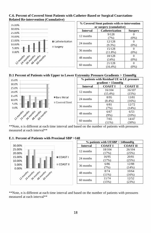
D.1 Percent of Patients with Upper to Lower Extremity Pressure Gradients > 15mmHg
% patients with Residual UE to LE pressure
gradient > 15mmHg
Interval COAST I COAST II
12 months 16/104
(15.4%)
16/107
(15%)
24 months 8/95
(8.4%)
13/81
(16%)
36 months 6/81
(7%)
12/72
(14%)
48 months 6/67 (9%)
6/53 (10%)
60 months 7/65
(11%)
14/47
(30%)
**Note, n is different at each time interval and based on the number of patients with pressures
measured at each interval**
E.1. Percent of Patients with Proximal SBP >140
0.00%5.00%
10.00%15.00%20.00%25.00%30.00%
COAST I
COAST II
% patients with UESBP > 140mmHg
Interval COAST I COAST II
12 months 18/104 (17%)
26/104 (25%)
24 months 16/95
(17%)
20/81
(25%)
36 months 6/86 (7%)
12/88 (14%)
48 months 8/74
(11%)
10/64
(16%)
60 months 11/74 (15%)
12/52 (23%)
**Note, n is different at each time interval and based on the number of patients with pressures
measured at each interval**
19
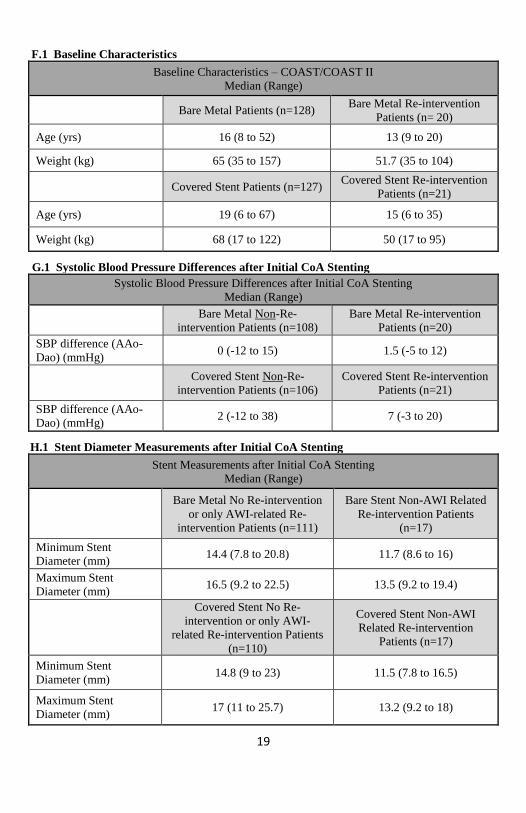
G.1 F.1 Baseline Characteristics
Baseline Characteristics – COAST/COAST II
Median (Range)
Bare Metal Patients (n=128) Bare Metal Re-intervention
Patients (n= 20)
Age (yrs) 16 (8 to 52) 13 (9 to 20)
Weight (kg) 65 (35 to 157) 51.7 (35 to 104)
Covered Stent Patients (n=127) Covered Stent Re-intervention
Patients (n=21)
Age (yrs) 19 (6 to 67) 15 (6 to 35)
Weight (kg) 68 (17 to 122) 50 (17 to 95)
H.1 G.1 Systolic Blood Pressure Differences after Initial CoA Stenting
Systolic Blood Pressure Differences after Initial CoA Stenting
Median (Range)
Bare Metal Non-Re-
intervention Patients (n=108)
Bare Metal Re-intervention
Patients (n=20)
SBP difference (AAo-
Dao) (mmHg) 0 (-12 to 15) 1.5 (-5 to 12)
Covered Stent Non-Re-
intervention Patients (n=106)
Covered Stent Re-intervention
Patients (n=21)
SBP difference (AAo-
Dao) (mmHg) 2 (-12 to 38) 7 (-3 to 20)
I.1 H.1 Stent Diameter Measurements after Initial CoA Stenting
Stent Measurements after Initial CoA Stenting
Median (Range)
Bare Metal No Re-intervention
or only AWI-related Re-
intervention Patients (n=111)
Bare Stent Non-AWI Related
Re-intervention Patients
(n=17)
Minimum Stent
Diameter (mm) 14.4 (7.8 to 20.8) 11.7 (8.6 to 16)
Maximum Stent
Diameter (mm) 16.5 (9.2 to 22.5) 13.5 (9.2 to 19.4)
Covered Stent No Re-
intervention or only AWI-
related Re-intervention Patients
(n=110)
Covered Stent Non-AWI
Related Re-intervention
Patients (n=17)
Minimum Stent
Diameter (mm) 14.8 (9 to 23) 11.5 (7.8 to 16.5)
Maximum Stent
Diameter (mm) 17 (11 to 25.7) 13.2 (9.2 to 18)

20
J.1 I.1 Upper Extremity Systolic Blood Pressures
Number (Percent) or Median (Range)
and Mean ± Standard Deviation
Upper Extremity Systolic Blood Pressures at 4
years
Bare Metal Patients
Upper and Lower
Extremity SBP
measured
Only Upper Extremity
SBP measured
Upper Extremity Systolic Blood
Pressure (mmHg) (n=67,7)
Median (range)
Mean ± standard deviation
123 (79 to 231)
125 + 19
120 (70 to 134)
113 + 21
Covered Stent Patients
Upper Extremity Systolic Blood
Pressure (mmHg) (n=59,5)
Median (range)
Mean ± standard deviation
126 (93 to 174)
126 + 15
124 (110 to 140)
123 + 11
Upper Extremity Systolic Blood Pressures at 5
years
Bare Metal Patients
Upper and Lower
Extremity SBP
measured
Only Upper Extremity
SBP measured
Upper Extremity Systolic Blood
Pressure (mmHg) (n=65,9)
Median (range)
Mean ± standard deviation
128 (106 to 165)
129 + 12
117 (100 to 160)
121 + 19
Covered Stent Patients
Upper Extremity Systolic Blood
Pressure (mmHg) (n=47,5)
Median (range)
Mean ± standard deviation
131 (101 to 159)
129 + 14
122 (121 to 134)
126 + 6
21
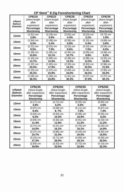
CP Stent™ 8 Zig Foreshortening Chart
Inflated Balloon
Diameter
CP8Z16 (Stent length
after expansion) Percentage Shortening
CP8Z22 (Stent length
after expansion) Percentage Shortening
CP8Z28 (Stent length
after expansion) Percentage Shortening
CP8Z34 (Stent length
after expansion) Percentage Shortening
CP8Z39 (Stent length
after expansion) Percentage Shortening
12mm (1.61) cm
2.8% (2.18) cm
0.8% (2.62) cm
4.4% (3.23) cm
3.1% (3.72) cm
1.9%
14mm (1.54) cm
6.5% (2.08) cm
5.4% (2.56) cm
6.8% (3.15) cm
5.4% (3.66) cm
3.6%
15mm (1.51) cm
8.5% (2.02) cm
7.9% (2.51) cm
8.6% (3.10) cm
7.0% (3.54) cm
6.6%
16mm (1.48) cm
10.6% (1.98) cm
10.1% (2.45) cm
10.7% (3.00) cm
9.8% (3.48) cm
8.2%
18mm (1.43) cm
13.7% (1.89) cm
14.0% (2.38) cm
13.3% (2.88) cm
13.5% (3.20) cm
15.6%
20mm (1.32) cm
20.0% (1.80) cm
17.9% (2.30) cm
16.3% (2.63) cm
20.9% (2.96) cm
21.9%
22mm (1.23) cm
25.4% (1.67) cm
23.9% (2.09) cm
24.0% (2.46) cm
26.0% (2.85) cm
25.0%
24mm (1.05) cm
36.4% (1.46) cm
33.8% (1.91) cm
30.3% (2.07) cm
37.9% (2.27) cm
40.1%
Inflated Balloon
Diameter
CP8Z45 (Stent length
after expansion) Percentage Shortening
CP8Z50 (Stent length
after expansion) Percentage Shortening
CP8Z55 (Stent length
after expansion) Percentage Shortening
CP8Z60 (Stent length
after expansion) Percentage Shortening
12mm (4.17) cm
3.8% (4.71) cm
6.2% (5.25) cm
5.0% (5.84) cm
4.5%
14mm (3.97) cm
8.4% (4.58) cm
8. 7% (5.11) cm
7.6% (5.67) cm
7.3%
15mm (3.94) cm
9.2% (4.50) cm
10.3% (4.98) cm
10.0% (5.55) cm
9.2%
16mm (3.84) cm
11.4% (4.42) cm
11.9% (4.91) cm
11.2% (5.43) cm
11.2%
18mm (3.71) cm
14.5% (4.21) cm
16.1% (4.70) cm
15.1% (5.20) cm
14.9%
20mm (3.27) cm
24.7% (3.96) cm
21.0% (4.43) cm
20.0% (4.92) cm
19.5%
22mm (3.15) cm
27.3% (3.71) cm
26.0% (4.09) cm
26.1% (4.55) cm
25.5%
24mm (2.83) cm
34.9% (3.33) cm
33.5% (3.72) cm
32.8% (4.14) cm
32.3%

22
CP Stent™ 10 Zig Foreshortening Chart
Inflated Balloon
Diameter
CP10Z39 (Stent length
after expansion) Percentage Shortening
CP10Z45 (Stent length
after expansion) Percentage Shortening
CP10Z50 (Stent length
after expansion) Percentage Shortening
CP10Z55 (Stent length
after expansion) Percentage Shortening
CP10Z60 (Stent length
after expansion) Percentage Shortening
26mm (3.17) cm 18.33%
(3.44) cm 22.09%
(4.10) cm 17.34%
(4.24) cm 23.32%
(4.85) cm 20.20%
28mm (2.96) cm 23.68%
(3.24) cm 26.75%
(3.71) cm 25.11%
(4.00) cm 27.58%
(4.39) cm 27.87%
30mm (2.58) cm 33.45%
(3.09) cm 30.16%
(3.26) cm 34.34%
(3.64) cm 34.17%
(4.11) cm 32.55%
CP Stent™ 8 Zig Balloon Sizing Chart Stent ID (mm)
Inner Balloon
Pressure (atm)
12mm Diameter RBP = 7.0
14mm Diameter RBP = 6.0
15mm Diameter RBP = 5.0
16mm Diameter RBP = 5.0
18mm Diameter RBP = 4.0
20mm Diameter RBP = 4.0
22mm Diameter RBP = 3.0
24mm Diameter RBP = 3.0
1.0 2.75 3.22 3.49 3.75 3.94 4.02 4.20 4.28
2.0 2.85 3.32 3.59 3.85 4.36 4.13 4.33 4.50
3.0 5.85 6.91 6.89 7.79 8.54 9.20 10.16 10.57
4.0 6.12 7.00 7.02 7.95 8.71 9.63 10.40 11.08
4.5 10.84 11.94
5.0 6.20 7.08 7.10 8.04 8.91 10.00 Outer
Balloon Pressure
(atm)
1.0 10.73 13.08 13.45 14.87 16.85 17.91 20.52 22.79
2.0 10.86 13.27 14.16 15.10 17.06 18.38 21.46 23.95
3.0 11.15 13.50 14.55 15.68 17.64 19.42 21.98 24.68
4.0 11.33 13.68 14.88 15.93 18.06 20.07
5.0 11.62 13.87 15.06 16.19
6.0 11.80 13.98
7.0 12.04
*This data is based on testing performed using the NuMED BIB® Stent Placement Catheter.
The figures in bold face represent the stent ID @ Rated Burst Pressure.
23
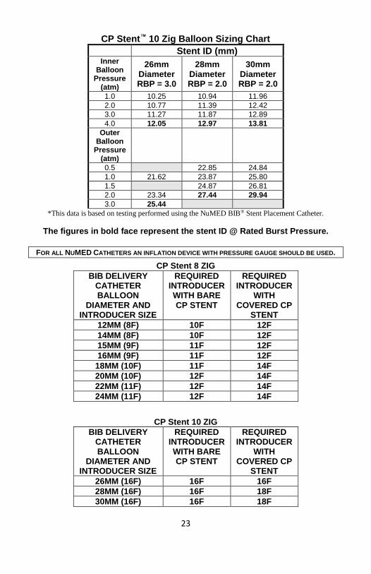
CP Stent™ 10 Zig Balloon Sizing Chart Stent ID (mm)
Inner Balloon
Pressure (atm)
26mm Diameter RBP = 3.0
28mm Diameter RBP = 2.0
30mm Diameter RBP = 2.0
1.0 10.25 10.94 11.96
2.0 10.77 11.39 12.42
3.0 11.27 11.87 12.89
4.0 12.05 12.97 13.81
Outer Balloon
Pressure (atm)
0.5 22.85 24.84
1.0 21.62 23.87 25.80
1.5 24.87 26.81
2.0 23.34 27.44 29.94
3.0 25.44
*This data is based on testing performed using the NuMED BIB® Stent Placement Catheter.
The figures in bold face represent the stent ID @ Rated Burst Pressure.
FOR ALL NUMED CATHETERS AN INFLATION DEVICE WITH PRESSURE GAUGE SHOULD BE USED.
CP Stent 8 ZIG
BIB DELIVERY CATHETER BALLOON
DIAMETER AND INTRODUCER SIZE
REQUIRED INTRODUCER WITH BARE CP STENT
REQUIRED INTRODUCER
WITH COVERED CP
STENT
12MM (8F) 10F 12F
14MM (8F) 10F 12F
15MM (9F) 11F 12F
16MM (9F) 11F 12F
18MM (10F) 11F 14F
20MM (10F) 12F 14F
22MM (11F) 12F 14F
24MM (11F) 12F 14F
CP Stent 10 ZIG
BIB DELIVERY CATHETER BALLOON
DIAMETER AND INTRODUCER SIZE
REQUIRED INTRODUCER WITH BARE CP STENT
REQUIRED INTRODUCER
WITH COVERED CP
STENT
26MM (16F) 16F 16F
28MM (16F) 16F 18F
30MM (16F) 16F 18F
24
RETURN OF EXPLANTED DEVICE:
NuMED, Inc. is interested in obtaining recovered CP Stents. Place the explanted device
in a container or vial immediately after excision. For further instructions on the return of
an explanted device, contact the RA Manager, NuMED, Inc. 2880 Main Street,
Hopkinton, New York, 12965. Phone number: 315-328-4491.
WARNING:
NuMED stents are placed in the extremely hostile environment of the human body.
Stents may fail to function for a variety of causes including, but not limited to, medical
complications or failure of stent by fracture and embolization. In addition, despite the
exercise of all due care in design, component selection, manufacture, and testing prior to
sale, stents may be easily damaged before, during, or after insertion by improper
handling, crimping or other intervening acts. Metal stents placed where there are
extrinsic forces of compression, i.e. right ventricular outflow tract, are especially prone to
fatigue fracture and embolization and should be avoided.
WARRANTY AND LIMITATIONS
Stents and accessories are sold in an 'as is' condition. The entire risk as to the quality and
performance of the stent is with the buyer. NuMED disclaims all warranties, expressed
or implied, with respect to catheters and accessories, including but not limited to, any
implied warranty of merchantability or fitness for a particular purpose. NuMED shall not
be liable to any person for any medical expenses or any direct or consequential damages
resulting from the use of any catheter or accessory or caused by any defect, failure, or
malfunction of any catheter or accessory, whether a claim for such damages is based
upon warranty, contract, tort, or otherwise. No person has any authority to bind NuMED
to any representation or warranty with respect to catheters and accessories.
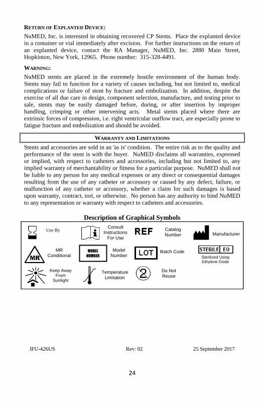
Description of Graphical Symbols
IFU-426US Rev: 02 25 September 2017
Use By
Consult Instructions
For Use
Catalog Number
Manufacturer
MR Conditional
Model Number
Batch Code Sterilized Using Ethylene Oxide
Keep Away From
Sunlight
Temperature Limitation
Do Not Reuse
MODEL
NUMBER