Moss mimesis par excellence: integrating previous and new ...
49
© 2020 The Linnean Society of London, Zoological Journal of the Linnean Society, 2020, XX, 1–49 1 This is an Open Access article distributed under the terms of the Creative Commons Attribution License (http://creativecommons.org/licenses/by/4.0/), which permits unrestricted reuse, distribution, and reproduction in any medium, provided the original work is properly cited. Zoological Journal of the Linnean Society, 2020, XX, 1–49. With 18 figures. Moss mimesis par excellence: integrating previous and new data on the life history and larval ecomorphology of long-bodied craneflies (Diptera: Cylindrotomidae: Cylindrotominae) YUME IMADA Graduate School of Science and Engineering, Ehime University, 2–5 Bunkyo-cho, Matsuyama, Ehime, 790-8577 Japan Received 17 June 2020; revised 12 November 2020; accepted for publication 22 November 2020 Different physical structures play a central role in animal camouflage. However, in evolutionary studies of mimicry, the ecological and evolutionary significance of such structures has been poorly investigated. Larvae of long-bodied craneflies, Cylindrotominae, are all obligate herbivores and resemble plants. They are distinctively characterized by possessing numerous elongated cuticular lobes on the integument. A comprehensive overview of the biology and morphology of cylindrotomids, particularly their larval stages, is laid out, providing original data on nine species. To explore the ecological background of moss resemblance, host-plants of most examined species are clarified, revealing that terrestrial moss-feeding species tend to use specific groups of mosses, either belonging to Bryales or Hypnales. However, the evolution of cryptic forms remains paradoxical, due to the apparent absence of visual predators. Based on histological examinations, extensive internal musculatures within the cuticular lobes on the lateral side are discovered, shedding new light on their function in locomotion. Traditional functional explanations for these lobes, particularly as devices for respiration, locomotion and attachment, are challenged. This study promotes our understanding of the ecomorphology of mimicry devices, which is an angle often dismissed in evolutionary studies of mimicry. ADDITIONAL KEYWORDS: bryophytivore – colour polymorphism – countershading – cryptic mimicry – fleshy projection –functional morphology – Mniaceae – Nematocera –Tipulomorpha – tracheal gill. INTRODUCTION Camouflage is a fascinating example of adaptive evolution (Darwin, 1859; Wallace, 1867). A wide range of animals camouflage in their surroundings to avoid detection or deceive keen-sighted natural enemies in their vicinity (Stevens & Merilaita, 2009; Ruxton et al., 2018). Many insects, typically of the orders Diptera, Hymenoptera, Lepidoptera, Mantodea, Orthoptera and Phasmatodea, disguise themselves as plants. The cryptic forms of insects that resemble plants have presumably evolved as a survival strategy for a long time, with evidence in the fossil record extending from the Middle Permian to the Recent (Scott et al., 1992; Wedmann et al., 2007; Wang et al., 2010, 2012; Wedmann, 2010; Garrouste et al. , 2016; Liu et al. , 2018). Coloration and patterning play a key role in various modes of mimicry and have historically been studied from ecological and evolutionary perspectives ( Poulton, 1890; Cott, 1940; Protas & Patel, 2008; Cuthill et al., 2017). Moreover, visual camouflage is also associated with physical structures (Robbins, 1980; Sabaj et al., 1999), behaviours (Kang et al., 2015; Suzuki & Sakurai, 2015) and the active uptake of materials from the environment by the camouflaged organisms ( Ruxton & Stevens, 2015). One famous example of such morphological structures is that of some lycaenid and riodinid butterflies, which bear narrow tails mimicking antennae on their hind wings – a so-called ‘false head’, which acts by deflecting the attention of birds (Robbins, 1980, 1981). Less obviously, larvae of the erebid moth genus Homodes Guenée, 1852 possess an anterior end with abundant long, bent, tentacle-like, spatulate setae, which apparently Corresponding author. E-mail: [email protected] Downloaded from https://academic.oup.com/zoolinnean/advance-article/doi/10.1093/zoolinnean/zlaa177/6048384 by Ehime University Library user on 25 February 2021
Transcript of Moss mimesis par excellence: integrating previous and new ...
© 2020 The Linnean Society of London, Zoological Journal of the Linnean Society, 2020, XX, 1–49 1This is an Open Access article distributed under the terms of the Creative Commons Attribution License (http://creativecommons.org/licenses/by/4.0/), which permits unrestricted reuse, distribution, and reproduction in any medium, provided the original work is properly cited.
Zoological Journal of the Linnean Society, 2020, XX, 1–49. With 18 figures.
Moss mimesis par excellence: integrating previous and new data on the life history and larval ecomorphology of long-bodied craneflies (Diptera: Cylindrotomidae: Cylindrotominae)
YUME IMADA
Graduate School of Science and Engineering, Ehime University, 2–5 Bunkyo-cho, Matsuyama, Ehime, 790-8577 Japan
Received 17 June 2020; revised 12 November 2020; accepted for publication 22 November 2020
Different physical structures play a central role in animal camouflage. However, in evolutionary studies of mimicry, the ecological and evolutionary significance of such structures has been poorly investigated. Larvae of long-bodied craneflies, Cylindrotominae, are all obligate herbivores and resemble plants. They are distinctively characterized by possessing numerous elongated cuticular lobes on the integument. A comprehensive overview of the biology and morphology of cylindrotomids, particularly their larval stages, is laid out, providing original data on nine species. To explore the ecological background of moss resemblance, host-plants of most examined species are clarified, revealing that terrestrial moss-feeding species tend to use specific groups of mosses, either belonging to Bryales or Hypnales. However, the evolution of cryptic forms remains paradoxical, due to the apparent absence of visual predators. Based on histological examinations, extensive internal musculatures within the cuticular lobes on the lateral side are discovered, shedding new light on their function in locomotion. Traditional functional explanations for these lobes, particularly as devices for respiration, locomotion and attachment, are challenged. This study promotes our understanding of the ecomorphology of mimicry devices, which is an angle often dismissed in evolutionary studies of mimicry.
ADDITIONAL KEYWORDS: bryophytivore – colour polymorphism – countershading – cryptic mimicry – fleshy projection –functional morphology – Mniaceae – Nematocera –Tipulomorpha – tracheal gill.
INTRODUCTION
Camouflage is a fascinating example of adaptive evolution (Darwin, 1859; Wallace, 1867). A wide range of animals camouflage in their surroundings to avoid detection or deceive keen-sighted natural enemies in their vicinity (Stevens & Merilaita, 2009; Ruxton et al., 2018). Many insects, typically of the orders Diptera, Hymenoptera, Lepidoptera, Mantodea, Orthoptera and Phasmatodea, disguise themselves as plants. The cryptic forms of insects that resemble plants have presumably evolved as a survival strategy for a long time, with evidence in the fossil record extending from the Middle Permian to the Recent (Scott et al., 1992; Wedmann et al., 2007; Wang et al., 2010, 2012; Wedmann, 2010; Garrouste et al., 2016; Liu et al.,
2018). Coloration and patterning play a key role in various modes of mimicry and have historically been studied from ecological and evolutionary perspectives (Poulton, 1890; Cott, 1940; Protas & Patel, 2008; Cuthill et al., 2017). Moreover, visual camouflage is also associated with physical structures (Robbins, 1980; Sabaj et al., 1999), behaviours (Kang et al., 2015; Suzuki & Sakurai, 2015) and the active uptake of materials from the environment by the camouflaged organisms (Ruxton & Stevens, 2015). One famous example of such morphological structures is that of some lycaenid and riodinid butterflies, which bear narrow tails mimicking antennae on their hind wings – a so-called ‘false head’, which acts by deflecting the attention of birds (Robbins, 1980, 1981). Less obviously, larvae of the erebid moth genus Homodes Guenée, 1852 possess an anterior end with abundant long, bent, tentacle-like, spatulate setae, which apparently Corresponding author. E-mail: [email protected]
applyparastyle “fig//caption/p[1]” parastyle “FigCapt”
Dow
nloaded from https://academ
ic.oup.com/zoolinnean/advance-article/doi/10.1093/zoolinnean/zlaa177/6048384 by Ehim
e University Library user on 25 February 2021

2 Y. IMADA
© 2020 The Linnean Society of London, Zoological Journal of the Linnean Society, 2020, XX, 1–49
work by mimicking aggressive ants (Kalshoven, 1961; Leong & D’Rozario, 2012). A myriad of alluring examples of camouflage, in which physical structures are combined with coloration, are familiar to most people (Wickler, 1968; Unno, 2015). Nevertheless, the function and evolutionary significance of such structures has, generally, not been tackled in the study of the evolutionary ecology of mimicry.
Cylindrotominae is a subfamily belonging to the smallest cranefly family, Cylindrotomidae. Larval morphology of Cylindrotominae, known for more than two centuries (De Geer, 1773), deviates greatly from typical cranefly larvae – they resemble plants to a remarkable degree. Their extraordinary appearance is likely associated with their biological features as free-living, obligate herbivores. Whereas the majority of cranefly species live in organic mud along stream banks or in rotten wood, and feed on decaying plant materials (Alexander, 1915; De Jong et al., 2008), all known larvae of the cylindrotomines live exposed on angiosperms or within the tufts of mosses and feed on living tissue of plants. Notably, the habitats vary among major clades, ranging from aquatic (Phalacrocera Schiner, 1863) to semi-aquatic (Triogma Schiner, 1863) to fully terrestrial (Cylindrotoma Macquart, 1834, Diogma Edwards, 1938, Liogma Osten Sacken, 1869, partly Triogma). Larval cylindrotomines apparently attain camouflage among plants by several means. Many species are tinged with green or brown hues and are dorsally decorated with dark markings. In addition, elongated cuticular appendages longitudinally aligned on the integument are particularly noteworthy in moss-feeding species; these species attach to, and remain motionless on, the plant substrate. In combination, these features of coloration, patterning, integumental structure and behaviour, make finding the larvae in the moss carpets that surround them notoriously difficult. The suit of morphological traits in larvae varies significantly among taxa and is seemingly linked with biological attributes (e.g. habitats and host-plants). Notably, the elongated cuticular appendages may also be physiological, mechanical (physical) or behavioural adaptations to habitats; specifically, they have been hypothesized to serve as tracheal gills in aquatic species (De Geer, 1773, 1776; Miall & Shelford, 1897; Müggenburg, 1901; Haake, 1922), an osmoregulatory organ (Brinkmann, 1991) or as a device for locomotion in both terrestrial and aquatic species (Zeller, 1842; Osten Sacken, 1869; Miall & Shelford, 1897; Peus, 1952), although these hypotheses do not conflict with one another. The highly cryptic form of the cylindrotomids is apparently associated with their habitats and biological interactions therein.
A question thus arises as to how a device for mimicry can evolve simultaneously alongside physiological,
mechanical and behavioural functions. Osten Sacken (1897) referred to the larvae of cylindrotomids, writing, ‘These larvae must, of course, show corresponding adaptations for such a mode of life, and it will be the task of future investigators to describe these structural differences in detail’.
To examine the link between ecological background and phenotypic variations of these complex traits, I herein provide an overview of the biology and key structures relevant to crypsis. Included are new data and analyses derived from my own investigations, as well as from relevant literature. Furthermore, devices and strategies of mimicry are challenged, with an emphasis on their functional aspects.
Background taxonomy and systematics
Cylindrotomidae is commonly regarded as an independent family in the superfamily Tipuloidea (Evenhuis, 1994; Bertone et al., 2008; Petersen et al., 2010; Zhang et al., 2016; Courtney et al., 2017), of which interfamilial relationships are debated (Yeates et al., 2007). Less commonly, Cylindrotomidae has been treated as a subfamily within the family Tipulidae (Brindle, 1967; Brodo, 1967; Alexander & Byers, 1981; Pritchard, 1983; Sinclair, 1992; Petersen et al., 2010). The cylindrotomids currently comprise 71 extant species in nine genera and are subdivided into two subfamilies: Cylindrotominae and Stibadocerinae (Alexander, 1927; Oosterbroek, 2020). The subfamily Cylindrotominae consists of five genera (Cylindrotoma, Diogma, Liogma, Triogma and Phalacrocera), which mainly occur in the Holarctic and Oriental realms (Ribeiro, 2009). The subfamily Stibadocerinae is composed of four genera (Stibadocera Enderlein, 1912, Stibadocerella Brunetti, 1918, Stibadocerina Alexander, 1929 and Stibadocerodes Alexander, 1928) and is distributed in the Oriental, Neotropical and Australasian regions and in part of the Eastern Palaearctic (Ribeiro, 2009). The monophyly of Cylindrotomidae is contentious due to the apparent lack of autapomorphies (Lukashevich & Ribeiro, 2018). There is robust support showing that Cylindrotomidae is sister to Tipulidae, according to some previous phylogenetic studies based on morphology (Hennig, 1954, 1973; Oosterbroek & Theowald, 1991; Starý, 1992; Oosterbroek & Courtney, 1995; Ribeiro, 2008; Neugart et al., 2009; Lukashevich & Ribeiro, 2018) and molecular data (Petersen et al., 2010; Zhang et al., 2016; Kang et al., 2017).
The early history of this family is scantily known due to its meager fossil record. The stem clade of Tipulomorpha dates back to the Late Triassic (Krzemiński, 1992; Krzemiński et al., 1994; Shcherbakov et al., 1995; Krzemiński & Krzemińska, 2003;
Dow
nloaded from https://academ
ic.oup.com/zoolinnean/advance-article/doi/10.1093/zoolinnean/zlaa177/6048384 by Ehim
e University Library user on 25 February 2021

MIMESIS OF CYLINDROTOMINAE CRANEFLIES 3
© 2020 The Linnean Society of London, Zoological Journal of the Linnean Society, 2020, XX, 1–49
Blagoderov et al., 2007; Bertone et al., 2008). However, the fossils identified or described as belonging to Cylindrotomidae have only been recorded from the Palaeogene, spanning from the Ypresian (c. 56–48 Mya) to the Rupelian (c. 33–28 Mya) (Cockerell, 1920; Séguy, 1934; Freiwald, 1991; Freiwald & Krzemiński, 1991; Evenhuis, 1994). Only 14 fossil species are named and assigned to this family (Krzemiński et al., 2019).
In Japan, seven species in four genera of cylindrotomids are known (Takahashi, 1960; Nakamura, 2001; The Editorial Committee of Catalogue of the Insects of Japan, 2014): Cylindrotoma japonica Alexander, 1919; Diogma caudata Takahashi, 1960; D. glabrata (Meigen, 1818); Liogma brevipecten Alexander, 1932; L. mikado (Alexander, 1919); L. serraticornis Alexander, 1919; and Triogma kuwanai (Alexander, 1913). Triogma kuwanai is subdivided into two subspecies, T. kuwanai subsp. kuwanai (Alexander, 1913) and subsp. limbinervis Alexander, 1953; occasionally they are treated as species (Oosterbroek, 2020). A recent phylogenetic analysis of major clades of Cylindrotominae using molecular (partial COI gene sequences) and morphological data has revealed discordance between the current taxonomy and phylogenetic relationships at both the species and genus level, suggesting that the taxonomic status of some Japanese and North American species should be changed (Kolcsár et al., unpubl. data). However, herein, the above-mentioned species and subspecies are treated following their current taxonomic status, as this does not cause any problems in demarcating these species.
MATERIAL AND METHODS
data collection
In this study, immature stages were investigated for nine species, which cover all the described genera of Cylindrotominae: C. japonica, D. glabrata, L. brevipecten, L. mikado, L. nodicornis (Osten Sacken, 1865), L. serraticornis, T. kuwanai, Phalacrocera replicata (Linnaeus, 1758) and P. tipulina Osten Sacken, 1865 (Supporting Information, Table S1). For the species that were not originally studied here, data on their biology and morphology were retrieved from the primary literature (Supporting Information, Table S2). The original source is always cited if the information was gleaned from previous studies; this clearly distinguishes it from data obtained in this study and avoids possible confusion and inconsistency. Larvae have previously been described for six species in five genera of Cylindrotominae: C. distinctissima (Meigen, 1818), D. glabrata, L. nodicornis, T. exsculpta Osten Sacken, 1865, T. trisulcata (Schummel, 1829) and P. replicata. Immature stages of Stibadocerinae have not been described to date.
sampling, rearing and oBservations
For exploring life histories of cylindrotomines, insect larvae were collected in Japan (Hokkaido, Honshu and Shikoku) and the United States (California, Maryland, Tennessee and Virginia), between 2014 and 2020. Behaviour of adults and larvae, host-plant species and natural enemies were investigated in the field or in the laboratory. Host-plant classification follows APG IV (Angiosperm Phylogeny Group, 2016) for flowering plants and Goffinet & Buck (2020) for bryophytes. Insects were searched for in clumps of moss on humid forest floors and stream banks, and from trickling waters, waterfalls and lakes, and were collected together with the host-plants occurring in their habitats. The larvae of these species were usually found in those moss patches in which moss shoots were frequently damaged with characteristic feeding marks (see ‘Biology and life cycle’). In order to collect L. nodicornis from the Great Smoky Mountains National Park in Tennessee, a research permit (GRSM-2017-SCI-2389) was acquired.
In order to identify the species at the larval stage, larvae obtained were reared until they became adults, where possible. In the laboratory, larvae were reared in plastic cases, which were occasionally moistened by a spray and provided with fresh host-plants. They were kept under laboratory conditions (15–24 °C). Rearing and observations of cylindrotomid larvae were performed either at Kyoto University or Ehime University (Japan), or at the Department of Paleobiology, National Museum of Natural History, Smithsonian Institution (USA).
To validate some functional hypotheses for locomotion of some terrestrial larvae, short films of larvae in motion were recorded with a Leica MC170HD camera mounted on a Leica M125C stereo microscope. The movement of the larval head of Cylindrotoma japonica was recorded from the ventral side (Supporting Information, Video S1; https://youtu.be/dMSjisOKzV8). The larva was placed on a glass Petri dish and then flipped over; conveniently, it was able to attach itself to the dish, using its own attachment mechanism (i.e. fluid secretion). A larva of Liogma brevipecten at late instar was also observed from the ventral side (Supporting Information, Video S2; https://youtu.be/S1gywcO3ces). Due to the translucent integument, movement of the internal structure was observable from outside the body. The larva of L. brevipecten was easily detached on dried substrates, and thus placed on a dish of which the bottom was moistened with water. For L. brevipecten, L. mikado and T. kuwanai, adult insects, particularly their mating and oviposition behaviour, were also observed in the field.
Dow
nloaded from https://academ
ic.oup.com/zoolinnean/advance-article/doi/10.1093/zoolinnean/zlaa177/6048384 by Ehim
e University Library user on 25 February 2021

4 Y. IMADA
© 2020 The Linnean Society of London, Zoological Journal of the Linnean Society, 2020, XX, 1–49
morphological and histological examinations
For examining the ultrastructure of the external morphology, larvae were preserved in 70–99% ethanol, observed and photographed with the camera mounted on a stereomicroscope (as above). Larvae of four species were examined: C. distinctissima, L. serraticornis, P. tipulina and T. kuwanai. A JSM-5600LV (JEOL Ltd.) scanning electron microscope (SEM) was used to examine the ultrastructure of the spiracular field and integumental surface. Specimens were completely dried via the critical point drying method using a Leica CPD 300 Critical Point Drier; later, sputter coating was provided with a JFC-1600 Auto Fine Coater (JEOL Ltd.).
For examining internal structures of the integument, histological methods were employed for L. brevipecten at the first instar (N = 4). The specimens used for this purpose were fixed either with Bouin’s solution or 5% glutaraldehyde solution; they were then cut into pieces shorter than 5 mm in length and embedded in paraffin. Serial sections, with a cross-section of 6–10 µm, were placed on slides, then deparaffinized by sequential immersion in the following for 2 min each: ethanol, 95%, 90%, 70%, 50%, 30% (v/v in distilled water) and distilled water; the sections were stained with Mayer’s haematoxylin and eosin and then observed under a light microscope.
For external morphology, the terminology generally follows Beutel et al. (2009) and Neugart et al. (2009). All examined specimens are housed at the Graduate School of Science and Engineering, Ehime University, Japan.
RESULTS
general account of life-history notes
Biological features, including behaviour of larvae (feeding, protection and defence), pupation and adult oviposition, greatly differ between terrestrial moss-feeders (Diogma, Liogma and Triogma), herbaceous plant-feeders (Cylindrotoma) and aquatic moss-feeders (Phalacrocera). This section is mainly devoted to terrestrial moss-feeders (Diogma, Liogma and Triogma). For the others (Cylindrotoma and Phalacrocera), see the section of each genus.
Biology and life cycle
Most studied species are univoltine and adults appear in spring; the genera Cylindrotoma (Schellenberg, 1803; Boie, 1838; Zeller, 1842; this study) and Phalacrocera (Miall & Shelford, 1897; this study) may have a second brood. The larvae grow slowly, for about 11 months (Alexander, 1920; this study).
The number of instars is not known exactly for many species. Cylindrotoma distinctissima distinctissima moulted four times (Brinkmann, 1991), as did Triogma trisulcata (Haake, 1922; but see the species descriptions below). Phalacrocera might moult eight to ten times (Bengtsson, 1897); L. brevipecten, L. mikado and L. serraticornis moult more than seven times (Imada, Y & Shindo, H, pers. obs.). According to Bengtsson’s (1897) observation of P. replicata, the mode of moulting was the same for each moulting event: the old larval skin ruptured in the midline of the elongated lobes through a dorsal, straight longitudinal slit, extending from abdominal segments I to IV (sometimes V).
Larvae firmly adhere to leaf epidermis most of the time. The feeding manoeuvres of first-instar larvae differ to those of the second or later instars. The first-instar larvae actively move around and pierce moss leaves with their mandibles oriented horizontally before sucking out cell contents. This feeding behaviour results in damaged leaf tissues with areas of white spots in which cells are devoid of chlorophyll (Fig. 1A, black arrowheads). The white spots on moss leaves are especially distinct on species of Mniaceae (Bryales), and are thus a useful signal by which the presence of Liogma spp. are detectable in the field; moss-feeding insects, other than cylindrotomine larvae, do not inflict such feeding damage. The feeding marks on other moss taxa, with small leaves, such as Thuidiaceae, are much less apparent and almost undetectable. Second- or later-instar larvae cause apparent chewing marks along the edges of leaves and stems (Fig. 1B). The larvae overwinter and then, in the next spring, pupate in the lower decomposed layers of moss carpets (Diogma, Liogma and T. kuwanai). Pupae (Fig. 1C) wiggle and crawl to actively escape from danger when disturbed.
Adult flies are sluggish and tend to stay among the vegetation where the larvae occur; they often hang down on branches or tree trunks, but do not dangle over them. The adult males usually emerge earlier than females and mate immediately after females eclose, before females have completely developed their coloured, chitinised skeleton (C. distinctissima, D. glabrata and L. mikado) (Mik, 1886; Müggenburg, 1901; this study, as in Fig. 1D). This behaviour is also known amongst some tipulids (Mik, 1886; Alexander, 1920). The females of Cylindrotoma cut the leaf epidermis near the leaf margin, using their cerci to make a slit in which single eggs are inserted (Fig. 1E); whereas in other species, the eggs are laid singly, attached to the surface of plant leaves or stems (Diogma, Liogma, Triogma and Phalacrocera) (e.g. Fig. 1F). Female oviposition behaviour is similar among the moss-feeding species (L. brevipecten, L. mikado and T. kuwanai). When ovipositing, the female bends its abdomen backward and places the cerci on the lower
Dow
nloaded from https://academ
ic.oup.com/zoolinnean/advance-article/doi/10.1093/zoolinnean/zlaa177/6048384 by Ehim
e University Library user on 25 February 2021

MIMESIS OF CYLINDROTOMINAE CRANEFLIES 5
© 2020 The Linnean Society of London, Zoological Journal of the Linnean Society, 2020, XX, 1–49
surface of a leaf so that the dorsal side of the cerci face the lower of the leaf; she then moves the cerci rapidly alternately and parallel to one another; the cerci are then slightly removed from the hypovalves, between which the egg appeared. The egg is released behind the moving cerci resting against the leaf.
The eggs have the lateral portion covered with a secretion fluid with which they adhere to substrates.
Without exception, the oviposition substrates are always plants, but not necessarily the host-plants of the larvae.
larval locomotion
The motion of final-instar larvae of Liogma brevipecten was investigated (Supporting Information, Video S2).
Figure 1. Life history of Cylindrotominae. (A) First-instar larva of Triogma kuwanai consuming fluid within a leaf of Plagiomnium vesicatum (Mniaceae), dorsal view; cells devoid of chlorophyll (black arrowheads) indicate the piercing-and-sucking feeding method. (B) Final-instar larva of Liogma brevipecten chewing a leaf margin of Rhizomnium tuomikoskii (Mniaceae), lateral view; note that the head capsule is fully retracted within the lip-like cuticular lobe. (C) Pupa of Liogma brevipecten with a green hue and displaying transverse black stripes in abdominal segments, dorsal view. (D) Copulating couple of Liogma mikado on moss tufts; the female, on the left, was dragged out of the pupal exuvium by the male and thus the body has not been completely sclerotised. (E) Leaf of Stellaria (Caryophyllaceae) with two characteristic incisions in the epidermis of the lower leaf surface (white arrowheads) due to endophytic oviposition; the eggs are absent. (F) Egg of L. mikado laid on a shoot of Hylocomnium splendens (Hylocomniaceae). (G) Larva of C. japonica, which is being preyed upon by what is presumably a third-instar nymph of Himacerus apterus (Hemiptera: Nabidae). Scale = 1 mm.
Dow
nloaded from https://academ
ic.oup.com/zoolinnean/advance-article/doi/10.1093/zoolinnean/zlaa177/6048384 by Ehim
e University Library user on 25 February 2021

6 Y. IMADA
© 2020 The Linnean Society of London, Zoological Journal of the Linnean Society, 2020, XX, 1–49
The larvae firmly attach to moss leaves or stems solely using the force of their mandibles. The larvae can easily detach from the substrate, as they do not have any special adhesive structure on the ventral side; nor do they use a sticky substance, unlike Cylindrotoma. The larvae move around by crawling. The crawling motion starts at the posterior end of the body and shifts to the anterior end; it is, therefore, anterograde locomotion (Hughes, 1965). Multiple waves of steps (i.e. the movement in which a part of the ventral surface of the body is raised from, and landed on to, the substrate) can arise simultaneously. The ventral side of the body is broad and flat. The lateral lobes are attached to the substrate in the stance phase, and they are neither overtly deformed nor retracted during locomotion. The gut in the mid-portion of the body moves from posterior to anterior, before the surrounding cuticle begins to move. Such gut movement is reported for some lepidopteran larvae (Simon et al., 2010; van Griethuijsen & Trimmer, 2014). During each wave, three or four adjacent body segments are lifted simultaneously. Throughout the movement, the ventral lobes are solely used as friction points, which probably provide grip.
natural enemies
Records of predators and parasites of cylindrotomids are scarce. De Rossi (1876) reported that an ichneumonid wasp emerged from an unidentified
species of cylindrotomid larva, but this is the only accessible record of parasitoid wasps of the Cylindrotomidae. The species of the infested larva was poorly described, as it was provided from memory. Various interpretations of the identity of this species by later authors as Triogma (Osten Sacken, 1897; Haake, 1922) or Diogma (Müggenburg, 1901; Alexander, 1920; Peus, 1952) are, therefore, not conclusive. Cameron (1918) examined over 100 larvae of C. distinctissima, which bore no parasitoids. Peus (1952) failed to find any parasitoids of any cylindrotomine genera, but he once observed numerous water striders, Gerris Fabricius, 1794 (Limnotrechus Stål, 1868 in the paper), feeding on a dead individual of P. replicata. I have observed a larva of C. japonica as the prey of, probably, a third-instar nymph of Himacerus apterus (Fabricius, 1798) (Hemiptera: Nabidae) on the upper surface of a dicot leaf (Fig. 1G).
general account of immature stages
Egg: About 1 mm in length, spindle-shaped, circular in cross-section, with a slightly tapered front pole; longitudinally most slender in Phalacrocera, and most bulbous in Diogma (Peus, 1952); colour ranges from pale yellow (Fig. 2A), beige (Fig. 2B) or pale grey (Fig. 2C) in some species of Liogma and Triogma, to nearly black in Liogma mikado (Fig. 1F) and Phalacrocera replicata (Peus, 1952). Exochorion forms
Figure 2. Eggs of Cylindrotominae (Cylindrotomidae). (A) Liogma brevipecten, dorsal (left) and ventral sides (right); micropyle at the distal end (black arrowhead) and possess a lateral adhesive region (white arrowhead). (B) Liogma brevipecten on the lower side of a liverwort thallus of Conocephalum conicum (Conocephalaceae). (C) Triogma kuwanai, on a dicot shoot. Scale = 500 µm.
Dow
nloaded from https://academ
ic.oup.com/zoolinnean/advance-article/doi/10.1093/zoolinnean/zlaa177/6048384 by Ehim
e University Library user on 25 February 2021
MIMESIS OF CYLINDROTOMINAE CRANEFLIES 7
© 2020 The Linnean Society of London, Zoological Journal of the Linnean Society, 2020, XX, 1–49
sculpture, consisting of convex, longitudinal rows of wrinkles; wrinkles coloured pale-greyish to pale-yellowish. Chorionic sculpture varies among species: smooth-edged in Cylindrotoma or irregularly jagged in the other genera; occur close-set in Diogma or protrusions isolated in the other genera; elongated and narrow with large spaces between each other in Triogma (Fig. 2C) and Liogma (Fig. 2A, B). Chorionic structures of Cylindrotominae significantly differ from Tipulidae and Limoniidae (Cramer, 1968; Hinton, 1981; Savchenko, 1983; Candan et al., 2005; this study). Non-sculptured region present on middle of dorsal side with adhesive substance (white arrowhead, Fig. 2A). The adhesive material present as a protrusion either strongly attached to, or belonging to, chorion (Hemmingsen, 1968) and also occurs on eggs in the female ovary; material structurally various, ranging from club-shaped (Phalacrocera), hyaline bump or protrusion (Diogma, Liogma and Triogma), to devoid of distinct structure (Cylindrotoma) (Hemmingsen, 1952, 1960, 1968). Micropyle lies at the anterior pole within the non-sculptured area and thus appears slightly brighter (Peus, 1952; this study).
First-instar larva: Newly hatched larva hyaline except head capsules (Fig. 1A). Elongated cuticular lobes present on dorsal, lateral and ventral sides; number and shape of lobes do not coincide with final-instar larvae.
Final-instar larva: Body elongate, subcylindrical (Liogma, Diogma, Triogma and Phalacrocera) or markedly dorsoventrally flattened (Cylindrotoma) (Fig. 3A–E). Body trunk homonomous, consisting of head, three thoracic and eight abdominal segments without distinct differentiation. Body colour light green with seven symmetrical pigmented marks along the sides of abdominal segments I–VII (or I–VI); five marks on abdominal segments II–VI large and conspicuous, on segment VII smallest and least distinct (see ‘Body coloration’). Head capsule prognathous and hemicephalic, completely retractable into prothorax; only weakly sclerotized, possessing paired dorsolateral incisions and a deep ventromedian incision at the posterior end. Antenna two-segmented; distal segment with diameter less than that of elongated basal segment. Maxilla apically narrow and pale. Maxillary palp with short, broad palp, with chitinised basal segment. Mandible toothed at adoral side, operating horizontally in first instar; in second or later instars, mandibles moving in vertical plane and working approximately parallel to one another (Bengtsson, 1897; Alexander, 1920; Peus, 1952; this study); prostheca arise on separate sclerotized lobe (Oosterbroek & Theowald, 1991; this study). Hypostoma
distinct, constituting well-sclerotized, multidentated plate; completely fused with subgenal bridge (Oosterbroek & Theowald, 1991); in later-instar larvae, due to vertical operation of mandibles, mandibles work against hypostoma. Labium has about seven teeth on either side; teeth near median line relatively larger. Labrum not strongly chitinised, bearing few scattered bristles. Prothorax frontally slopes from anterior end, comprising a pronotal ridge lined with some tubercles or conical outgrowths; anterior opening forms a lip-like lobe (except Cylindrotoma). Integumental surface structure differs among species and body regions (see ‘Integumental structure’). Elongated cuticular lobes occur on dorsal, lateral and ventral sides of thoracic and abdominal segments with various size and form (see ‘Elongated cuticular lobes’). Ventral creeping welts absent. Abdominal segment VIII (hereafter, anal segment) with one pair of spiracles typically surrounded by four lobes on dorsal and ventral sides (see ‘Spiracular field’); each of ventral lobes possesses a sclerotized apical hook; anal lobes present on ventral side with two or four anal papillae or without anal papilla.
key structures of larvae
Body colorationThe matured larvae exhibit two different components of body coloration: green or brown hues occurring over the whole body, and dark pigmentation on the integumental surface. These features are typically apparent in larvae in the second or later instars. The body colour appears after the first moult and the instar in which it becomes more distinctive varies among species. The green hue is attained mainly by haemolymph, whereas the integument is nearly transparent with a range of yellowish tints, on which the patterning of dark pigmentation occurs (Fig. 4A, B). Fat bodies surrounding the simple, cylindrical digestive tract, are pale yellow and do not significantly contribute to the hue. In some later-instar larval individuals, the hue changes from green to pale brown. This colour change through ontogeny is seen in C. distinctissima (Alexander, 1920), C. japonica, T. trisulcata (Bengtsson, 1897; Haake, 1922; Hemmingsen, 1968), L. mikado (Fig. 4C, D), L. serraticornis and P. replicata (Wesenberg-Lund, 1915; Brinkmann, 1997; Pujante et al., 2016); whereas a brown morph has not been found in D. glabrata (Müggenberg, 1901; Wesenberg-Lund, 1915; Alexander, 1920; this study), L. nodicornis (Alexander, 1915, 1920) or L. brevipecten. For T. kuwanai, only brown larvae were found.
The dark pigmentation on the dorsal integumental surface is found in larvae at various instars. Occurrence
Dow
nloaded from https://academ
ic.oup.com/zoolinnean/advance-article/doi/10.1093/zoolinnean/zlaa177/6048384 by Ehim
e University Library user on 25 February 2021

8 Y. IMADA
© 2020 The Linnean Society of London, Zoological Journal of the Linnean Society, 2020, XX, 1–49
Figure 3. Larvae of Cylindrotominae, showing diverse sizes and forms of elongated cuticular appendages. (A ) Cylindrotoma distinctissima, ventral (left), lateral (middle), and dorsal (right) views. (B) Diogma glabrata, oblique ventral (left) and lateral (right) views. (C) Triogma trisulcata, oblique ventral (left) and dorsal (right) views. (D) L. mikado, lateral (left) and dorsal (right) view. (E) Phalacrocera tipulina, dorsal view. A–C are redrawn from Peus (1952).
Dow
nloaded from https://academ
ic.oup.com/zoolinnean/advance-article/doi/10.1093/zoolinnean/zlaa177/6048384 by Ehim
e University Library user on 25 February 2021

MIMESIS OF CYLINDROTOMINAE CRANEFLIES 9
© 2020 The Linnean Society of London, Zoological Journal of the Linnean Society, 2020, XX, 1–49
of the pigmentation shows a well-organized patterning consistent with each body segment, especially on the abdominal segments I–VII; this also occurs on the thoracic segments in some species, but in a less organized manner. The pigmentation represents ontogenetic changes: in species of Liogma and Diogma, it appears in the second or later instars and is most conspicuous in the final instar. However, in C. japonica this marking is present only in larvae in early instars and later disappears. The patterning varies
significantly among species; the coloration ranges from grey to nearly black, and the size of the shaded area can vary even within species due to unknown mechanisms that affect the entire body coloration.
Elongated cuticular lobesFinal-instar larvae are most easily characterized by their elongated cuticular lobes, which are integumental outgrowths. A set of several lobes of various shapes
Figure 4. Coloration and patterning of larval Cylindrotominae. (A) Dissected integuments of larvae with a green hue of L. brevipecten (left) and L. mikado (right). Note that there is a marked difference in integument colouration between them and that L. mikado has a yellowish tint. (B) Final-instar larva of Cylindrotoma japonica drowned in a wet spot on a leaf. The transparency of the integument is evident, with a shiny, silvery ventral side. (C–D) Colour dimorphism in Liogma mikado. (C) Late-instar larva of Liogma mikado with a green hue, blending in well in a tuft of Plagiothecium euryphyllum (Plagiotheciaceae). (D) Late-instar individual with a brown hue, occurring on the same moss patch as (C).
Dow
nloaded from https://academ
ic.oup.com/zoolinnean/advance-article/doi/10.1093/zoolinnean/zlaa177/6048384 by Ehim
e University Library user on 25 February 2021

10 Y. IMADA
© 2020 The Linnean Society of London, Zoological Journal of the Linnean Society, 2020, XX, 1–49
(e.g. conical and rose-thorn) are regularly arranged on the thoracic and abdominal segments and occur on the dorsal, lateral and ventral sides (Fig. 3). A great deal of variation in size, number, form and ultrastructure of the lobes can be found across species (Fig. 5A–K). Previously, arrangements of these elongated cuticular appendages were described for some species, including C. distinctissima (Cameron, 1918; Lenz, 1919; Peus, 1952), D. glabrata (Müggenburg, 1901; Alexander, 1920; Peus, 1952), L. nodicornis (Alexander, 1915; Lenz, 1919), T. trisulcata (Lenz, 1919; Alexander, 1920; Haake, 1922) and P. replicata (Lenz, 1919; Alexander, 1920). Configuration patterns of the lobes are categorized into six groups of consecutive body segments: (1) prothorax, (2) meso- and metathorax, (3) abdominal segment I, (4) abdominal segment II, (5) abdominal segments III–VII and (6) abdominal segment VIII (Fig. 5A–K). In many species, cuticular lobes in (4) and (5) display the same pattern (except P. replicata). General features shared by many species are provided herein.
Dorsal lobes occur on thoracic and abdominal segments; they are particularly well-developed and markedly differentiated among species on the abdominal segments II–VII (Figs 3A–E, 5A–K). The dorsal lobes are longitudinally aligned in a double row (Diogma, Liogma, Phalacrocera and Triogma, as in Fig. 6B–H), or, at least partly, in a single row (Cylindrotoma, as in Fig. 6A), along the central axis of the body. They are grouped in an ordered, repetitive manner, corresponding with each horizontal section on each body segment. The lobes on the pronotum are either tuberculate or conical and arranged in two different ways: dorsal lobes constitute a crown on the pronotal ridge (pr), except Cylindrotoma; in Cylindrotoma, the frontal edge of the pronotal opening is surrounded with the anterolateral lobes, whereas in other genera, it often bears many tubercles and constitutes a smaller crown (L. serraticornis and T. kuwanai) or it can be simple without such tubercles (L. brevipecten). On meso-, metathorax and abdominal segment I, each segment carries two pairs of lobes – one at the anterior and one at the posterior of the segment. These pairs tend to be close to each other due to the short segment length; and the lobes tend to be simple and conical, but in some species carrying auxiliary branches. On abdominal segments II–VII, each segment essentially possesses four pairs of lobes (except P. replicata, possessing two pairs), and the abdominal-segment lobes successively increase in size and complexity of shape from anterior to posterior. These forms vary, ranging from short and tuberculate (Cylindrotoma; Fig. 6A), to conical with blunt or sharp apex, with a various number of auxiliary outgrowths at the frontal side (Diogma, Liogma and Triogma; Fig. 6B–G), to extremely long and filiform (Phalacrocera;
Fig. 6H). On the anal segment, they project laterally (Cylindrotoma), are conical (Diogma, Liogma and Triogma) or are fully developed (Phalacrocera).
Lateral lobes are simple, finger- or rose-thorn-shaped, flat laterally and bent backwards with a broad base (Diogma, Liogma and Triogma). They can also be long and filiform (Phalacrocera) or appear gently undulating (Cylindrotoma). On the thoracic segments, two pairs of lateral lobes are present. On the abdominal segments, three to four pairs occur per segment or at the border between two serial segments; in species with four pairs of lobes, usually the first (anteriormost) pair is small and associated with the second pair.
Ventral lobes can be absent on the prothorax in many species, but are present on meso-, metathoracic and abdominal segments I–VIII. In Phalacrocera, some of these lobes can be nearly as long as the dorsal lobes. The lobes on the abdominal segments represent small, rounded protuberances (Cylindrotoma), backwardly bent cones (Diogma, Liogma and Triogma) or filiform, as in the case of dorsal lobes (Phalacrocera). On meso- and metathoracic segments, the number of ventral lobes varies among species. Abdominal segment I bears three pairs (except Cylindrotoma and P. replicata). On abdominal segments II–VII, each segment bears five pairs of ventral lobes (except Cylindrotoma and P. replicata) arranged in a similar order to the dorsal ones, corresponding to each horizontal section, increasing in size from anterior to posterior.
The anal segment possessses cuticular lobes with varied number and form (see ‘Spiracular field’). In all examined species, the spiracular field is surrounded by one pair each of dorsal and ventral lobes. The dorsal and ventral lobes are traditionally considered to be homologous to elongated cuticular lobes in other body segments (Lenz, 1919; Haake, 1922; Peus, 1952). For these lobes, the affinity to the spiracular lobes of Tipulidae is unclear (e.g. Young, 2004). Anal lobes with anal papillae at the apices, which are often soft, finger-like or filiform; they are retractable except in the cases of Cylindrotoma and Phalacrocera.
Integumental structureThe integument is unsclerotized and moderately elastic; the surface bears bumps, tubercles and macrosetae. The ultrastructure of the dorsal integument greatly differs between Cylindrotoma japonica, Liogma brevipecten and Phalacrocera tipulina. In these three species, the ultrastructure of the elongated cuticular lobes is conspicuously differentiated from that of the general dorsal integument. In C. japonica, evenly spaced, button-shaped microtubercles are present on the dorsal integument (Fig. 7A). These microtubercles become conspicuously swollen on elongated dorsal
Dow
nloaded from https://academ
ic.oup.com/zoolinnean/advance-article/doi/10.1093/zoolinnean/zlaa177/6048384 by Ehim
e University Library user on 25 February 2021

MIMESIS OF CYLINDROTOMINAE CRANEFLIES 11
© 2020 The Linnean Society of London, Zoological Journal of the Linnean Society, 2020, XX, 1–49
Figure 5. Schematic diagram illustrating diverse arrangements and numbers of the elongated cuticular lobes in 11 cylindrotomine species. Each diagram represents a simplified larval body, longitudinally grouped into six, based on the patterned arrangements of the lobes: (1) prothorax, (2) meso- and metathorax, (3) abdominal segment I, (4) abdominal segment II, (5) abdominal segments III–VII, and (6) abdominal segment VIII (anal segment). Each square partitioned by double lines in a row denotes the integument on the dorsal (dl), lateral (la), ventral (vl) sides of the corresponding segment(s). Each black dot symbolizes a cuticular lobe, of which position/size in a given space express the relative position/size of the lobe. Anal papillae are not shown. Lobe forms, auxiliary outgrowths, and relative length of segments are not shown here (see each species' own description). Each taxon name is abbreviated. For G and I, diagrams are based on the description in Brodo
Dow
nloaded from https://academ
ic.oup.com/zoolinnean/advance-article/doi/10.1093/zoolinnean/zlaa177/6048384 by Ehim
e University Library user on 25 February 2021

12 Y. IMADA
© 2020 The Linnean Society of London, Zoological Journal of the Linnean Society, 2020, XX, 1–49
lobes (Fig. 7B). In P. tipulina, the dorsal integument bears similar microtubercles on a smoothly undulated, nearly glabrous, cuticular surface (Fig. 7G); each microtubercle possesses a minute micropapilla near the centre (Fig. 7H). The lobes in Phalacrocera are entirely armoured with conical micropapillae (Fig. 7I), as previously illustrated for P. replicata (Bengtsson, 1897: fig. 21). In L. brevipecten, the dorsal integument is markedly different from those of Cylindrotoma and
Phalacrocera; it bears extensive wrinkles and becomes reticulated at the base of elongated lobes (Fig. 7D); the surface of the elongated lobes is smoothly undulated (Fig. 7E), and has nearly transparent, coarse, lateral spinules (Fig. 7F). The spinules in L. brevipecten and the micropapillae in P. tipulina are similar, in that both are unicellular, but each spinule of L. brevipecten has a base and is seemingly separate from the cuticular surface, whereas the micropapillae in P. tipulina lack
Figure 6. Comparative morphology of dorsal elongated cuticular appendages on abdominal segment in cylindrotomine larvae at late instar. (A) Cylindrotoma japonica. (B) Diogma glabrata. (C) Liogma brevipecten. (D) L. mikado. (E) L. nodicornis. (F) L. serraticornis. (G) Triogma kuwanai. (H) Phalacrocera replicata. For C, E and H, body colour is faded away due to preservation in alcohol. Scale = 1 mm.
(1967) and Peus (1952), respectively; caution is needed when these are compared with the species examined herein (A–F, I–K), because the lobe characteristics could be treated differently in the previous studies. For K, a single lobe is present at the location of asterisk (*) only in the abdominal segment VII but not in the other segments.
Dow
nloaded from https://academ
ic.oup.com/zoolinnean/advance-article/doi/10.1093/zoolinnean/zlaa177/6048384 by Ehim
e University Library user on 25 February 2021

MIMESIS OF CYLINDROTOMINAE CRANEFLIES 13
© 2020 The Linnean Society of London, Zoological Journal of the Linnean Society, 2020, XX, 1–49
such differentiation (Fig. 7F). The microtubercles and micropapillae do not show an internal structure such as air canals.
Internal structuresFundamental internal structures of Liogma brevipecten at the first-instar larvae are characterized here, based on serial sections of body segments: thoracic segments (Fig. 8A–C); anterior (Fig. 8D) and posterior (Fig. 8E) parts of abdominal segments; and anal segment (Figs 8F, 9A–D). The internal space (haemocoel) is compartmentalized into three regions in cross-section: dorsal sinus, held by a dorsal diaphragm (ddph); ventral sinus, defined by ventral diaphragms (vdph); and the space in-between – the perivisceral sinus (Fig. 8E). The dorsal sinus contains the dorsal
blood vessel (dv), surrounded by thick fibres of dorsal longitudinal muscles. The ventral sinus contains the ventral nerve cord (ne) with segmented ganglia, extensively surrounded by ventral, longitudinal muscles (e.g. Fig. 8A). The longitudinal muscles contained in the dorsal and ventral sinuses can also be extended into the cavities of the dorsal and ventral cuticular lobes, respectively.
The integument is internally striated; two cuticular layers consisting of a thin, hyaline, exocuticle layer and an unsclerotized, achromatic, endocuticle layer and an underlying epidermal cell sheet are recognized (Fig. 8C). The thickness of the cuticle is heterogenous across different body regions, culminating in dorsal cuticular lobes (Fig. 8D).
The cuticle forms lobes on the dorsal, lateral and ventral sides, which do not have a basal articulation
Figure 7. Ultrastructure of dorsal integument, SEM views. Dorsal integument with elongated cuticular lobes of abdominal segment at larval stage. Cylindrotoma japonica (A–C). (A) General dorsal integument. (B) Dorsal elongated lobe. (C) Ditto, closer view. Liogma brevipecten (D–F). (D) General dorsal integument. (E) Dorsal elongated lobe, lateral side. (F) Ditto, frontal side. Phalacrocera tipulina (G–I). (G) General dorsal integument to which diatoms and debris are sparsely attached. (H) Ditto, closer view. (I) Dorsal elongated lobe. Scales are shown in each image.
Dow
nloaded from https://academ
ic.oup.com/zoolinnean/advance-article/doi/10.1093/zoolinnean/zlaa177/6048384 by Ehim
e University Library user on 25 February 2021

14 Y. IMADA
© 2020 The Linnean Society of London, Zoological Journal of the Linnean Society, 2020, XX, 1–49
Figure 8. Histological characterisation of internal structure of Liogma brevipecten at first instar, in crosssection. Different body segments are represented in each section: thoracic segments (A–C), showing foregut and midgut region; anterior (D) and posterior (E) parts of abdominal segments, representing anterior hindgut; and anal segment (F), illustrating posterior hindgut. Note that integumental thickness is heterogeneous across body parts. Black arrowheads and arrows denote intrinsic and extrinsic muscles, respectively. Abbreviations: cm, circular muscle; dcl, dorsal cuticular lobe; ddph, dorsal diaphragm; dv, dorsal blood vessel; ed, endocuticle; epd, epidermis; ept, gut epithelium; ex, exocuticle; fb, food bolus; ft, fat body; lcl, lateral cuticular lobe; lm, longitudinal muscle; lu, gut lumen; mdm, midgut microvilli; mlp, malpighian tubule; mtb, microtubercle; ne, ventral nerve cord; pr, peritrophic membrane; pro, proventriculus; re, rectum; sal, salivary duct; stm, stomodeal valve; tr, trachea; vdph, ventral diaphragm; vcl, ventral cuticular lobe. Scale = 50 µm.
Dow
nloaded from https://academ
ic.oup.com/zoolinnean/advance-article/doi/10.1093/zoolinnean/zlaa177/6048384 by Ehim
e University Library user on 25 February 2021

MIMESIS OF CYLINDROTOMINAE CRANEFLIES 15
© 2020 The Linnean Society of London, Zoological Journal of the Linnean Society, 2020, XX, 1–49
with the body wall. Dorsal and ventral cuticular lobes are characterized by extremely thickened endocuticles (Fig. 8C); they do not receive intrinsic muscles, but contain longitudinal muscles, sometimes with attachment points (Fig. 9A). In contrast, muscles fully contained within the narrow cavity sheltered by lateral cuticular lobes are found, of which both
ends are attached to the lobes (i.e. intrinsic muscles), throughout thoracic and abdominal segments; several short fibres are positioned closely from the apical to basal part of the lobes (black arrowheads, Fig. 8A–F). Although the intrinsic muscles near the distal end of the lobes are dorsoventrally oriented, some fibres near the base of the lobes are inserted in oblique directions
Figure 9. Histological characterisation of internal structure of anal segment of Liogma brevipecten at first instar, in cross-section. Selected cross-sections of anal segment from anterior (A) to posterior (D), showing spiracular field and surroundings. Black arrowheads denote intrinsic muscles of lateral lobes. Abbreviations: anp, anal papilla; at, atrium chamber; re, rectum; stg, stigmal ring. Scale = 50 µm.
Dow
nloaded from https://academ
ic.oup.com/zoolinnean/advance-article/doi/10.1093/zoolinnean/zlaa177/6048384 by Ehim
e University Library user on 25 February 2021

16 Y. IMADA
© 2020 The Linnean Society of London, Zoological Journal of the Linnean Society, 2020, XX, 1–49
(i.e. extrinsic muscles; black arrows in Fig. 8B, E, F). The intrinsic and extrinsic muscles in the lateral lobes are thus clearly distinguished from the longitudinal and circular muscles based on their positions and orientation. The internal structures of the dorsal, lateral and ventral lobes between those in the thoracic (Fig. 9A, B) and abdominal (Fig. 9C–F) segments are similar to each other, except in the anal segment. In the anal segment, the dorsal sinus contains muscle fibres oriented dorsolaterally, as well as a pair of dorsoventral muscle fibres surrounding the dorsal vessel, which are attached to the dorsal diaphragm (Fig. 9A).
The digestive tract is nestled in the perivisceral sinus as an elongated tube with little differentiation in external appearance. The foregut is composed of the pharynx, crop and proventriculus. Salivary glands are strongly developed behind the head (Fig. 8A). The stomodeal valve (stm) (Fig. 8B) connects the posterior end of the proventriculus and anterior end of the midgut. The midgut (Fig. 8C) is simple without conspicuous diverticula. The epithelium bears microvilli (mdm) in the lumen (lu). The food bolus (fb), which is composed of fine particles, is enveloped within the peritrophic membrane (pr). The hindgut appears to consist of two parts – the ileum to the anterior (Fig. 8D) and the rectum to the posterior (Fig. 8E). The wall of the hindgut bears Malpighian tubules (mlp), which loop and diverge posteriorly (Fig. 8D). The digestive tract is surrounded by numerous fat bodies (ft) and tracheae (tr) (Fig. 8D). At the posterior end, four anal papillae (anp) are present near the anus (Fig. 9A); the tissue is histologically distinguished by a hyaline cuticle and ramifying, fine muscle fibres.
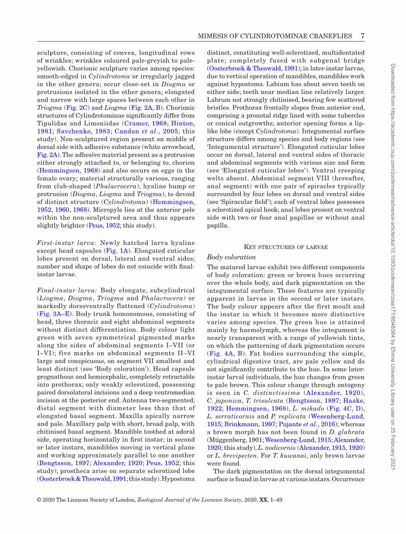
Spiracular fieldLarvae are metapneustic. A pair of spiracles (sp) is present at the dorsodistal end of the anal segment. In Cylindrotoma, spiracles are located dorsally without a spiracular disc (Fig. 10A). In most species (Diogma, Liogma, Phalacrocera and Triogma), the spiracles lie in the retractable cavity of the spiracular field (i.e. flattened posterior face of the anal segment) (Fig. 10C–E). When the spiracular dics are closed, the spiracles face each other (Fig. 10B, E). Typically, each pair of dorsal (dl) and ventral lobes (vl) are present on the spiracular field, but additionally, dorsomedial lobes (dm) are present in some species (D. glabrata, L. mikado, L. nodicornis, T. exsculpta and T. trisulcata), of which size and position various. Each of the ventral lobes possesses a sclerotized apical hook; the posterior side of the ventral lobe are pigmented as a dark line. In Phalacrocera, each of the dorsal lobes bears a small accessory lobe (accessory dorsal lobe; dac) near its base.
The external structure of the spiracles is similar to that of other cranefly larvae. The spiracular field of Cylindrotoma and P. replicata is glabrous (Fig. 10A), whereas those of other species bear a hair fringe (ha) (Fig. 10C, E). A central scar plug lies at the centre of each spiracle as a uniformly sclerotized circular disc, which is surrounded by aeropyles. Scanning electron microscope observations reveal that the central plug has an aperture and the aeropyles have slit-like openings in three species: Cylindrotoma japonica (Fig. 11A, B), Liogma brevipecten (Fig. 11C, D) and Phalacrocera tipulina (Fig. 11E, F).
The stigmatic ring internally has a developed spiracular atrium (at) (Fig. 9A–C). The atrium is lined with numerous tracheae (Fig. 9C, D), constituting a ‘felt chamber’ (Keilin, 1944). The spiracular structure differs between aquatic (P. replicata) and semi-aquatic (T. trisulcata) species (Haake, 1922). In Triogma trisulcata, the central scar plug is thin and heavily sclerotized; internally, the atrium chamber was reported to be wide and relatively short (Haake, 1922). The spiracles of Phalacrocera replicata are characterized externally by an extremely thick, weakly sclerotized, central scar plug surrounded by a stigmatic ring; the atrium chamber was relatively narrow and long by Haake’s (1922) observation.
diversity in the Biology and morphology in cylindrotomine species
genus Cylindrotoma macquart, 1834
Notes: Two species (including two subspecies), Cylindrotoma distinctissima subsp. americana Osten Sacken, 1865 and C. d. subsp. distinctissima, and C. japonica have been studied so far. These species are closely related to each other, and the life history and the behaviour of adults and larvae do not significantly differ among these taxa, and thus are given collectively below.
Life historyThe typical habitat of Cylindrotoma is shaded, humid forest floor in marshy woodlands. The larvae are found on leaves and stems of herbaceous plants. Cylindrotoma distinctissima and C. japonica are polyphagous and they use a wide range of dicots and some monocots. The larvae of these species occur densely in patchy, small populations, where they feed on leaf tissues of various herbaceous plants with soft, green, unlignified stems; some larvae even feed on young shoots of woody plants with short heights. Adult flies appear in spring; for some populations of C. d. distinctissima, two broods occur per year (Brinkmann, 1991): one
Dow
nloaded from https://academ
ic.oup.com/zoolinnean/advance-article/doi/10.1093/zoolinnean/zlaa177/6048384 by Ehim
e University Library user on 25 February 2021
MIMESIS OF CYLINDROTOMINAE CRANEFLIES 17
© 2020 The Linnean Society of London, Zoological Journal of the Linnean Society, 2020, XX, 1–49
in spring (Schellenberg, 1803; Zeller, 1842) and the other in autumn (Boie, 1838). Larvae moult four times (Brinkmann, 1991). At the first instar, the larvae feed on upper leaf tissues, leaving the lower epidermis. At the second instar or later, the larvae skeletonize the leaf tissues by opening holes and leaving only the hyaline membrane and leaf veins. The larvae live on a flat, smooth leaf substrate, which provides no supporting structures to hold on to, and so they ensure their attachment via the suction-cup principle and sticky gland secretions (as noted in ‘Larval behaviour’). The late-instar larvae become apathetic, do not eat and do not noticeably move. The larvae cease feeding after the final moult and overwinter among fallen leaves (Brodo, 1967), which presumably occur at the third instar (Brinkmann, 1991). The larvae pupate
on a leaf or a stem, holding on via the sheath of the adhered larval exuvia (Zeller, 1842; Alexander, 1920; Peus, 1952).
Adult behaviour and mechanism of endophytic ovipositionFemales exclusively insert eggs under the leaf epidermis (Fig. 1E); endophytic oviposition is only known so far from this genus among other genera of Cylindrotomidae. The female makes use of its ovipositor, which consists of two types of valves: an upper set of double-bladed cutting valves bearing a serrated blade; and a lower set of hypogynial valves that deliver eggs with an unserrated blade. The female fly places the upper side of her cerci against
Figure 10. Spiracular lobes of cylindrotomine larvae. (A) Cylindrotoma japonica, dorsal view. (B) Liogma nodicornis, caudal view. (C) L. brevipecten, oblique dorso-caudal view. (D) Phalacrocera replicata, caudal view. (E) P. tipulina, caudal view. Abbreviations: anp, anal papilla; dac, accessory dorsal lobe; dl, dorsal lobe; dm, dorsomedial lobe; ha, hair fringe; sp, spiracle; spd, spiracular disc; vl, ventral lobe. Scale = 2 mm.
Dow
nloaded from https://academ
ic.oup.com/zoolinnean/advance-article/doi/10.1093/zoolinnean/zlaa177/6048384 by Ehim
e University Library user on 25 February 2021

18 Y. IMADA
© 2020 The Linnean Society of London, Zoological Journal of the Linnean Society, 2020, XX, 1–49
the lower leaf surface, near to the edge. She then slits the leaf epidermis using the cutting valves, whilst the lower valve guides the egg into the resulting slit. The bifurcated valve on the lower side apparently serves to keep the leaf in position during the act of ovipositing, since the leaf margin was held securely between this valve and the cutting valves (Cameron, 1918). When cutting a slit in the leaf epidermis, the cerci are kept close to one another and are moved
alternately parallel to one another, which moves the abdomen from side to side (Peus, 1952). The eggs are partly concealed beneath the leaf epidermis and dorsally exposed through the slit; they were generally laid in close, parallel series (Cameron, 1918). The eggs were covered with adhesive material when oviposited (Peus, 1952) and became solid after drying, but unlike other genera, any adhesive substance was not observable (Hemmingsen, 1960).
Figure 11. Ultrastructure of spiracular field of cylindrotomine larvae, SEM views. (A–B) Cylindrotoma japonica. (A) Spiracular field is devoid of hair fringe. (B) Ditto, closer view. (C–D) Liogma brevipecten. (C) Spiracular field is present on the lobe fringed with hairs. (D) Ditto, closer view. (E–F) Phalacrocera tipulina. (E) Spiracular field on the lobe with hair fringe. (F) Ditto, closer view. White arrowheads denote apertures in the central plugs. Scales are shown in each image.
Dow
nloaded from https://academ
ic.oup.com/zoolinnean/advance-article/doi/10.1093/zoolinnean/zlaa177/6048384 by Ehim
e University Library user on 25 February 2021

MIMESIS OF CYLINDROTOMINAE CRANEFLIES 19
© 2020 The Linnean Society of London, Zoological Journal of the Linnean Society, 2020, XX, 1–49
Figure 12. Biology and morphology of Cylindrotoma japonica. (A) Anterior body of the final instar; body colour faded due to ethanol preservation: anteriormost part of prothorax to abdominal segment III, dorsal view. (B) Abdominal segments III–V showing pairs of lobes, ventral view. (C) Thick prothoracic integument in which the larval head is covered, which constitutes a ‘suction cup’. (D) Crown-shaped material covering the larval prothoracic integument, which is presumably composed of silk and debris. (E) Anal segment, ventral view. (F) Second-instar larva carrying its own faecal pellets on the dorsal and lateral sides of the integument. (G) Early-instar larva with green hue and grey pigmentation on the dorsal segments. (H) Lateinstar larva with brown hue. (I–K) Snapshots showing a sequence of the looping locomotion of a late-instar larva when
Dow
nloaded from https://academ
ic.oup.com/zoolinnean/advance-article/doi/10.1093/zoolinnean/zlaa177/6048384 by Ehim
e University Library user on 25 February 2021
20 Y. IMADA
© 2020 The Linnean Society of London, Zoological Journal of the Linnean Society, 2020, XX, 1–49
Larval behaviourDebris carrying: Larvae of Cylindrotoma carry debris on the dorsal integument of the abdominal segments (Fig. 12F). This is done by newly hatched larvae, the bodies of which are nearly transparent and only the fresh gut content has a green tint; in later larval stages, as their haemolymph becomes green, this behaviour becomes less common. The larvae deposit faecal pellets on the dorsal integument by bending the distal abdominal segments forwards. The anal segment can only reach over the front of the metathorax, which, therefore, receives the densest accumulation of faeces. Peus (1952) described the debris-carrying behaviour in detail, as below. The pronotum was not covered with faeces because the anus cannot reach over the two high processes of the mesonotum (Peus, 1952). The posterior segments indirectly supplied with faeces as these pellets already deposited on the dorsal segments rolled over (Peus, 1952). The faecal pellets were dull black in colour, cylindrical to roundish in shape and adhesive (Peus, 1952). Larvae sticked firmly to the substrate with an anal secretion, which covered the entire ventral surface of abdominal segments VI–VIII (Peus, 1952).
Defensive behaviour: The larvae of Cylindrotoma are sensitive to the motion of surrounding objects and they show defensive behaviour when threatened. When disturbed, in response they bend the anterior part of the body backwards tightly, making an acute angle with the caudal end, and then demonstrate katalepsis; namely, they hold a frozen pose for ten seconds to a minute. In cases where the danger is within touching distance, the larvae on leaves display thanatosis (i.e. death feigning) by tumbling down from the substrate to the ground.
Larval locomotion of Cylindrotoma: The larvae live on the leaf surface making use of two, especially developed, attachment mechanisms that are used in combination: a suction cup and a sticky secretion. The head capsule is retracted into the thorax and thereby a cavity is created; this acts as a suction cup when it is tightly pressed against the flat leaf surface. The head in Cylindrotoma is the bead-like, soft, front edge of the prothoracic integument, and the rim is pressed against the substrate (Supporting Information, Video S1). The interior cavity is filled with a thin film of salivary gland secretion, which covers the larval head and prothorax (Fig. 12D), but with an opening (white
arrowhead, Fig. 13A). When the secretion completely dries, it becomes a solid, reticulated aggregation, which is composed of delicate, thread-like strands (Fig. 13B), presumably silk. The fluid is drawn by a capillary between the integumental ring and the substrate, thus producing a tight seal. Although the silk-like threads are widespread on the ventral side of the prothorax, those occurring on the body elsewhere than the head capsule are coarse and have a greater diameter (Fig. 13C). When feeding or resting, the larvae normally keep themselves adhered to the substrate in such way that the head capsule remains entirely within the thorax but the mandibles are kept in a position to access the leaf surface.
The suction-cup mechanism also has a role in locomotion. The larvae move around by inching; when they transit from one leaf to another, they fix themselves with their caudal end, straighten up sharply and then move forwards the fore-body often accompany with a slow back and forth motion (Fig. 12I–K). The ventral cuticular lobes tend to be small, rounded protubercles and they also are involved in larval movement. The ventral lobes of the abdominal segment VII (Fig. 13D) have a stamp-like broad surface end, and they work in cooperation with the ventral lobes on the anal segment (Osten Sacken, 1869; Peus, 1952); these lobes aid the movement of larvae up and down the leaves and stems of plants. The secretion from the salivary glands causes the larvae on the leaf surface to leave a slight, silvery, creeping trail, which fades after a few hours as a result of drying (Cameron, 1918; this study).
Cylindrotoma distinCtissima suBsp. ameriCana osten sacken, 1865
Egg: See C. d. distinctissima.
Final-instar larva: See C. d. distinctissima.
Host-plants: Recorded host-plants encompass eight angiospermous families: two monocot species, Allium L. (Asparagales: Amaryllidaceae) and Maianthemum dilatatum (Alph.Wood) A.Nelson & J.F.Macbr. (Asparagales: Asparagaceae); and six eudicot families, Sanicula europaea L. (Apiales: Apiaceae), Valeriana officinalis L. (Dipsacales: Caprifoliaceae), Stellaria nemorum L. (Caryophyllales: Caryophyllaceae), Anemone nemorosa L., Caltha palustris L., Trautvetteria
ascending a plant stem. (I) The larva arches its body forward using both its mouth (as a sucker) and anal segment attached to the substrate . (J) The larva releases the mouth while the anal segment is wrapped around the substrate to get a grip. (K) The larva then wobbles from side to side to crawl forward. Abbreviations: dl, dorsal lobe; la, lateral lobe; vl, ventral lobe. Scale = 1 mm.
Dow
nloaded from https://academ
ic.oup.com/zoolinnean/advance-article/doi/10.1093/zoolinnean/zlaa177/6048384 by Ehim
e University Library user on 25 February 2021

MIMESIS OF CYLINDROTOMINAE CRANEFLIES 21
© 2020 The Linnean Society of London, Zoological Journal of the Linnean Society, 2020, XX, 1–49
caroliniensis (Walter) Vail (Ranunculales: Ranunculaceae), Chrysosplenium L. (Saxifragales: Saxiflagaceae) and Viola biflora L. (Malpighiales: Violaceae).
Cylindrotoma distinCtissima distinCtissima (meigen, 1818)
(fig. 3a)
Egg: Spindle-shaped, circular in cross-section, with a slightly tapered front pole. Chorionic sculpture smooth-edged. Egg covered with adhesive fluid right after oviposition and becoming solid after drying (Peus, 1952); adhesive area absent (Hemmingsen,
1960). Micropyle usually hidden under leaf epidermis; region around micropyle not differentiated well from surrounding area; instead, showing fine granulation (Peus, 1952).
Final-instar larva: Length 20 mm (Peus, 1952). Light green, darker dorsally; brown individuals unknown (Peus, 1952). Larval trunk dorsolaterally flattened, dorsally with a single row of small, conical lobes along its body. Prothorax with eight tubercles lining at lateral margin and with one pair of small, simple, conical lobes on dorsal side (Peus, 1952). Spiracular field without obvious spiracular disc without hair fringe; spiracles present dorsally (Peus, 1952). Elongated cuticular
Figure 13. Ultrastructure of Cylindrotoma japonica final-instar larva, SEM views. (A) larval head loaded with a mass of secretion (presumably silk material); secretion is reticulated and entirely conceals the head capsule but a small, circular opening is present near the centre (white arrowhead), which leads to the mouth underneath. (B) Close-up view of a secretion mass, which provides a cap for the head capsule, revealing delicate reticulated threads. Bright flaring is due to sample charge. (C) Isolated threads of the ventral thoracic segment; note that the diameter is greater than that of head part threads in image B. (D) Ventral lobe on abdominal segment VII, in which the cuticular surface is worn out due to abrasion. Scales as shown in each image.
Dow
nloaded from https://academ
ic.oup.com/zoolinnean/advance-article/doi/10.1093/zoolinnean/zlaa177/6048384 by Ehim
e University Library user on 25 February 2021

22 Y. IMADA
© 2020 The Linnean Society of London, Zoological Journal of the Linnean Society, 2020, XX, 1–49
lobes internally without containing tracheal branches (Peus, 1952).
Host-plants: The known host-plants encompass nine angiospermous families: two monocot families and diverse eudicots consisting of seven families. Following genera and species were recorded as host-plants (Lenz, 1921; Peus, 1952; Tarasova, 1981; Stubbs, 2006; Uffen & Chandler, 2010; Paramonov, 2013): Allium ursinum L. (Asparagales: Amaryllidaceae), Maianthemum dilatatum (Asparagales: Asparagaceae), Anemone nemorosa, Caltha palustris, Ranunculus repens L., Trautvetteria caroliniensis, Trollius asiaticus L. (Ranunculales: Ranunculaceae), Cirsium kamtschaticum Ledeb. ex DC., Petasites Mill., Saussurea pseudotilesii Lipsch. (Asterales: Asteraceae), Lysimachia europaea (L.) U.Manns & Anderb. (Ericales: Primulaceae), Acer pseudoplatanus L. (Sapindales: Sapindaceae), Sanicula sp. (Apiales: Apiaceae), Valeriana officinalis (Dipsacales: Caprifoliaceae), Stellaria media (L.) Vill. (Calryophyllales: Caryophyllaceae) and Viola biflora (Violales: Violaceae). All but one species listed here are herbaceous plants and a maple species, Acer pseudoplatanus, is a tree.
Cylindrotoma japoniCa alexander, 1919
(figs 1e, 4B, 5a, 6a, 7a–c, 10a, 11a, B, 12a–k, 13a–d)
Egg: Unknown.
Final-instar larva: (N = 3) Larvae similar to C. distinctissima distinctissima, with few minor characters not described for other taxa. Body apparently green (Fig. 12G) or brown hue (Fig. 12H) when moderately dried, but nearly transparent with silvery shining ventral region when wet (Figs 4B, 12F). Dorsal integument with tadpole-shaped, dark pigment in young larvae (presumably at second instar; Fig. 12G); pigment absent in late instars (Fig. 12H). Larval trunk dorsolaterally flattened. Prothorax with eight tubercles lining at lateral margin and with one pair of small, conical, dorsal lobes (Fig. 12C); integument at frontal margin often wrapped with larval secretion (presumably silk) trapping debris (Figs 12D, 13A). Dorsal elongated lobes on abdominal segments small, simple and tuberculate, arranged longitudinally in a single row (Figs 5A, 12A). Lateral lobes on meso-, metathoracic and abdominal segments corrugated at the margins (Fig. 12A). Ventral lobes on meso-, metathoracic and abdominal segments I–VII as one pair of rounded tubercles, lined near posterior margin (Fig. 12B). Anal segment possesses one pair of dorsal lobes, oriented laterally (Fig. 12E); two pairs of lateral lobes, with bases near the anterior end of the segment; one pair of ventral lobes, papilla-like,
with apices projecting downward; anal papilla absent. Posterior spiracles present dorsally, without forming an obvious spiracular disc (Fig. 10A) and devoid of hair fringe (Fig. 11A, B). Elongated cuticular lobes internally, without containing tracheal branches.
Host-plants: The host-plant species recorded so far encompass 16 angiospermous families: two magnoliids, Asarum sieboldii Miq. and A. caulescens Maxim. (Piperales: Aristolochiaceae); three monocots, Tricyrtis latifolia Maxim. (Liliales: Liliaceae), Maianthemum dilatatum (Asparagales: Asparagaceae) and Dioscorea japonica Thunb. (Dioscoreales: Dioscoreaceae); some basal eudicots, Ranunculus japonicus Thunb., R. silerifolius Lév. var. glaber (H.Boissieu) Tamura and Trautvetteria caroliniensis (Ranunculales: Ranunculaceae); and many herbacrous core eudicots, including Astilbe microphylla Knoll (Saxifragales: Saxifragaceae), Potentilla spp. (Rosales: Rosaceae), Viola spp. (Violaceae), Sambucus sieboldiana Blume var. pinnatisecta G.Y.Luo & P.H.Huang (Dipsacales: Adoxaceae), Angelica sachalinensis Maxim. var. glabra (Koidz.) T.Yamaz., Conioselinum filicinum (H.Wolff) Hara, Sanicula chinensis Bunge (Apiales: Apiaceae), Hydrocotyle sp. (Apiales: Araliaceae), Artemisia indica Willd. var. maximowiczii (Nakai) H.Hara, Cirsium lucens Kitam. (locally known as ‘Cirsium taishakuense’ ined.), Erigeron annuus (L.) Pers., E. philadelphicus L., Eupatorium makinoi T.Kawahara & Yahara, Nemosenecio nikoensis (Miq.) B.Nord., Parasenecio adenostyloides (Maxim.) H.Koyama, Pa. delphiniifolius (Siebold & Zucc.) H.Koyama, Petasites japonicus (Siebold & Zucc.) Maxim. (Asterales: Asteraceae), Silene gracillima Rohrb. (Caryophyllales: Caryophyllaceae), Scutellaria brachyspica Nakai & H.Hara (Lamiales: Lamiaceae), Torenia crustacea (L.) Cham. & Schltdl. (Lamiales: Linderniaceae) and Plantago asiatica L. (Lamiales: Plantaginaceae); also, young shoots of a woody eudicot, Orixa japonica Thunb. (Sapindales: Rutaceae), which is only recorded from one local population. Among the taxa listed above, some widespread taxa of Asteraceae are commonly targeted. The result of tentative non-choice experiments indicates that C. japonica apparently does not accept some common species that co-occur in their populations, such as Commelina communis L. (Commelinales: Commelinaceae).
genus diogma edwards, 1938
diogma glabrata (meigen, 1818)
(figs 3B, 5B, 14a–c)
Life history: The whole life-cycle of this species is completed in one year (Müggenburg, 1901); in Japan,
Dow
nloaded from https://academ
ic.oup.com/zoolinnean/advance-article/doi/10.1093/zoolinnean/zlaa177/6048384 by Ehim
e University Library user on 25 February 2021

MIMESIS OF CYLINDROTOMINAE CRANEFLIES 23
© 2020 The Linnean Society of London, Zoological Journal of the Linnean Society, 2020, XX, 1–49
the adult flying period is in summer (July to August), which is much later than the other co-occurring species (Liogma mikado and Liogma serraticornis). Larvae are found in terrestrial mosses growing on stones in limestone woodlands, and also in moss mats growing on soil. In Europe, they were found in forests in grassy, humid areas with Rhytidiadelphus squarrosus (Hedw.) Warnst. (Hypnales: Hylocomniaceae) (Zeller, 1842; Müggenburg, 1901). The number of moults is unknown. Final-instar larvae apparently do not have a preference for pupation sites and pupae were found anywhere in the lower damp layers of moss carpets (Alexander, 1920).
Oviposition: A female of this species laid about 60 eggs in total (Müggenburg, 1901), depositing them singly on the lower surface of leaves or stems of mosses (Hemmingsen, 1960).
Egg: Length 1–1.3 mm (Müggenburg, 1901). Spindle-shaped, circular in cross-section, with a slightly
tapered front pole; longitudinally bulbous (Peus, 1952). Chorionic sculpture irregularly jagged, occuring close-set; reticulated near micropyle (Peus, 1952).
First-instar larva: Newly hatched larvae ashy grey, but become moss green in colour at later stages (Müggenburg, 1901).
Final-instar larva: Length 17–18 mm (N = 3). Body colour with dark green hue; dark pigmentation appears as small blotches on lateral sides, with various forms ranging from rhombus to trapezoid; also, a single, small, isolated blotch near the distal corner of the anterior end is present in the abdominal segment; size of pigmented area increases from abdominal segments I to VI; pigmentation small and pale on abdominal segment VII (Fig. 14A, B). Brown individual unknown (Müggenberg, 1901; Wesenberg-Lund, 1915; Alexander, 1920). Prothorax rugulose; dorsal lobes conical with sharply pointed apices, two pairs along
Figure 14. Biology and morphology of Diogma glabrata. (A) Final instar larva of D. glabrata. (B) Later-instar larva feeding on Bryhnia cf. tenerrima (Brachytheciaceae). (C) Characters of anal segment. Abbreviations: dl, dorsal lobe; dm, dorsomedial lobe; vl, ventral lobe. Scale = 1 mm.
Dow
nloaded from https://academ
ic.oup.com/zoolinnean/advance-article/doi/10.1093/zoolinnean/zlaa177/6048384 by Ehim
e University Library user on 25 February 2021

24 Y. IMADA
© 2020 The Linnean Society of London, Zoological Journal of the Linnean Society, 2020, XX, 1–49
pronotal ridge, anterior pair longer than posterior pair (Fig. 5B); lateral lobes one pair; ventral lobes two pairs. Meso- and metathoracic segments have two pairs of dorsal lobes; lateral lobes two pairs; ventral lobes two pairs, papilla-like, anterior pair closer to each other than posterior pair (Fig. 5B). Dorsal lobes on abdominal segments with bluntly pointed apices, third and fourth pairs bearing two, rounded auxiliary outgrowths (Fig. 6B); segment I with two pairs of lobes, each with two auxiliary outgrowths at the front; segments II–VII with four pairs, each of the posterior two pairs carry two teeth-like auxiliary outgrowths at the front, fourth pair longest and stout; segment VIII with a pair of small, simple, conical lobes without auxiliary outgrowth. Lateral lobes on abdominal segments simple, finger-shaped; segments I and II–VI with three and four pairs, respectively. Ventral lobes on abdominal segments rounded. Anal segment with one pair of dorsal lobes long; dorsomedial lobes (dm) one pair, short and conical (Fig. 14C); lateral lobes one pair, papilla-like; ventral lobes two pairs, anterior pair papilla-like. Spiracular field with hair fringe.
Host-plants: Larvae feed on Rhytidiadelphus squarrosus (Hypnales: Hylocomiaceae) and Hypnum cupressiforme Hedw. (Hypnales: Hypnaceae) (Zeller, 1842; Müggenburg, 1901). At mixed forest near Mt. Kisokomagatake (Nagano, Japan), at least two larvae have been confirmed to feed on Bryhnia cf. tenerrima (Hypnales: Brachytheciaceae) in the field.
genus liogma osten sacken, 1869
liogma brevipeCten alexander, 1932
(figs 1B, c, 2a, B, 4a, 5c, 6c, 7d–f, 8a–f, 9a–d, 10c, 11c, d, 15a–f)
Life history: This species is univoltine; adults emerge during May–July, in central Japan. Larvae of this species occur in well-moistened Mniaceae moss tufts in wet spots of forest or woodland, such as stream banks, trickling waters, rocks near waterfalls and lake margins. In Japan, they frequently co-occur with some other terrestrial moss-feeding species, such as L. serraticornis, L. mikado and T. kuwanai. In winter, the larva buries itself beneath the moss patches and sometimes its body is even embedded in the soft ground.
Egg: Length 0.9 mm (N = 1). Spindle-shaped, circular in cross-section, with a tapered front pole (Fig. 2A). Greyish yellow, apparently darker than T. kuwanai. Chorionic sculpture elongated and narrow, arranged as dotted dash lines; reticulated near micropyle (black arrowhead in Fig. 2A).
First-instar larva: Length 2.8 mm (N = 3). Body hyaline (Fig. 15E). Dorsal elongated cuticular lobes on thoracic and abdominal segments simple, outer surface covered with simple microtubercles (mtb) (Fig. 7C). Dorsal lobes on thoracic and abdominal segments with spinules present but less apparent than in later-instar larvae.
Final-instar larva: Length 14 mm (N = 47). Body colour dark green dorsally, due to oblique dark pigmentation on lateral side (Fig. 15A); colour generally uniform among individuals, brown individual unknown. Integumental surface rugulose due to undulated or reticulated ridges (Fig. 7D). Prothorax with two pairs of dorsal lobes along pronotal ridge, small and conical, with needle-like apices; anterior opening relatively smooth, without distinct tubercles (Fig. 15B); one pair of lateral lobes near the posterior end of the segment, small. Meso- and metathoracic segments with one pair of dorsal lobes; lateral lobes two pairs; ventral lobes two pairs, small and conical. Dorsal lobes on abdominal segments with translucent, sharply pointed apices; lobe covered with numerous sharp, translucent spinules on anterior and posterior sides of the lobes (Figs 6C, 7F, 15C); abdominal segments I and II–VII with two and four pairs, respectively. Lateral lobes on abdominal segments with sharp apices; abdominal segment I with three pairs; abdominal segments II–VII with four pairs (Fig. 5C); lobes bearing spinules as of dorsal lobes, but with lower density. Ventral lobes on abdominal segments small and conical. Anal segment with one pair of dorsal lobes bearing spinules; one pair of ventral lobes, devoid of spinules; dorsomedial lobe absent; lateral lobe absent; two pairs of ventral lobes, anterior pair papilla-like; four anal papillae present. Spiracular field with hair fringe (Fig. 10C). Elongated cuticular lobes internally containing tracheal branches (Fig. 15D).
Oviposition: Behaviour of four female individuals was observed in the field (5–13 May 2016, at Kibune, Kyoto, Japan). The adults occur in humid moss patches in woodlands, where ground water exudes continually. At the locality, several acrocarpous mosses and Chrysosplenium grayanum Maxim. (Saxifragaceae) abundantly grow on the mucky soil. Females lay eggs singly, inserting the abdomen into the tufts of Rhizomnium and Mnium mosses. The eggs (N = 19) are laid exclusively on moss shoots.
Host-plants: Larvae are found from tufts of moss species belonging to Mniaceae (Bryales). Recorded host-plants in the field include Plagiomnium acutum (Lindb.) T.J.Kop., Pl. cuspidatum (Hedw.) T.J.Kop., Pl. maximoviczii (Lindb.) T.J.Kop., Pl. vesicatum (Besch.) T.J.Kop., Rhizomnium hattorii T.J.Kop. and Rh. tuomikoskii T.J.Kop.
Dow
nloaded from https://academ
ic.oup.com/zoolinnean/advance-article/doi/10.1093/zoolinnean/zlaa177/6048384 by Ehim
e University Library user on 25 February 2021

MIMESIS OF CYLINDROTOMINAE CRANEFLIES 25
© 2020 The Linnean Society of London, Zoological Journal of the Linnean Society, 2020, XX, 1–49
Figure 15. Biology and morphology of Liogma brevipecten. (A) Final-instar larva, dorsolateral view. (B) Prothoracic integument sheltering the head capsule, frontal view. (C) Spinules on a dorsal lobe, dorsolateral view; for ultrastructure, see Fig. 6F. (D) Tracheal branches inserted into a lateral lobe. (E) First-instar larva, lateral view. (F) Female laying an egg onto the abaxial side of a leaf of Bryhnia novae-angliae (Brachytheciaceae). Scale = 1 mm.
Dow
nloaded from https://academ
ic.oup.com/zoolinnean/advance-article/doi/10.1093/zoolinnean/zlaa177/6048384 by Ehim
e University Library user on 25 February 2021

26 Y. IMADA
© 2020 The Linnean Society of London, Zoological Journal of the Linnean Society, 2020, XX, 1–49
liogma mikado (alexander, 1919)
(figs 1d, 1f, 3d, 4c, d, 5d, 6d, 16a–c)
Life history: Liogma mikado is univoltine, having the adult stage in spring. This species markedly differs in habitat preference from other Liogma species, although it frequently co-occurs with L. serraticornis on a small geographic scale (within a range of a few metres). The larvae are most frequently found on arid moss tufts on rocks or on the ground, but also on decomposed tree trunks. The larvae are exposed on the surface of moss patches, unlike other species of Liogma (e.g. L. brevipecten and L. serraticornis), which sometimes bury deeply beneath the moss patches.
Mating and oviposition: Two adult females of L. mikado were observed during 13:00–16:10 on 19 May 2016, at Ikawa-touge (Shizuoka, Japan). The site was a forest floor with gentle slope facing a forest path toward a mountain pass. The males emerged before the females and waited beside the female pupae for the emergence of their mates. Mating took place mainly resting on the branches and leaves of the understory vegetation (Fig. 1D). When pairs are in copula, they readily take flight. The oviposition was observed once on the same day. One female flit about the tufts of mosses and lay an egg on a branch leaf of Hylocomnium splendens (Hedw.) Schimp. (Hylocomniaceae) (Fig. 1F).
Egg: Length 0.74 mm (N = 1). Nearly black in colour (Fig. 1F). Spindle-shaped, circular in cross-section, with a tapered front pole (Fig. 2F). Chorionic surface nearly black without noticeable wrinkles; sculptures unexamined.
First-instar larva: Length 1.3 mm (N = 2). Body hyaline (Fig. 16B). Dorsal elongated cuticular lobes on thoracic and abdominal segments simple and stout.
Final-instar larva: Length 8–10 mm (N = 3). Body colour polymorphic, with light green (Fig. 4C) or brown hue (Fig. 4D); dark pigmentation making a narrow band, appearing as an oblique line in lateral view, from mesothoracic through abdominal segment VII; also, tips of small lobes tinged, including lobes on thoracic segments, first and second pairs and auxiliary outgrowths (particularly, second and third teeth) of third and fourth pairs of lobes on abdominal segments II–VII (Fig. 16A). Integument slightly tinged yellow (Fig. 4A), surface smooth. Prothorax dorsally with two, conical pairs of lobes on pronotal ridge with sharply pointed apices, posterior pairs longer than anterior pairs; two pairs of lateral lobes, papilla-like. Meso- and metathoracic segments with two pairs of dorsal lobes,
each with two conical auxilliary outgrowths at the front; two pairs of lateral lobes, anterior pair longer than posterior pair; one pair of ventral lobes, papilla-like (Fig. 5D). Dorsal elongated lobes on abdominal segments generally short, longest lobe shorter than body width in ventrolateral view (Fig. 6D); segment I with two pairs: anterior pair conical, with two tiny teeth-shaped auxiliary outgrowths at the front; posterior pair long, with three teeth-shaped outgrowths at the front; segments II–VII with four pairs of lobes, anterior two pairs conical and simple, third pair with two teeth at the front and fourth pair with three teeth near the base at the front; apices of teeth-like auxiliary outgrowths obtuse and tuberculate when dried, sharply pointed when wet. Lateral lobes on abdominal segments with sharply pointed apices; segment I with three pairs, posterior two pairs longer than anteriormost pair; segments II–VII with four pairs, middle two pairs longer than others. Ventral lobes on abdominal segments small, rounded or conical. Anal segment with one pair of dorsal lobes, long; one pair of dorsomedial lobes, short and conical; lateral lobes papilla-like; two pairs of ventral lobes, anterior pair papilla-like. Spiracular field with hair fringe.
Host-plants: The host-plant range of this species varies considerably among populations. Typically, the larvae are found and reared on some species of Thuidium (Hypnales: Thuidiaceae), such as T. tamariscinum (Hedw.) Schimper, T. kanedae Sakurai, T. delicatulum (Hedw.) Schimp., T. cymbifolium (Dozy & Molk.) Dozy & Molk. and T. pristocalyx (Müll. Hall.) Jaeger. In some populations, the larvae feed on Eurhynchium savatieri Schimp. ex Besch. (Hypnales: Brachytheciaceae), Hylocomiopsis ovicarpa (Besch.) Card. (Hypnales: Leskeaceae), Ctenidium capillifolium Brotherus, Hylocomnium splendens (Hypnales: Hylocomniaceae), Hypnum oldhamii Jaeger (Hypnales: Hypnaceae), Thamnobryum sp. (Hypnales: Neckeraceae) , Plagiothecium euryphyllum (Card. & Thér.) Iwats. and Pseudotaxiphyllum pohliaecarpum (Sull. & Lesq.) Iwats. (Hypnales: Plagiotheciaceae) (Figs 4C, D, 16I). Under rearing conditions, the following moss species are also detected from the faecal pellets of larvae: Leucobryum neilgherrense Müll. Hal. (Dicranales: Dicranaceae) and Fauriella tenuis (Mitt.) Cardot (Hypnales: Theliaceae). Thus, L. mikado potentially uses a wide range of mosses belonging to Hypnales, although no larva has ever been obtained from Bryales. From the faecal pellets, the larval diet exclusively consists of moss species, even though some liverworts, including Cephalozia (Jungermanniales: Cephaloziaceae) and Calypogeia (Jungermanniales: Calypogeiaceae), commonly occurred in the larval habitat.
Dow
nloaded from https://academ
ic.oup.com/zoolinnean/advance-article/doi/10.1093/zoolinnean/zlaa177/6048384 by Ehim
e University Library user on 25 February 2021

MIMESIS OF CYLINDROTOMINAE CRANEFLIES 27
© 2020 The Linnean Society of London, Zoological Journal of the Linnean Society, 2020, XX, 1–49
Figure 16. Biology and morphology of Liogma mikado, L. nodicornis, L. serraticornis. (A–C) L. mikado. (A) Late-instar larva, lateral view. (B) First-instar larva (scale = 0.5 mm). (C) Early-instar larva on a tuft of Plagiothecium euryphyllum (Plagiotheciaceae). (D) Late-instar larva of L. nodicornis, feeding on Plagiomnium ciliare (Mniaceae). (E–I) L. serraticornis. (E) Late-instar larvae with green hue, feeding on Plagiomnium vesicatum (Mniaceae). (F) Late-instar larvae with brown hue feeding on Mnium lycopodioides (Mniaceae), found in the same population as the individual shown in (B). (G) Habitus of late-instar, dorsal view; mesoand metathoracic segments are entirely dark and inverted Y-shaped pattern is evident in abdominal segments I–VII. (H) Characters of head. (I) Characters of anal segment. Black arrows indicate papilla-like, anterior pair of the ventral lobes. Abbreviations: anp, anal papilla; dl, dorsal lobe; pr, pronotal ridge; vl, ventral lobe. Scale (except B) = 1 mm.
Dow
nloaded from https://academ
ic.oup.com/zoolinnean/advance-article/doi/10.1093/zoolinnean/zlaa177/6048384 by Ehim
e University Library user on 25 February 2021

28 Y. IMADA
© 2020 The Linnean Society of London, Zoological Journal of the Linnean Society, 2020, XX, 1–49
liogma nodiCornis (osten sacken, 1865)
(figs 5e, 6e, 16d)
Life history: This species is univoltine (Alexander, 1920). It commonly occurs in damp, swampy woods, in gorges of creeks and on moist, shaded hillsides. Larvae are found on small patches of Mniaceae mosses along the slope of forest paths, dead tree trunks and detritus underneath.
Oviposition: Females deposited eggs singly on the leaves or branches; or lightly attached eggs to the axils of the leaves of Rhytidiadelphus squarrosus (Alexander, 1915).
Final-instar larva: Body with green hue, and pale-grey pigmentation patterning on the integument (Fig. 14A); brown individuals unknown; dark pigmentation making a narrow band, appearing as an oblique line in lateral view, on abdominal segments I–VII, but absent on cuticular lobes; pigmented area larger on posterior body segment. Larval trunk cylindrical. Integumental surface smooth. Prothorax with two pairs of dorsal, lateral and ventral lobes. Meso- and metathorax with two pairs of dorsal lobes, anterior pair small and conical, and posterior pair bearing single, small, tooth-like auxilliary outgrowth at the front; two pairs of lateral lobes, anterior pair larger and posterior pair conical and smaller; two pairs of ventral lobes. Dorsal lobes of abdominal segments short, apices sharply pointed backwards (Fig. 5D): abdominal segment I with two pairs, anterior pair conical, bearing a tiny tooth at the front, posterior pair longer, with a small tooth at the front; abdominal segments II–VII with four pairs of lobes, posterior two bearing a small tooth-like auxiliary outgrowth at the front. Lateral lobes of abdominal segments conical; three and four pairs on segment(s) I and II–VII, respectively. Ventral lobes small, three and five pairs on segment(s) I and II–VII, respectively. Anal segment with one pair each of dorsal, dorsomedial lobes; two pairs of ventral lobes, anterior pair papilla-like; four anal papillae present. Spiracular field with hair fringe. Elongated cuticular lobes internally containing tracheal branches.
Host-plants: Larvae are reared on Rhizomnium punctatum (Hedw.) T.J.Kop. (Bryales: Mniaceae) and Hypnum cupressiforme Hedw. (Hypnales: Hypnaceae) (Brodo, 1967; Byers, 2002). This study confirms that larvae can also be reared on Rhizomnium appalachianum T.J.Kop. and Plagiomnium ciliare (Müller Hal.) T.J.Kop., Pl. cuspidatum and Pl.
drummondii (Bruch & Schimper) T.J.Kop. (Bryales: Mniaceae).
liogma serratiCornis alexander, 1919
(figs 5f, 6f, 16e–i)
Life history: This species is univoltine; adults emerge during May–July, in central Japan. Larvae occur on moss tufts of Mniaceae in relatively wet spots in the forest or woodland. The matured larvae often are found lying underneath thalli of thallose liverworts, especially Conocephalum conicum (L.) Dum. (Conocephalaceae), or amongst shed old pine needles of Cryptomeria japonica (Thunb. ex Lf.) Don (Pinales: Cupressaceae) into which individuals with dark brown hue blend well.
Final-instar larva: Length 20 mm (N = 10). Body colour various with light green (Fig. 16E) or dark brown hue (Fig. 16F). Dorsal pigmentation pattern well organized: prothorax lacks pigmentation; meso- and metathorax nearly entirely pigmented; in abdominal segments I–VII, dark blotches appear as inverted Y-shape, broadly diffused anteriorly (Fig. 16G); in some individuals, small blotches also occur ventrally. Integument translucent, except for pigmented parts, surface rugulose (Fig. 6F). Prothorax dorsally with six lobes along pronotal ridge having blunt apices; middle two longer and conical, outer two shorter and tuberculate (Fig. 16H). Meso- and metathorax with two pairs of dorsal lobes, both bearing two button-shaped auxiliary outgrowths at the front; two pairs of lateral lobes, anterior pair larger; two pairs of ventral lobes. Dorsal lobes on abdominal segments slender and each pair clinging to each other with bluntly pointed apices (Fig. 6F): segment I with three pairs, posteriormost pair carrying two auxiliary outgrowths; segments II–VII with four pairs, posterior two pairs bearing two auxiliary outgrowths (Fig. 6F). Lateral lobes on abdominal segments relatively long, apices obtuse and bent backwards; segments I and II–VII with three and four pairs, respectively. Ventral lobes on abdominal segments relatively long, apices significantly oriented backwards. Anal segment with one pair of dorsal lobes; dorsomedial lobe absent; two pairs of ventral lobes, anterior pair papilla-like (black arrowheads, Fig. 16I); four anal papillae (anp) present (Fig. 16I). Spiracular field with hair fringe.
Host-plants: The larvae feed on several moss species belonging to Mniaceae (Bryales), including Mnium lycopodioides (Hook.) Schwägr., Plagiomnium
Dow
nloaded from https://academ
ic.oup.com/zoolinnean/advance-article/doi/10.1093/zoolinnean/zlaa177/6048384 by Ehim
e University Library user on 25 February 2021

MIMESIS OF CYLINDROTOMINAE CRANEFLIES 29
© 2020 The Linnean Society of London, Zoological Journal of the Linnean Society, 2020, XX, 1–49
maximoviczii, Pl. tezukae (Sakurai) T.J.Kop., Pl. vesicatum, Rhizomnium hattorii and R. tuomikoskii T.J.Kop.
genus triogma schiner, 1863
triogma exsCulpta osten sacken, 1865
(fig. 5g)
Life history: The habitat of this species is ‘seepage areas and along short marshy spring rills and are often found in wet mossy meadows where there is no woody vegetation’ (Brodo, 1967).
Egg: Unknown.
Final-instar larva: Following description based on Brodo (1967). Length 16 mm. Body colour undescribed. Prothorax glabrous; two and one pair(s) of dorsal and lateral lobes present. Meso- and metathoracic segments with two pairs of dorsal lobes, lobes of posterior pair with one small protuberance at the base; two pairs of lateral lobes; three pairs of ventral lobes, the middle pair most widely separated to each other. Dorsal lobes on abdominal segments as follows: segment I with two pairs of lobes, each with one anterior protuberance; segments II–VII with four pairs, first pair small, second pair slightly larger, third pair with two frontal tooth-like protuberances, fourth pair longest with one frontal tooth. Lateral lobes on abdominal segments simple; two and four pairs on segments I and II–VII, respectively. Ventral lobes simple, conical. Anal segment with three pairs of lobes, inclined toward each other, one small dorsomedial pair and one pair of dorsal lobes; one pair of dorsomedial lobes; two pairs of ventral lobes, anterior pair shorter. Ventral surface of anal segment possesses protuberances.
Host-plants: Larvae were collected among the tufts of the aquatic moss Fontinalis antipyretica Hedw. (Hypnales: Fontinalaceae) (Brodo, 1967).
triogma kuwanai (alexander, 1913)
(figs 1a, 2c, 5h, 6g, 17a–f)
Notes: Triogma kuwanai used to be subdivided into two subspecies, T. kuwanai kuwanai (Honshu) and T. kuwanai limbinervis (Shikoku), but T. limbinervis Alexander, 1953 can be treated as a species (Oosterbroek 2020). The accounts below are based on T. kuwanai kuwanai.
Life history: In Japan, adults often fly together with species of L. mikado, L. serraticornis and L. brevipecten
at the same locality and about the same period of time (May–June, in Honshu). Larvae are found in moss patches on the forest floor. The cuticular surface is heavily armoured with debris and epibionts, which seems to be related to the cryptic habitat.
Adult behaviour: Three adults were observed on 18 May 2016, Ikawa-touge, Shizuoka, Japan. They occasionally flew over the shaded forest floor when disturbed, but soon returned to the original position. Adult males generally perched on a moss mat, whereas females usually rested hanging on branches or near the damp soil. Eggs were laid on the leaves of the following plant species: Pohlia longicolla (Hedw.) Lindb. (Bryaceae) (four eggs), Mnium heterophyllum (Hook.) Schwaegr. (Mniaceae) (two eggs), Plagiothecium curvifolicum Limpricht (Plagiotheciaceae) (one egg), young shoots of Stellaria media (two eggs) and a sprout of an unidentified species of fern (one egg). Two eggs were laid on the shallow layer of soil. Larvae hatched 10–11 days later from 20 out of 29 eggs. Three larvae perforated and fed on the leaves of M. heterophyllum. All reared larvae died before reaching the second instar.
Egg: Length 1.3 mm (N = 1). Spindle-shaped, circular in cross-section, with a tapered front pole. Pale grey due to exochorionic sculpture, with yellowish tint of egg content (Fig. 2C), yet apparently lighter than L. brevipecten. Chorionic sculpture elongated and narrow, arranged as irregularly jagged, dotted dash lines; reticulated near micropyle.
First-instar larva: Length 2.9 mm (N = 4). Hyaline (Fig. 17D). Dorsal elongated cuticular lobes on thoracic and abdominal segments acutely bent backwards (or occasionally forward).
Final-instar larva: Body with dark brown hue (Fig. 17A). Integument entirely rugulose with velvet-like texture, retaining high opacity in alcohol; dark pigmentation diffused on dorsal side, densest near dorsal lobes, making stripes in parallel with longitudinal body axis; small patch with purple lustre at bases of dorsal lobes (Fig. 17E). Prothorax dorsally with six tubercles along pronotal ridge; one pair of lateral lobes; ventral lobe absent. Meso- and metathorax with two pairs of dorsal lobes, posterior pair bearing two auxiliary outgrowths at the front; two pairs of lateral lobes, anterior pair longer; two pairs of ventral lobes, anterior pair more widely separated from each other than the posterior pair. Dorsal lobes on abdominal segments slender and apices acutely bent backwards (Fig. 6G); four pairs on abdominal segments I–VII, two anterior pairs smallest, and
Dow
nloaded from https://academ
ic.oup.com/zoolinnean/advance-article/doi/10.1093/zoolinnean/zlaa177/6048384 by Ehim
e University Library user on 25 February 2021

30 Y. IMADA
© 2020 The Linnean Society of London, Zoological Journal of the Linnean Society, 2020, XX, 1–49
fourth pair longest with three auxiliary outgrowths at front. Lateral lobes relatively long and broad, apices strongly bent backwards. Ventral lobes on abdominal
segments relatively long and finger-like (Fig. 17E). Anal segment (Fig. 17C) with one pair of dorsal lobes; dorsomedial lobes absent; two pairs of ventral lobes,
Figure 17. Larvae of Triogma kuwanai. (A) Specimen of late-instar larva preserved in alcohol, dorsal view. (B) Characters of head. (C) Characters of anal segment. (D) First-instar larva. (E) Late-instar larva, ventral view. (F) Adult female ovipositing a single egg on a young shoot of Pohlia longicolla (Bryales: Bryaceae). Abbreviations: dl, dorsal lobe; pr, pronotal ridge; vl, ventral lobe. Scale = 1 mm.
Dow
nloaded from https://academ
ic.oup.com/zoolinnean/advance-article/doi/10.1093/zoolinnean/zlaa177/6048384 by Ehim
e University Library user on 25 February 2021

MIMESIS OF CYLINDROTOMINAE CRANEFLIES 31
© 2020 The Linnean Society of London, Zoological Journal of the Linnean Society, 2020, XX, 1–49
anterior pair papilla-like. Spiracular field with hair fringe.
Host-plants: Although larvae are found amid moss tufts of Mniaceae (Bryales), together with Liogma serraticornis, this species probably does not feed on them. One late-instar larva was found feeding on Brachythecium brotheri Paris (Hypnales: Brachytheciaceae). Under rearing conditions, first instar does not survive on the host-plants used for female oviposition (as noted above), including three moss species, Pohlia longicolla (Hedw.) Lindb. (N = 4), Mnium heterophyllum (Hook.) Schwaegr. (Bryales: Mniaceae) (N = 2), Plagiothecium curvifolicum Limpricht (Hypnales: Plagiotheciaceae) (N = 1), although they chew on some leaves of these plants.
triogma trisulCata (schummel, 1829)
(figs 3c, 5i)
Life history: Larvae of this species occur in semi-aquatic mosses on moorlands, such as those of Hypnum cuspidatum (Brindle, 1967), in stagnant waters on bogs (Brinkmann, 1997). They have also been found in slowly running water and springs (Steinmann, 1907–08; Hemmingsen, 1968), and in small alpine streams (Steinmann, 1907–08; Müller, 1908; Alexander, 1915). According to Haake (1922), the larvae underwent moulting four times until pupation (Haake, 1922); however, his explanation had a contradictory statement. As Haake (1922) noted, stages I, II and III have one, two and no moulting, respectively, which does not make the total of four instars that the author recorded. First-instar larvae were whitish without distinct colour (Haake, 1922). The larva lives among the stems of the aquatic moss Fontinalis antipyretica, to which it clings firmly with two strong chitinised hooks at the caudal end of the body (Haake, 1922). On pupating, it comes to the water surface and remains immobile for the first few days of pupation; after 8–10 days, it becomes more mobile (Haake, 1922).
Egg: Spindle-shaped, circular in cross-section, with a tapered front pole. Pale yellow. Chorionic sculpture elongated and narrow, arranged as irregularly jagged, dotted dash lines; reticulated near micropyle.
Final-instar larva: Length 19 mm (Alexander, 1920). Body colour light green with dark blotches (Alexander, 1920). Prothorax with three pairs of dorsal lobes along pronotal ridge; one pair of lateral lobes; three pairs of ventral lobes as tubercles (Peus, 1952). Meso- and metathoracic segments with each
two pairs of dorsal and lateral lobes; lateral and ventral lobes two and three pairs, respectively (Alexander, 1920). Dorsal elongated lobes on abdominal segments shorter than body width in ventrolateral view, c. 1.5 mm in length (Alexander, 1920): abdominal segments II–VII with four pairs of lobes, posteriormost pair longest, bearing three or four teeth-like auxiliary outgrowths at the front (Alexander, 1915, 1920). Lateral lobes on abdominal segments conical; on segments I and II–VII, three and four pairs, respectively, anteriormost pair smallest (Alexander, 1920). Ventral lobes small, finger-shapd (Alexander, 1920). Anal segment with one pair each of dorsal, lateral and ventral lobes. Spiracular field with hair fringe (Alexander, 1920).
Host-plants: Larvae are found on the submerged moss Fontinalis antipyretica (Hypnales: Fontinalaceae), and may also use Hypnum (Hypnales: Hypnaceae) and Amblystegium (Hypnales: Amblystegiaceae) as hosts (Wesenberg-Lund, 1943). Brinkmann (1991) reported that one adult emerged from Mnium hornum Hedw. (Bryales: Mniaceae).
genus phalaCroCera schiner, 1863
Notes: The life history of Phalacrocera replicata has been intensively examined (e.g. Bengtsson, 1897; Miall & Shelford, 1897; Peus, 1952). The larval life-history and behaviour are somewhat similar to Phalacrocera tipulina, and combined details are thus provided below.
Larval behaviourDefensive behaviour: Larvae of P. tipulina are usually passive and not responsive, even when poked. However, larvae of P. replicata curl up their body and clasp the moss shoot they are on when alarmed; that way, the larvae may avoid being washed away in flowing water (Haake, 1922; Peus, 1952). Larval cuticles of Phalacrocera are often armoured with ectobionts, such as algae, diatoms and other freshwater microorganisms (Fig. 6G), and even with gastropods (e.g. Planorbis O.F. Müller, 1774) (Miall & Shelford, 1897).
Locomotion and attachment mechanisms: Larvae live underwater and cling to aquatic mosses, making use of their ventral lobes. When feeding or resting, the larvae normally keep themselves adhered to the substrate in such way that the head capsule remains entirely within the thorax, but making the mandibles accessible to the leaf surface. This attachment mechanism also plays a role in locomotion. Larvae fix themselves to the substrate at the caudal end, straighten up sharply and
Dow
nloaded from https://academ
ic.oup.com/zoolinnean/advance-article/doi/10.1093/zoolinnean/zlaa177/6048384 by Ehim
e University Library user on 25 February 2021

32 Y. IMADA
© 2020 The Linnean Society of London, Zoological Journal of the Linnean Society, 2020, XX, 1–49
move the fore-body with a slow back and forth motion (Fig. 16D, E).
phalaCroCera repliCata (linnaeus, 1758)
(figs 5J, 10d, 18a, B)
Life history: Larvae were found to live in stagnant, dystrophic waters or acid pools of moors or woodlands where aquatic mosses, such as Hypnum, Fontinalis and Sphagnum, were abundant (Bengtsson, 1897; Brindle, 1967; Brinkmann, 1997; Stubbs, 2010). Phalacrocera replicata had a single brood in a year (Bengtsson, 1897; Müggenburg, 1901; Alexander, 1920); admittedly, they might have a second brood (Miall & Shelford, 1897). Bengtsson (1897) confirmed that P. replicata moult at least eight and possibly ten times during their development. The first moult occurred 4–6 days after leaving the eggs (Bengtsson, 1897), which did not significantly affect the external or internal appearance. The second moult occurred at about 18–20 days, and the larvae dramatically changed in appearance (Bengtsson, 1897). First-instar larvae started feeding on the host-plants immediately after hatching; they scraped only the upper layer of leaves (Peus, 1952). They overwintered as larvae and thus endured long and severe cold under the ice (De Geer, 1773; Miall & Shelford, 1897; Wesenberg-Lund, 1915). The larvae were also able to live a long period of time (c. 5 days) in anoxic conditions (Miall & Shelford, 1897) and they could sit down deep in water 1 m in depth for a while in autumn (Wesenberg-Lund, 1915). Pupae were much more active than larvae and when disturbed, wriggled about and bent their bodies almost into a circle by flexion of the abdomen (Miall & Shelford, 1897). Usually, the pupae rested vertically, with the mesothoracic horns reaching the surface of the water (Miall & Shelford, 1897; Hemmingsen, 1952; Peus, 1952), by firmly grasping floating plants with its dorsal abdominal hooks (Miall & Shelford, 1897). Although the pupae could survive asphyxiation for six hours (Miall & Shelford, 1897), they occasionally floated at the water surface through weeds by abdominal movement, and then recovered their original position for respiration (Miall & Shelford, 1897).
Oviposition behaviour: Larvae were found in ponds in which moss vegetation flourishes and currents keep the water in a constant motion (Hemmingsen, 1952). Females walked on the water surface with the posterior end of the abdomen pricking into the water or into submerged moss (Hemmingsen, 1952). Female adults deposited the eggs in the leaf axils of submerged moss, usually singly (Miall & Shelford, 1897), or four to six egg batchs in each pocket of concave leaves of
Fontinalis moss (Wesenberg-Lund, 1943); the number of eggs laid in each leaf seemed to depend on the leaf size (Hemmingsen, 1952). Females laid c. 60 eggs in total (Miall & Shelford, 1897).
Egg: Spindle-shaped. Eggs of Phalacrocera appearently darkest among all genera (Peus, 1952). Chorionic surface finely granulated, with numerous small pits (Peus, 1952). Adhesive material delivered by the heads of club-shaped structures, and their necks stretched into apparent fibres when eggs are detached (Hemmingsen, 1952).
Final-instar larva: Length 25 mm (Brindle, 1967; Alexander, 1920). Body colour on dorsal side dark brown or lighter, showing dimorphism among individuals at final instar (Wesenberg-Lund, 1915; Brinkmann, 1997; Pujante et al., 2016); dorsally with pale, indistinct stripes (Miall & Shelford, 1897), and ventrally whitish. Body with extremely long cuticular lobes (Fig. 16A). Prothorax with each one pair of dorsal and lateral lobes; ventral lobe absent. Meso- and metathorax with dorsal and lateral lobes, filiform and long, two and one pair(s), respectively; one pair of ventral lobes, small and conical. Dorsal lobes on abdominal segments filiform and long, with length exceeding length of segment; segments I–VII with two pairs of lobes, anterior pair simple and posterior pair deeply bifurcated (Fig. 5H). Lateral lobes on abdominal segments simple without bifurcation, filiform and long; abdominal segment I with two pairs; abdominal segments II–VII with three pairs, anteriormost and posteriormost pairs shorter than middle pair, with longest pair approximate dorsal lobes in length. Ventral lobes on abdominal segments simple; segments I, II and III–VII with two, three, four pairs of lobes, respectively, posteriormost pair longest; additionally, on segments I–VII, single papilla-like lobe present near posterior end of segment. Anal segment with three pairs of lobes in total (Fig. 16B): one pair of dorsal lobes (dl), filiform and simple, bearing a short, sclerotized accessory lobe (dac) at the base with dark pigmentation; lateral lobe absent; one pair of ventral lobes (vl), blackened distally. Anal papillae soft and filiform, as single papilla (anp) on each side of the anus (Fig. 8D). Spiracular field without hair fringe (Fig. 10D). Elongated cuticular lobes internally containing tracheal branches (Bengtsson, 1897; Peus, 1952).
Host-plants: This species feeds on submerged or aquatic mosses, including Sphagnum sp. (Sphagnales: Sphagnaceae) (Clymo & Hayward, 1982), Amblystegium exanulatum De.Not., A. fluitans Hedw. (Hypnales: Amblystegiaceae), Warnstorfia exannulata (Schimp.) Loeske (Hypnales: Calliergonaceae),
Dow
nloaded from https://academ
ic.oup.com/zoolinnean/advance-article/doi/10.1093/zoolinnean/zlaa177/6048384 by Ehim
e University Library user on 25 February 2021

MIMESIS OF CYLINDROTOMINAE CRANEFLIES 33
© 2020 The Linnean Society of London, Zoological Journal of the Linnean Society, 2020, XX, 1–49
Figure 18. Biology and structures of Phalacrocera replicata and P. tipulina. (A–B) P. replicata. (A) Specimen of late-instar larva preserved in alcohol, dorsal view. (B) Characteristic lobes in anal segment, lateral view. (C–I) P. tipulina. (C) Specimen preserved in alcohol. (D) Late-instar larva curling its posterior body anchored with anal segment the Sphagnum mosses. (E) Late-instar larva floating near water surface with ventral side up, anchoring its mandibles to a moss shoot. (F) Morphological features of anal segment, oblique lateral view. (G) Ultrastructure of ventral lobe in anal segment, SEM view. (H) Ultrastructure of accessory dorsal lobe, SEM view. Abbreviations: anp, anal papilla; dac, accessory dorsal lobe; dl, dorsal lobe; dm, dorsomedial lobe; spd, spiracular disc; vl, ventral lobe. Scale (except G and H) = 1 mm.
Dow
nloaded from https://academ
ic.oup.com/zoolinnean/advance-article/doi/10.1093/zoolinnean/zlaa177/6048384 by Ehim
e University Library user on 25 February 2021

34 Y. IMADA
© 2020 The Linnean Society of London, Zoological Journal of the Linnean Society, 2020, XX, 1–49
Hypnum elodes F.Weber & D.Mohr (unsolved name) (Hypnales: Hypnaceae), Fontinalis antipyretica and F. dalecarlica Schimp. var. microphylla (Schimp.) Limpr. (Hypnales: Fontinalaceae) (Miall & Shelford, 1897; Hemmingsen, 1952; Brinkmann, 1997). Miall & Shelford (1897) and Osten Sacken (1897) cited Engel (1884) and Giard (1895), who reported that the larvae not only eat some mosses (e.g. A. exanulatum), but also aquatic angiosperms like Ranunculus fluitans Lam. (Ranunculales: Ranunculaceae), but this was not confirmed by later authors.
Larval behaviour: Larvae cling to moss stems using their large anal hooks to secure the body and sway it from side to side, as if to promote respiration (Miall & Shelford, 1897). They creep from stem to stem by grasping with the mandibles and the anal hook alternately; when alarmed, they curl up like a caterpillar (Miall & Shelford, 1897).
phalaCroCera tipulina osten sacken, 1865
(figs 3e, 5k, 7g–i, 10e, 11e, f, 18c–h)
Life history: This species occur in shaded seepage areas surrounded by deep grasses and clumps of ferns; the larvae occur in stagnant pools with brown and murky water due to the acidification by Sphagnum mosses (Byers, 2002; this study). The number of moults is unknown. This species can be multivoltine, as larvae with greatly variable sizes are observed both in February and November. The larvae are motionless and hang on shoots of Sphagnum mosses; they pupate near water surface.
Egg: Unknown.
Final-instar larva: Length 25 mm (N = 3). Body pale brown to yellowish on dorsal side, without distinct markings (Fig. 18A, B); whitish on ventral side (Fig. 18C). Prothorax devoid of cuticular lobe. Meso- and metathorax with two pairs of dorsal and lateral lobes, filiform; one pair of ventral lobes, small and conical. Dorsal lobes on abdominal segments filiform and simple, longest one as long as body width; segments I and II–VII with two and four pairs, respectively. Lateral lobes simple; abdominal segment I with three pairs of lobes, second pair longest; segments II–VII with four pairs of lobes, third pair longest. Ventral lobes on abdominal segments simple; segments I and II–VII with three and five pairs of lobes, respectively, posteriormost pair longest; additionally, only on abdominal segment VII, single papilla-like lobe present near posterior end of segment. Anal segment possesses five pairs of lobes in total (Fig. 18F): one pair of dorsal lobes (dl) bear accessory lobes (dac) at
the base, with sclerotized area at the inner part and with macrosetae at the tip (Fig. 18H), and also basally connects with dorsomedial lobe (dm) projecting laterad; lateral lobes one pair; ventral lobes one pair, conspicuously blackened on distal side, bearing one sharp, recurved apical hook with three macrosetae (Fig. 18G). Anal papilla (anp) single, short and stout, placed at proximal end of anus (Fig. 18F). Spiracular field with hair fringe (Fig. 10E). Elongated cuticular lobes internally containing tracheal branches.
Host-plants: This species feeds on an unidentified species of Sphagnum (Sphagnales: Sphagnaceae) in a population at Mountain Lake, Virginia, USA. At the same locality, Fontinalis dalecarlica (Hypnales: Fontinalaceae) is common in a gently flowing stream nearby, but is not used by P. tipulina.
DISCUSSION
Based on the bio logical and morphological characteristics of cylindrotomines, I herein explore how their series of mimicry-related traits can be associated with the ecological background of this group. First, the ecological significance of how these species visually resemble their moss substrate (hereafter, ‘moss mimesis’ for the sake of simplicity) is argued for here, in contrast to Thienemann (in Lenz, 1919), who was opposed to this view. Second, biological interactions in which cylindrotomines are involved are discussed in the light of two possible selective agents (i.e. natural enemies and host-plants). Third, mechanisms and functions of camouflage devices are examined from the perspectives of physiology, physical structure and behaviour.
a dismissed model of moss camouflage
The validity of moss mimesis in Cylindrotominae is revisited, which Thienemann (in Lenz, 1919) previously cast doubt on. He weighed up two possibilities for explaining why the cylindrotomine larvae visually resemble mosses: the resemblance to mosses ‘is causally related to the life in the mosses’, by which he meant ‘adaptive resemblance’ (Sykes, 1905; Starrett, 1993); the alternative is that the moss-mimetic forms evolved coincidentally (‘non-adaptive resemblance’). Thienemann consequently disputed the first possibility; namely, he alleged that the cryptic traits (i.e. body colour and special integument) are not the products of cryptic mimicry favoured by natural selection, but are the incidental result of developmental inertia. Such an idea was based on two main reasons related to larval structures and ecological background. First, similar cuticle structures are seen in some
Dow
nloaded from https://academ
ic.oup.com/zoolinnean/advance-article/doi/10.1093/zoolinnean/zlaa177/6048384 by Ehim
e University Library user on 25 February 2021

MIMESIS OF CYLINDROTOMINAE CRANEFLIES 35
© 2020 The Linnean Society of London, Zoological Journal of the Linnean Society, 2020, XX, 1–49
distantly related dipteran groups that are not moss-feeders. Second, Thienemann pointed out the lack of evidence for predation by visually oriented predators. Birds are apparently absent in cylindrotomine habitats (i.e. moss tufts), which is inconsistent with an evolutionary scenario of anti-predator adaptation in such environments. Although Thienemann (1919) was the first to explore the link between ecological background and evolutionary consequences, the anti-predator argument has long been dismissed by many authors subsequently (Alexander, 1920, 1927; Haake, 1922; Brindle, 1967; Hemmingsen, 1968; Brinkmann, 1997; but see: Peus, 1952).
The ‘non-adaptive resemblance’ of Thienemann is revisited, focusing on two pertinent issues. First, moss resemblance is not achieved by just the elongated cuticular lobes, but by complex character sets: coloured haemolymph, cuticular appendages and dark patterning, as well as protective and defensive behaviour. The cuticular appendages are among the most unique characters of Cylindrotominae, which are not seen in bryophyte-feeders in Tipulidae and Limoniidae, such as Dolichopeza, Tipula (Tipulidae), Limonia, Erioptera and Gonomyia (Limoniidae) (Byers, 1961; Coulson, 1962; Coulson & Whittaker, 1978; Caspers, 1980; Leffler, 1988; Todd, 1993; Smith et al., 2001; Arroyo-Rodríguez et al., 2007; Stubbs, 2010; Glime, 2017a). A parallel dipteran example is in the Syrphidae (Cyclorrhapha), the larvae of which use certain colours and shapes to blend into particular substrates, such as bark, pine needles and even bird droppings (Rotheray, 1986, 2019). The evolution of complex traits implies that they facilitate the overall resemblance to plants as a product of natural selection under the threat of natural enemies. In fact, protective concealment is often achieved by complex traits, including coloration, patterning, texture, outline modification and shadow reduction/disruption (Waldbauer, 1988; Ruxton et al., 2018), which can be consistent with the case of cylindrotomines.
Second, adaptive appendages do not necessarily entail evolutionary novelties. Even those structures considered novelties can be generated through accumulated orchestrated changes in multiple pattern elements (Brigandt & Love, 2012; Almundí & Pescual-Anaya, 2019), as in the example of wing patterning in dead leaf butterflies (Suzuki, 2013; Suzuki et al., 2014; Suzuki, 2017). Conversely, ventral prolegs and similar structures are obtained, with different developmental mechanisms, in many pterygote orders (Nagy & Grbic, 1999; Suzuki & Palopoli, 2001; Bitsch, 2012). The lack of evidence for evolutionary novelty per se thus does not rule out the possibility that a structure is favoured by natural selection. In fact, the homology of the special cuticle modification of Cylindrotominae with similar structures in dipterans is highly questionable.
Some findings on the structures of the cuticular lobes (see ‘Morphological variation of cuticular lobes’) imply that the cuticular lobes in Cylindrotominae can be a unique structure among other dipterans.
These aspects undermine ‘non-adaptive resemblance’ of cylindrotomines, although the effectiveness of moss mimicry has not been empirically tested. Development and maintenance of the cuticular outgrowths are bound to come at a cost to efficiency in both manoeuvrability and metabolism (Kaiser et al., 2007; Maddrell, 2018). The larvae, therefore, are likely to gain benefits from having these structures over possible costs, otherwise they would just be disadvantageous hinderances. Admittedly, protection against predators may not be the only driving force for the evolution of the elongated cuticular lobes, but physiological reasons may also exist. Peus (1952) suggested that these structures may primarily be used for physiological or behavioural purposes and may play only a secondary role in mimicry. The mechanical and functional explanations of mimicry-related traits are provided later.
natural enemies
The moss mimesis in cylindrotomines is paradoxical, due to the scarcity of natural enemies, as Thienemann pointed out. Drozdová et al. (2009) investigated predators in two microhabitats (moss cushions vs. litter) using bait traps, demonstrating that no birds are seen around the moss cushions, but some arthropods, including millipedes, spiders and ants, frequently predated the bait. Bryophytes are believed to be much less associated with herbivores (Gerson, 1969, 1982; Frankland, 1974; Longton, 1992; Vanderpoorten & Goffinet, 2009) and decomposers (Lang et al., 2009) than lichen and vascular plants. They are believed to provide an enemy-free space for small arthropod herbivores (Parker et al., 2007). Nevertheless, astonishing examples of moss mimesis in some insect clades are known, such as the mossy walking-stick Trychopeplus laciniatus (Westwood, 1874) (Diapheromeridae) (Robinson, 1969), peloridiid moss bugs (Helmsing & China, 1937), micropterigid moths (Imada et al., 2011; Davis & Landry, 2012; Gibbs, 2014) and Adelpha caterpillars (Greeney et al., 2012). Although the food-web structure on bryophyte carpets is under-investigated, these bryophyte-mimicking insects may only provide circumstantial evidence for predation pressure by keen-sighted predators on insects associating with bryophytes (Glime, 2017b).
Possible selective agents that favour moss mimesis in cylindrotomines remain speculative. There should be carnivores or omnivores that employ a colour visual system and present active foraging behaviour in moss-laden environments. As in the case for many insect larvae, birds are the most influential selective
Dow
nloaded from https://academ
ic.oup.com/zoolinnean/advance-article/doi/10.1093/zoolinnean/zlaa177/6048384 by Ehim
e University Library user on 25 February 2021

36 Y. IMADA
© 2020 The Linnean Society of London, Zoological Journal of the Linnean Society, 2020, XX, 1–49
agents (Holmes et al., 1979; Sekercioglu, 2006). Some foliage- or ground-foraging bird species use patches of epiphytic or ground-dwelling bryophytes (Glime, 2017d). Recently, bird foraging in bryophyte patches has been found to be more common among various forest passerines than previously believed and, thereby, plays an important role in moss dispersal (Chmielewski & Eppley, 2019). Many observational records do show that ground-foraging species at least occasionally attack insects in moss patches covering the ground (Davison, 1976; Sabo & Holmes, 1983; Antos et al., 2008; Osorio-Zuñiga et al., 2014; Glime, 2017d, e). Other visually oriented predatory vertebrates common in mosses include salamanders (Przyrembel et al., 1995; Kelber et al., 2003; Glime, 2017c; Glime & Boelema, 2017), which are reported as predators of liverwort-feeding micropterigid moths (Tuskes & Smith, 1984), and lizards, which also rely on visual cues to search for food (Boyden, 1966; Hespenheide, 1986; Beneš & Veselý, 2017). Some arthropods of higher trophic levels are also candidate selective agents, including tachinid flies (Stireman, 2002; Dindo & Nakamura, 2018) and jumping spiders (Taylor et al., 2014, 2016; Zurek et al., 2015). Some parasitoid wasps are visual hunters (Rotheray, 1981; Peitsch et al., 1992; Osorio & Vorobyev, 2005) and, particularly, some Ichneumonidae use Tipulidae as hosts (Humala, 2002). Few studies also report that Tipulidae are parasitized by some dipterans, including Tachinidae, Phoridae and Pipunculidae (Arnaud, 1978; Carter et al., 1981; Shaw & Askew, 2010), although their searching behaviours are poorly understood.
host-plants
From the standpoint of the evolution of mimicry in Cylindrotominae, it is important to understand whether the moss-feeding species maximize their resemblance to host-plant species, as previously believed by many authors (Osten Sacken, 1897; Müggenberg, 1901; Alexander, 1915), or they only resemble mosses in general. This shall be viewed from two different perspectives: (1) body form matches with host-plants on an evolutionary timescale, or (2) plastic characters are influenced by host-plants on a developmental timescale.
First, the resemblance to a specific model requires the high degree of host-plant specificity in moss-feeding species. Whereas Cylindrotoma are associated with an extremely broad range of low-growing herbaceous angiosperms, each of the moss-feeding species in Diogma, Liogma and Triogma tend to be restricted to one specific family or order of mosses. Based on the extensive field survey and rigorous identification presented in this work, many species of Liogma feed on several Mniaceae genera (Plagiomnium, Mnium
and Rhizomnium), which belong to Bryales; in contrast, L. mikado, Diogma glabrata and T. kuwanai are associated with various moss taxa belonging to Hypnales.
However, in examined terrestrial moss-feeding species, host-plants for feeding, and by adults for oviposition, are not necessarily coupled. In the case of T. kuwanai, the larvae did not even survive on the plants on which they are laid. The host-plant fidelity of moss-feeders thus are not expected to be high, unlike some herbivorous insect lineages that show the correlation between host-plants as oviposition substrates and larval foods (Jaenike, 1990; Thompson & Pellmyr, 1991; Gripenberg et al., 2010). Additionally, the appearances of some terrestrial moss-feeding species, which exhibit most elaborate patterns and forms, do not seem to be matched to those of their principal host-plants, i.e. Mniaceae. The ‘evolutionary fit’ hypothesis is, therefore, not supported by my observations.
Second, the larval body colour can be adjustable to that of their host-plants on a developmental timescale. Host-plant use is one of the key factors determining body coloration in Lepidoptera (Poulton, 1892; Fink, 1995; Canfield et al., 2009; Greene et al., 2009; Boege et al., 2019). Plasticity in body colour allows larvae to be polyphagous (Poulton, 1892; Skelhorn & Ruxton, 2011). In some cylindrotomine species, the body colour is, in fact, a plastic character. Host-plant attributes should be taken into account as a potential factor affecting body colours and patterns.
camouflage devices
The cylindrotomines may use several strategies for reducing the probability of detection by predators: the coloured haemolymph, the dark pigmentation patterning and the elongated lobes. The utility of these cuticular lobes in crypsis is not fully understood, but they may altogether break up the outline of the animal and facilitate disruptive coloration, in collaboration with coloration and pattern. Two modes of mimicry, countershading and crypsis (sensu Quicke, 2017), are likely involved as mechanism of the moss resemblance. Countershading helps to make an organism seem less three-dimensional or make its shadows less conspicuous (Ruxton et al., 2004; Rowland et al., 2008; Rowland, 2009). A substantial level of variation can be found in these camouflage-related traits, such as the pigmentation patterns, and both arrangement and form of elongated cuticular lobes.
cryptic coloration and patterning
There was an inconsistency among previous authors in the views on which organs or tissues determine
Dow
nloaded from https://academ
ic.oup.com/zoolinnean/advance-article/doi/10.1093/zoolinnean/zlaa177/6048384 by Ehim
e University Library user on 25 February 2021

MIMESIS OF CYLINDROTOMINAE CRANEFLIES 37
© 2020 The Linnean Society of London, Zoological Journal of the Linnean Society, 2020, XX, 1–49
the body coloration. The green hue was previously thought to be the consequence of transparency of the food bolus in the alimentary canal (Bengtsson, 1897; Miall & Shelford, 1897; Alexander, 1920; Peus, 1952), fat bodies (Bengtsson, 1897) or the uppermost zone of the integumental epicuticle (Bengtsson, 1897; Müggenburg, 1901).
This study clearly demonstrates that body coloration of larvae fundamentally consists of two separable elements: (1) a green or brown hue and (2) dark pigmentation. The body colour is attained mainly by the haemolymph, whereas the integument is nearly transparent (Fig. 4A, B). Notably, some individuals change their body colour, from green to brown, at later stages. Even within local populations, the coloration can markedly vary (Figs 4C, D, 12G, H, 16E, F). The mechanisms underlying changes in body hue are currently unknown, but it has been hypothesized that these changes are triggered by the colour of the surrounding mosses (Müggenburg, 1901; Lenz, 1919; Haake, 1922) or by seasonal differences in temperature (Alexander, 1920).
Another element of coloration is the dark (grey-brown to nearly black) pigmentation on the dorsal surface. In most species, the cuticular pigmentation becomes visible after the second or later instars, in concert with increased coloration of the haemolymph. The pigmented line pattern is organized in moss-feeding species (Diogma, Liogma and Triogma) in the final instar (e.g. Fig. 4A) but is less evident in Phalacrocera (Fig. 18A, C) and Cylindrotoma (Fig. 12G). The dark markings are likely to be a countershading strategy (Kiltie, 1988; Ruxton et al., 2018). Dorsal darkening is commonly seen as an anti-predator defence, which improves background-matching camouflage when viewed from above against dark backgrounds under sunlight (Behrens, 2009; Rowland, 2009; Allen et al., 2012; Cuthill et al., 2016), although it may also pertain to other physiological functions, such as the protection from UV or abrasion and thermoregulation (Rowland, 2009), especially in some species with diffused pigmentation patterns (T. kuwanai and L. serraticornis).
The variation and polymorphism of colour elements between and within species presumably play an important role in concealment (Turner, 1961; Allen, 1988; Booth, 1990). These two elements of colour – green (brown) hue and dark pigmentation patterning – may enhance each other to achieve disruptive camouflage, possibly in combination with the dorsal elongated lobes. Although most tipulid larvae lack such distinct coloration and markings, mature larvae of Dolichopeza spp. (Tipulidae: Dolichopezinae), which feed on terrestrial mosses, also possess a similar green hue and dark pigmentation on the dorsal integument (Byers, 1961). The co-occurrence of similar body
coloration and patterning in distantly related clades of Tipuloidea suggests that these traits are effective as a survival strategy, especially for terrestrial moss-feeders.
morphological variation of cuticular loBes
The arrangement of cuticular lobes is, below, compared to similar structures, such as other integumentary outgrowths or appendages such as prolegs, in other dipteran groups and in endopterygotes. The cuticular lobes can be viewed in two ways: from longitudinal and horizontal aspects. The number, position and form of the lobes are longitudinally divided into six groups of consecutive body segments (Fig. 5). The lobes on prothoracic and anal segments often exhibit irregular patterns, presumably because they are associated with feeding and excretion, respectively. The lobes on thoracic and abdominal segments are not soundly differentiated. The dorsal lobes on the abdominal segment I tend to represent the intermediate state of the neighbouring body segments (i.e. metathoracic and abdominal segment II) in number and form. The lateral lobes also are arranged continuously in a row from the mesothorax through abdominal segment VII, without marked differences. The longitudinal configuration of the lobes in cylindrotomines does not follow the general pattern in Diptera. In most dipterans, the prolegs are absent on the meso- and metathoracic segments (Hinton, 1955a). From the horizontal aspect, each of the dorsal, lateral and ventral lobes in cylindrotomines have some distinct features: in particular, only the lateral lobes have both intrinsic and extrinsic muscles, whereas the dorsal and ventral lobes are basically thickened cuticles. Thus, the lateral lobes are essentially different from the dorsal and ventral lobes (see also ‘Insight into locomotory and attachment mechanisms’).
B a s e d o n t h e a b o v e - m e n t i o n e d u n u s u a l characteristics, it is suggested that the cuticular lobes in the cylindrotomines may represent a unique structure among Diptera. Such proleg-like structures repeatedly originated across many lineages in Panorpida (Hinton, 1955a; Bitsch, 2012), although the internal structures are poorly understood. The integumental outgrowths have been found in many species of Bibionidae (Plecia Wiedemann, 1828 and Penthetria Meigen, 1803), Phoridae and Fanniidae (Fannia Robineau-Desvoidy, 1830), which tend to live under layers of decomposed leaves or in leaf mould (Banks, 1912; Morris, 1921, 1922; Hardy, 1945; Hartley, 1960; Lyneborg, 1970; Ferrar, 1987; Rotheray & Gilbert, 1999). The fleshy projections are also apparently similar to those in some mecopteran larvae, such as Panorpa Linnaeus, 1758 (Panorpidae) and Bittacus Latreille, 1805 (Bittacidae); these lobes
Dow
nloaded from https://academ
ic.oup.com/zoolinnean/advance-article/doi/10.1093/zoolinnean/zlaa177/6048384 by Ehim
e University Library user on 25 February 2021
38 Y. IMADA
© 2020 The Linnean Society of London, Zoological Journal of the Linnean Society, 2020, XX, 1–49
on dorsal, lateral and ventral sides lacked intrinsic muscles but had extrinsic muscles connecting with endosternal apodeme (Suzuki, 1991; Bitsch, 2012).
It should be noted that, in two fully aquatic species, Phalacrocera replicata and P. tipulina, the lobe characteristics are remarkably different from each other. Phalacrocera replicata have fewer and longer lobes than P. tipulina on dorsal, lateral and ventral sides (Fig. 5J). Phalacrocera tipulina lacks prothoracic lobes and has a pair of well-developed additional lobes (dorsomedial lobes) in the anal segment. The lobes of P. tipulina tend to be more similar to terrestrial moss-feeders than to P. replicata, in terms of number and configuration (Fig. 5K). The morphological variation between these species implies that even fully aquatic species can be at different levels of dependency on the aquatic habitat, although it remains unclear. Both species have a pair of accessory dorsal lobes (dac) near the bases of the dorsal lobes on the anal segment, the presence of which can be regarded as an autapomorphy of Phalacrocera. The cuticular surface of the accessory dorsal lobes (Fig. 18H) are found to be different from those of the dorsal lobes in the anal segment. They were suspected to be a sensory organ (Bengtsson, 1897), but this has not been tested.
respiratory gill hypothesis revisited
The possible function of cuticular lobes as a device for respiration is discussed in the light of three common methods for gas exchange: tracheal system connected to the spiracles (Klowden, 2013); cuticular surface, including plastrons, which are water-repellent structures that allow arthropods to use their air-filled tracheal system underwater (Thorpe, 1950; Marx & Messner, 2012); and tracheal gills (Wigglesworth, 1945). In general, terrestrial species of tipulids have a pair of posterior spiracles on the anal segment, through which they acquire oxygen (Brown, 1910; Gerbig, 1913; Whitten, 1959, 1960). However, the structure of spiracles in cranefly larvae tend to differ depending on the habitat (aquatic vs terrestrial), and spiracles can even be non-functional in some aquatic tipulids (Brown, 1910; Gerbig, 1913; Wardle, 1926; Keilin, 1944; Pritchard & Stewart, 1982).
The respiratory methods of cylindrotomids was, somewhat arbitrarily, believed to be various according to the wide range of habitats. Particularly, there was an extensive argument over whether the spiracles in Phalacrocera are functional or not (Bengtsson, 1897; Miall & Shelford, 1897; Haake, 1922). Previous authors claimed that P. replicata does not take in atmospheric oxygen (Miall & Shelford, 1897; Haake, 1922). The spiracles in Phalacrocera were once assumed to be completely closed (Miall & Shelford, 1897), but later authors found that they are open at the central plug
(Bengtsson, 1897; Haake, 1922) and thus concluded that they are probably functional.
The SEM images of both terrestrial (Cylindrotoma, Liogma) and aquatic (Phalacrocera) species show that all examined species maintain the posterior spiracles opening both at the central scar plug and aeropyles (Fig. 11A–F). Importantly, the SEM images of the spiracles of P. tipulina are consistent with the idea that they are functional. This is also supported by the larval behaviour, as they occasionally surface (Fig. 18E). In P. tipulina, the spiracles lie in the retractable spiracular field (Fig. 10E) and are surrounded by a hair fringe (Fig. 11E); these structures are generally thought to prevent water getting into spiracles (Pritchard & Stewart, 1982). However, in this regard, P. replicata does not bear hair fringe near the spiracular field (Fig. 10D).
The ultrastructure of the integument (Fig. 7G–I) shows that the cuticular surface of P. tipulina does not bear bristles, hairs or microtrichia, which function for retaining air (Thorpe & Crisp, 1947; Thorpe, 1950). The integument apparently lacks any potential aerial chambers (e.g. a three-dimensional meshwork structure with minute pores) that are internally connected to the tracheal system (Crowe & Magnus, 1974; Fielden et al., 2011; Davis & Landry, 2012). Some characteristic ultrastructures of the integument, such as microtubercles and micropapillae, are hard to interpret, as they are not comparable to any orthodox plastron structures known in arthropods (Hinton, 1947, 1955b, 1957, 1967, 1968, 1976, 1981; Marx & Messner, 2012). Hence, any convincing evidence for cutaneous respiration with plastrons cannot be found in Phalacrocera. The lack of cuticular plastrons may reflect their fairly unusual habitat. Phalacrocera inhabits low-oxygen, acidic water, including Sphagnum-dominated peatlands (Brauns, 1954; Desrochers & Van Duinen, 2006; Stubbs, 2010). Their host-plants, Sphagnum mosses, acidify water during the process of peat production (Schofield, 1985) and these mosses favour stagnant pools with low levels of dissolved oxygen where their spores have higher germinability (Feng et al., 2018). The plastron of insects only operates in well-oxygenated water, because when insects are in water with low oxygen levels, oxygen diffuses from air bubbles trapped in the plastron into the water, which is the reverse of its function as a gill (Klowden, 2013).
The elongated cuticular appendages as devices for gas exchange have been substantially debated (De Geer, 1773; Zeller, 1842; Bengtsson, 1897; Miall & Shelford, 1897; Brinkmann, 1997). This hypothesis was proposed particularly for aquatic species of Phalacrocera, which were armoured with extremely long lobes (Fig. 18A). The lobes in Phalacrocera are morphologically comparable to tracheal gills in some truly aquatic moths (Crambidae: Acentropinae), of
Dow
nloaded from https://academ
ic.oup.com/zoolinnean/advance-article/doi/10.1093/zoolinnean/zlaa177/6048384 by Ehim
e University Library user on 25 February 2021

MIMESIS OF CYLINDROTOMINAE CRANEFLIES 39
© 2020 The Linnean Society of London, Zoological Journal of the Linnean Society, 2020, XX, 1–49
which the gills are filamentous, simple or branched processes (Welch, 1922; Wichard et al., 2002; Lancaster & Downes, 2013; Graça & Solis, 2018; De-Freitas et al., 2019), and each lobe contains a trachea (De Geer, 1773; Peus, 1952; Miall & Shelford, 1897; this study). However, structural aspects of the lobes cast doubt on their role as respiratory appendices. In general, cutaneous respiration occurs through thin-walled, delicate parts of the cuticle (Snodgrass, 1935). In Phalacrocera, the cuticle is thick and solid, so it is unlikely that it allows oxygen to pass through, as Miall & Shelford (1897) pointed out. Furthermore, the tracheae contained in the appendages are not ramified as much as those in the anal gills (i.e. anal papillae) of some craneflies (Alexander, 1920; Young, 2004). Another possibility is that they act as blood gills, as in some caddisflies (Morgan & O’Neil, 1931; Thorpe, 1933), although this has not been sufficiently examined either. It is thus suggested that both terrestrial and aquatic species can acquire oxygen through the spiracles, although the possibility of elongated lobes in Phalacrocera acting as respiratory device have not been tested.
insight into locomotory and attachment mechanisms
Historically, much attention has been paid to the function of cuticular lobes in locomotion. Ventral lobes were previously regarded as a major attachment mechanism (Haake, 1922; Peus, 1952). In particular, the roles of the ventral lobes on the anal segment, often called ‘pseudopodium’ or ‘fixing apparatus’, were emphasized (Osten Sacken, 1869; Alexander, 1920; Haake 1922; Peus, 1952), as they are apparently used for attaching the body to plant substrates. Hinton (1955a) found that the ventral lobes of cylindrotomines lacked intrinsic muscles and thereby concluded that they were not prolegs; internal structure of the dorsal and lateral lobes was not mentioned, or perhaps it might not have been examined.
This study reveals that lateral lobes are distinguished from dorsal and ventral lobes by the presence of intrinsic and extrinsic muscles (see ‘Internal structures’). The lateral lobes contain some fibres of intrinsic muscles throughout the thoracic and abdominal segments (Fig. 8A–D). The intrinsic muscles near the tip of the lobes are closely positioned in a dorsoventral direction, whereas those near the base are inserted in an oblique position (Fig. 7C). The extrinsic muscles are inserted into the lobes at one end at an oblique angle (black arrows in Fig. 8B, E, F). However, the dorsal and ventral lobes tend to be simply thickened cuticles (Fig. 8C, D), except that the ventral hooks on the anal segment contain a huge amount of intrinsic muscle internally (Fig. 9C).
The musculature is particularly relevant to locomotion, casting new light on the traditional functional scenarios of the lobes. Terrestrial moss-feeding species are crawlers (Supporting Information, Videos S1, S2), as is a typical locomotion strategy for many legless dipteran larvae (Roberts, 1971; Pritchard, 1985; Rotheray, 2019). The crawling can be explained by three phases of muscle contractions, in refenrece to the well-studied examples in caterpillars (e.g. Pryor, 1951; Hughes, 1965; Gillott, 1995). First, when the dorsal longitudinal muscle and transverse muscles contract, a body segment is shortened dorsally and the posterior end is lifted up. This results in the segment behind being lifted from the substrate. Second, intrinsic and extrinsic muscles within the lateral lobes can contract, which results in lifting the ventral side of the segment from the substrate. At this moment, the intrinsic muscles in cylindrotomines can be comparable to dorsoventral muscles in caterpillars’ prolegs. The extrinsic muscles, comparable to retractor muscle of prolegs, can particularly be important in propelling the larva forward (Law et al., 2014). Finally, contraction of the ventral longitudinal muscles, in combination with the relaxation of intrinsic and extrinsic muscles, moves the segment forward and down to the substrate. As above, the lateral lobes can be a functional analogue of ventral prolegs in the caterpillar.
The larval locomotion indicates that the ventral lobes serve only as friction points (Fig. 13D) and the ventral hooks are not fully engaged, at least when the larva is crawling on a wet, flat surface (Supporting Information, Video S2). The use of the ventral hooks as fixing apparatus may be conditional, for example, when larvae ascend moss stems in arid conditions.
The reason why intricate musculature does not occur in the ventral lobes, but does occur in the lateral lobes, is unclear. One possible explanation for this can be that the distribution of muscles on the lateral side may enable the larvae to stabilize their body when they lift it, as it may contribute to keeping the centre of mass relatively low. Such a design can be efficient in terms of balance, unlike lepidopteran larvae of which the prolegs have to support the body mass from the ventral side (Van Griethuijsen & Trimmer, 2014). Studies of comparative anatomy, cytology of musculatures and ontogeny are warranted to understand how the cuticular lobes evolved. The musculature of tipuloids is poorly studied and thus do not allow us to make a comparison yet (e.g. Eldridge, 1966).
In contrast, Cylindrotoma move by inching, which is a unique locomotory strategy for cranefly larvae (Fig. 11I–K). Cylindrotoma use the prothoracic integument (Fig. 11C; Supporting Information, Video S1) and ventral anal lobe (Fig. 11E) as anterior and
Dow
nloaded from https://academ
ic.oup.com/zoolinnean/advance-article/doi/10.1093/zoolinnean/zlaa177/6048384 by Ehim
e University Library user on 25 February 2021

40 Y. IMADA
© 2020 The Linnean Society of London, Zoological Journal of the Linnean Society, 2020, XX, 1–49
posterior anchors, respectively. The ‘suction-cup’ principle involving the prothoracic integument can be seen as a strategy for attachment and feeding on herbs. The internal structure of Cylindrotoma is not described herein.
Taken together, some lobes partly constitute the device for locomotion. Especially, the lateral lobes can assist locomotion in terrestrial moss-feeding species. However, the lobes lack any special, single-purpose attachment mechanisms, such as crochets, dense setae/hairs and welts. The attachment mechanisms are instead associated anteriorly with their feeding apparatus. The larvae use the mandibles effectively for attaching to a moss shoot, while the head is completely sheltered in the prothoracic integument (Fig. 2B).
other possiBle functions of cuticular loBes
Other than the potential functions touched on above (i.e. respiratory gills, locomotory and attachment devices), some other functions for the modified integument have been deduced from similar structures in examples of insects. Thick cuticles can also be a useful aid for feeding (Nation, 2008), as mechanical protection against predators (Gross, 1993; Gorb, 2002; Boevé & Angeli, 2010) or abiotic stress (e.g. desiccation and abrasion) (Smith, 1989; Gorb, 2002; Nation, 2008). Integuments tend to be tougher and thicker in terrestrial species (Diogma, Liogma) than in aquatic species (Phalacrocera spp. and Triogma trisulcata). Although tremendous efforts have been made in pursuit of the parasitoids (Cameron, 1918; Peus, 1952; this study), there is only a single record known (De Rossi, 1876); the low infection loads may be a consequence of the extremely thick cuticle in terrestrial species, which can prevent parasitoids attacking the larvae. The integumental surface is distinctively warty in many species. In particular, Phalacrocera spp. and L. brevipecten have minute unicellular spicules (acanthae) on the surface of the dorsal lobes (Fig. 7F, I). Although the physical properties and function of these spicules are so far unknown, they may be related to water saturation because these species live in aquatic or frequently inundated habitats.
It is noteworthy that the anal papillae are hypothesized to be used for osmoregulation (Brinkmann, 1997) or respiration (Wardle, 1926). The osmoregulatory function was originally proposed for Tipulidae, mainly since the number and size of anal papillae seem correlated with aspects of their habitats (Chiswell, 1956; Brindle, 1957), although this has not been empirically tested (Pritchard, 1983). The anal papillae in Tipula greatly differ from those in non-biting midges and mosquitos, which are known to have an osmoregulatory function (Thorpe, 1933; Wigglesworth, 1933, 1938; Koch, 1938; Lawson, 1951; Bradley, 1987;
Te Velde et al., 1988). Recently, Krivosheina (2005) concluded that the structure and function of anal papillae could vary among dipteran groups.
In the case of Cylindrotominae, the anal papillae vary in form and in number between species and they may be partly retractile (Diogma, Liogma and Triogma) or not (Cylindrotoma and Phalacrocera). In L. brevipecten, the anal papillae tissue is stained differently from that of the integument (Fig. 10A), which may correspond to a hind gut origin of the anal papillae (Edwards & Harrison, 1983). Moreover, anal papillae are not extensively tracheated, nor is considerable circulation of haemolymph seen. The anal papillae in cylindrotomines, therefore, are not comparable to the ‘anal gills’ of Tipulidae (Brown, 1910; Chiswell, 1956; Pritchard, 1983).
synthetic view on herBivory, camouflage and ecomorphology
The biology and larval morphology of cylindrotomines give insight into two aspects of evolutionary ecology of mimicry: interactions with host-plants and enemies, and functional morphology of the camouflage devices.
Evolution of herbivory generally accompanies a series of adaptations, such as morphology, behaviour and physiology: devices and strategies for locomotion and attachment or efficient ingestive/digestive systems (Strong et al., 1984; Bernays, 1998). Some morphological features in larvae and adults are apparently linked with the host-plants. In the terrestrial moss-feeding larvae, the body form and crawling movement may be correlated with their habit of clinging to mosses and slowly crawling on the slippery moss mat within some small patches. The female adult ovipositor (cerci) of Cylindrotoma, used for endophytic oviposition (Hemmingsen, 1952, 1960; Peus, 1952), can also be seen as an effective strategy for angiosperm-feeding of the larvae. Endophytic oviposition is nearly impossible for moss-feeding taxa because moss leaves consist of one cell-layer thick and the stems are too small and weak to support the load of the female’s terminalia. The differences in a series of characters – morphology, modes of attachment and locomotion in larvae, and that of oviposition in adults – underscore how colonization to some different plant groups (angiosperms and mosses) have an impact on insect evolution.
With regard to the evolutionary background of moss mimesis, it is hardly explicable, as ground moss patches are regarded as an enemy-free space. The lack of visual predators attacking cylindrotomines can be explained by two circumstances: such predators may have simply been overlooked because the ground moss patches have been more poorly studied than other vegetation types; otherwise, there may be virtually
Dow
nloaded from https://academ
ic.oup.com/zoolinnean/advance-article/doi/10.1093/zoolinnean/zlaa177/6048384 by Ehim
e University Library user on 25 February 2021

MIMESIS OF CYLINDROTOMINAE CRANEFLIES 41
© 2020 The Linnean Society of London, Zoological Journal of the Linnean Society, 2020, XX, 1–49
no visual predators that attack cylindrotomine larvae in the present, due to the successful camouflage. The larvae of Cylindrotoma show a stereotypical behaviour for protection and defence when disturbed, whereas the larvae of terrestrial, moss-feeding species are sluggish and do not respond to disturbances. The larvae of Cylindrotoma, living in open habitat, may be more prone to predation than moss-feeders (Shaw & Askew, 2010; Humphreys & Ruxton, 2018).
The cylindrotomines provide a unique opportunity to study the function of proleg-like appendages in an ecological context. It is surprising that, for soft, turgid appendages in many insect larvae, structures and functions are poorly known. Disentangling the link between ecological setting and functional structures, properly interlinked by mechanistic explanations, is a future task for understanding how these curious designs evolved.
CONCLUSION
The larvae of Cylindrotominae achieve complex crypsis via multiple elements, including coloured haemolymph, dark dorsal patterning, elongated cuticular lobes, as well as defensive and protective behaviour. The moss-mimetic forms of cylindrotomines are thus considered to offer a protective resemblance to their surroundings. However, the evolution of mimicry in cylindrotomines is paradoxical because knowledge about visual predators is lacking. The orthodoxy of ground moss patches as an enemy-free space should be challenged, in a broader context.
The morphological and behavioural traits vary among taxa in concert with their habitats. Species within Cylindrotoma constitutes the group of terrestrial herb-feeders that use a wide range of angiosperm hosts, altogether encompassing 15 orders, and exhibit evident stereotypical protective behaviour (i.e. debris-carrying, thanatosis and katalepsis). This study clarifies that two distantly related moss groups are used by the terrestrial moss-feeders: Mniaceae (Bryales) are the principal host-plant clade, which are associated with Liogma (except L. mikado); in constrast, most of the recorded host-plants of D. glabrata, L. mikado and T. kuwanai belong to Hypnales. Nevertheless, the random ways of female oviposition suggest that they do not show high fidelity to their host-plants. This study thus implies that the cylindrotomines only imitate the general appearance of mosses, but not some particular moss taxa. Evolution of moss resemblance may be operated by mechanisms on evolutionary and developmental timescales. Future studies should address the question as to how the structural elements (e.g. forms of cuticular lobes) and body coloration come into play.
Some functional hypotheses for elongated cuticular lobes are challenged. Although the extremely long lobes in Phalacrocera were believed to be tracheal gills, they are not likely to be the primary respiratory method; however, it is still possible as a subordinary method, as functional spiracles are present and any apparent plastrons are not found within the cuticular lobes. The possible roles of the lateral lobes in locomotion are emphasized, based on observation of the musculature and movement. The integumental appendages may also play some other roles, such as avoidance of parasitoid attack. Overall, the modified integuments (elongated cuticular lobes) may not only facilitate the resemblance of cylindrotomines to host-plants but also simultaneously may be used for different purposes.
To address the evolution of cryptic mimicry in Cylindrotominae, future research should investigate the ecological network (i.e. interactions with predators and host-plants) they are part of, and infer the trait evolution built on robust phylogenetic framework. Moreover, the biology and morphology of Stibadocerinae, especially the immature stages, should be explored.
ACKNOWLEDGEMENTS
I express my gratitude to Dr Makoto Kato for igniting my interest in these fascinating bryophyte-feeding insects and for providing some materials with valuable biological information. This paper greatly benefited from the help of Dr Levente-Péter Kolcsár who shared unpublished data, identified adult craneflies in field photos and provided critical comments in many fruitful discussions. I also thank Takayuki Ohgue for identifying some bryophytes; Honoka Shindo for her dedication to collections and rearing of larval cylindrotomids; Noriyuki Muro and Esko Viitanen for providing some larval specimens; Yoshiko Yamane, Dr Conrad C. Labandeira, Yumiko Imada and Yasuharu Imada for assisting in fieldwork; Dr Takeshi Kaneta was instrumental in training histological techniques; Dr Kazutaka Yamada and Dr Tadashi Ishikawa for identifying the hemipteran nymph; Dr Makiko Fukui, Manabu Sawa and Manami Nitta for initial assistance of SEM operation and visualization; Dr Nikolai Paramonov for sharing with me a copy of a Russian paper. I thank two anonymous reviewers who made a tremendous effort to improve clarity and accuracy of the previous versions of the manuscript. I gratefully acknowledge the support of the interlibrary loan staff of Ehime University Library for collecting literature. I am also grateful to the staff of the Great Smoky Mountains National Park and of the biological station of Mountain Lake, Virginia, USA. I also thank Dr Robert Davis for improvement of the English of
Dow
nloaded from https://academ
ic.oup.com/zoolinnean/advance-article/doi/10.1093/zoolinnean/zlaa177/6048384 by Ehim
e University Library user on 25 February 2021

42 Y. IMADA
© 2020 The Linnean Society of London, Zoological Journal of the Linnean Society, 2020, XX, 1–49
this manuscript. This study was made possible with support from the Yoshida Scholarship Foundation, a research grant for Environmental Field Research by the Asahi Glass Foundation (Asahi Glass Co., Ltd.) and Grant-in-Aid for Scientific Research (KAKENHI) by Japan Society for the Promotion of Science (JSPS) Grant Number 18H06077 and 20K15852. The author declare no conflicts of interest.
REFERENCES
Alexander CP. 1915. Biology of the North American crane-flies (Tipulidae, Diptera). II. Liogma nodicornis Osten Sacken. Journal of Entomology and Zoology 6: 105–120.
Alexander CP. 1920. The crane-flies of New York. Part II. Biology and phylogeny. Cornell University Agricultural Experiment Station Memoir 38: 695–1133.
Alexander CP. 1927. Diptera fam. Tipulidae subfam. Cylindrotominae. Genera Insectorum 187: 1–16.
Alexander CP, Byers GW. 1981. Tipulidae. In: McAlpine JF, Peterson BV, Shewell GE, Teskey HJ, Vockeroth JR, Wood DM, eds. Manual of Nearctic Diptera, Vol. 1. Research Branch, Agriculture Canada, Monograph 27: 153–190.
Allen L. 1988. Frequency-dependent selection by predators. Philosophical Transactions of the Royal Society of London, Series B: Biological Sciences 319: 485–503.
Allen WL, Baddeley R, Cuthill IC, Scott-Samuel NE. 2012. A quantitative test of the predicted relationship between countershading and lighting environment. The American Naturalist 180: 762–776.
Almundí I, Pescual-Anaya J. 2019. How do morphological novelties evolve? Novel approaches to define novel morphologies. In: Martín-Durán JM, Vellutini BC, eds. Old questions and young approaches to animal evolution. Fascinating life sciences. Cham: Springer International Publishing, 107–132.
Angiosperm Phylogeny Group. 2016. An update of the Angiosperm Phylogeny Group classification for the orders and families of flowering plants: APG IV. Botanical Journal of the Linnean Society 181: 1–20.
Antos M, Bennett A, White J. 2008. Where exactly do ground-foraging woodland birds forage? Foraging sites and microhabitat selection in temperate woodlands of southern Australia. Emu 108: 201–211.
Arnaud PH. 1978. A host-parasite catalog of North American Tachinidae (Diptera). Miscellaneous Publication No. 1319. Washington DC: United States Department of Agriculture.
Arroyo-Rodríguez V, Puyana-Eraso J, Bernecker-Lüching A, Hanson P. 2007. Observations of Geranomyia recondita (Diptera: Tipuloidea: Limoniidae) larvae feeding on epiphyllous liverworts in Costa Rica. Journal of New York Entomological Society 114: 170–175.
Banks N. 1912. The structure of certain dipterous larvae with particular reference to those in human foods. Washington, DC: National Agricultural Library, US Department of Agriculture, 1–44.
Behrens RR. 2009. Revisiting Abbott Thayer: non-scientific reflections about camouflage in art, war and zoology. Philosophical Transactions of the Royal Society B: Biological Sciences 364: 497–501.
Beneš J, Veselý P. 2017. The ability of lizards to identify an artificial Batesian mimic. Zoology 123: 46–52.
Bengtsson S. 1897. Bidrag till Kännedomen om Larven af Phalacrocera replicata (Lin.). Acta Universitatis Lundensis 33: 1–102 [in Swedish].
Bernays EA. 1998. Evolution of feeding behavior in insect herbivores. BioScience 48: 35–44.
Bertone MA, Courtney GW, Wiegmann BM. 2008. Phylogenetics and temporal diversification of the earliest true flies (Insecta: Diptera) based on multiple nuclear genes. Systematic Entomology 33: 668–687.
Beutel RG, Kristensen NP, Pohl H. 2009. Resolving insect phylogeny: the significance of cephalic structures of the Nannomecoptera in understanding endopterygote relationships. Arthropod Structure & Development 38: 427–460.
Bitsch J. 2012. The controversial origin of the abdominal appendage-like processes in immature insects: are they true segmental appendages or secondary outgrowths? (Arthropoda hexapoda). Journal of Morphology 273: 919–931.
Blagoderov V, Grimaldi DA, Fraser NC. 2007. How time flies for flies: diverse Diptera from the Triassic of Virginia and early radiation of the order. American Museum Novitates 3572: 1–9.
Boege K, Agrawal AA, Thaler JS. 2019. Ontogenetic strategies in insect herbivores and their impact on tri-trophic interactions. Current Opinion in Insect Science 32: 61–67.
Boevé J-L, Angeli S. 2010. Ecophysiology of dorsal versus ventral cuticle in flattened sawfly larvae. Naturwissenschaften 97: 595–599.
Boie F. 1838. Zur Verwandlungsgeschichte inländischer Zweiflügler. Naturhistorisk Tidsskrift 1: 234–248 [in German].
Booth CL. 1990. Evolutionary significance of ontogenetic colour change in animals. Biological Journal of the Linnean Society 40: 125–163.
Boyden TC. 1966. Butterfly palatability and mimicry: experiments with Ameiva lizards. Evolution 30: 73–81.
Bradley T. 1987. Physiology of osmoregulation in mosquitoes. Annual Review of Entomology 32: 439–462.
Brauns A. 1954. Terricole Dipterenlarven. Berlin: Musterschmidt.
Brigandt I, Love AC. 2012. Conceptualizing evolutionary novelty: moving beyond definitional debates. Journal of Experimental Zoology Part B: Molecular and Developmental Evolution 318B: 417–427.
Brindle A. 1957. The ecological significance of the anal papillae of Tipula larvae (Diptera, Tipulidae). Entomologist’s Monthly Magazine 93: 202–204.
Brindle A. 1967. The larvae and pupae of the British Cylinrotominae and Limoniidae (Diptera, Tipulidae). Transactions of the Society of British Entomology 17: 151–216.
Dow
nloaded from https://academ
ic.oup.com/zoolinnean/advance-article/doi/10.1093/zoolinnean/zlaa177/6048384 by Ehim
e University Library user on 25 February 2021

MIMESIS OF CYLINDROTOMINAE CRANEFLIES 43
© 2020 The Linnean Society of London, Zoological Journal of the Linnean Society, 2020, XX, 1–49
Brinkmann R. 1991. Zur Habitatpräferenz und Phänologie der Limoniidae, Tipulidae und Cylindrotomidae (Diptera) im Bereich eines norddeutschen Tieflandbaches. Faunistisch-Ökologische Mitteilungen Supplement 11: 1–156.
Brinkmann R. 1997. Diptera Cylindrotomidae. In: Nilsson AN, ed. Aquatic insects of North Europe, a taxonomic handbook, Vol. 2. Vester Skerninge: Apollo Books, 99–104.
Brodo F. 1967. A review of the subfamily Cylindrotominae in North America (Diptera: Tipulidae). University of Kansas Science Bulletin 3: 71–115.
Brown J. 1910. Some points in the anatomy of the larva of Tipula maxima. A contribution to our knowledge of the respiration and circulation in insects. Transactions of the Linnean Society of London 11: 125–135.
Byers GW. 1961. The cranefly genus Dolichopeza in North America. University of Kansas Science Bulletin 42: 665–924.
Byers GW. 2002. Summer crane flies (Tipulidae) of the Mountain Lake vicinity, Virginia. Banisteria 20: 4–30.
Cameron AE. 1918. Life history of the leaf-eating crane-fly Cylindrotoma splendens, Doane (Diptera, Tipulidae). Proceedings of the Entomological Society of America 11: 9–12.
Candan S, Suludere Z, Koc H, Kuyucu N. 2005. External morphology of eggs of Tipula (Lunatipula) decolor, Tipula (Lunatipula) dedecor, and Tipula (Acutipula) latifurca (Diptera: Tipulidae). Annals of the Entomological Society of America 98: 346–350.
Canfield M, Chang S, Pierce N. 2009. The double cloak of invisibility: phenotypic plasticity and larval decoration in a geometrid moth, Synchlora frondaria, across three diet treatments. Ecological Entomology 34: 412–414.
Carter JB, Griffiths C, Smith AD. 1981. A high level of parasitism by Siphona geniculata (De Geer) (Diptera: Tachinidae) in a population of Tipula paludosa (Meigen) (Diptera: Tipulidae). Entomologist’s Gazette 32: 257–258.
Caspers VN. 1980 . Zur Larva lentwick lung und Produktionsoekologie von Tipula maxima Poda (Diptera, Nematocera, Tipulidae). Archiv für Hydrobiologie, Supplement 58: 273–309 [in German].
Chiswell JR. 1956. A taxonomic account of the last instar larvae of some British Tipulinae (Diptera, Tipulidae). Transactions of the Royal Entomological Society of London 108: 409–484.
Chmielewski MW, Eppley SM. 2019. Forest passerines as a novel dispersal vector of viable bryophyte propagules. Proceedings of the Royal Society B: Biological Sciences 286: 20182253.
Clymo RS, Hayward PM. 1982. The ecology of Sphagnum. In: Smith AJE, ed. Bryophyte ecology. London: Springer, 229–289.
Cockerell TDA. 1920. Fossil arthropods in the British Museum – IV. Annales and Magazine of Natural History 6: 211–214.
Cott HB. 1940. Adaptive colouration in animals. London: Methuen.
Coulson JC. 1962. The biology of Tipula subnodicornis (Zetterstedt) with comparative observations on T. paludosa (Meigen). Journal of Animal Ecology 31: 1–21.
Coulson JC, Whittaker JB. 1978. Ecology of moorland animals. In: Heal OW, Perkins DF, eds. Production ecology of British moors and montane grasslands. Berlin, Heidelberg: Springer, 52–93.
Courtney GW, Pape T, Skevington JH, Sinclair BJ. 2017. Biodiversity of Diptera. In: Footit RG, Adler PH, eds. Insect biodiversity: science and society, volume I. Second edition. New Jersey: John Wiley & Sons Ltd, 229–278.
Cramer E. 1968. Die Tipuliden des Naturschutzparkes Hoher Vogelsberg. (Ein Beitrag zur Biologie, Ökologie und Entwicklung der Tipuliden sowie zur Kenntnis der Limoniinenlarven und -puppen). Deutsche Entomologische Zeitschrift (neue Folge) 15: 133–232 [in German].
Crowe J, Magnus K. 1974. Studies on acarine cuticles – II. Plastron respiration and levitation in a water mite. Comparative Biochemistry and Physiology – Part A: Physiology 49: 301–309.
Cuthill I, Sanghera NS, Penacchio O, Lovell PG, Ruxton GD, Harris JM. 2016. Optimizing countershading camouflage. Proceedings of the National Academy of Sciences of the USA 113: 13093–13097.
Cuthill IC, Allen WL, Arbuckle K, Caspers B, Chaplin G, Hauber ME, Hill GE, Jablonski NG, Jiggins CD, Kelber A, Mappes J, Marshall J, Merrill R, Osorio D, Prum R , Roberts NW , Roulin A , Rowland HM , Sherratt TN, Skelhorn J, Speed MP, Stevens M, Stoddard MC, Stuart-Fox D, Talas L, Tibbetts E, Caro T. 2017. The biology of color. Science 357: eaan0221.
Darwin C. 1859. On the origin of species by means of natural selection, or the preservation of favoured races in the struggle for life. London: John Murray.
Davis D, Landry J. 2012. A review of the North American genus Epimartyria (Lepidoptera, Micropterigidae) with a discussion of the larval plastron. ZooKeys 183: 37–83.
Davison GWH. 1976. Role of birds in moss dispersal. British Birds 69: 65–66.
De Geer C. 1773. Tipula, fusca, antennis simplicibus, alis longitudinaliter plicatis. Nova Acta Regiae Societatis Scientiarum Upsaliensis 1: 66–77 [in Latin].
De Geer C. 1776. Mémoires pour servir à l’histoire des insects, Vol. 6. Stockholm: P. Hesselberg [in French].
De Jong H, Oosterbroek P, Gelhaus J, Reusch H, Chen Y. 2008. Global diversity of craneflies (Insecta, Diptera: Tipulidae or Tipulidae sensu lato). Hydrobiologia 595: 457–467.
De-Freitas I, Agostini JD, Stefani V. 2019. The aquatic lepidopterans: a mysterious and unknown fauna. In: Del-Claro K, Guillermo R, eds. Aquatic insects. Cham: Springer International Publishing, 341–347.
De Rossi G. 1876. Vermischtes. Entomologische Nachrichten 2: 30–31 [in German].
Desrochers A, Van Duinen G. 2006. Peatland fauna. In: Wieder RK, Vitt DH, eds. Boreal peatland ecosystems. Berlin, Heidelberg: Springer–Verlag, 67–100.
Dindo ML, Nakamura S. 2018. Oviposition strategies of tachinid parasitoids: two Exorista species as case studies. International Journal of Insect Science 10: 1–6.
Drozdova M, Sipoš J, Drozd P. 2009. Predation risk for insects living in moss cushions: comparison between different strata of mountain forest. Nowellia Bryologica 38: 31–35.
Dow
nloaded from https://academ
ic.oup.com/zoolinnean/advance-article/doi/10.1093/zoolinnean/zlaa177/6048384 by Ehim
e University Library user on 25 February 2021

44 Y. IMADA
© 2020 The Linnean Society of London, Zoological Journal of the Linnean Society, 2020, XX, 1–49
Edwards H, Harrison J. 1983. An osmoregulatory syncytium and associated cells in a freshwater mosquito. Tissue and Cell 15: 271–280.
Eldridge M. 1966. The suspensory muscles in a tipulid larva, Tipula abdominalis (Diptera: Tipulidae). Ohio Journal of Science 66: 209–219.
Engel EO. 1884. Ueber einige individuelle Färbungsdifferenzen der Tipulide: Ctenophora pectinicornis L. Entomologische Nachrichten 10: 2590–260 [in German].
Evenhuis NL. 1994. Catalogue of the fossil flies of the world (Insecta: Diptera). Leiden: Backhuys Publishers.
Feng L, Sundberg S, Ooi MKJ, Wu Y-H, Wang M, Bu Z-J. 2018. Oxygen-deficiency and allelochemicals affect Sphagnum spore persistence in peatlands. Plant and Soil 432: 403–413.
Ferrar P. 1987. A guide to the breeding habits and immature stages of Diptera Cyclorrhapha. Copenhagen: Scandinavian Science Press.
Fielden L, Knolhoff L, Villarreal S. 2011. Underwater survival in the dog tick Dermacentor variabilis (Acari: Ixodidae). Journal of Insect Physiology 57: 21–26.
Fink LS. 1995. Foodplant effects on colour morphs of Eumorpha fasciata caterpillars (Lepidoptera: Sphingidae). Biological Journal of the Linnean Society 56: 423–437.
Frankland JC. 1974. Decomposition of lower plants. In: Dickinson CH, Pugh GJF, eds. Biology of plant litter decomposition, Vol. 1. London, New York: Academic Press, 3–36.
Freiwald A , Krzemiński W. 1991. Cylindrotomidae (Diptera, Tipulomorpha) from the Paleogene of Bolshaya Svetlovodnaya (eastern Asiatic USSR). Paläontologische Zeitschrift 65: 339–344.
Freiwald A. 1991. Insekten aus der Fur-Formation von Dänemark (Moler, ob. Paleozän/unt. Eozän?). 5. Cylindrotomidae (Diptera: Tipulomorpha). Meyniana 43: 97–123 [in German].
Garrouste R, Hugel S, Jacquelin L, Rostan P, Steyer J, Desutter-Grandcolas L, Nel A. 2016. Insect mimicry of plants dates back to the Permian. Nature Communications 7: 13735.
Gerbig F. 1913. Über Tipuliden-Larven mit besonderer Berücksichtigung der Respirationsorgane. Zoologische Jahrbücher. Abteilung für Systematik, Geographie und Biologie der Tiere 35: 127–184 [in German].
Gerson U. 1969. Moss-arthropod associations. Bryologist 72: 495–500.
Gerson U. 1982. Bryophytes and invertebrates. In: Smith A, ed. Bryophyte ecology. London, New York: Chapman and Hall, 291–332.
Giard A. 1895. Note sur l’accouplement du Tipula rufina Meig. (Dipt.). Bulletin de la Société Entomologique de France 64: cxci–cxcii [in French].
Gibbs GW. 2014. Micropterigidae (Insecta: Lepidoptera). Fauna of New Zealand, Vol. 72. Lincoln: Manaaki Whenua Press.
Gillott C. 1995. Entomology. New York: Springer Science + Business Media.
Glime JM. 2017a . Chapter 11 Aquat i c insec t s . Subchapter 11-13a Holometabola – Diptera, Suborder Nematocera. In: Glime JM, ed. Bryophyte ecology, Vol. 2. Bryological interaction. Ebook sponsored by Michigan Technological University and the International Association of Bryologists. Available at: http://digitalcommons.mtu.edu/bryophyte-ecology2/ (accessed 21 April 2017).
Glime JM. 2017b. Chapter 12 Terrestrial insects. Subchapter 12-1 Habitat and adaptations. In: Glime JM, ed. Bryophyte ecology, Vol. 2. Bryological interaction. Ebook sponsored by Michigan Technological University and the International Association of Bryologists. Available at: http://digitalcommons.mtu.edu/bryophyte-ecology2/ (accessed 21 April 2017).
Glime JM. 2017c. Chapter 14 Amphibians. Subchapter 14–6 Salamanders and adaptations. In: Glime JM, ed. Bryophyte ecology, Vol. 2. Bryological interaction. Ebook sponsored by Michigan Technological University and the International Association of Bryologists. Available at: http://digitalcommons.mtu.edu/bryophyte-ecology2/ (accessed 21 April 2017).
Glime JM. 2017d. Chapter 16 Birds. Subchapter 16-1 Birds and bryophytes intersect. In: Glime JM, ed. Bryophyte ecology, Vol. 2. Bryological interaction. Ebook sponsored by Michigan Technological University and the International Association of Bryologists. Available at: http://digitalcommons.mtu.edu/bryophyte-ecology2/ (accessed 21 April 2017).
Glime JM. 2017e. Chapter 16 Birds. Subchapter 16-2 Birds and bryophytic food sources. In: Glime JM, ed. Bryophyte ecology, Vol. 2. Bryological interaction. Ebook sponsored by Michigan Technological University and the International Association of Bryologists. Available at: http://digitalcommons.mtu.edu/bryophyte-ecology2/ (accessed 21 April 2017).
Glime JM, Boelema WJ. 2017. Chapter 14 Amphibians. Subchapter 14–8 Salamander mossy habitats. In: Glime JM, ed. Bryophyte ecology, Vol. 2. Bryological interaction. Ebook sponsored by Michigan Technological University and the International Association of Bryologists. Available at: http://digitalcommons.mtu.edu/bryophyte-ecology2/ (accessed 21 April 2017).
Goffinet B, Buck WR. 2020. Classification of the Bryophyta. Available at: http://bryology.uconn.edu/classification/ (accessed 20 November 2020).
Gorb S. 2002. Attachment devices of insect cuticle. Dordrecht: Kluwer Academic Publishers.
Graca MB, Solis MA. 2018. Order Lepidoptera. In: Hamada N, Thorp JH, Rogers DC, eds. Thorp and Covich’s freshwater invertebrates: Vol. 3: keys to Neotropical Hexapoda, 4th edn. Cambridge: Academic Press, 325–338.
Greene E, Canfield M, Ehmer A. 2009. Developmental flexibility, phenotypic plasticity, and host-plants: a case study with Nemoria caterpillars. In: Whitman DJ, Ananthakrishman TN, eds. Phenotypic plasticity of insects: mechanisms and consequences. Enfield: Science Publishers, 135–146.
Greeney HF, Dyer LA, Smilanich AM. 2012. Feeding by lepidopteran larvae is dangerous: a review of caterpillars’ chemical, physiological, morphological, and behavioral
Dow
nloaded from https://academ
ic.oup.com/zoolinnean/advance-article/doi/10.1093/zoolinnean/zlaa177/6048384 by Ehim
e University Library user on 25 February 2021
MIMESIS OF CYLINDROTOMINAE CRANEFLIES 45
© 2020 The Linnean Society of London, Zoological Journal of the Linnean Society, 2020, XX, 1–49
defenses against natural enemies. Invertebrate Survival Jornal 9: 7–34.
Gripenberg S, Mayhew PJ, Parnell M, Roslin T. 2010. A meta-analysis of preference–performance relationships in phytophagous insects. Ecology Letters 13: 383–393.
Gross P. 1993. Insect behavioral and morphological defenses against parasitoids. Annual Review of Entomology 38: 251–273.
Haake B. 1922. Die Metamorphose von Triogma trisulcata. Zoologische Jahrbücher, Abteilung für Systematik, Geographie und Biologie der Tierre 45: 459–500 [in German].
Hardy DE. 1945. Revision of Nearctic Bibionidae including Neotropical Plecia and Penthetria (Diptera). University of Kansas Science Bulletin 30: 367–547.
Hartley J. 1960. A taxonomic account of the larvae of some British Syrphidae. Proceedings of the Zoological Society of London 136: 505–573.
Helmsing IW, China WE. 1937. On the biology and ecology of Hemiodoecus veitchi Hacker (Hemiptera, Peloridiidae). Annals and Magazine of Natural History 19: 473–489.
Hemmingsen AM. 1952. The oviposition of some crane-fly species (Tipulidae) from different types of localities. Videnskabelige Meddelelser Dansk Naturhistorisk Forening 114: 265–430.
Hemmingsen AM. 1960. The function of some remarkable crane-fly ovipositors. Entomologiske Meddelelser 29: 221–247.
Hemmingsen AM. 1968. The role of Triogma trisulcata (Schmmel) (Diptera, Tipulidae, Cylindrotominae) in the adaptive radiation of the Cylindrotominae. Folia Limnologica Scandinavica 15: 1–30.
Hennig W. 1954. Flügelgeäder und system der Dipteren unter Berücksichtigung der aus dem Mesozoikum beschriebenen Fossilien. Beiträge zur Entomologie 4: 245–338 [in German].
Hennig W. 1973. Ordnung Diptera (Zweiflügler). In: Helmcke J-G, Starch D, Wermuth H, eds. Handbuch der Zoologie. IV. Band: Arthropoda – 2. Häfte: Insecta (Zweite Auflage). 2. Teil: Spezielles. Berlin: W. De Gruyter [in German].
Hespenheide HA. 1986. Mimicry of ants of the genus Zacryptocerus (Hymenoptera: Formicidae). Journal of the New York Entomological Society 94: 394–408.
Hinton HE. 1947. On the reduction of functional spiracles in the aquatic larvae of the Holometabola, with notes on the moulting process of spiracles. Transactions of the Royal Entomological Society of London 38: 449–473.
Hinton HE. 1955a. On the structure, function, and distribution of the prolegs of the Panorpoidea, with a criticism of the Berlese–Imms theory. Proceedings of the Royal Entomological Society of London 106: 455–540.
Hinton HE. 1955b. The structure and function of the spiracular gill of the genus Lipsothrix (Tipulidae) with some observations on the living epithelium isolated in the gill at the pupa-adult moult. Proceedings of the Royal Entomological Society of London, Series A 30: 1–14.
Hinton HE. 1957. The structure and function of the spiracular gill of the fly, Taphrophila vitripennis. Proceedings of the Royal Society of London, Series B 147: 99–120.
Hinton HE. 1967. Structure of the plastron in Lipsothrix, and the polyphyletic origin of plastron respiration in Tipulidae. Proceedings of the Royal Entomological Society of London, Series A 42: 35–38.
Hinton HE. 1968. Spiracular gills. Advances in Insect Physiology 5: 65–162.
Hinton HE. 1976. The fine structure of the pupal plastron of simuliid flies. Journal of Insect Physiology 22: 1061–1070.
Hinton HE. 1981. Biology of insect eggs. Oxford: Pergamon Press.
Holmes RT, Schultz JC, Nothnagle P. 1979. Bird predation on forest insects: an exclosure experiment. Science 206: 462–463.
Hughes GM. 1965. Locomotion: terrestrial. In: Rockstein M, ed. The physiology of Insecta, Vol. III. Cambridge: Academic Press, 335–379.
Humala AE. 2002. A review of parasitic wasps of the genera Cylloceria Schiodte, 1838 and Allomacrus Förster, 1868 (Hymenoptera, Ichneumonidae) of the Fauna of Russia. Entomological Review 82: 301–313.
Humphreys RK, Ruxton GD. 2018. A review of thanatosis (death feigning) as an anti-predator behaviour. Behavioral Ecology and Sociobiology 72: 22.
Imada Y, Kawakita A, Kato M. 2011. Allopatric distribution and diversification without niche shift in a bryophyte-feeding basal moth lineage (Lepidoptera: Micropterigidae). Proceedings of the Royal Society B: Biological Sciences 278: 3026–3033.
Jaenike J. 1990. Host specialization in phytophagous insects. Annual Review of Ecology and Systematics 21: 243–273.
Kaiser A, Klok CJ, Socha JJ, Lee W-K, Quinlan MC, Harrison JF. 2007. Increase in tracheal investment with beetle size supports hypothesis of oxygen limitation on insect gigantism. Proceedings of the National Academy of Sciences of the USA 104: 13198–13203.
Kalshoven LGE. 1961. Larvae of Homodes mimicking the aggressive Oecophylla ant in Southeast Asia (Lepidoptera, Noctuidae). Tijdschrift voor Entomologie 104: 43–50.
Kang C, Stevens M, Moon JY, Lee SI, Jablonski PG. 2015. Camouflage through behavior in moths: the role of background matching and disruptive coloration. Behavioral Ecology 26: 45–54.
Kang Z, Zhang X, Ding S, Tang C, Wang Y, De Jong H, Cameron SL, Wang M, Yang D. 2017. Transcriptomes of three species of Tipuloidea (Diptera, Tipulomorpha) and implications for phylogeny of Tipulomorpha. PLoS One 12: e0173207.
Keilin D. 1944. Respiratory systems and respiratory adaptations in larvae and pupae of Diptera. Parasitology 36: 1–66.
Kelber A, Vorobyev M, Osorio D. 2003. Animal colour vision – behavioural tests and physiological concepts. Biological Review 78: 81–118.
Kiltie RA. 1988. Countershading: universally deceptive or deceptively universal? Trends in Ecology and Evolution 3: 321–332.
Klowden MJ. 2013. Physiological systems in insects, 3rd edn. Cambridge: Academic Press.
Dow
nloaded from https://academ
ic.oup.com/zoolinnean/advance-article/doi/10.1093/zoolinnean/zlaa177/6048384 by Ehim
e University Library user on 25 February 2021

46 Y. IMADA
© 2020 The Linnean Society of London, Zoological Journal of the Linnean Society, 2020, XX, 1–49
Koch B. 1938. The absorption of chloride ions by the anal papillae of Diptera larvae. Journal of Experimental Biology 15: 152–160.
Krivosheina M. 2005. Structure and role of larva anal papillae in Diptera (Insecta). Zoologicheskii Zhurnal 84: 207–217 [n Russian].
Krzemiński W. 1992. Triassic and Lower Jurassic stage of Diptera evolution. Bullentin de la Société Entomologique Suisse 65: 39–39.
Krzemiński W, Krzemińska E. 2003. Triassic Diptera: descriptions, revisions and phylogenetic relations. Acta Zoologica Cracoviensia 46: 153–184.
Krzemiński W , Krzemińska E , Francine P. 1994. Grauvogelia arzvilleriana sp. n. – the oldest Diptera species (Lower/Middle Triassic of France). Acta Zoologica Cracoviensia 37: 95–99.
Krzemiński W, Blagoderov V, Azar D, Lukashevich E, Szadziewski R, Wedmann S, Nel A, Collomb FM, Waller A, Nicholson DB. 2019. True flies (Insecta: Diptera) from the late Eocene insect limestone (Bembridge Marls) of the Isle of Wight, England, UK. Earth and Environmental Science Transactions of the Royal Society of Edinburgh 110: 495–554.
Lancaster J, Downes BJ, eds. 2013. Aquatic entomology. Oxford: Oxford University Press.
Lang SI, Cornelissen JHC, Klahn T, Van Logtestijn RSP, Broekman R, Schweikert W, Aerts R. 2009. An experimental comparison of chemical traits and litter decomposition rates in a diverse range of subarctic bryophyte, lichen and vascular plant species. Journal of Ecology 97: 886–900.
Law CJ, Dorgan KM, Rouse GW. 2014. Relating divergence in Polychaete musculature to different burrowing behaviors: a study using opheliidae (Annelida). Journal of Morphology 275: 548–571.
Lawson J. 1951. The anatomy and morphology of the early stages of Culicoides nubeculosus Meigen (Diptera: Ceratopogonidae = Heleidae). Transactions of the Royal Entomological Society of London 102: 511–574.
Leffler TR. 1988. Invertebrates associated with the pincushion moss (Leucobryum glaucum) and underlying soil. Unpublished MSc Thesis, Eastern Illinois University. https://thekeep.eiu.edu/theses/2578. Accessed 23 November 2020.
Lenz FR. 1919. Die Metamorphose der Cylindrotomiden. Archiv für Naturgeschichte 85: 113–146 [in German].
Lenz FR. 1921. Die Eiablage von Cylindrotoma distinctissima (Mg.). Archiv für Naturgeschichte 87A: 128–135.
Leong TM, D’Rozario V. 2012. Mimicry of the weaver ant Oecophylla smaragdina by the moth caterpillar, Homodes bracteigutta, the crab spider, Amyciaea lineatipes, and the jumping spider, Myrmarachne plataleoides. Nature in Singapore 5: 39–56.
Liu X, Shi G, Xia F, Lu X, Wang B, Engel MS. 2018. Liverwort mimesis in a Cretaceous lacewing larva. Current Biology 28: 1475–1481.
Longton RE. 1992. The role of bryophytes and lichens in terrestrial ecosystems. In: Bates JF, Farmer AM, eds. Bryophytes and lichens in a changing environment. Oxford: Clarendon Press.
Lukashevich ED, Ribeiro GC. 2018. Mesozoic fossils and the phylogeny of Tipulomorpha (Insecta: Diptera). Journal of Systematic Palaeontology 17: 635–652.
Lyneborg L. 1970. Taxonomy of European Fannia larvae (Diptera, Fannidae). Stuttgarter Beiträge zur Naturkunde 215: 1–28.
Maddrell SHP. 2018. How the simple shape and soft body of the larvae might explain the success of endopterygote insects. Journal of Experimental Biology 221: jeb177535.
Marx MT, Messner B. 2012. A general definition of the term ‘plastron’ in terrestrial and aquatic arthropods. Organisms Diversity & Evolution 12: 403–408.
Miall LC, Shelford R. 1897. The structure and life-history of Phalacrocera replicata. Transactions of the Entomological Society of London 45: 343–361.
Mik J. 1886. Eine dipterologische Notiz. Entomologische Nachrischten 12: 315–316 [in German].
Morgan A, O’Neil H. 1931. The function of the tracheal gills in larvae of the caddis fly, Macronema zebratum Hagen. Physiological Zoology 4: 361–379.
Morris HM. 1921. The larval and pupal stages of the Bibionidae. Bulletin of Entomological Research 12: 221–232.
Morris HM. 1922. The larval and pupal stages of the Bibionidae. Part II. Bulletin of Entomological Research 13: 189–195.
Müggenburg FH. 1901. Larve und Puppe von Cylindrotoma glabrata (Meigen) 1818, ein Beitrag zur Kenntnis der Tipuliden. Archiv fur Naturgeschichte 67: 114–136 [in German].
Müller G. 1908. Über die Larve von Triogma trisulcata Schumm. Annales de Biologie Lacustre 3: 15 [in German].
Nagy LM, Grbic M. 1999. Cell lineages in larval development and evolution of holometabolous insects. In: Hall BK, Wake MH, eds. The origin and evolution of larval forms. San Diego: Academic Press, 275–300.
Nakamura T. 2001. Cylindrotominae of Tochigi Prefecture, Japan (Diptera, Tipulidae). Bulletin of Tochigi Prefecture Museum 18: 23–30 [in Japanese].
Nation JL. 2008. Insect physiology and biochemistry, 2nd edn. Boca Raton: CRC Press.
Neugart C, Schneeberg K, Beutel RG. 2009. The morphology of the larval head of Tipulidae (Diptera, Insecta) – The dipteran groundplan and evolutionary trends. Zoologischer Anzeiger 248: 213–235.
Oosterbroek P. 2020. Catalogue of the Craneflies of the World (Diptera, Tipuloidea: Pediciidae, Limoniidae, Cy l indro t omidae, Tipu l idae ) . Amsterdam: ETI BioInformatics, Available at: https://ccw.naturalis.nl/. Accessed 20 November 2020.
Oosterbroek P, Courtney GW. 1995. Phylogeny of the nematocerous families of Diptera (Insecta). Zoological Journal of the Linnean Society 11: 267–311.
Oosterbroek P, Theowald B. 1991. Phylogeny of the Tipuloidea based on characters of larvae and pupae (Diptera, Nematocera), with an index to the literature except Tipulidae. Tijdschrift voor Entomologie 134: 211–267.
Osorio D, Vorobyev M. 2005. Photoreceptor spectral sensitivities in terrestrial animals: adaptations for
Dow
nloaded from https://academ
ic.oup.com/zoolinnean/advance-article/doi/10.1093/zoolinnean/zlaa177/6048384 by Ehim
e University Library user on 25 February 2021

MIMESIS OF CYLINDROTOMINAE CRANEFLIES 47
© 2020 The Linnean Society of London, Zoological Journal of the Linnean Society, 2020, XX, 1–49
luminance and colour vision. Proceedings of the Royal Society B: Biological Sciences 272: 1745–1752.
Osorio-Zuñiga F, Fontúrbel FE, Rydin H. 2014. Evidence of mutualistic synzoochory between cryptogams and hummingbirds. Oikos 123: 553–558.
Osten Sacken CR. 1869. Monograph of the Diptera of North America. Part IV. Smithsonian Miscellaneous Collections 8: 1–345.
Osten Sacken CR. 1897. Remarks on the literature of the earlier stages of the Cylindrotomina, a section of the Tipulidae. Transactions of the Entomological Society of London 1897: 362–366.
Parker JD, Burkepile DE, Collins DO, Kubanek J, Hay ME. 2007. Stream mosses as chemically-defended refugia for freshwater macroinvertebrates. Oikos 116: 302–312.
Paramonov L. 2013. New host plants for larvae of Cylindrotoma distinctissima (Meigen, 1818) (Diptera: Cylindrotomidae). Far Eastern Entomologist 258: 6–8.
Peitsch D, Fietz A, Hertel H, de Souza J, Ventura DF, Menzel R. 1992. The spectral input systems of hymenopteran insects and their receptor-based colour vision. Journal of Comparative Physiology A 170: 23–40.
Petersen MJ, Bertone MA, Wiegmann BM, Courtney GW. 2010. Phylogenetic synthesis of morphological and molecular data reveals new insights into the higher-level classification of Tipuloidea (Diptera). Systematic Entomology 35: 526–545.
Peus F. 1952. Cylindrotomidae. Fliegen der Palaearktischen Region 3: 1–80 [in German].
Poulton SEB. 1890. The colours of animals: their meaning and use, especially considered in the case of insects. New York: D. Appleton and Company.
Poulton SEB. 1892. Further experiments upon the colour-relation between certain lepidopterous larvae, pupae, cocoons, and imagines and their surroundings. Transactions of the Royal Entomological Society of London 1892: 293–487.
Pritchard G. 1983. Biology of Tipulidae. Annual Review of Entomology 28: 1–22.
Pritchard G. 1985. On the locomotion of cranefly larvae (Tipulidae: Tipulinae). Journal of the Kansas Entomological Society 58: 152–156.
Pritchard G, Stewart M. 1982. How cranefly larvae breathe. Canadian Journal of Zoology 60: 310–317.
Protas M, Patel N. 2008. Evolution of coloration patterns. Annual Review of Cell and Developmental Biology 24: 425–446.
Pryor M. 1951. On the abdominal appendages of larvae of Trichoptera, Neuroptera, and Lepidoptera, and the origins of jointed limbs. Journal of Cell Science 3: 351–376.
Przyrembel C, Keller B, Neumeyer C. 1995. Trichromatic color vision in the salamander (Salamandra salamandra). Journal of Computational Physiology A 176: 575–586.
Pujante AM, Rodríguez S, Torrijos L, Gracia R, Gómez L. 2016. Primera cita de Phalacrocera replicata (Linnaeus, 1758) (Diptera, Cylindrotomidae) para la península ibérica. Boletín de la Asociación Española de Entomología 40: 531–533 [in Spanish]
Quicke DLJ. 2017. Mimicry, crypsis, masquerade and other adaptive resemblances. Hoboken: Wiley-Blackwell.
Ribeiro G. 2008. Phylogeny of the Limnophilinae (Limoniidae) and early evolution of the Tipulomorpha (Diptera). Invertebrate Systematics 22: 627–694.
Ribeiro G. 2009. The Neotropical genus Stibadocerina Alexander and its phylogenetic relationship to other Stibadocerinae genera: further evidence of an ancestral trans-Pacific biota (Diptera: Cylindrotomidae). Systematic Entomology 34: 324–333.
Robbins RK. 1980. The lycaenid ‘false head’ hypothesis: historical review and quantitative analysis. Journal of the Lepidopterists’ Society 34: 194–208.
Robbins RK. 1981. The ‘false head’ hypothesis: predation and wing pattern variation in lycarnid butterflies. The American Naturalist 118: 770–775.
Roberts M. 1971. On the locomotion of cyclorrhaphan maggots (Diptera). Journal of Natural History 5: 583–590.
Robinson MH. 1969. The defensive behaviour of some orthopteroid insects from Panama. Transactions of the Royal Entomological Society of London 121: 281–303.
Rotheray GE. 1981. Host searching and oviposition behaviour of some parasitoids of aphidophagous Syrphidae. Ecological Entomology 6: 79–87.
Rotheray GE. 1986. Colour, shape and defense in aphidophagous syrphid larvae (Diptera). Zoological Journal of the Linnean Society 88: 201–216.
Rotheray GE. 2019. Ecomorphology of Cyclorrhaphan larvae (Diptera). Zoological Monographs 4. Cham: Springer International Publishing.
Rotheray GE, Gilbert F. 1999. Phylogeny of Palaearctic Syrphidae (Diptera): evidence from larval stages. Zoological Journal of the Linnean Society 127: 1–112.
Rowland HM. 2009. From Abott Thayer to the present day: what have we learned about the function of countershading? Philosophical Transactions of the Royal Society B: Biological Sciences 364: 519–527.
Rowland HM , Cuthill IC , Harvey IF , Speed MP , Ruxton GD. 2008. Can’t tell the caterpillars from the trees: countershading enhances survival in a woodland. Proceedings of the Royal Society B 275: 2539–2545.
Ruxton GD, Stevens M. 2015. The evolutionary ecology of decorating behaviour. Biology Letters 11: 20150325.
Ruxton GD, Speed MP, Kelly DJ. 2004. What, if anything, is the adaptive function of countershading? Animal Behaviour 68: 445–451.
Ruxton GD, Allen WL, Sherratt TN, Speed MP, eds. 2018. Avoiding attack. The evolutionary ecology of crypsis, aposematism, and mimicry, 2nd edn. Oxford: Oxford University Press.
Sabaj MH, Armbruster JW, Page LM. 1999. Spawning in Ancistrus (Siluriformes: Loricariidae) with comments on the evolution of snout tentacles as a novel reproductive strategy: larval mimicry. Ichthyological Exploration of Freshwaters 10: 217–229.
Sabo SR, Holmes RT. 1983. Foraging niches and the structure of forest bird communities in contrasting montane habitats. Condor 85: 121–138.
Savchenko E. 1983. Family Tipulidae. General part and beginning of taxonomic part. Subfamilies Dolichopezinae
Dow
nloaded from https://academ
ic.oup.com/zoolinnean/advance-article/doi/10.1093/zoolinnean/zlaa177/6048384 by Ehim
e University Library user on 25 February 2021
48 Y. IMADA
© 2020 The Linnean Society of London, Zoological Journal of the Linnean Society, 2020, XX, 1–49
and Tipulinae (Beginning). Diptera. Fauna of the USSR 2: 1–584. Leningrad: Nauka [in Russian].
Schellenberg. 1803. Genres der Mouches Diptères. Zürich: Orell, Füssli und Compagnie, 22–23 [in French text with caption in German].
Schofield WB. 1985. Introduction to bryology. Caldwell: Blackburn Press.
Scott A, Stephenson J, Chaloner W. 1992. Interaction and coevolution of plants and arthropods during the Palaeozoic and Mesozoic. Philosophical Transactions of the Royal Society B: Biological Sciences 335: 129–165.
Séguy E. 1934. Un nouveau Cylindrotomine fossile (Tipulidae). Encyclopédie Entomologique, Série B 2: 47–48 [in French].
Sekercioglu CH. 2006. Increasing awareness of avian ecological function. Trends in Ecology and Evolution 21: 464–471.
Shaw MR, Askew RR. 2010. Hymenopterous parasitoids of Diptera. In: Chandler P, ed. A dipterist’s handbook, Vol. 15, 2nd edn. Orpington: Amateur Entomologists’ Society, 347–361.
Shcherbakov DE, Lukashevich ED, Blagoderov VA. 1995. Triassic Diptera and initial radiation of the order. International Journal of Dipterological Research 6: 75–115.
Simon MA, Woods WA, Serebrenik YV, Simon SM, Van Griethuijsen LI, Socha JJ, Lee WK, Trimmer BA. 2010. Visceral-locomotory pistoning in crawling caterpillars. Current Biology 20: 1–6.
Sinclair BJ. 1992. A phylogenetic interpretation of the Brachycera (Diptera) based on the larval mandible and associated mouthpart structures. Systematic Entomology 17: 233–252.
Skelhorn J, Ruxton G. 2011. Mimicking multiple models: polyphenetic masqueraders gain additional benefits from crypsis. Behavioral Ecology 22: 60–65.
Smith K. 1989. An introduction to the immature stages of British flies: Diptera larvae, with notes on eggs, puparia and pupae. Handbook for the Identification of British Insects 10: 1–280.
Smith RM, Young MR, Marquiss M. 2001. Bryophyte use by an insect herbivore: does the cranefly Tipula montana select food to maximise growth? Ecological Entomology 26: 83–90.
Snodgrass RE. 1935. Principles of insect morphology. New York: McGraw-Hill.
Starrett A. 1993. Adaptive resemblance: a unifying concept for mimicry and crypsis. Biological Journal of the Linnean Society 48: 299–317.
Starý J. 1992. Phylogeny and classification of Tipulomorpha, with special emphasis on the family Limoniidae. Acta Zoologica Cracoviensia 35: 11–36.
Steinmann P. 1907–08. Die Tierwelt der Gebirgsbäche, eine faunistisch-biologiche Studie. Annales de Biologie Lacustre 2: 107–108 [in German].
Stevens M, Merilaita S. 2009. Animal camoufage: current issues and new perspectives. Philosophical Transactions of the Royal Society B: Biological Sciences 364: 423–427.
Stireman JO.III 2002. Host location and selection cues in a generalist tachinid parasitoid. Entomologia Experimentalis et Applicata 103: 23–34.
Strong DR, Lawton JH, Southwood R. 1984. Insects on plants. Community patterns and mechanisms. Oxford: Blackwell.
Stubbs AE. 2006. Observations of craneflies (Diptera, Cylindrotomidae and Limoniidae) feeding on leaf-surfaces. Dipterists’ Digest 12: 134.
Stubbs AE. 2010. Mosses, liverworts, lichens and algae. In: Chandler PJ, ed. A dipterist’s handbook, Vol. 15. Orpington: Amateur Entomologists’ Society, 442–443.
Suzuki N. 1991. Embryology of the Mecoptera (Panorpidae, Panorpodidae, Bittacidae and Boreidae). Bulletin of the Sugadaira Montane Research Center University of Tsukuba 11: 1–87.
Suzuki TK. 2013. Modularity of a leaf moth-wing pattern and a versatile characteristic of the wing-pattern ground plan. BMC Evolutionary Biology 13: 158.
Suzuki TK. 2017. On the origin of complex traits: progress since the Darwin versus Mivart debate. Journal of Experimental Zoology Part B: Molecular and Developmental Evolution 328: 304–320.
Suzuki Y, Palopoli M. 2001. Evolution of insect abdominal appendages: are prolegs homologous or convergent traits? Development Genes and Evolution 211: 486–492.
Suzuki T, Sakurai R. 2015. Bent posture improves the protective value of bird dropping masquerading by caterpillars. Animal Behaviour 105: 79–84.
Suzuki TK, Tomita S, Sezutsu H. 2014. Gradual and contingent evolutionary emergence of leaf mimicry in butterfly wing patterns. BMC Evolutionary Biology 14: 229.
Sykes ML. 1905. Protective resemblance. Nature 71: 520–521.Takahashi M. 1960. A revision of Japanese Cylindrotominae
(Diptera: Tipulidae). Transactions of the Shikoku Entomological Society 6: 81–91.
Tarasova TA. 1981. Material k fauna Cylindrotomidae (Diptera) Pribaikalya. In: Kulik SA, ed. Fauna i ekologiya nazemnikh chlenistonogikh Sibiri. Zhdanova, Irkutsk: Irkutskiy gosudarstvennyy universitet imeni A.A., 91–93 [in Russian]
Taylor LA, Maiser EB, Byrne KJ, Amin Z, Morehouse NI. 2014. Colour use by tiny predators: jumping spiders show colour biases during foraging. Animal Behavior 90: 149–157.
Taylor LA, Amin Z, Maier EB, Byrne KJ, Morehouse NI. 2016. Flexible color learning in an invertebrate predator: Habronattus jumping spiders can learn to prefer or avoid red during foraging. Behavioral Ecology 27: 520–529.
Te Velde J, Molthoff C, Scharloo W. 1988. The function of anal papillae in salt adaptation of Drosophila melanogaster larvae. Journal of Evolutionary Biology 1: 139–153.
The Editorial Committee of Catalogue of the Insects of Japan. 2014. Catalogue of the insects of Japan. Fukuoka: Entomological Society of Japan [in Japanese].
Thompson J, Pellmyr O. 1991. Evolution of oviposition behavior and host preference in Lepidoptera. Annual Review of Entomology 36: 65–89.
Thorpe WH. 1933. Tracheal and blood gills in aquatic insect larvae. Nature 131: 549–550.
Thorpe WH. 1950. Plastron respiration in aquatic insects. Biological Reviews 25: 344–390.
Dow
nloaded from https://academ
ic.oup.com/zoolinnean/advance-article/doi/10.1093/zoolinnean/zlaa177/6048384 by Ehim
e University Library user on 25 February 2021

MIMESIS OF CYLINDROTOMINAE CRANEFLIES 49
© 2020 The Linnean Society of London, Zoological Journal of the Linnean Society, 2020, XX, 1–49
Thorpe WH, Crisp DJ. 1947. Studies on plastron respiration II. The respiratory efficiency of the plastron in Aphelocheirus. Journal of Experimental Biology 24: 270–303.
Todd CM. 1993. The feeding ecology of certain larvae in the genus Tipula (Tipulidae, Diptera), with special reference to their utilisation of bryophytes. Unublished PhD dissertation, Durham University.
Turner ERA. 1961. Survival values of different methods of camouflage as shown in a model population. Proceedings of the Zoological Society of London 136: 273–284.
Tuskes P, Smith N. 1984. The life history and behavior of Epimartyria pardella (Micropterigidae). Journal of the Lepidopterists’ Society 38: 40–46.
Uffen R, Chandler P. 2010. Higher plants. In: Chandler PJ, ed. A dipterist’s handbook, Vol. 15. Orpington: Amateur Entomologists’ Society, 443–467.
Unno K. 2015. Camouflage and mimicry of insects. Tokyo: Seibundo [in Japanese].
Vanderpoorten A, Goffinet B. 2009. Ecological significance of bryophytes. In: Introduction to bryophytes. Cambridge: Cambridge University Press, 26–42.
Van Griethuijsen LI, Trimmer BA. 2014. Locomotion in caterpillars. Biological Reviews 89: 656–670.
Waldbauer GP. 1988. Aposematism and Batesian mimicry: measuring mimetic advantage in natural habitats. Evolutionary Biology 22: 227–259.
Wallace AR. 1867. Mimicry, and other protective resemblances among animals. The Westminster Review (London Edition) 1: 1–43.
Wang Y, Liu Z, Wang X, Shih C, Zhao Y, Engel MS, Ren D. 2010. Ancient pinnate leaf mimesis among lacewings. Proceedings of the National Academy of Sciences of the USA 107: 16212–16215.
Wang Y, Labandeira C, Shih C, Ding Q, Wang C, Zhao Y, Ren D. 2012. Jurassic mimicry between a hangingfly and a ginkgo from China. Proceedings of the National Academy of Sciences of the USA 109: 20514–20519.
Wardle RP. 1926. The respiratory system of contrasting types of cranefly larvae. Proceedings of the Zoological Society of London 96: 25–48.
Wedmann S. 2010. A brief review of the fossil history of plant masquerade by insects. Paleontolographica Abteilung B 283: 175–182.
Wedmann S, Bradler S, Rust J. 2007. The first fossil leaf insect: 47 million years of specialized cryptic morphology and
behavior. Proceedings of the National Academy of Sciences of the USA 104: 565–569.
Welch P. 1922. The respiratory mechanism in certain aquatic Lepidoptera. Transactions of the American Microscopical Society 41: 29–50.
Wesenberg-Lund C. 1915. Insektlivet i ferske vande. Copenhagen: Gyldendal [in Danish].
Wesenberg-Lund C. 1943. Biologie der Süsswasserinsekten. Berlin: Springer, 380–397 [in German].
Whitten JM. 1959. The tracheal system as a systematic character in larval Diptera. Systematic Zoology 8: 130–139.
Whitten JM. 1960. The tracheal pattern in selected Diptera Nematocera. Journal of Morphology 107: 233–257.
Wichard W, Arens W, Eisenbeis G. 2002. Biological atlas of aquatic insects. Vester Skerninge: Apollo Books.
Wickler W. 1968. Mimikry. Nachahmung und Täuschung in der Natur. Munich: Kinder Verlag [in German].
Wigglesworth VB. 1933. The function of the anal gills of the mosquito larva. Journal of Experimental Biology 10: 16–26.
Wigglesworth VB. 1938. The regulation of osmotic pressure and chloride concentration in the haemolymph of mosquito larvae. Journal of Experimental Biology 15: 235–247.
Wigglesworth VB. 1945. Transpiration through the cuticle of insects. Journal of Experimental Biology 21: 97–114.
Yeates DK, Wiegmann BM, Courtney GW, Meier R, Lambkin C, Pape T. 2007. Phylogeny and systematics of Diptera: two decades of progress and prospects. Zootaxa 1668: 565–590.
Young CW. 2004. Insecta: Diptera, Tipulidae. In: Yule CM, Yong HS, eds. Freshwater invertebrates of the Malaysian region. Kuala Lumpur: Academy of Sciences, 774–784.
Zeller PC. 1842. Dipterologische Beiträge. Part 2. En beskrifning af larven af Cylindrotoma distinctissima. Isis von Oken 1842: 807–810 [in German and Swedish].
Zhang X, Kang Z, Mao M, Li X, Cameron SL, De Jong H, Wang M, Yang D. 2016. Comparative mt genomics of the Tipuloidea (Diptera: Nematocera: Tipulomorpha) and its implications for the phylogeny of the Tipulomorpha. PLoS One 11: e0158167.
Zurek DB, Cronin TW, Taylor LA, Byrne K, Sullivan MLG, Morehouse NI. 2015. Spectral filtering enables trichromatic vision in colorful jumping spiders. Current Biology 25: R403–R404.
SUPPORTING INFORMATION
Additional Supporting Information may be found in the online version of this article at the publisher’s web-site.
Table S1. Specimens used for morphological and/or biological assessment in this study, with host-plant records. Taxon name in host-plant list followed by asterisk (*) were detected as fragments in faeces of reared larvae; those without asterisks are confirmed to feed on in the field and by rearing experiments.Table S2. List of literature cited for describing biology, larval morphology, and behaviour of adults and larvae.Video S1. Movie showing presumable suction cup of Cylindrotoma japonica making use of circular rim of prothorax, ventral view. https://youtu.be/dMSjisOKzV8Video S2. Movie showing peristaltic movement of Liogma brevipecten on a wet glass petri dish, ventral view. https://youtu.be/S1gywcO3ces
Dow
nloaded from https://academ
ic.oup.com/zoolinnean/advance-article/doi/10.1093/zoolinnean/zlaa177/6048384 by Ehim
e University Library user on 25 February 2021