
Morphometricchangesandsexsteroidlevelsduringthe ...7 650W). After collection, fish were transported...
12
Morphometric changes and sex steroid levels during the annual reproductive cycle of the Lusitanian toadfish, Halobatrachus didactylus Teresa Modesto * and Adelino V.M. Can ario Centro de Ci^ encias do Mar, Universidade do Algarve, Campus de Gambelas, 8000-810 Faro, Portugal Accepted 12 December 2002 Abstract The Lusitanian toadfish has group synchronous oocytes, which grow from November until June–July when they are released probably as a single batch. Blood plasma levels of estradiol-17b (E 2 ) and testosterone (T) increase during vitellogenesis and drop rapidly during final maturation and ovulation, when 17,20b, 21-trihydroxy-4-pregnen-3-one (17,20b,21-P) levels increase. The male reproductive apparatus is composed of paired testes and multichambered accessory glands, which secrete mucosubstances and are connected to the spermatic duct. Changes in the gonadosomatic index of males paralleled the females but started to drop slightly earlier. The swimbladder and accessory glands also underwent important seasonal changes in weight reaching a maximum at spawning. T, 11-ketotestosterone (11-KT) and 17,20a-dihydroxy-4-pregnen-3-one (17,20a-P) were generally low except for a sharp peak in June. 17,20b,21-P also peaked in June and then declined slowly. 17,20b-dihydroxy-4-pregnen-3-one (17,20a-P) was unde- tectable in males and females. As with other species of the family two types of males were identified: type I males with smaller testes (ca. 7-fold) and larger accessory glands (ca. 3-fold) and swimbladders than type II. Type I males also had significantly higher (ca. 6- fold) 11-KT levels than type II males. This suggests a role for 11-KT in the development of structures important for reproductive behaviour. Ó 2003 Elsevier Science (USA). All rights reserved. Keywords: Lusitanian toadfish; Halobatrachus didactylus; Reproductive cycle; Gametogenesis; Sex steroids; Male dimorphism 1. Introduction The Batrachoididae family, that includes toadfishes and midshipmen, is considered one of the most highly evolved groups of marine teleosts (Lagler et al., 1977) and has been the subject of many studies of life history, toxicology, ethology, neurophysiology, cardiology, and endocrinology (see Palaz on-Fern andez et al., 2001, for references). The Lusitanian toadfish, Halobatrachus di- dactylus, is an eastern Atlantic member of the family that inhabits estuaries and coastal lagoons, partly buried or concealed in rock crevices (Roux, 1986). The spawning season extends from March to August with a peak in May–June (Palaz on-Fern andez et al., 2001). Eggs (227–1233 eggs/female, Palaz on-Fern andez et al., 2001) are deposited to the roof of a nest where they attach by an adhesive disk and are guarded by a male (Maigret and Ly, 1986). During the breeding season Lusitanian toadfish males produce sounds with the swimbladder, similar to the acoustic emissions used as matting and/or agonistic calls by other batrachoidids (dos Santos et al., 2000). Seasonal ovarian development has been described in detail for the Bay of C adiz (Southern Spain) and has included histological, histo- chemical, and biochemical characteristics of the oocytes (Blanco, 1991; Gonz alez de Canales et al., 1992; Rosety et al., 1992; Mu~ noz-Cueto et al., 1996). However, no information is available on seasonal testicular develop- ment and on the endocrine control of annual repro- ductive events. Gametogenesis in teleosts is known to be dependent on several steroid hormones produced by the gonads in General and Comparative Endocrinology 131 (2003) 220–231 www.elsevier.com/locate/ygcen GENERAL AND COMPARATIVE ENDOCRINOLOGY * Corresponding author. Fax: +351-289-818353. E-mail address: [email protected] (T. Modesto). 0016-6480/03/$ - see front matter Ó 2003 Elsevier Science (USA). All rights reserved. doi:10.1016/S0016-6480(03)00027-3
Transcript of Morphometricchangesandsexsteroidlevelsduringthe ...7 650W). After collection, fish were transported...
Morphometric changes and sex steroid levels during theannual reproductive cycle of the Lusitanian toadfish,
Halobatrachus didactylus
Teresa Modesto* and Adelino V.M. Can�aario
Centro de Cieencias do Mar, Universidade do Algarve, Campus de Gambelas, 8000-810 Faro, Portugal
Accepted 12 December 2002
Abstract
The Lusitanian toadfish has group synchronous oocytes, which grow from November until June–July when they are released
probably as a single batch. Blood plasma levels of estradiol-17b (E2) and testosterone (T) increase during vitellogenesis and drop
rapidly during final maturation and ovulation, when 17,20b, 21-trihydroxy-4-pregnen-3-one (17,20b,21-P) levels increase. The male
reproductive apparatus is composed of paired testes and multichambered accessory glands, which secrete mucosubstances and are
connected to the spermatic duct. Changes in the gonadosomatic index of males paralleled the females but started to drop slightly
earlier. The swimbladder and accessory glands also underwent important seasonal changes in weight reaching a maximum at
spawning. T, 11-ketotestosterone (11-KT) and 17,20a-dihydroxy-4-pregnen-3-one (17,20a-P) were generally low except for a sharp
peak in June. 17,20b,21-P also peaked in June and then declined slowly. 17,20b-dihydroxy-4-pregnen-3-one (17,20a-P) was unde-tectable in males and females. As with other species of the family two types of males were identified: type I males with smaller testes
(ca. 7-fold) and larger accessory glands (ca. 3-fold) and swimbladders than type II. Type I males also had significantly higher (ca. 6-
fold) 11-KT levels than type II males. This suggests a role for 11-KT in the development of structures important for reproductive
behaviour.
� 2003 Elsevier Science (USA). All rights reserved.
Keywords: Lusitanian toadfish; Halobatrachus didactylus; Reproductive cycle; Gametogenesis; Sex steroids; Male dimorphism
1. Introduction
The Batrachoididae family, that includes toadfishes
and midshipmen, is considered one of the most highly
evolved groups of marine teleosts (Lagler et al., 1977)
and has been the subject of many studies of life history,
toxicology, ethology, neurophysiology, cardiology, and
endocrinology (see Palaz�oon-Fern�aandez et al., 2001, for
references). The Lusitanian toadfish, Halobatrachus di-
dactylus, is an eastern Atlantic member of the familythat inhabits estuaries and coastal lagoons, partly buried
or concealed in rock crevices (Roux, 1986). The
spawning season extends from March to August with a
peak in May–June (Palaz�oon-Fern�aandez et al., 2001).
Eggs (227–1233 eggs/female, Palaz�oon-Fern�aandez et al.,
2001) are deposited to the roof of a nest where theyattach by an adhesive disk and are guarded by a male
(Maigret and Ly, 1986). During the breeding season
Lusitanian toadfish males produce sounds with the
swimbladder, similar to the acoustic emissions used as
matting and/or agonistic calls by other batrachoidids
(dos Santos et al., 2000). Seasonal ovarian development
has been described in detail for the Bay of C�aadiz(Southern Spain) and has included histological, histo-chemical, and biochemical characteristics of the oocytes
(Blanco, 1991; Gonz�aalez de Canales et al., 1992; Rosety
et al., 1992; Mu~nnoz-Cueto et al., 1996). However, no
information is available on seasonal testicular develop-
ment and on the endocrine control of annual repro-
ductive events.
Gametogenesis in teleosts is known to be dependent
on several steroid hormones produced by the gonads in
General and Comparative Endocrinology 131 (2003) 220–231
www.elsevier.com/locate/ygcen
GENERAL AND COMPARATIVE
ENDOCRINOLOGY
* Corresponding author. Fax: +351-289-818353.
E-mail address: [email protected] (T. Modesto).
0016-6480/03/$ - see front matter � 2003 Elsevier Science (USA). All rights reserved.
doi:10.1016/S0016-6480(03)00027-3

response to stimulation by pituitary gonadotrophins(GtHs). Among those steroids, estradiol-17b (E2) is
involved in vitellogenesis, whereas 17,20b-dihydroxy-4-pregnen-3-one (17,20b-P) and/or 17a,20b,21-trihydroxy-4-pregnen-3-one (17,20b,21-P) are required for final
oocyte maturation and sperm function (Nagahama,
1994). Recently 17,20b,21-P has been identified as the
probable maturation inducing steroid in Lusitanian
toadfish (Modesto and Canario, 2002). 17,20a-Dihydr-oxy-4-pregnen-3-one (17,20a-P) has been associated
with male germ cells in a number of species although no
specific function has been ascribed to it (e.g., Verme-
irssen et al., 2000). Testosterone (T) and 11-ketotestos-
terone (11-KT) have an important role on male teleost
gametogenesis, development of secondary sexual char-
acters and induction of reproductive behaviours (Borg,
1994).The purpose of the present study was to detail in
Lusitanian toadfish the seasonal hormonal cycle, relate
gonadal development with plasma levels of sex steroids
(E2, T, 11-KT, 17,20a-P, 17,20b-P, and 17,20b,21-P)and to obtain insights on their roles in gametogenesis.
Attention was given to the histological and histochem-
ical characterisation of the male reproductive apparatus
and to evidence for the existence of two reproductivetypes of males based on their morphometric and endo-
crine characteristics.
2. Materials and methods
2.1. Sampling
Adult Lusitanian toadfishes were collected monthly,
except in October because of bad weather, by beam
trawler in Ria Formosa (south of Portugal 37 �000N;
7 �650W). After collection, fish were transported alive
to the laboratory and immediately processed. Animals
were anaesthetised with 2-phenoxyethanol (0.15ml l�1)
and a blood sample collected from the caudal vein inheparinised syringes. Plasma was separated by centri-
fugation (13,000 rpm for 5min) and stored at )20 �Cuntil analysis. After sacrifice by spinal transection,
specimens were measured to the nearest mm in total
length (LT), to the nearest 0.1 g in total (WT) and
eviscerated weights (WE), and to the nearest 0.01 g in
gonad (WG), accessory testicular organs (WA), swim-
bladder (WS), and liver weights (WL). Condition factor(K) was calculated as 100WEðL3TÞ
�1. Hepatosomatic
index (IH), gonadosomatic index (IG), accessory glands
index (IS), and swimbladder index (Is) were calculated
as 100WHðWEÞ�1, 100WGðWEÞ�1, and 1000WA ðWEÞ�1,
100WS ðWEÞ�1, respectively. IA was calculated using
the weight of fixed material without correction for
dehydration. Oocyte diameter (mm) was measured
with an ocular micrometer of a stereomicroscope from
15 randomly chosen oocytes of each of 37 ripe fe-males.
2.2. Histology and histochemistry
Fragments of the mid testicular and ovarian region
and the whole accessory testicular organs of males were
preserved in Bouin�s fluid for 48 h and then transferred
into ethanol. Paraffin sections (6–10 lm thick) werestained in Harris�s haematoxylin and in a staining so-
lution containing light green SF yellowish, orange G,
and acid fuchsin (V.O.F., Guti�eerrez, 1967). Ovarian
development was classified according to Blanco (1991)
and Gonz�aalez de Canales et al. (1992). The presence of
glycogen in paraffin sections was demonstrated by Pe-
riodic Acid-Schiff (PAS, McManus and Best�s glycogendetection in Cook, 1990). Sulphated and non-sulphatedmucosubstances were stained with a combined deami-
nation-Alcian blue-PAS technique at pH 1.0 and pH 2.5
(Cook, 1990). Proteins were stained using the mercury
bromophenol blue method (Pearse, 1985).
2.3. Hormone assays
Sex steroids were measured by radioimmunoassays(RIAs). RIA methodology and cross reactions for
17,20b-P and 17,20b,21-P assays were described in Ca-
nario et al. (1989); cross reactions for T, 11-KT, E2, and
17,20a-P were described, respectively, in Scott et al.
(1984), Kime and Manning (1982), and Moore et al.
(2000). Intra-assay and inter-assay precision (coefficient
of variation) were 4.6 and 15.5% for 17,20b-P, 5.0 and
12.0% for 17,20a-P, 1.5 and 10.4% for 17,20b,21-P, 7.5and 12.4% for T, 8.2 and 11.6% for 11-KT, 8.0 and 8.8%
for E2, respectively. The limit of detection of assays was
100 pgml�1 for E2 and T, 160 pgml�1 for 11-KT,
17,20aP, and 17,20bP, and 320 pgml�1 for 17,20b,21-P.Individual plasma samples (50 ll) of 10 females and
of 10 males taken in January, March, May, June, July,
September, and November were diluted 1:20 in 0.1M
phosphate BSA buffer, pH 7.6, and heat-treated for 1 hat 80 �C. After cooling, samples were stored at )20 �Cuntil hormone assay. A preliminary study comparing
heat-treated and diethyl ether extracted samples showed
no significant differences in the levels of steroids quan-
tified by the two methodologies.
2.4. Statistical analysis
Deviation from 1:1 sex ratio monthly and at each of
20mm total length intervals were tested by Chi-square.
All data were expressed as the means� SEM (standard
error of the mean). Normality and homogeneity of
variance was obtained by transforming data before
statistical tests were applied: inverse sine square root
transformation for percentages and log transformation
T. Modesto, A.V.M. Can�aario / General and Comparative Endocrinology 131 (2003) 220–231 221

for steroid concentrations and fish or tissue weights.Significant differences between monthly mean values of
the various indices and plasma steroid levels were tested
by analysis of variance (ANOVA) followed by Tukey�shonestly significant difference test. Analysis of covari-
ance (ANCOVA) was used for comparison of morpho-
metric parameters. Plots in figures are based on
untransformed data. Statistical significance was consid-
ered at the 5% level.
3. Results
3.1. Morphometric changes
Of a total of 312 specimens examined, 139 were fe-
males and 173 were males. Sex ratio did not deviatesignificantly from 1:1 when calculated for each month or
for all the sampled individuals (v2 ¼ 3:705; p > 0:05).The range of the length distribution of females and
males was 128–266mm and 130–372mm, respectively.
There were no statistical differences in the number of
females and males at length classes < 220mm (mean¼224.3 g WE) ðp > 0:05Þ but males dominated in number
above 220mm ðp < 0:05Þ.Mean K for females and males showed a similar
seasonal pattern. Mean K was significantly higher dur-
ing the period from February to April and decreased
thereafter until June–July (p < 0:001; Fig. 1a). The
mean IH exhibited two peaks for each sex, one in April
(females) or June (males) and the other in September
(females and males). Sex differences in IH during ovarian
growth was significantly higher than at post-spawning(p < 0:05; Fig. 1b).
During May to September the IS in males was sig-
nificantly elevated compared to the remainder of the
year ðp < 0:001Þ, with a peak in June. In females,
however, no significant changes in IS were observed
(P ¼ 0:187) throughout the year. IS values were signifi-
cantly lower in females compared to males at every
month except February, April, and December (Fig. 1c).
3.2. Ovarian development
In females the IG was significantly elevated from
April to June, when it reached its maximum (p < 0:001;Fig. 2a). Spawning occurred in June and July and the
period of resting and endogenous vitellogenesis was for
most females from July to January. In February, oo-cytes in exogenous vitellogenesis were dominant, in-
creased dramatically in size throughout May, and in
June most of the females showed large hyaline oocytes
(5:54� 0:24mm). A second, very distinct, batch of oo-
cytes arrested at endogenous vitellogenesis was also
present which probably represented those that would be
recruited in the next spawning season.
3.3. Testicular development
In the Lusitanian toadfish testes are paired elongated
and well-separated organs. The anterior edges of two
testes diverge and extend along the ventral–lateral sides
of the swimbladder. On the medial–ventral side, testic-
ular ducts and blood vessels are prominent (Fig. 3).
Fig. 1. Monthly changes in (a) condition factor (K), in (b) hepatoso-
matic index (IH) and in (c) swimbladder index (IS) of females and males
of H. didactylus. Type I males are those with low gonadosomatic index
(IG) and large accessory glands index (IA) and type II males is the
reverse (see details in text). Vertical bars represent the SEM.
222 T. Modesto, A.V.M. Can�aario / General and Comparative Endocrinology 131 (2003) 220–231

The IG of males began to increase in January and
peaked in May (Fig. 2a). From April to June the IG was
significantly elevated compared to the remainingmonths. Spawning occurred during May and June and
at this time the testes were large and white structures
soft to the touch with sperm running out of the genital
pore upon slight pressure to the flanks. From July to
December the testicular volume decreased considerably
and the testes became very thin. On the basis of the
histological changes, the annual testicular development
could be divided as follows:(1) Immature (virgin)—spermatogonia proliferation (Fig.
4a)—small transverse section of lobules compared to
resting males. Many groups of spermatogonia in the
periphery of seminal lobules.
(2) Maturation:
Early spermatogenesis (Fig. 4b)—the number of
spermatogonia declined, while seminal lobules were fil-
led with cysts containing primary and secondary sper-matocytes. A small population of spermatids present.
Mid-spermatogenesis (Fig. 4c)—the number of cysts
with spermatids increased and small populations of
spermatozoa were present in the central part of lobules.
Late spermatogenesis (Fig. 4d)—cysts with sperma-
tocytes showed a considerable decrease while the num-
ber of cysts with spermatids and spermatozoa increasedenormously in relative abundance.
(3) Spawning (Fig. 4e)—ripe spermatozoa in large quan-
tities filled the lumen of all the lobules.
(4) Post-spawning (Fig. 4f)—Residual spermatozoa re-
mained in the lobules. The lobule walls become
folded.
(5) Resting (Fig. 4g)—Some residual spermatozoa,
though less than in post-spawning stage. There isan increased presence of connective tissue in the lob-
ule walls and apparent cellular disorganisation of
lobule periphery. Sertoli cells and spermatogonia
are visible.
3.4. Accessory testicular organs
Connected to the posterior part of each of the maintesticular ducts there was a well-developed accessory
structure of a glandular nature (Fig. 3), which could be
macroscopically divided into a fan shaped anterior yel-
low coloured region (AR) and a rounded dark brown to
black posterior region (PR). Connecting these two re-
gions there was a thin pale yellow middle region (MR).
The accessory gland was connected laterally through the
three regions with the spermatic duct, which openedto the exterior through the genital pore placed at the
Fig. 2. Monthly changes in (a) gonadosomatic index (IG) and in (b)
accessory gland index (IA) of H. didactylus. At the bottom is the
progress of gametogenesis during the annual cycle for females and
males. For definitions of type I and type II males see Fig. 1 and the
text. Vertical bars represent SEM.
Fig. 3. Aspect of the male reproductive apparatus of H. didactylus. T,
testes; AC, accessory glands; SD, spermatic duct; AR, MR, and PR,
anterior, middle, and posterior regions of accessory glands.
T. Modesto, A.V.M. Can�aario / General and Comparative Endocrinology 131 (2003) 220–231 223
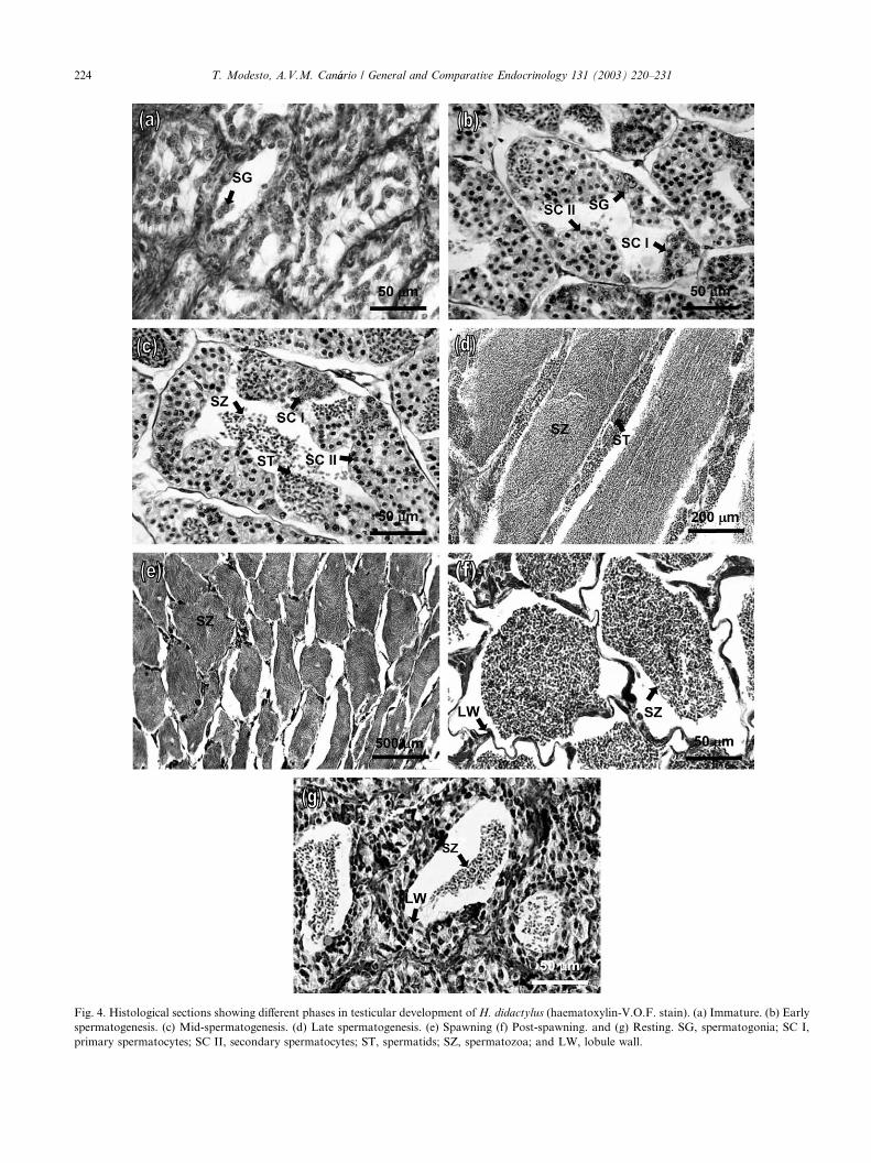
Fig. 4. Histological sections showing different phases in testicular development of H. didactylus (haematoxylin-V.O.F. stain). (a) Immature. (b) Early
spermatogenesis. (c) Mid-spermatogenesis. (d) Late spermatogenesis. (e) Spawning (f) Post-spawning. and (g) Resting. SG, spermatogonia; SC I,
primary spermatocytes; SC II, secondary spermatocytes; ST, spermatids; SZ, spermatozoa; and LW, lobule wall.
224 T. Modesto, A.V.M. Can�aario / General and Comparative Endocrinology 131 (2003) 220–231

extremity of an elongated papilla. The accessory glandswere well defined during testicular development and had
numerous septa forming chambers. During the repro-
ductive season these glands produced an abundant fluid,
which could be expressed through the genital pore by
gentle pressure applied to the surrounding area. In the
more developed glands this fluid was mostly dark, while
in the less developed glands the fluid was yellowish.
Chambers of the AR were elongated and their wallswere thicker when compared to the PR. Septa were
specially folded in the region adjacent to the PR. The
AR epithelium consists of columnar cells, which prob-
ably secretes a homogeneous fluid which can be visual-ised inside the chambers. In chambers with a large
amount of fluid the height of epithelial cells declined.
Various degrees of glandular activity, depending on the
amount of stored secretory material, could usually be
found in this part of the accessory glands. Positive re-
action to histochemical stains indicated the presence of
weakly sulphated mucosubstances and proteins inside
the chambers. No glycogen was detected (Figs. 5a andc). In the most apical zone of the AR, columnar cells
were taller and the peripheral end of the cells were
pinched. The coarse secretion of vesiclous non-homo-
Fig. 5. Histological appearance of accessory testicular organs. (a) Chambers of anterior region filled with secretion. Combined deamination-Alcian
blue, pH 2.5 – PAS. (b) Chambers of the posterior region. Mercury bromophenol blue stain. (c) Detail of anterior region. (d) Detail of posterior
region. (e) Anterior region during the non-reproductive period. (f) Posterior region during the non-reproductive period. Haematoxylin-V.O.F. stain.
EP, epithelium; SE, secretion; CT, connective tissue.
T. Modesto, A.V.M. Can�aario / General and Comparative Endocrinology 131 (2003) 220–231 225
geneous material that could be identified with brom-ophenol blue but reacted weakly to PAS or alcian blue
at pH 2.5 was probably produced by the epithelium.
The MR is histologically and histochemically similar
to AR but with larger chambers filled with fluid.
The dark colour of the PR of the accessory organs
was given by a pigment accumulated in rounded lobules.
Microscopically, the septa had cuboid cells. As cham-
bers became full of a dark extremely fine granular se-cretion, cells became progressively smaller and flat.
Epithelial cells reacted strongly to mercury bromophe-
nol blue and moderately to PAS and alcian blue at pH
2.5 (no reaction was detected at pH 1.0). Secretory
material in the chambers reacted weakly to PAS and
alcian blue at pH 2.5 and did not react with mercury
bromophenol blue. No positive reaction to glycogen was
detected in any structure (Figs. 5b and d). The IA fol-lowed a similar profile to that of the IG and reached their
maximum size in June (Fig. 2b) corresponding also to
the maximum enlargement of the chambers and thinning
of the septa. After the spawning season a reduction in
the total size of the accessory glands took place with loss
of fluid secretion and an increase of connective tissue in
the septa. In the AR, chambers became quite folded with
no visible secretion. In the PR, secretion of the darkfluid was minimal and chambers became round and
small (Figs. 5e and f). In January, the secretory material
reappeared in the chambers of both regions and fluid
could be expressed from the genital pore when the
glands were pressurised. From February to June, the
septa became progressively thinner, the chambers filled
with secretory material and fluid could be expressed.
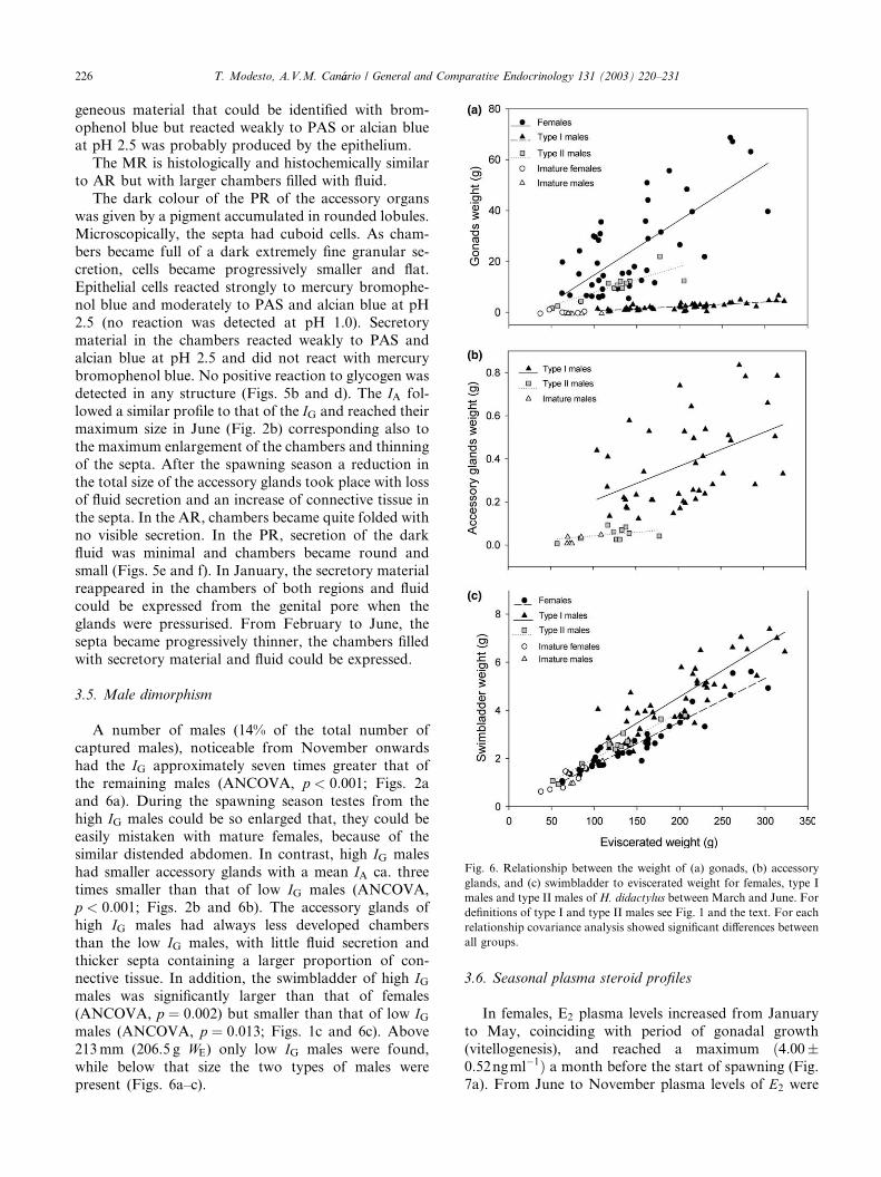
3.5. Male dimorphism
A number of males (14% of the total number of
captured males), noticeable from November onwards
had the IG approximately seven times greater that of
the remaining males (ANCOVA, p < 0:001; Figs. 2a
and 6a). During the spawning season testes from the
high IG males could be so enlarged that, they could beeasily mistaken with mature females, because of the
similar distended abdomen. In contrast, high IG males
had smaller accessory glands with a mean IA ca. three
times smaller than that of low IG males (ANCOVA,
p < 0:001; Figs. 2b and 6b). The accessory glands of
high IG males had always less developed chambers
than the low IG males, with little fluid secretion and
thicker septa containing a larger proportion of con-nective tissue. In addition, the swimbladder of high IGmales was significantly larger than that of females
(ANCOVA, p ¼ 0:002) but smaller than that of low IGmales (ANCOVA, p ¼ 0:013; Figs. 1c and 6c). Above
213mm (206.5 g WE) only low IG males were found,
while below that size the two types of males were
present (Figs. 6a–c).
3.6. Seasonal plasma steroid profiles
In females, E2 plasma levels increased from January
to May, coinciding with period of gonadal growth
(vitellogenesis), and reached a maximum ð4:00�0:52ngml�1Þ a month before the start of spawning (Fig.
7a). From June to November plasma levels of E2 were
Fig. 6. Relationship between the weight of (a) gonads, (b) accessory
glands, and (c) swimbladder to eviscerated weight for females, type I
males and type II males of H. didactylus between March and June. For
definitions of type I and type II males see Fig. 1 and the text. For each
relationship covariance analysis showed significant differences between
all groups.
226 T. Modesto, A.V.M. Can�aario / General and Comparative Endocrinology 131 (2003) 220–231
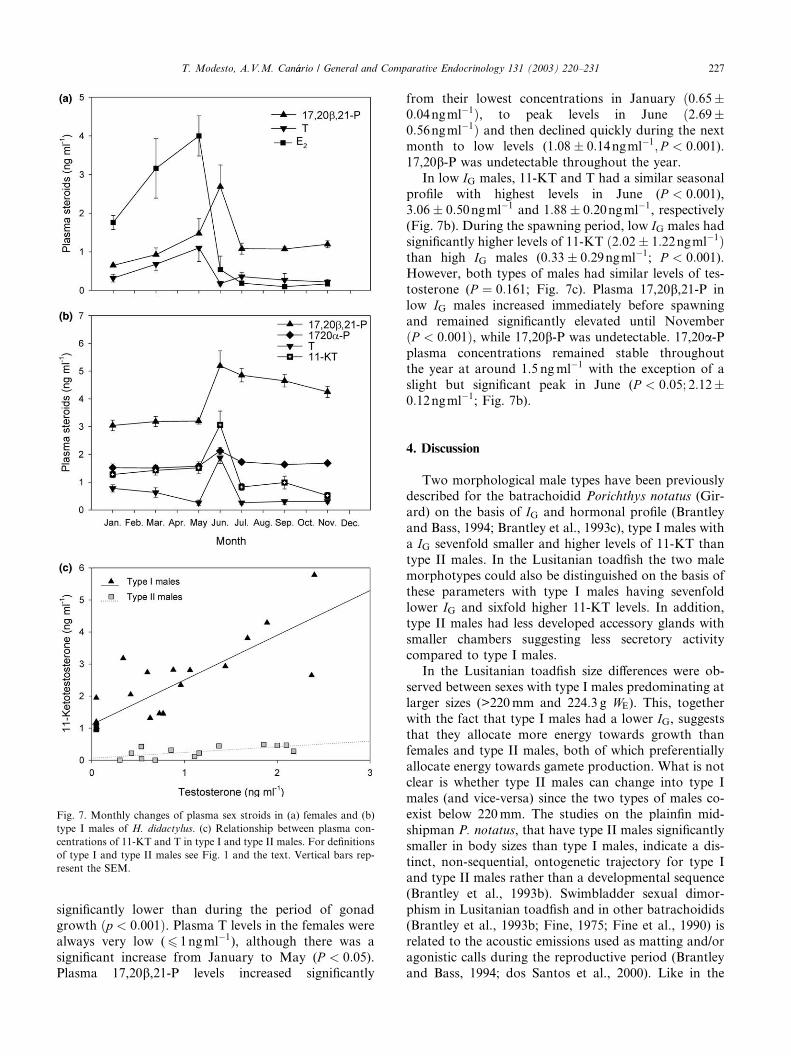
significantly lower than during the period of gonad
growth ðp < 0:001Þ. Plasma T levels in the females were
always very low (6 1ngml�1), although there was a
significant increase from January to May (P < 0:05).Plasma 17,20b,21-P levels increased significantly
from their lowest concentrations in January ð0:65�0:04ngml�1Þ, to peak levels in June ð2:69�0:56ngml�1Þ and then declined quickly during the next
month to low levels (1:08� 0:14ngml�1; P < 0:001).17,20b-P was undetectable throughout the year.
In low IG males, 11-KT and T had a similar seasonal
profile with highest levels in June (P < 0:001),3:06� 0:50ngml�1 and 1:88� 0:20ngml�1, respectively
(Fig. 7b). During the spawning period, low IG males hadsignificantly higher levels of 11-KT ð2:02� 1:22ngml�1Þthan high IG males (0:33� 0:29ngml�1; P < 0:001).However, both types of males had similar levels of tes-
tosterone (P ¼ 0:161; Fig. 7c). Plasma 17,20b,21-P in
low IG males increased immediately before spawning
and remained significantly elevated until November
ðP < 0:001Þ, while 17,20b-P was undetectable. 17,20a-Pplasma concentrations remained stable throughoutthe year at around 1.5 ngml�1 with the exception of a
slight but significant peak in June (P < 0:05; 2:12�0:12ngml�1; Fig. 7b).
4. Discussion
Two morphological male types have been previouslydescribed for the batrachoidid Porichthys notatus (Gir-
ard) on the basis of IG and hormonal profile (Brantley
and Bass, 1994; Brantley et al., 1993c), type I males with
a IG sevenfold smaller and higher levels of 11-KT than
type II males. In the Lusitanian toadfish the two male
morphotypes could also be distinguished on the basis of
these parameters with type I males having sevenfold
lower IG and sixfold higher 11-KT levels. In addition,type II males had less developed accessory glands with
smaller chambers suggesting less secretory activity
compared to type I males.
In the Lusitanian toadfish size differences were ob-
served between sexes with type I males predominating at
larger sizes (>220mm and 224.3 g WE). This, together
with the fact that type I males had a lower IG, suggeststhat they allocate more energy towards growth thanfemales and type II males, both of which preferentially
allocate energy towards gamete production. What is not
clear is whether type II males can change into type I
males (and vice-versa) since the two types of males co-
exist below 220mm. The studies on the plainfin mid-
shipman P. notatus, that have type II males significantly
smaller in body sizes than type I males, indicate a dis-
tinct, non-sequential, ontogenetic trajectory for type Iand type II males rather than a developmental sequence
(Brantley et al., 1993b). Swimbladder sexual dimor-
phism in Lusitanian toadfish and in other batrachoidids
(Brantley et al., 1993b; Fine, 1975; Fine et al., 1990) is
related to the acoustic emissions used as matting and/or
agonistic calls during the reproductive period (Brantley
and Bass, 1994; dos Santos et al., 2000). Like in the
Fig. 7. Monthly changes of plasma sex stroids in (a) females and (b)
type I males of H. didactylus. (c) Relationship between plasma con-
centrations of 11-KT and T in type I and type II males. For definitions
of type I and type II males see Fig. 1 and the text. Vertical bars rep-
resent the SEM.
T. Modesto, A.V.M. Can�aario / General and Comparative Endocrinology 131 (2003) 220–231 227
plainfin midshipman, the swimbladder (IS) is less devel-oped in type II males than in type I males. In P. notatus
the two male morphotypes maintain alternative repro-
ductive tactics. Type I males are territorial and use their
muscular swimbladders to make specific ‘‘humming’’
sounds to attract females into depositing eggs in nests
guarded by these males until the young become free
swimming. In contrast, type II males mimic females
behaviourally and morphologically and sneak or sa-tellite-spawn at the nests of type I males and only produce
short duration grunts that are similar in spectral, tem-
poral and amplitude characteristics to the grunts of fe-
males (Brantley and Bass, 1994). Altogether evidence
points towards territoriality and sound communication
having evolved together in the family. Whether the
swimbladder muscle organisation and microscopic ap-
pearance, such as area ofmitochondria-filled sarcoplasm,and myofiber number of the sonic muscles, which enable
the types of sound observed in P. notatus (Brantley et al.,
1993b), exist in the two types of Lusitanian toadfish
males still requires to be investigated. Also, although no
behavioural observations of the Lusitanian toadfish have
been made, the morphological evidence suggests that
alternative reproductive tactics may be present in Lusit-
anian toadfish. Like P. notatus, Lusitanian toadfish fe-males deposit eggs in nests, which are fertilised and
guarded by territorial males, possibly of type I, which
have smaller testis and heavier swimbladders. The larger
testis and lighter swimbladders in type II Lusitanian
toadfish are consistent with a male (sneaker/satellite)
which would emit opportunistically large amounts of
sperm to fertilise eggs from a distance without requiring
development of heavy sound producing muscular struc-tures to attract females.
The spawning season of Lusitanian toadfish in Ria
Formosa (June–July) lasts little more than a month.
From the group synchronous distribution pattern of the
oocytes and the short spawning period there is strong
indication that each female spawns only once. In Ria
Formosa spawning is also a month delayed compared to
the population in C�aadiz, southern Spain (Blanco, 1991;Palaz�oon-Fern�aandez et al., 2001; Rosety et al., 1992),
possibly related to the lower yearly average temperature
in Ria Formosa. The differences in spawning time be-
tween the two locations are also apparent in the IG and
IH, suggesting that alterations in the two indices are
related. This is also indicated by the fact that total lipids
and phospholipids in the ovary are correlated to IHduring ovarian growth (Blanco, 1991; Rosety et al.,1992; Mu~nnoz-Cueto et al., 1996). The liver is the organ
of synthesis of precursors of egg components (Wallace
et al., 1987) and its mass started to decline when vitel-
logenesis became more intense in April, with a minimum
in July at the end of spawning. In agreement with the
role of the liver in oogenesis, the IH in males, although
showing important monthly changes, was generally
smaller than in females and continued to grow untilJune when it underwent a marked reduction to its
minimum in July, at the end of spawning. The decrease
of IH in males is probably related to the metabolic needs
of territorial defence, partner calling and nest guarding
which lasts at least 30 days until fry become free living
(Modesto, unpublished observations). Both in C�aadizand in Ria Formosa males mature early than females;
this apparent asynchronism between sexes has also beenshown in other teleost species (e.g., Htun-Han, 1978).
The testicular organisation of the Lusitanian toadfish
is typical of most teleosts and corresponds to the ‘‘un-
restricted’’ or ‘‘lobular’’ type (Billard, 1986; Grier,
1981). Spermatogenesis occurs within cysts where germ
cells undergo synchronous differentiation. The cysts
break down at the end of spermatogenesis and sper-
matozoa are released into the lumen of lobules. Thetesticular accessory glands have many of the morpho-
logical and histological features of those of the toadfish
Opsanus tau (L.) and P. notatus (Barni et al., 2001;
Hoffman, 1963a,b). In all cases they are paired exten-
sions of the posterior portions of main testicular ducts
differentiated in well-defined portions, which are or-
ganised in chambers filled with secretory material.
However, the dark brown coloured anterior region ofthe accessory gland of O. tau (Hoffman, 1963a) corre-
sponds to the most posterior region of the accessory
gland of H. didactylus. Mature spermatozoa tend to
concentrate inside the cysts of the testes and do not need
to cross these secretory structures to reach the spermatic
duct. Prior to release, however, spermatozoa receive in
the final portion of the paired spermatic ducts the se-
cretion from the different regions of the accessoryglands. These accessory glands do not appear to have a
nutritional function for the sperm, since in H. didactylus
and in any of the other batrachoidids studied so far
there is no evidence for glycogen production (Barni et al.,
2001). Also, mixing sperm with the accessory organ se-
cretions does not change its mobility or behaviour
(Hoffman, 1963a). The AR of the accessory glands of
H. didactylus can produce mucosubstances and proteinswhile in the PR the production of these substances is not
so intense. The exocrine production of mucosubstances
by the accessory glands seems to be a common feature of
batrachoidids and of many other teleost species: some
blennies produce sialomucins (Lahnsteiner et al., 1990),
while gobies produce sialomucins and sulphomucins
(Cinquetti, 1997; Lahnsteiner et al., 1992). These mu-
cosubstances have been suggested to increase the vis-cosity of the seminal fluid and help to agglutinate the
sperm (Cinquetti, 1997; Lahnsteiner et al., 1990; Mann,
1964). Recently, Barni et al. (2001) suggested that the
mucosubstances released by nesting P. notatus type I
males, which have active large accessory glands, may
embed sperm and help produce sperm trails or layers
that slowly dilute in water reducing sperm dispersion
228 T. Modesto, A.V.M. Can�aario / General and Comparative Endocrinology 131 (2003) 220–231

and allowing the presence of sperm in water more con-stantly over time. This would increase the efficacy of
fertilisation of the sticky benthic eggs, and allow pa-
rental males to remain guarding nests. Type II males,
however, having less developed accessory glands (poor
in mucosubstances) and large testes, would need and are
adapted to release large quantities of free moving sperm
quickly outside the type I male nest entrance or sneaking
inside to try to reach the eggs inside the nest.E2 plasma concentrations increased with IG as ex-
pected, considering the group synchronous pattern of
oocyte development. However, E2 levels dropped
markedly a month before the peak IG, when T and
17,20b,21-P levels surged. This pattern of hormone
changes in females E2 ! T ! progestagen is common to
most teleosts and is the result of a GtH-mediated shift in
steroidogenesis, in which there is a progressive decreasein aromatase activity and concomitant activation of
20b-hydroxysteroid dehydrogenase (20b-HSD) to pro-
duce the maturation-inducing steroid (MIS, Kobayashi
et al., 1987; Nagahama et al., 1995; Scott et al., 1983). In
Lusitanian toadfish 17,20b,21-P, has been identified as
the probable MIS by in vitro studies (Modesto and
Canario, 2002) and that is supported in the current
study by the elevated 17,20b,21-P during spawning andthe simultaneous absence of 17,20b-P.
Circulating levels of androgens in H. didactylus type I
males underwent marked seasonal changes and peaked
during the spawning period. Similar observations have
been made for other species specially those that display
conspicuous reproductive behaviour: e.g., garibaldi,
Hypsypops rubinculus (Girard) (Sikkel, 1993), bluegill,
Lepomis macrochirus (Rafinesque) (Kindler et al., 1989),and demoiselle Chromis dispilus (Griffin) (Barnett and
Pankhurst, 1994). For many other teleosts (rainbow
trout, Salmo gairdneri (Richardson), Scott et al., 1980;
white sucker, Catostomus commersoni (Lacep�eede), Scottet al., 1984; Artic charr, Salvelinus alpinus (L.), Mayer
et al., 1992) 11-KT and T peak during the pre-spawning
period possibly related to the role of 11-KT in sper-
matogenesis (Miura et al., 1991a). Furthermore, type Imales had significantly higher plasma levels of 11-KT
than type II males. This seems to be a common pattern
of teleosts that have male dimorphism and alternative
reproductive tactics in which elevated 11-KT characte-
rise the territorial courting morph relative to its non-
courting counterpart (see Brantley et al., 1993c; Oliveira
et al., 2001a,b). For example, in Salaria pavo (Risso)
nest-holders had a higher 11-KT metabolisation indices(11-KT relative to total 11-KT plus T) than floaters both
in the testis and in the testicular gland suggesting that
nest holding promotes the conversion of T into 11-KT
(Oliveira et al., 2001a). The higher levels of 11-KT, but
not T, inH. didactylus type I males suggests also that 11-
KT must be the androgen responsible for the increase in
size of sonic muscle. Swimbladder muscle hypertrophy
during the matting season has been identified in threesound-generating species, the haddock Melanogrammus
aeglefinus (L.) (Templemen and Hodder, 1958), the
midshipman Porichthys sp (Mommsen & Nickolichuck
in Walsh et al., 1995) and the weakfish Cynoscion regalis
(Bloch and Schneider) (Connaughton et al., 1997;
Connaughton and Taylor, 1995b). In C. regalis T im-
plants induced sonic muscle growth but 11-KT was not
tested (Connaughton and Taylor, 1995a). In P. notatusand in toadfish O. tau implants of both T and 11-KT
androgens were effective in inducing sonic muscle
growth (Brantley et al., 1993a; Fine and Pennypacker,
1986). Both studies are consistent with a role for 11-KT
in the process of sonic muscle growth.
Accessory gland development also appears to be de-
pendent on 11-KT, both its growth and secretory ac-
tivity. The fact that 11-KT levels in type II males wererelatively low, even though their relative testes size was
larger than that of type I males suggests however that
spermatogenesis may not require very high levels of that
androgen. Whether the extraordinary growth of the
testis has per se any direct influence on accessory gland
growth is not known. However, it can be hypothesised
that the lower 11-KT levels in type II males may release
a feedback to the hypophysis to increase the secretion ofa hormone (possibly prolactin, Bartke et al., 1975) re-
sponsible for stimulating testis hypertrophy.
Therefore, it would appear most likely that elevated
serum levels of 11-KT in type I males could be asso-
ciated with the development of reproductive behaviour
and related swimbladder growth and mate calls, female
courtship, development of accessory testicular organs
for controlled release of sperm, while similarities be-tween the plasma levels of testosterone among the type
I and type II males seem to indicate that this hormone
may be associated with an aspect common to both
types of males, such as gonadal development and/or
spermiation.
In males, 17,20b,21-P but not 17,20b-P or 17,20a-P,was elevated during the spawning and post-spawning
period. While until now 17,20b-P has been shown to beinvolved in sperm function in teleosts (Baynes and Scott,
1985; Miura et al., 1991b, 1992), for those species in
which this hormone is lacking a similar role may be
taken by 17,20b,21-P (Thomas et al., 1997) or 17,20a-P(Vermeirssen et al., 2000). However, in Lusitanian
toadfish 17,20b,21-P stays elevated for a long period and
follows the pattern of swimbladder hypertrophy. Whe-
ther it has any significance in this process requires fur-ther studies.
In conclusion, two male morphotypes (type I and
type II) could be distinguished in Lusitanian toadfish on
the basis of differences in relative size of testis, testicular
accessory glands and swimbladder as well as androgen
levels. Although confirmation is required, it is probable
that type II males correspond to a sneaker/satellite
T. Modesto, A.V.M. Can�aario / General and Comparative Endocrinology 131 (2003) 220–231 229
morph with alternative reproductive tactics. Thus, thisspecies which combines alternative reproductive tactics,
parental care and a diversified repertoire of mating vo-
calisations can be a useful model in the investigation of
endocrine mechanisms that control reproductive be-
haviour and in the understanding of the evolution of
reproductive systems within teleosts.
Acknowledgments
This work was supported by Junta Nacional de In-
vestigac�~aao Cient�ııfica e Tecnol�oogica project STDRB/C/MAR/231/92. TM benefited from the Programme for
Educational Development in Portugal (PRODEP 5.2).
References
Barnett, C.W., Pankhurst, N.W., 1994. Changes in plasma levels of
gonadal steroids and gonad morphology during the spawning cycle
of male and female demoiselles Chromis dispilus (Pisces: Pomacen-
tridae). Gen. Comp. Endocrinol. 93, 260–274.
Barni, A., Mazzoldi, C., Rasotto, M.B., 2001. Reproductive apparatus
and male accessory structures in two batrachoid species (Teleostei,
Batrachoididae). J. Fish Biol. 58, 1557–1569.
Bartke, A., Croft, B.T., Dalterio, S., 1975. Prolactin restores plasma
testosterone levels and stimulates testicular growth in hamsters
exposed to short day-length. Endocrinology 97, 1601–1604.
Baynes, S.M., Scott, A.P., 1985. Seasonal variations in parameters of
milt production and in plasma concentration of sex steroids of male
rainbow trout (Salmo gairdneri). Gen. Comp. Endocrinol. 57, 150–
160.
Billard, R., 1986. Spermatogenesis and spermatology of some teleost
fish species. Reprod. Nutr. Dev. 26, 877–920.
Blanco, M.A., 1991. Estudio histol�oogico, histoqu�ıımico y bioqu�ıımico
durante la reproducci�oon del pez sapo, Halobatrachus didactylus
(Schneider, 1801) de la Bah�ııa de C�aadiz Ph.D. Thesis, University of
C�aadiz, C�aadiz, Spain.
Borg, B., 1994. Androgens in teleost fishes. Comp. Biochem. Physiol.
109C, 219–245.
Brantley, R.K., Bass, A.H., 1994. Alternative male spawning tactics
and acoustic signals in the plainfin midshipman fish Porichthys
notatus Girard (Teleostei, Batrachoididae). Ethology 96, 213–232.
Brantley, R.K., Marchaterre, M.A., Bass, A.H., 1993a. Androgen
effects on vocal muscle structure in a teleost fish with inter- and
intra-sexual dimorphism. J. Morphol. 216, 305–318.
Brantley, R.K., Tseng, J., Bass, A.H., 1993b. The ontogeny of inter-
and intrasexual vocal muscle dimorphisms in a sound-producing
fish. Brain Behav. Evol. 42, 336–349.
Brantley, R.K., Wingfield, J.C., Bass, A.H., 1993c. Sex steroid levels in
Porichthys notatus, a fish with alternative reproductive tactics, and
a review of the hormonal bases for male dimorphism among teleost
fishes. Horm. Behav. 27, 332–347.
Canario, A.V.M., Scott, A.P., Flint, A.P.F., 1989. Radioimmunoassay
investigations of 20b-hydroxylated steroids in maturing/ovulating
female rainbow trout (Salmo gairdneri). Gen. Comp. Endocrinol.
74, 77–84.
Cinquetti, R., 1997. Histochemical, enzyme histochemical and ultra-
structural investigation on the sperm-duct glands of Padogobius
martensi (Pisces, Gobiidae). J. Fish Biol. 50, 978–991.
Connaughton, M.A., Fine, M.L., Taylor, M.H., 1997. The effects of
seasonal hypertrophy and atrophy on fiber morphology, metabolic
substrate concentration and sound characteristics of the weakfish
sonic muscle. J. Exp. Biol. 200, 2449–2457.
Connaughton, M.A., Taylor, M.H., 1995a. Effects of exogenous
testosterone on sonic muscle mass in the weakfish, Cynoscion
regalis. Gen. Comp. Endocrinol. 100, 238–245.
Connaughton, M.A., Taylor, M.H., 1995b. Seasonal and daily cycles
in sound production associated with spawning in the weakfish,
Cynoscion regalis. Env. Biol. Fish. 42, 233–240.
Cook, H.C., 1990. Carbohydrates. In: Bancroft, J.D., Stevens, A.,
Turner, D.R. (Eds.), Theory and Practice of Histological Tech-
niques, third ed.. Churschill Livingstone, New York, pp. 177–213.
dos Santos, M.E., Modesto, T., Matos, R.J., Grober, M.S., Oliveira,
R.F., Canario, A.V.M., 2000. Sound production by the Lusitanian
toadfish, Halobatrachus didactylus. Bioacoustics 10, 309–321.
Fine, M.L., 1975. Sexual dimorphism of the growth rate of the
swimbladder of the toadfish Opsanus tau. Copeia, 438–490.
Fine, M.L., Burns, N.M., Harris, T.M., 1990. Ontogeny and sexual
dimorphism of sonic muscle in the oyster toadfish. Can. J. Zool. 68,
1374–1381.
Fine, M.L., Pennypacker, K.R., 1986. Hormonal basis for sexual
dimorphism of the sound-producing apparatus of the oyster
toadfish. Exp. Neurol. 92, 289–298.
Gonz�aalez de Canales, M.L., Blanco, M., Sarasquete, M.C., 1992.
Carbohydrate and protein histochemistry during oogenesis in
Halobatrachus didactylus (Schneider, 1801) from the Bay of C�aadiz
(Spain). Histochem. J. 24, 337–344.
Grier, H.J., 1981. Cellular organization of the testis and spermato-
genesis in fishes. Am. Zool. 21, 345–357.
Guti�eerrez, M., 1967. Coloraci�oon histol�oogica para ovarios de peces,
crust�aaceos y moluscos. Invest. Pesq. 3, 265–271.
Hoffman, R.A., 1963a. Acessory glands and their ducts in the
reproductive system of the male toadfish, Opsanus tau. Chesapeake
Sci. 4, 30–37.
Hoffman, R.A., 1963b. Gonads, spermatic ducts, and spermatogenesis
in the reproductive system of male toadfish, Opsanus tau. Chesa-
peake Sci. 4, 21–29.
Htun-Han, M., 1978. The reproductive biology of the dab Limanda
limanda (L.) in the North Sea: gonosomatic index, hepatosomatic
index and condition factor. J. Fish Biol. 13, 369–378.
Kime, D.E., Manning, N.J., 1982. Seasonal patterns of free and
conjugated androgens in the brown trout Salmo trutta. Gen. Comp.
Endocrinol. 48, 222–231.
Kindler, P.M., Philipp, D.P., Gross, M.R., Bahr, J.M., 1989. Serum
11-ketotestosterone and testosterone concentrations associated
with reproduction in male bluegill (Lepomis macrochirus, Centrar-
chidae). Gen. Comp. Endocrinol. 75, 446–453.
Kobayashi, M., Aida, K., Hanyu, I., 1987. Hormone changes during
ovulation and effects of steroid hormones on plasma gonadotropin
levels and ovulation in goldfish. Gen. Comp. Endocrinol. 67, 24–
32.
Lagler, K.F., Bardach, J.E., Miller, R.R., Passino, D.R.M., 1977.
Ichthyology, second ed. Wiley, New York.
Lahnsteiner, F., Richtarski, U., Patzner, R.A., 1990. Functions of the
testicular gland in two blenniid fishes, Salaria (¼Blennius) pavoand Lipophrys (¼Blennius) dalmatinus (Blennidae, Teleostei) as
revealed by electron microscopy and enzyme histochemistry. J.
Fish Biol. 37, 85–97.
Lahnsteiner, F., Seiwald, M., Patzner, R.A., Ferrero, E.A., 1992. The
seminal vesicles of the male grass goby. Fine structure and
histochemistry, Zosterisessor ophiocephalus (Teleostei, Gobiidae).
Zoomorphology 111, 239–248.
Maigret, J., Ly, B., 1986. �Les poissons de mer Mauritanie. Science
Naturel, Compi�eegne.
Mann, T., 1964. The Biochemistry of Semen and of the Male
Reproductive Tract. Methuen, London.
Mayer, I., Schmitz, M., Borg, B., Schulz, R., 1992. Seasonal endocrine
changes in male and female Artic charr (Salvelinus alpinus). I.
230 T. Modesto, A.V.M. Can�aario / General and Comparative Endocrinology 131 (2003) 220–231
Plasma levels of three androgens, 17a-hydroxy-20b-dihydropro-gesterone, and 17b-estradiol. Can. J. Zool. 70, 37–42.
Miura, T., Yamauchi, K., Takahashi, H., Nagahama, Y., 1991a.
Hormonal induction of all stages of spermatogenesis in vitro in the
male Japanese eel (Anguilla japonica). Proc. Natl. Acad. Sci. USA
88, 5774–5778.
Miura, T., Yamauchi, K., Takahashi, H., Nagahama, Y., 1991b.
Involvement of steroid-hormones in gonadotropin-induced testic-
ular maturation in male Japanese eel (Anguilla japonica). Biomed.
Res. 12, 241–248.
Miura, T., Yamauchi, K., Takahashi, H., Nagahama, Y., 1992. The
role of hormones in the acquisition of sperm motility in salmonid
fish. J. Exp. Zool. 261, 359–363.
Modesto, T., Canario, A.V.M., 2002. 17a,20b,21-Trihydroxy-4-preg-nen-3-one: the probable maturation-inducing steroid of the Lusi-
tanean toadfish. J. Fish Biol. 60, 637–648.
Moore, R.K., Scott, A.P., Collins, P.M., 2000. Circulating C-21
steroids in relation to reproductive condition of a viviparous
marine teleost, Sebastes rastrelliger (grass rockfish). Gen. Comp.
Endocrinol. 117, 268–280.
Mu~nnoz-Cueto, J.A., Alvarez, M., Blanco, M., Gonzalez de Canales,
M.L., Garcia-Garcia, A., Sarasquete, C., 1996. Histochemical and
biochemical study of lipids during the reproductive cycle of the
toadfish, Halobatrachus didactylus (Schneider, 1801). Sci. Mar. 60,
289–296.
Nagahama, Y., 1994. Endocrine regulation of gametogenesis in fish.
Int. J. Dev. Biol. 38, 217–229.
Nagahama, Y., Yoshikuni, M., Yamashita, M., Tokumoto, T., Katsu,
Y., 1995. Regulation of oocyte growth and maturation in fish.
Curr. Top. Dev. Biol. 30, 103–145.
Oliveira, R.F., Almada, V.C., Goncalves, E.J., Forsgren, E., Canario,
A.V.M., 2001a. Androgen levels and social interactions in breeding
males of the peacock blenny. J. Fish Biol. 58, 897–908.
Oliveira, R.F., Canario, A.V.M., Grober, M.S., Santos, R.S., 2001b.
Endocrine correlates of male polymorphism and alternative repro-
ductive tactics in the Azorean rock-pool blenny, Parablennius
sanguinolentus parvicornis. Gen. Comp. Endocrinol. 121, 278–288.
Palaz�oon-Fern�aandez, J.L., Arias, A.M., Sarasquete, C., 2001. Aspects of
the reproductive biology of the toadfish, Halobatrachus didactylus
(Schneider, 1801) (Pisces: Batrachoididae). Sci. Mar. 65, 131–138.
Pearse, A.G.E., 1985. Histochemistry, theoretical and applied, analyt-
ical technology. Churschill Livingstone, Edinburgh.
Rosety, M., Blanco, M., Gonz�aalez de Canales, M.L., Grau, A.,
Sarasquete, M.C., 1992. Histochemical parameters during repro-
duction of the toadfish,Halobatrachus didactylus (Schneider, 1801).
Sci. Mar. 56, 87–94.
Roux, C., 1986. Batrachoididae. In: Whitehead, P.J.P., Bauchot, M.-
L., Hureau, J.C., Nielsen, J., Tortonese, E. (Eds.), Fishes of the
North-Easten Atlantic and the Mediterranean, vol. 3. UNESCO,
Paris, pp. 1360–1361.
Scott, A.P., Baynes, S.M., Springate, J.R., 1980. Seasonal variation in
plasma concentrations of 11-ketotestosterone and testosterone in
male rainbow trout (Salmo gairdineri) Richardson. J. Fish Biol. 17,
495–506.
Scott, A.P., MacKenzie, D.S., Stacey, N.E., 1984. Endocrine changes
during natural spawning in the white sucker, Catostomus commer-
soni. II. Steroid hormones. Gen. Comp. Endocrinol. 56, 349–359.
Scott, A.P., Sumpter, J.P., Hardiman, P.A., 1983. Hormone changes
during ovulation in the rainbow trout (Salmo gairdneri Richard-
son). Gen. Comp. Endocrinol. 49, 128–134.
Sikkel, P.C., 1993. Changes in plasma androgen levels associated with
changes in male reproductive behavior in a brood cycling marine
fish. Gen. Comp. Endocrinol. 89, 229–237.
Templemen, W., Hodder, V.M., 1958. Variations with fish length, sex,
stage of sexual maturity and season in the appearance and volume
of the drumming muscles of the swim-bladder in the haddock
Melanogrammus aeglefinus (L.). J. Fish. Res. Bd. Can. 15, 355–
390.
Thomas, P., Breckenridge-Miller, D., Detweiler, C., 1997. Binding
characteristics and regulation of the 17,20b,21-trihydroxy-4-preg-nen-3-one (20b-S) receptor on testicular and sperm plasma
membranes of spotted sea trout (Cynoscion nebulosus). Fish
Physiol. Biochem. 17, 109–116.
Vermeirssen, E.L.M., Shields, R.J., de Quero, C.M., Scott, A.P., 2000.
Gonadotrophin-releasing hormone agonist raises plasma concen-
trations of progestogens and enhances milt fluidity in male Atlantic
halibut (Hippoglossus hippoglossus). Fish Physiol. Biochem. 22, 77–
87.
Wallace, R.A., Selman, K., Greeley, M.S., Begovac, P.C., Lin, Y.W.P.,
McPherson, R., Petrino, T.R., 1987. Current status of oocyte
growth. In: Idler, D.R., Crim, L.W., Walsh, J.M. (Eds.), Proceed-
ings of the Third International Symposium on the Reproductive
Physiology of Fish. Memorial University of Newfoundland, 167-
177.
Walsh, P.J., Mommsen, T.P., Bass, A.H., 1995. Biochemical and
molecular aspects of singing in batrachoidid fishes. In: Hochachka,
P.W., Mommsen, T.P. (Eds.), Metabolic Biochemistry. Elsevier,
Amsterdam, pp. 279–289.
T. Modesto, A.V.M. Can�aario / General and Comparative Endocrinology 131 (2003) 220–231 231


![650W 180mm Tile cutter manual[EN] - Free …Original Instructions_MNL _ MTC650_GB_V01_180102 MTC650 650W Tile Cutter EAN: 3663602797913 WARNING! Read the instructions before …](https://static.fdocuments.in/doc/165x107/5ee27ebaad6a402d666ced7e/650w-180mm-tile-cutter-manualen-free-original-instructionsmnl-mtc650gbv01180102.jpg)


