Morphological, anatomical and physiological leaf - Botanical Studies

Available online at www.pelagiaresearchlibrary.com
Pelagia Research Library
Asian Journal of Plant Science and Research, 2016, 6(4):69-80
ISSN : 2249-7412
CODEN (USA): AJPSKY
69 Pelagia Research Library
Morphological, Epidermal and Anatomical Properties of Datura Linn. Leaf in Sana'a City-Yemen and its Taxonomical Significance
Hassan M. Ibrahim¹*, Nasser A. Abdo1, Esraa S. Al Masaudi1 and Abdul Nasser A. Al-Gifri²
1Department of Biology, Faculty of Science, Sana’a University, Yemen
2Department of Biology Faculty of Education, University of Aden, Yemen _____________________________________________________________________________________________
ABSTRACT The morphological, epidermal and anatomical characters of two wild Datura taxa leaves grown in Sana’a city were investigated. Morphologically; the shape, apex, margin, base, texture, size and venation of lamina were studied. The epidermal characters including the properties of epidermal cells (shape, size & frequency), stomata properties (type of stomata complex, size, frequency, index and ratio) and type of trichomes were determined. Anatomically the characters of mesophyll and main midrib were investigated. The lamina morphological characters of the two studied Datura taxa leaves (shape, base, apex, margin, texture and type of venation) as well as epidermal characters including the properties of epidermal cells (length, size and frequency), stomata properties (size and frequency) & type of trichomes and the anatomical characters ( thickness of Mesophyll and number of abaxial collenchyma layers in the midrib) shows a high significant in taxonomic value for the separation between the two studied Datura taxa . Key words: Datura, Lamina, Stomata, Trichome, Mesophyll. _____________________________________________________________________________________________
INTRODUCTION
The genus Datura Linn. comprises about 10 species distributed mainly in tropical and warm temperate regions, especially tropical American and Australia [1], only 3 species are known from Yemen, Datura metel, D. innoxia & D. stramonium [2] and only 2 species of Datura taxa so far recorded from Sana'a governorate, D. innoxia & D. stramonium [3 , 4] Few studies have been done on the morphological [1, 5, 6, 7], epidermal [8, 9] and anatomical [8] features of Datura taxa leaves. However, no attempts seem to have been made to evaluate the taxonomical significance of those features. Therefore, the present study aims to investigate the morphological and anatomical features as well as epidermal features of Datura taxa leaves in Sana'a city and to evaluate their significance as key characters for differentiation.
MATERIALS AND METHODS
Fresh samples of Datura taxa were collected from different localities in Sana'a city during the May 2015 to August 2015 (Table 1) and identified according to Chaudhary [1], Wood [7] & Collenette [10] and compared with samples from the Herbarium of the Faculty of Science Sana'a University. For leaf morphology at least five to seven matured and well expanded leaves were investigated to record the leaf architecture characters of each species which were based on the terminology of (Approaches to identification of Angiosperm leaf remain) Dilcher [11]. The characters described were leaf petiole and lamina features (size, shape, apex, base, margin, texture & venation). To study the epidermal characters fresh matured and well expanded leaves of the Datura taxa were cut at the median portion, the specimens were soaked in concentrated Nitric acid for 2 to10 hrs depending on the texture of the

Hassan M. Ibrahim et al Asian J. Plant Sci. Res., 2016, 6(4):69-80 _____________________________________________________________________________
70 Pelagia Research Library
leave. The appearance of the air bubbles indicated the readiness of epidermises to be separated. The samples were then transferred to Petri dish containing water and with the use of fine forceps and dissecting needle the upper (Adaixal) and lower (Abaxial) epidermis were separated. These were then cleaned with camel hair brush in water [12]. The two epidermal layers (Adaxial & Abaxial) were stripped and stained with Saffranin, Excess stain was rinsed off with clean water and mounted in glycerol on clean slides then covers by cover slide [13]. The slides were observed by using Leica (ATC 2000) microscope to determine lamina epidermal (adaxial & abaxial) characters of each species which were based on the terminology of Dilcher [11]. The characters determined were stomata complex features (stomata type, size & frequency); epidermal cell features (shape, size & frequency) and Trichomes features (Type & frequency). Photographs of lamina epidermis (Adaxial & Abaxial) characters were taken by Canon (IXUS255 HS) digital camera. The stomata frequency, epidermis cell frequency and trichome frequency were based on average obtained from observation of 10 microscope field with an area 625µm2 at x 200, the stomata index (SI) was calculated using the formula of Salisbury [14]; SI= [S \ (S+E)] x 100, where S= No. of stomata in an area of 625µm2 & E= No. of epidermal cells in an area of 625µm2. The stomata ratio (SR) was helpful in defining the type of leaf. It is the ratio of the number of stomata on the abaxial epidermis to the number of stomata on adaxial epidermis, if SR>1 the leaves are classified as amphistomatic, if 0.1<SR<1 as hypoamphistomatic and if SR<0.1 as hypostomatic [15]. The stomata size (length x width); epidermal cell size (length x width) and guard cells area (length x width x Franc'os constant which is 0.78525) were based on average obtained from observation of 40 individual, by the help of ocular micrometer calibrated with stage micrometer (value of 400x 1ocular small division = 0.25 µm) and Image j program. For the anatomical studies leaf lamina were cut to small samples each sample were fixed in formalin acetic acid-alcohol solution for two days. After removing the fricative by distilled water, they were dehydrated with ethyl alcohol solution of 30%, 50%, 60%, 70%, 85%, 90%, and 100% before being embedded into paraffin and sectioned by using a rotary microtome. The sections were stained in a Saffranin O/Fast Green combination [16]. The anatomical characters (structure of the lamina, lamina epidermis and midrib) were examined by using Leica (ATC 2000) microscope and by utilizing the available anatomical literatures of Fahn [17] and Esau [18]. Photographs of the leaf sections were taken by Canon (IXUS255 HS) digital camera. The thickness of the Mesophyll was measured by the help of ocular micrometer calibrated with stage micrometer (value of 400x 1ocular small division = 0.25 µm) and Image j program, in addition to that the thickness of the Midrib was measured by the help of ocular micrometer calibrated with stage micrometer (value of 100 x 1ocular small division = 1 µm) and Image j program.
Table 1: Locality and Date of Collection of the two investigated Datura taxa
Date Location Coordinates
Elevation Taxa Longitude Latitude
May
1 44°11'16.3"E 15°21'49.7"N 2271m asl. Datura innoxia
2015 & Datura stramonium
June 2 44°11'41.5"E 15°21'16.8"N 2266m asl. Datura stramonium 2015 3 44°11'13.9"E 15°22'07.8"N 2269m asl. Datura innoxia July
4 44°11'42.8"E 15°20'38.2"N 2275m asl. Datura innoxia
2015 & Datura stramonium
August 5 44°11'20.3"E 15°21'55.1"N 2313m asl.
Datura innoxia &
2015 Datura stramonium
6 44°12'23.3"E 15°20'1.44"N 2280m asl. Datura stramonium
The taxonomical value of quantitative leaf morphological, epidermal and anatomical features were determined by T. test using Graph Pad Prism 6.01 program, if P- value P < 0.05 then the quantitative leaf features is significantly different.
RESULTS AND DISCUSSION
Morphological Analysis: Table 2 &3 and Figure 1 demonstrate the main morphological properties of the studied Datura taxa leaves The leaf architecture shows kind of difference aspects.

Hassan M. Ibrahim et al Asian J. Plant Sci. Res., 2016, 6(4):69-80 _____________________________________________________________________________
71 Pelagia Research Library
Lamina is, ovate – lanceolate, asymmetrical, coriaceous, grey green, pubescent, with a Campotodromous (Brochidodromous) venation ( secondary veins joined together in a series of prominent arches, never terminating at the margin), up to 15.7 x 9.6cm, acute at apex, repeand to sinuate at margin, oblique, asymmetrical at base in Datura innoxia, while it is ovate, asymmetrical, chartaceous, yellowish green, glabrescent, with a Craspedodromous venation (secondary veins terminating at the margin) , up to 16.9 x 11.7 cm, acute to acumenate at apex, coarsely dentate to lobed at margin, oblique, asymmetrical at base in Datura stramonium, leaves of Datura innoxia and Datura stramonium are petiolate leaves with a normal petiole. Identification key to the studied Datura taxa leaves based on their morphological characters: + Lamina ovate – lanceolate, coriaceous, Grey green, pubescent, Campotodromous venation, repand to sinuate at margin.......................................... Datura innoxia. - Lamina ovate, chartaceous, Yellowish green, glabrescent, Craspedodromous venation, coarsely dentate to lobed at margin............................................ Datura stramonium.
Epidermal Analysis: Table 2 & 3 and Figure 2 shows the main epidermal characters of the studied Datura taxa leaves as clarified by light microscope. Epidermis cell shape is rectangular with undulate cell wall in Datura innoxia and D. stramonium (Figure 2). The mean of epidermal cells size is largest in the adaxial surface of D. stramonium (22.9 µm2) and the smallest mean of epidermal cell size (11µm2) was recorded in the abaxial surface of D. innoxia furthermore, the highest mean of epidermal cells density was found in the abaxial surface of D. innoxia (335 epidermal cell /625 µm2) followed by 175 epidermal cell / 625 µm2 on the adaxial surface of D. innoxia; while the lowest mean of epidermal cells density (93 epidermal cell / 625 µm2) was recorded on the adaxial surface of D. stramonium (Table 3). Amonotetracytic (four cells enclosing guard cell in an irregular and variable pattern) and anisocytic (single ring of 3 cells -2 larger & 1 smaller- enclosing the guard cells) stomata complex types were occurred in the adaxial and abaxial surface of D. innoxia and D. stramonium (Table 2 & Figure 2) and this agrees with what Solereder [8] and Hameed & Hussain [9] recorded.

Hassan M. Ibrahim et al Asian J. Plant Sci. Res., 2016, 6(4):69-80 _____________________________________________________________________________
72 Pelagia Research Library
A-C: Datura innoxia leaf, A: Adaxial surface, B: Abaxial surface & C: Campotodromous (Brochidodromous) venation; D-F: Datura stramonium leaf: D: Adaxial surface, E: Abaxial
surface & F: Craspedodromous venation.
2.6 cm
A B C
3.6 cm
D E F
Figure 1: General Morphological Characteristic of Datura taxa Studied Leaf.

Hassan M. Ibrahim et al Asian J. Plant Sci. Res., 2016, 6(4):69-80 _____________________________________________________________________________
73 Pelagia Research Library
The largest mean of stomata size was in the abaxial surface of D. stramonium (4.5µm2) and the smallest mean of stomata size (2.7 µm2) was recorded in the abaxial surface of D. innoxia (Table 3) on the other hand the largest mean of guard cell area was found in the adaxial surface of D. stramonium (1.6µm2) and the smallest mean of guard cell area (0.9µm2) was recorded in the adaxial and abaxial surface of D. innoxia (Table 3) All the observed stomata are small size (Table 3) this is because, stomata whose guard cells less than 15 µm long are designated -small while those are more than 38 µm long are termed large [19]. The Stomata ratio (SR) of D. innoxia and D. stramonium were 0.54 and 0.73 (0.1<SR<1) respectively this shows that leaves of the studied Datura taxa are hypoamphistomatic (leaves that have stomata on both surface, with more on the abaxial surface than the adaxial surface)
The highest mean of stomata density was observed on the abaxial surface of D. innoxia (102 stomata complex /625 µm2) whose mean of stomata index was 23.2 followed by 55 stomata complex /625 µm2 on the adaxial surface of D. innoxia whose mean of stomata index was 23.9; while the lowest mean of stomata density (30 stomata complex /625 µm2) was recorded on the adaxial surface of D. stramonium whose stomata index was 24 (Table 3) Two main types of trichomes were identified in the two Datura taxa studied leaves: glandular trichomes and non-glandular trichomes. According to observation about 7 and 3 morphological categories of glandular trichomes were recorded in D. innoxia and D. stramonium correspondingly. Furthermore; in D. innoxia 7 and 5 morphological categories of glandular trichomes were recorded on the adaxial and abaxial surface respectively; while in D. stramonium 1 and 3morphological categories of glandular trichomes were observed, the former on adaxial surface and the latter on abaxial surface (Table 2 & Figure 3). On the other hand, the non-glandular trichomes were classified according to their surface in two types: smooth non-glandular trichomes and rough non-glandular trichomes, the former occurs in D. innoxia and the latter occurs in D. stramonium. Furthermore; the smooth non-glandular trichomes and rough non - glandular trichomes are subdivided structurally in to 4 and 6 morphological categories correspondingly (Table 2 & Figure 3).
A-B: Datura innoxia leaf, A: Adaxial surface, B: Abaxial surface; C-D: D stramonium Leaf, C: Adaxial surface, D: Abaxial surface; Am: Amonotetracytic stomata, An: Anisocytic
stomata
A B
C D
Am
Am
Am
Am
Am
Am
Am
An
An
An
An
An
An
An
An
An
An
An
An
Am
2.8µm 1.7µm
2.2µm 3.4µm
Figure 2: Types of Stomata in the Adaxial and Abaxial surface of Datura taxa leaves

Hassan M. Ibrahim et al Asian J. Plant Sci. Res., 2016, 6(4):69-80 _____________________________________________________________________________
74 Pelagia Research Library
In addition to that; the 4 types of smooth non-glandular trichomes were only observed on the adaxial surface of D. innoxia leaf; while 4 types of rough non - glandular trichomes were recorded on the adaxial & abaxial surface of D. stramonium leaf respectively (Table 2). Generally, the trichomes accrues on both surface of studied Datura taxa leaves with more density on the adaxial surface than the abaxial surface (Table 3), the highest mean of trichome density was recorded on the adaxial surface of D. innoxia (16 trichome/625 µm2) followed by 8 trichome/625 µm2 on the abaxial surface of D. innoxia, while the lowest mean of trichome density (2 trichome /625 µm2) was observed on the abaxial surface of D. stramonium (Table 3). Identification key to the studied Datura taxa leaves based on their epidermal characters: + Presence of unicellular head with tricellular uniseriate stalk and unicellular head with tetracellular uniseriate stalk on the adaxial and abaxial surface of the leaf, presence of smooth non-glandular trichomes on the adaxial surface of the leaf.................................................... Datura innoxia. - Absence of unicellular head with tricellular uniseriate stalk and unicellular head with tetracellular uniseriate stalk from the adaxial and abaxial surface of the leaf, presence rough non - glandular trichomes on the adaxial and abaxial surface of the leaf................................ Datura stramonium.

Hassan M. Ibrahim et al Asian J. Plant Sci. Res., 2016, 6(4):69-80 _____________________________________________________________________________
75 Pelagia Research Library
TaTable 2: Qualitative Morphological & Epidermal Characteristics of Datura taxa studied leaves.
Characters Datura innoxia Datura stramonium
Mo
rph
olog
y C
hara
cte
ristic
s
Lam
ina
Shape Ovate – Lanceolate Ovate Texture Coriaceous Chartaceous Colour Grey green Yellowish green Surface Pubescent Glabrescent Venation Campotodromous Craspedodromous Apex Acute Acute to Acumenate
Margin Repeand to Sinuate Coarsely dentate to Lobed
Base Oblique- asymmetrical Oblique- asymmetrical
Petiole Normal petiole Normal petiole
Ep
ide
rma
l Ch
ara
cte
ristic
s
Type of stomata Ad
Amonotetracytic & Anisocytic
Amonotetracytic & Anisocytic
Ab Amonotetracytic & Anisocytic
Amonotetracytic & Anisocytic
Typ
e o
f T
richo
me
s
Gla
ndu
lar
Unicellular head with unicellular stalk Ad Present Present Ab Absent Present
Unicellular head with bicellular uniseriate stalk Ad Present Absent Ab Present Present
Unicellular head with tricellular uniseriate stalk. Ad Present Absent Ab Present Absent
Unicellular head with tetracellular uniseriate stalk. Ad Present Absent Ab Present Absent
Bicellular head with unicellular stalk. Ad Present Absent Ab Absent Absent
Multicellular head with unicellular stalk. Ad Present Absent Ab Present Present
Multicellular head with bicellular uniseriate stalk. Ad Present Absent Ab Present Absent
No
n-g
land
ula
r
Sm
oot
h
Smooth, bicellular uniserate with a round apical cell.
Ad Present Absent Ab Absent Absent
Smooth, tricellular, uniserate with a round apical cell.
Ad Present Absent Ab Absent Absent
Smooth, tricellular, uniserate with a long round apical cell.
Ad Present Absent Ab Absent Absent
Smooth, tricellular, uniserate with an obtuse apical cell.
Ad Present Absent Ab Absent Absent
Rou
gh
Rough, simple unicellular. Ad Absent Absent Ab Absent Present
Rough, simple uniserate. Ad Absent Present Ab Absent Absent
Rough, bicellular uniserate with a hooked apical cell.
Ad Absent Present Ab Absent Absent
Rough, bicellular uniserate with a long acute apical cell.
Ad Absent Present Ab Absent Present
Rough, tricellular, uniserate uniserate with an acute apical cell.
Ad Absent Present
Ab Absent Present
Rough, tetracellular, uniserate withalong hooked apical cell.
Ad Absent Absent Ab Absent Present
Datura taxa

Hassan M. Ibrahim et al Asian J. Plant Sci. Res., 2016, 6(4):69-80 _____________________________________________________________________________
76 Pelagia Research Library
Characters Datura taxa
P- Value D. innoxia D. stramonium
Lam
ina
Length cm Min (Mean ±SD) Max 4.1 ( 9.8± 4.5) 15.7 6.9 ( 11.9± 3.7)16.9 0.438 Width cm Min (Mean ±SD) Max 2.768 ( 6.5± 2.7) 9.6 4.3( 8.8± 2.8) 11.7 0.2157 Size µm 2 Min (Mean ±SD) Max 11.3 ( 73 ± 55.2) 150.3 30.1( 112.8± 62) 198.4 0.3151
Ep
ide
rmis
Epidermis cells Length µm Min (Mean ±SD) Max Ad 1.3 ( 3.9 ± 1.3) 6.5 2.4 (5.5 ±1.8) 11 < 0.0001 Ab 2.3 ( 3.9 ±0.8 ) 5.8 1.6 (5.1±2.1)10.2 0.0011
Epidermis cells Width µm Min (Mean ±SD) Max Ad 1.3 ( 3 ±1.3 ) 6.6 2.2(4.1 ±1.3) 8 0.0002 Ab 1.5 ( 2.8 ± 0.8 ) 5.4 1.8 (3.8 ±1.3)7.4 0.0001
Epidermis cells Size µm 2 Min (Mean ±SD) Max Ad 1.2 (12.5 ± 9.5) 42.9 6.2( 22.9 ±10.6) 49.1 < 0.0001 Ab 4.2(11 ±4.3 ) 22.6 2.9 ( 20.2 ± 12) 47.9 < 0.0001
Frequency of Epidermis cells in an Area of 625 µm2 Min (Mean ±SD) Max
Ad 129 ( 175 ±41.4) 287 85( 93 ± 6) 104 < 0.0001 Ab 300 ( 335 ± 21.6) 372 112( 131 ±14.1) 150 < 0.0001
Guard cells Length µm Min (Mean ±SD) Max Ad 1.4 (1.9 ±0.4) 3 1.6 (2.6±0.5) 3.5 < 0.0001 Ab 1.1(1.7± 0.3 ) 2.2 1.5 (2.4 ±0.5) 3.4 < 0.0001
Guard cells Width µm Min (Mean ±SD) Max Ad 0.4(0.6±0.1) 0.8 0.6(0.8±0.2) 1.2 < 0.0001 Ab 0.3 (0.6 ±0.2) 0.9 0.4(0.7 ± 0.2)1 0.0076
Guard cells Area µm 2 Min (Mean ±SD) Max Ad 0.4 (0.9±0.3 )1.7 0.8 (1.6±0.5) 3.2 < 0.0001 Ab 0.3(0.9±0.4)1.5 0.5(1.4±0.6) 2.6 < 0.0001
Stomata complex Size µm 2 Min (Mean ±SD) Max Ad 1.5 (3 ± 1.1 ) 5.7 2.7(5.2 ± 1.4) 9.2 < 0.0001 Ab 1.3 (2.7 ±0.8 ) 4.2 1.8 (4.5±1.8) 8.4 < 0.0001
Frequency of Stomata in an Area of 625 µm2 Min (Mean ±SD) Max
Ad 48 ( 55 ±9.2) 80 24 ( 30 ±3.5) 34 < 0.0001 Ab 72 ( 102 ±14.4) 122 30( 41 ±5.4) 48 < 0.0001
Stomata ratio - 0.54 0.73 -
Stomata index Min (Mean ±SD) Max Ad 21.8 (23.9 ±1.7) 27.5 20.3 (24 ±1.9) 26.2 0.9603 Ab 18.7 (23.2±2.6 ) 25.9 19.6(23.6 ±2.1)27.7 0.7203
Frequency of Trichome in an Area of 625 µm2 Min (Mean ±SD) Max
Ad 7 ( 16 ±5) 25 3 ( 5 ±1.3) 7 < 0.0001 Ab 1 ( 8 ±4.7) 15 1 ( 2 ±0.8) 4 0.0029
Me
soph
yll
Layers of Palisade parenchyma - 1 1 - Layers of Spongy parenchyma - 5 (5.9 ± 0.7) 7 5 (5.4 ±0.5) 6 0.2183 Mesophyll thickness µm Min (Mean ±SD) Max - 12 ( 17.2 ± 2.8)20 9.8( 13.1 ± 2.2)15.5 <0.0001
Mid
rib
Number of Collenchyma Layers Min (Mean ±SD) Max Ad 4 (4.4 ±0.5) 5 5(5.6 ±1.1) 8 0.0167 Ab 2( 2.2 ±0.4) 3 3( 3.3 ±0.5) 4 0.0001
Number of Paranchyma Layers Min (Mean ±SD) Max Ad 5( 6.2 ±1.1) 8 6 ( 8 ±1.7)10 0.0163 Ab 6( 7.2 ±0.7) 8 7 (8 ±1.3)10 0.1348
Midrib thickness Min (Mean ±SD) Max - 48.2 ( 61± 8.1) 69.3 52.1( 74.5± 20.4) 111.8 0.0449
Table 3: Quantitative Characteristics Datura taxa studied Leaves
Ad: Adaxial surface of the leaf, Ab: Abaxial surface of the leaf, SD: Stander Deviation. Significantly different (P < 0.05)
Ad: Adaxial surface of the leaf, Ab: Abaxial surface of the leaf
Hassan M. Ibrahim et al Asian J. Plant Sci. Res., 2016, 6(4):69-80 _____________________________________________________________________________
77 Pelagia Research Library
Type I: Glandular Trichomes (A-G):
A- Unicellular head and unicellular stalk. B- Unicellular head with bicellular uniseriate stalk. C- Unicellular head with tricellular uniseriate stalk. D- Unicellular head with tetracellular uniseriate stalk. E- Bicellular head with unicellular stalk. F- Multicellular head with unicellular stalk. G- Multicellular head with bicellular uniseriate stalk.
Type II: Non-glandular Trichomes (H-Q):
H- Smooth, bicellular uniserate with a round apical cell. I- Smooth, tricellular, uniserate with a round apical cell. J- Smooth, tricellular, uniserate with a long round apical cell. K- Smooth, tricellular, uniserate with an obtuse apical cell. L- Rough, simple unicellular. M- Rough, simple uniserate. N- Rough, bicellular uniserate with a hooked apical cell. O- Rough, bicellular uniserate with a long acute apical cell. P- Rough, tricellular, uniserate with an acute apical cell. Q Rough, tetracellular, uniserate with a long hooked apical cell.
A 1.7µm
K
1.8µm
F 1.7µm G 1.2µm H 1.1µm I 1.4µm J 1.2µm
D 1.4µm
O 1.7µm Q 3.1µN 1.8µm M 2.4µm P 1.6µL 3.3µm
B 1.1µm C 1µm E 1.6µm
Figure 3: Types of Trichomes in Datura spp.

Hassan M. Ibrahim et al Asian J. Plant Sci. Res., 2016, 6(4):69-80 _____________________________________________________________________________
78 Pelagia Research Library
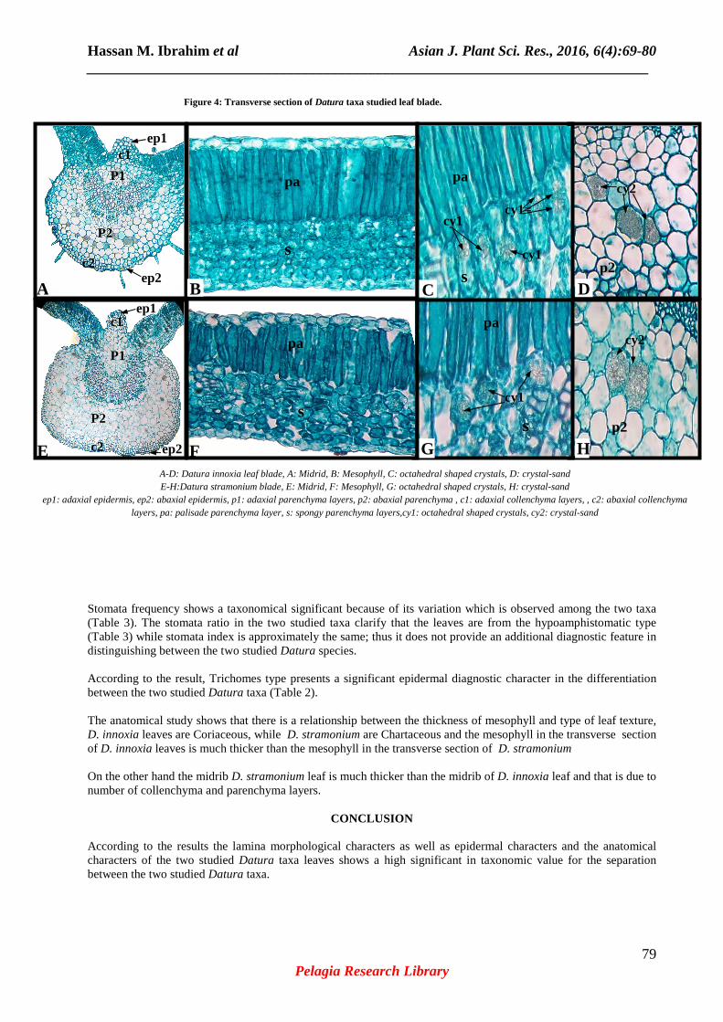
Anatomical Analysis: Table 3 & Figure 4 Illustrate the main anatomical characters of the two Datura taxa studied leaves as clarified by light microscope. The transverse section of the two Datura taxa studied leaves (Figure 4) shows the presence of cuticle on both adaxial and abaxial surface, the adaxial and the abaxial epidermis composed of uniseriate oval to rectangular cells. Although the transverse section of the two Datura taxa studied leaves confirms that the leaves are from the type bifacial (Dorsiventral). The mesophyll in D. innoxia consist of one layer of elongate of Palisade parenchyma arranged like a row of stakes without air-space and 5-7 layers of irregular Spongy parenchyma with a few air-space; while in D. stramonium it composed of one layer of elongate Palisade parenchyma arranged like a row of stakes with a few air-space and 5-6 layers of irregular Spongy parenchyma with air-space. According to observation the mesophyll in D. innoxia is thicker then the mesophyll D. stramonium with average thickness 17.2 µm and13.1 µm correspondingly and this agrees with type of leaf texture in D. innoxia and D. stramonium (Table 2 & 3 and Figure 4). On the other hand the midrib in the transverse section of the two Datura taxa studied leaves consist of cuticle on both adaxial and abaxial surface, adaxial and abaxial epidermis composed of uniseriate oval to rectangular cells followed by adaxial and abaxial collenchyma, in D. innoxia the adaxial layers and the abaxial layers of collenchyma consist of 4 -5 layers & 2-3 layers, while in D. stramonium there is 5-8 adaxial layers and 3- 4abaxial layers of collenchymas (Figure 4). The main vascular bundle in the transverse section of the two Datura taxa studied leaves is surrounded by adaxial and abaxial layers of parenchyma, in D. innoxia there is 5-8 layers of adaxial paranchyma and 6- 8 layers of abaxial parenchyma, while in D. stramonium there is 6-10 layers of adaxial paranchyma and 7 -10 layers of abaxial paranchyma. According to observation the thickness of the D. stramonium midrib is much thicker than the midrib of D. innoxia, with average thickness 74.5 µm and 61 µm respectively (Table 3 & Figure 4). Identification key to the studied Datura taxa leaves based on their Anatomical characters: + In the Midrib section of the leaf the adaxial and abaxial collenchyma consist of 4 - 5 and 2- 3 layers respectively....................................................................... Datura innoxia. - In the Midrib section of the leaf the adaxial and abaxial collenchyma consist of 5 -8and 3- 4 layers respectively ............................................................................Datura stramonium. The results, proved that leaf architecture character are good taxonomic markers in plant identification and classification. The laminar shape, base, apex, margin and texture are the most useful morphological characters in separating the two studied species from each other. Also; this study shows that the type of venation is an important diagnostic character in distinguishing between the two species morphologically. Epidermal characters have potential for taxonomic use as additional taxonomic characters [20]. The result of examining the epidermal cells shows that the length, size and frequency of epidermal cells vary from the leaf of one species to another among the genera. The results shows that size of stomata is taxonomically important and can be used in designation between the two studied taxa, on the other hand stomata complex type in the two Datura taxa, was amonotetracytic and anisocytic; this is in agreement with observations recorded by Solereder, [8] and Hameed & Hussain [9].
Hassan M. Ibrahim et al Asian J. Plant Sci. Res., 2016, 6(4):69-80 _____________________________________________________________________________
79 Pelagia Research Library
Stomata frequency shows a taxonomical significant because of its variation which is observed among the two taxa (Table 3). The stomata ratio in the two studied taxa clarify that the leaves are from the hypoamphistomatic type (Table 3) while stomata index is approximately the same; thus it does not provide an additional diagnostic feature in distinguishing between the two studied Datura species. According to the result, Trichomes type presents a significant epidermal diagnostic character in the differentiation between the two studied Datura taxa (Table 2). The anatomical study shows that there is a relationship between the thickness of mesophyll and type of leaf texture, D. innoxia leaves are Coriaceous, while D. stramonium are Chartaceous and the mesophyll in the transverse section of D. innoxia leaves is much thicker than the mesophyll in the transverse section of D. stramonium On the other hand the midrib D. stramonium leaf is much thicker than the midrib of D. innoxia leaf and that is due to number of collenchyma and parenchyma layers.
CONCLUSION
According to the results the lamina morphological characters as well as epidermal characters and the anatomical characters of the two studied Datura taxa leaves shows a high significant in taxonomic value for the separation between the two studied Datura taxa.
6.3µm
3.7µm
1.8µm
1.2µm 2.8µm
1µm 1.3µm 1.6µm
A-D: Datura innoxia leaf blade, A: Midrid, B: Mesophyll, C: octahedral shaped crystals, D: crystal-sand E-H:Datura stramonium blade, E: Midrid, F: Mesophyll, G: octahedral shaped crystals, H: crystal-sand
ep1: adaxial epidermis, ep2: abaxial epidermis, p1: adaxial parenchyma layers, p2: abaxial parenchyma , c1: adaxial collenchyma layers, , c2: abaxial collenchyma layers, pa: palisade parenchyma layer, s: spongy parenchyma layers,cy1: octahedral shaped crystals, cy2: crystal-sand
A B
E F
C D
G a
H a
cy2 pa
p2
P2
P1
c2
c1
ep2
ep1
cy2
p2
pa
s
cy1= cy1
cy1 s
pa
s P2
P1
c1
c2 ep2
ep1
cy1
s
pa
Figure 4: Transverse section of Datura taxa studied leaf blade.

Hassan M. Ibrahim et al Asian J. Plant Sci. Res., 2016, 6(4):69-80 _____________________________________________________________________________
80 Pelagia Research Library
REFERENCES
[1] A. S. Chaudhary, Flora of the kingdom of Saudi Arabia Illustrated, Ministry of Agriculture & Water, National Herbarium, National Agriculture Research Centre, Riyadh, 2001, 2 (2), 120-122. [2] A.A. Al Khulaidi, Flora of Yemen, Sustainable Natural Resource Management Project, Sana’a Yemen, 2013. [3] I. H. Al-Seragy, MSc, Thesis, Biology Dept. Faculty of Science, Sana’a Univ., 2009. [4] A. M. A. Dahmash, Univ. Aden J. and Appl. Sc., 2013, 17 (2): 435-444. [5] A. S. Chaudhary, R. Revri, Weeds of North Yemen, Deutsche Gesellschaft fur Technische Zusammenarbeit (GTZ), German, 1983, 370-371. [6] A. M. Migahid, Flora of Saudi Arabia, 4th, King Saud University, University Libraries, Saudi Arabia, 1996, 2, 145-148. [7] J.R.I. Wood, A Handbook of the Yemen Flora. Royal Botanic Gardens, Kew, UK, 1997, 226-230. [8] H. Solereder, Systematic Anatomy of the Dicotyledons, Ajay Book Service, New Delhi, India, 1986, 1, 573-583. [9] I. Hameed, F. Hussain, Journal of medicinal plants research, 2011, 5 (18), 4525-4529. [10] S. Collenette, Wildflowers of Saudi Arabia, National Commission for Wildlife Conservation and Development, Riyadh, Saudi Arabia, 1999, 697-698. [11] D.L. Dilcher, The Botanical Review, 1974, 40 (1), 2-157. [12] J.A. Ibrahim, A.E. Ayodele, World Applied Science Journal, 2013, 24 (9), 1172-1179. [13] V.V. Sreelakshmi, E. Sruthy, J. Shereena, International Journal of Research in Applied, National and Social Science, 2014, 2 (7), 53-60. [14] E.J. Salisbury, Phil. Trans. Roy.Soc. Kind., 1927, Ser. B., 216: 1-65. [15] M. Szymura, K. Wolski, Taxain Poland, ACTA Biological Cracoviensia Series Botanical, 2011, 53 (1): 38-46. [16] H.N. Nur Fatihah, M. Nashriyah, A. Nor Zaimah, M. Khairil, A. Ali, Turkish Journal of Botany, 2014, 38: 677-685. [17] A. Fahn, Plant Anatomy, 3rd, Maxwell Macmillan International Editions, Singapore, 1989, 208-249. [18] K. Esau, Plant Anatomy, 3rd ed., John Wiley & Sons, Inc., Hoboken, New Jersey, 2006,175-466. [19] A. Pataky, In: Metcalfe C R, Chalk L (Ed.) Leaf Epidermis of Salix. Anatomy of the Dicotyledons. 2nd, Clarendon Press, Oxford, 1969, 1, pp. 110. [20] R.S. Shavvon, S.S. Mehrvarz, N. Golmohammadi, Turk. J. Bot., 2012, 36: 655-666.