
Morphological and Molecular Characterization of ... · phenotypic characters like colony colour and...
6
International Journal of Science and Research (IJSR) ISSN (Online): 2319-7064 Impact Factor (2012): 3.358 Volume 3 Issue 7, July 2014 www.ijsr.net Licensed Under Creative Commons Attribution CC BY Morphological and Molecular Characterization of Trichoderma Isolates: An Antagonist against Soil Borne Pathogens Mukesh Srivastava, Anuradha Singh * , D. K. Srivastava 1 Biocontrol Laboratory, Department of Plant Pathology, C. S. Azad University of Agriculture & Technology, Kanpur, U.P., India, 1 Joint Director, Council of Science & Technology, U.P., Lucknow, India * Corresponding Author: Anuradha Singh, Biocontrol Laboratory, Department of Plant Pathology, C.S.A. University of Agriculture & Technology, Kanpur-208002, Uttar Pradesh, India Abstract: Trichoderma has attained importance as a substitute of chemical pesticides all over the world. Hence, an attempt was intended to corroborate the positive relatedness of molecular and morphological characters with antagonistic ability. Among many isolates of Trichoderma atroviride isolated from rhizospheric soils from different parts of U.P. has brought attention due to its highly antagonistic activity. Eight isolates of Trichoderma atroviride were assessed for their mycoparasitic and molecular variability. These were isolated on PDA medium by serial dilution and identified based on phenotypic characters like colony colour, growth, shape of conidiophore, phialides and conidia. Antagonistic variability of the isolates of T. atroviride revealed significant suppression in the radial growth of Fusarium oxysporum f. sp. ciceri, F. oxysporum f. sp. lentis and F. oxysporum f. sp. udum. Maximum inhibition (60.26%) of mycelial growth was recorded in case of TH 1 isolate against Fusarium oxysporum f. sp. lentis (F.o.l.) which was isolated from the soil sample of Bilgram (Hardoi), followed by TE 8 (50%) isolated from Bharthana (Etawah). TS 5 (39.76%) isolate from Lainbua (Sultanpur) was found to be least effective against F.o.l. Similarly, in case of Fusarium oxysporum f. sp. ciceri (F.o.c), the maximum inhibition (55.08%) of mycelial growth was shown by the isolate TH 3 , which was isolated from the soil sample of Bilgram (Hardoi). The next effective isolate was TS 6 (53.65%) isolated from Kadipur (Sultanpur). Isolate TSi 4 (37.69%) of Misrikh (Sitapur) was found least effective against F.o.c. Maximum inhibition in case of Fusarium oxysporum f. sp. udum (F.o.u) was recorded with isolate TA U8 (47.91%) from Azeetmal (Auraiya). Next effective isolate was TB a1 (47.25%) isolated from Ballaha (Bahraich). The least effective isolates were TS i3 (41.66%) and TKD 3 (41.66%) isolated from Misrikh (Sitapur) and Maitha (Kanpur Dehat). Molecular variability among the isolates showed 74 amplified bands out of which 65 were polymorphic and 19 were monomorphic. The size of amplified product varied from 0.1kb to 0.75kb. Keywords: Trichoderma atroviride, antagonism, morphological and molecular character, DNA extraction, RAPD 1. Introduction Plant disease epidemics have created an ecologically imbalance system in modern agriculture. Deterrence of such epidemics for the most part achieved through the use of chemical fungicides has greater repercussion on environment and human health. Also, progressive confrontation in a midst of pathogen resistance to accessible chemical plant protectants has engrossed the need of alternative methods of disease control. Fungi of the genus Trichoderma are important biocontrol agent against several soil borne phytopathogens (Benitez et.al.2004). Trichoderma spp. are free living rapid growing fungi that are common in soil and root eco-system. The fungi are exceptionally good model for biocontrol more importantly as bioagent, which is accomplished by means of mycoparasitism, antibiosis and competition. Several species of Trichoderma have been isolated from various substrates and locations (Bilgrami et.al.1971, Nagamani et.al. 2002). Several articles have also been published to identify the Trichoderma spp. based on the molecular or physiological bases (Samuels, 1996, Hermosa et.al. 2000). Kiffer and Morelet (2000) have recognized several species of Trichoderma based on molecular characters. The molecular techniques like Random Amplified Polymorphic DNA (RAPD) developed by Williams et al. (1990) has been used for genetic and taxonomic studies for several fungi including Trichoderma sp. (Muthumeenashi and Mills, 2004). Most of the species of Trichoderma are effective against soil borne pathogens that cause diseases in leguminous crop. Fusarium wilt is the most important disease that significantly damages the crop. A high level of genetic diversity in Trichoderma spp. has been reported (Chakraborty et.al. 2010) that can be used to produce a wide range of products of commercial and ecological interest. 2. Materials and Methods 2.1 Identification and Morphological characterization of Trichoderma sp. Isolates of Trichoderma were isolated from soil samples collected from rhizospheric of chickpea, pigeonpea and lentil crop from different places of Uttar Pradesh, India. All the isolates were isolated on PDA medium by following serial dilution plate technique as described by Johnson and Curl [8] and isolates were identified up to species level based on phenotypic characters like colony colour and growth; size and shape of conidiophore, phialides and conidia. The cultures were identified using the available literature [9-12] and confirmed by morphological characters and also confirmed by ITCC, Division of Plant Pathology IARI, New Delhi-12. The morphological characterization was based on monographic contribution provided by Bissett (1991 a, b). Paper ID: 020141330 2399
Transcript of Morphological and Molecular Characterization of ... · phenotypic characters like colony colour and...
International Journal of Science and Research (IJSR) ISSN (Online): 2319-7064
Impact Factor (2012): 3.358
Volume 3 Issue 7, July 2014 www.ijsr.net
Licensed Under Creative Commons Attribution CC BY
Morphological and Molecular Characterization of Trichoderma Isolates: An Antagonist against Soil
Borne Pathogens
Mukesh Srivastava, Anuradha Singh*, D. K. Srivastava1
Biocontrol Laboratory, Department of Plant Pathology, C. S. Azad University of Agriculture & Technology, Kanpur, U.P., India,1Joint Director, Council of Science & Technology, U.P., Lucknow, India
*Corresponding Author: Anuradha Singh, Biocontrol Laboratory, Department of Plant Pathology, C.S.A. University of Agriculture & Technology, Kanpur-208002, Uttar Pradesh, India
Abstract: Trichoderma has attained importance as a substitute of chemical pesticides all over the world. Hence, an attempt was intended to corroborate the positive relatedness of molecular and morphological characters with antagonistic ability. Among many isolates of Trichoderma atroviride isolated from rhizospheric soils from different parts of U.P. has brought attention due to its highly antagonistic activity. Eight isolates of Trichoderma atroviride were assessed for their mycoparasitic and molecular variability. These were isolated on PDA medium by serial dilution and identified based on phenotypic characters like colony colour, growth, shape of conidiophore, phialides and conidia. Antagonistic variability of the isolates of T. atroviride revealed significant suppression in the radial growth of Fusarium oxysporum f. sp. ciceri, F. oxysporum f. sp. lentis and F. oxysporum f. sp. udum. Maximum inhibition (60.26%) of mycelial growth was recorded in case of TH1 isolate against Fusarium oxysporum f. sp. lentis (F.o.l.) which was isolated from the soil sample of Bilgram (Hardoi), followed by TE8 (50%) isolated from Bharthana (Etawah). TS5 (39.76%) isolate from Lainbua (Sultanpur) was found to be least effective against F.o.l. Similarly, in case of Fusarium oxysporum f. sp. ciceri (F.o.c), the maximum inhibition (55.08%) of mycelial growth was shown by the isolate TH3, which was isolated from the soil sample of Bilgram (Hardoi). The next effective isolate was TS6 (53.65%) isolated from Kadipur (Sultanpur). Isolate TSi4 (37.69%) of Misrikh (Sitapur) was found least effective against F.o.c. Maximum inhibition in case of Fusarium oxysporum f. sp. udum (F.o.u) was recorded with isolate TAU8 (47.91%) from Azeetmal (Auraiya). Next effective isolate was TBa1 (47.25%) isolated from Ballaha (Bahraich). The least effective isolates were TSi3 (41.66%) and TKD3 (41.66%) isolated from Misrikh (Sitapur) and Maitha (Kanpur Dehat). Molecular variability among the isolates showed 74 amplified bands out of which 65 were polymorphic and 19 were monomorphic. The size of amplified product varied from 0.1kb to 0.75kb. Keywords: Trichoderma atroviride, antagonism, morphological and molecular character, DNA extraction, RAPD 1. Introduction Plant disease epidemics have created an ecologically imbalance system in modern agriculture. Deterrence of such epidemics for the most part achieved through the use of chemical fungicides has greater repercussion on environment and human health. Also, progressive confrontation in a midst of pathogen resistance to accessible chemical plant protectants has engrossed the need of alternative methods of disease control. Fungi of the genus Trichoderma are important biocontrol agent against several soil borne phytopathogens (Benitez et.al.2004). Trichoderma spp. are free living rapid growing fungi that are common in soil and root eco-system. The fungi are exceptionally good model for biocontrol more importantly as bioagent, which is accomplished by means of mycoparasitism, antibiosis and competition. Several species of Trichoderma have been isolated from various substrates and locations (Bilgrami et.al.1971, Nagamani et.al. 2002). Several articles have also been published to identify the Trichoderma spp. based on the molecular or physiological bases (Samuels, 1996, Hermosa et.al. 2000). Kiffer and Morelet (2000) have recognized several species of Trichoderma based on molecular characters. The molecular techniques like Random Amplified Polymorphic DNA (RAPD) developed by Williams et al. (1990) has been used for genetic and taxonomic studies for several fungi including Trichoderma sp. (Muthumeenashi and Mills, 2004). Most of the species of
Trichoderma are effective against soil borne pathogens that cause diseases in leguminous crop. Fusarium wilt is the most important disease that significantly damages the crop. A high level of genetic diversity in Trichoderma spp. has been reported (Chakraborty et.al. 2010) that can be used to produce a wide range of products of commercial and ecological interest. 2. Materials and Methods 2.1 Identification and Morphological characterization of Trichoderma sp. Isolates of Trichoderma were isolated from soil samples collected from rhizospheric of chickpea, pigeonpea and lentil crop from different places of Uttar Pradesh, India. All the isolates were isolated on PDA medium by following serial dilution plate technique as described by Johnson and Curl [8] and isolates were identified up to species level based on phenotypic characters like colony colour and growth; size and shape of conidiophore, phialides and conidia. The cultures were identified using the available literature [9-12] and confirmed by morphological characters and also confirmed by ITCC, Division of Plant Pathology IARI, New Delhi-12. The morphological characterization was based on monographic contribution provided by Bissett (1991 a, b).
Paper ID: 020141330 2399

International Journal of Science and Research (IJSR) ISSN (Online): 2319-7064
Impact Factor (2012): 3.358
Volume 3 Issue 7, July 2014 www.ijsr.net
Licensed Under Creative Commons Attribution CC BY
2.2 Confrontation assays in vitro The antagonistic potentiality of the isolates of T. atroviride was determined against three strains of Fusarium by dual culture technique described by Morton and Stroube (1955). A disc of 5 mm diameter was made from 7 days old culture of different isolates of T. atroviride and placed at one point leaving 1 cm distance from the periphery of one side of Petri plate and on the opposite site, disc (5 mm dia.) of F.o.u, F.o.c and F.o.l were placed separately. Plate was kept without antagonist served as control. These Petri plates were incubated at 23 ± 20C for 7 days in three replications (Table 3). Observations on colony growth were recorded and percent inhibition was measured by using the following formula:-
(C )100
TI
C
2.3 Molecular characterization Genomic DNA was extracted from each isolates of Trichoderma atroviride grown in 1000 ml conical flask containing 400 ml of PDB medium. Two agar plugs from actively growing colony of T. atroviride were transferred to each flask aseptically in a laminar flow. The flask was incubated at 23±20C for 7 days. The mycelial mat was collected by passing the fluid through three layers cheese cloth. The fungal cell wall was disrupted by grinding with pestle and mortar in liquid nitrogen. The DNA was extracted by CTAB method of fungal DNA extraction as used by Kumar et al. (2011). Quantification of DNA was done with 0.8% agarose gel electrophoresis. Working concentration of DNA was adjusted to 20 ng/μl and stored at 40C. The DNA from all isolates produced clear and sharp bands, indicating good quality of DNA.
(a)
(b)
Figure 1: Trichoderma atroviride: (a) Growth on PDA media, (b) Microscopic observation at 100x
2.4 Random Amplified Polymorphic DNA The procedure described by Williams et al. with minor modification like in PCR cycles standardization was done for carrying out PCR reaction to produce RAPD profiles. Amplification of DNA fragments was carried out by the PCR using 10-mer arbitrary primers. The reaction mixture consisted of 300ng of 200 μmM of dNTP mix (Fermentas company), 15 pmol of primer (Operon), 5 U/μl of Taq polymerase (Fermentas) and 25 mM MgCl2. DNA amplifications were performed in thermocycler with one cycle of initial denaturation at 94C for 15 min, followed by 40 cycles of denaturation at 940C for 1 min, annealing at 35C for 2 min, extension at 72C for 1 min and final extension at 72C for 10 min. Amplified products together with uncut lambda marker double digest; Bangalore, Genei) were resolved by 1.5% agarose gel electrophoresis (60 V for 1 hr). Gels were photographed by (Uvitec) Gel documentation system (Figure 1). 2.5 Data Analysis Comparison of each profile for each primer was done on the basis of the presence or absence (1/0) of amplified bands. Bands of the same size (in bp) were scored as identical. All reproducible polymorphic bands were scored and analysed following UPGMA cluster matrix using NTSYSpc (Numerical Taxonomy System Biostatistics, version 2.02e). 3. Results and Discussion 3.1 Morphological characterization The growth patterns of Trichoderma isolates after four days of incubation at 23 ± 20C on PDA media showed significant differences in nature of culture growth and sporulation patterns. The colony colour changes from light green shade to dull green with the production of conidia. The conidial wall patterns and shape were rough, subglobose and smooth (Fig.1). The growth characters of culture and sporulation patterns varied noticeably within and between the isolates (Table 2).
Paper ID: 020141330 2400

International Journal of Science and Research (IJSR) ISSN (Online): 2319-7064
Impact Factor (2012): 3.358
Volume 3 Issue 7, July 2014 www.ijsr.net
Licensed Under Creative Commons Attribution CC BY
Table 1: Key morphological description used for characterization of T. isolates Isolate Codes/
Characters 6 CP 24 CP 71 L 115 L 52 L 75 PP 126 PP 105 CP
Colony growth rate (cm/day)
7-8 in 5 days 7-8 in 5 days 6-7 in 5 days 7-8 in 5 days 7-8 in 5 days
7-8 in 5 days 7-8 in 5 days 6-7 in 5 days
Colony colour Light Green to yellowish green
Light Green to dark green
Light Green to dark green
Light Green to yellowish
green
Light Green Light Green to dark green
Light Green to yellowish green
Light Green to
dark greenReverse colony colour
Yellow Light yellow Colourless Light yellow Yellow Light yellow Colourless Light yellow
Colony edge Smooth Smooth Smooth Smooth Smooth Smooth Smooth Smooth Culture smell Malt Malt Malt Malt Malt Malt Malt Malt Mycelial form Arachnoid Floccose to
arachnoid Floccose to arachnoid
Floccose to arachnoid
Arachnoid Arachnoid Arachnoid Floccose toarachnoid
Conidiation Crusty in surface
Crusty in surface
Crusty in surface
Crusty in surface
Crusty in surface
Crusty in surface
Crusty in surface
Crusty in surface
Phialide shape Ampulliform Ampulliform Oblong Ampulliform Oblong Ampulliform Ampulliform Ampulliform
Conidial shape Subglobose & Smooth
Subglobose , Smooth
Subglobose, Smooth
Subglobose, Smooth
Subglobos, Smooth
Subglobose, Smooth
Subglobose, Smooth
Subglobose, Smooth
Conidial wall Rough Rough Rough Rough Rough Rough Rough Rough Conidial colour Yellowish
green Yellowish
green Green Yellowish
green Yellowish
green Green Green Yellowish
green Chlamydosopores Not observed Not observed Present
globose Not observed Not
observed Not
observed Not observed Not
observed 3.2 Confrontation assays in vitro The isolates of Trichoderma viz. 6 CP, 24 CP, 71 L, 115 L, 52 L, 75 PP, 126 PP and 105 CP belong to T. atroviride. In order to evaluate the antagonistic potential of T. atroviride, dual culture technique was carried out with three legume pathogens viz. F. oxysporum f. sp. ciceri, F. oxysporum f. sp. lentis and F. oxysporum f. sp. udum. Effects of different isolates of T. atroviride with respect to suppression of mycelial growth of the three test pathogens were recorded. It is evident from the data that T. atroviride suppressed the radial growth of F. oxysporum ciceri, F. oxysporum lentis and F. oxysporum udum significantly (Table 3). Maximum inhibition (60.26%) by T. atroviride TH1, isolate, which was isolated from soil sample of Bilgram block of Hardoi district, followed by TE8 (50.00%) isolated from Bharthana block of Etawah district. TS5 (39.76%) isolate of Lainbua block of
Sultanpur was found least effective against the pathogen. The findings clearly show the wide range of antagonistic effect of different isolates of T. atroviride. The maximum inhibition (47.9%) of mycelial growth of F. udum was recorded in case of TAU8 isolate, which is isolated from the soil sample of Azeetmal block of Auraiya. TSI3 and TKD3 isolate of Misrikh and Maitha were found to be least effective against F. udum among all the isolates of T. atroviride. Joshi et al. (2010) also found the antagonistic variability in different isolates of Trichoderma spp. collected from different places of India. The present finding was also supported by several workers (Obaiua Oti, 2007). Singh et al. (2013) also revealed that 30 isolates of Trichoderma viride collected from various districts of U.P. were found highly antagonist against three test pathogens (F.o.u, F.o.c and F.o.l).
Table 2: In vitro evaluation of different isolates of T. isolates against soil borne pathogens
I.D. No. Culture No. % inhibition of Trichoderma isolates against F. oxysporum
f. sp. udum
% inhibition of Trichoderma isolates
against F. oxysporum f. sp.ciceri
% inhibition of Trichoderma isolates
against F. oxysporum f. sp. lentis
% inhibition of Trichoderma
isolates against S. rolfsii
ITCC-7442/09 06 CP 45.83 53.65 39.76 54.83 ITCC-7443/09 24CP 41.66 37.69 44.88 58.03 ITCC-7445/09 71L 45.83 55.08 60.26 62.26 ITCC-7446/09 115L 47.25 52.17 42.30 45.16 ITCC-7447/09 52L 43.75 40.00 43.07 41.93 ITCC-7448/09 75PP 47.91 43.47 48.07 51.61 ITCC-7449/09 126PP 41.66 43.43 46.92 55.08 ITCC-7451/09 105PP 45.83 43.91 50.00 52.00
CD at 5 % 2.71 2.52 2.93 1.06 SE 1.28 1.19 1.38
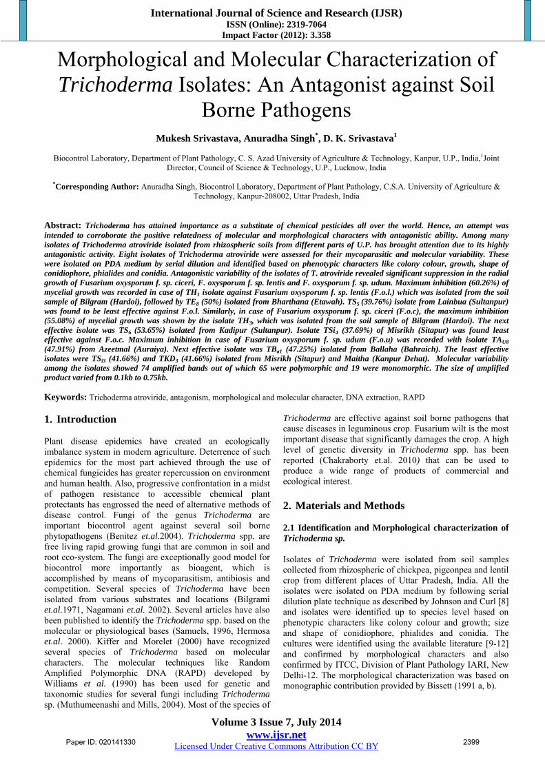
3.2 Molecular Characterization The results presented in Fig. 2 showed that the total number of reproducible bands amplified were 94, out of which 75 were found to be polymorphic and 19 were monomorphic, hence the percentage of polymorphism is 79.78. The number
of bands per primer ranged from the maximum of 12 (given by OPC-13) to a minimum of 3 (OPC-8) with an average of 7 bands per primer.
Paper ID: 020141330 2401
International Journal of Science and Research (IJSR) ISSN (Online): 2319-7064
Impact Factor (2012): 3.358
Volume 3 Issue 7, July 2014 www.ijsr.net
Licensed Under Creative Commons Attribution CC BY
Figure 2: RAPD analysis of T. atroviride isolates with 20 primers (OPC-1-OPC-20)
The experimental findings also revealed that the 7 RAPD primers (OPC 10, 11, 13, 14, 15, 19 & 20) produced average or above average amplified products (Table 4).
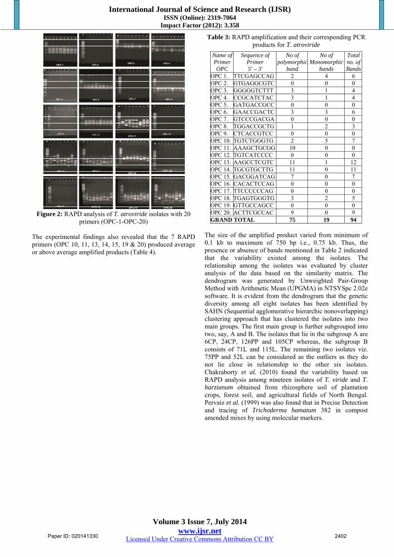
Table 3: RAPD amplification and their corresponding PCR products for T. atroviride
Name of Primer OPC
Sequence of Primer 5' – 3'
No of polymorphic
band
No of Monomorphic
bands
Total no. of Bands
OPC 1. TTCGAGCCAG 2 4 6 OPC 2. GTGAGGCGTC 0 0 0 OPC 3. GGGGGTCTTT 3 1 4 OPC 4. CCGCATCTAC 3 1 4 OPC 5. GATGACCGCC 0 0 0 OPC 6. GAACCGACTC 3 3 6 OPC 7. GTCCCGACGA 0 0 0 OPC 8. TGGACCGCTG 1 2 3 OPC 9. CTCACCGTCC 0 0 0 OPC 10. TGTCTGGGTG 2 5 7 OPC 11. AAAGCTGCGG 10 0 0 OPC 12. TGTCATCCCC 0 0 0 OPC 13. AAGCCTCGTC 11 1 12 OPC 14. TGCGTGCTTG 11 0 11 OPC 15. GACGGATCAG 7 0 7 OPC 16. CACACTCCAG 0 0 0 OPC 17. TTCCCCCCAG 0 0 0 OPC 18. TGAGTGGGTG 3 2 5 OPC 19. GTTGCCAGCC 0 0 0 OPC 20. ACTTCGCCAC 9 0 9 GRAND TOTAL 75 19 94
The size of the amplified product varied from minimum of 0.1 kb to maximum of 750 bp i.e., 0.75 kb. Thus, the presence or absence of bands mentioned in Table 2 indicated that the variability existed among the isolates. The relationship among the isolates was evaluated by cluster analysis of the data based on the similarity matrix. The dendrogram was generated by Unweighted Pair-Group Method with Arithmetic Mean (UPGMA) in NTSYSpc 2.02e software. It is evident from the dendrogram that the genetic diversity among all eight isolates has been identified by SAHN (Sequential agglomerative hierarchic nonoverlapping) clustering approach that has clustered the isolates into two main groups. The first main group is further subgrouped into two, say, A and B. The isolates that lie in the subgroup A are 6CP, 24CP, 126PP and 105CP whereas, the subgroup B consists of 71L and 115L. The remaining two isolates viz. 75PP and 52L can be considered as the outliers as they do not lie close in relationship to the other six isolates. Chakraborty et al. (2010) found the variability based on RAPD analysis among nineteen isolates of T. viride and T. harzianum obtained from rhizosphere soil of plantation crops, forest soil, and agricultural fields of North Bengal. Pervaiz et al. (1999) was also found that in Precise Detection and tracing of Trichoderma hamatum 382 in compost amended mixes by using molecular markers.
Paper ID: 020141330 2402

International Journal of Science and Research (IJSR) ISSN (Online): 2319-7064
Impact Factor (2012): 3.358
Volume 3 Issue 7, July 2014 www.ijsr.net
Licensed Under Creative Commons Attribution CC BY
Figure 3: Dendrogram showing the genetic relationships among 8 Trichoderma isolates based on RAPD analysis
4. Conclusion It may be concluded from the present findings that antagonistic, morphological and molecular variability exist among eight isolates of T. atroviride, collected from rhizosphere soil of different places of Uttar Pradesh. It is also concluded that there is good genetic diversity and these are strong possibility to get the isolates specific primers that will be utilized for particular Trichoderma isolates with good biological potential form the field isolates without going the cumbersome bioassay. 5. Acknowledgements The authors are grateful for the financial support granted by the Indian Council of Agriculture Research (ICAR) Govt. of India under the Niche Area of Excellence on “Exploration and Exploitation of Trichoderma as an antagonist against soil borne pathogens” running in the Biocontrol Laboratory, Department of Plant Pathology, C.S.A. University of Agriculture and Technology, Kanpur, India. References [1] Benitez T (2004). Increased antifungal and chitinase
specific activates of Trichoderma harzianum CECT 2413 by addition of a cellulose binding-domain. Appl. Microbiol. Biotechnol. 64: 675-685.
[2] Bilgrami KS, Jamaluddin and Rizvi MA (1971). Fungi of India. Today and Tomorrow Publication. New Delhi.
[3] Bisset J (1991a). A revision of the genus Trichoderma II. Infragenric classification Can. J. Bot. 69: 2373-2417.
[4] Bisset J (1991b). A revision of the genus Trichoderma III. Sect. Pachybasium. Can. J. Bot. 69: 2373-2417.
[5] Chakraborty BN, Chakraborty U, Saha A, Dey PL, Sunar K (2010). Molecular characterization of Trichoderma viride and T. harzianum isolated from soil from North Bengal based on rDNA markers and analysis of their PCR-RAPD profiles. Global J. Biotech. And Biochem. 5 (1): 55-61.
[6] Hermosa MR, Gronoda I, Iturriaga A, Diaz- Minguez JM, Castro C, Monte E, Garcia-Acha I (2000). Molecular Characterization and Identification of Biocontrol Isolates of Trichoderma spp. App. & Env. Microbiology. 66(5): 1890-1898.
[7] Johnson LF, Curl EA (1972). Methods for Research on the Ecology of Soil borne Plant Pathogens. Burgess Publishing Company. Minneapolis. 247pp.
[8] Joshi BB, Bhatt RP, Bahukhandi D (2010). Antagonistic and Plant growth activity of Trichoderma isolates of Western Himalayas. J. of Environ. Biology. 6: 921-928.
[9] Kiffer E, Morelet E (2000). The Deuteromycetes Mitosporic Fungi Classification and Generic Keys. Science Publications. Inc. USA. pp 1152.
[10] Kumar Vipul, Mohd. Shahid, Singh Anuradha, Srivastava Mukesh, Biswas SK (2011). RAPD analysis of Trichoderma logibrachiatum isolated from pigeonpea fields of Uttar Pradesh. Indian J. Agric. Biochem. 24(1): 80-82.
[11] Morton DT, Stroube NH (1955). Antagonistic and stimulatory effects of microorganism upon Sclerotium rolfsii. Phytopathology. 45: 419-420.
[12] Muthumeenakshi S, Mills PR, Brown AE, Seaby Da (1994). Intraspecific molecular variation among Trichoderma harzianum isolates colonizing mushroom compost in British Isles. Microbiology. 140: 769-777.
[13] Nagamani A, Kunwar IK, Manoharachary C, Pranti Reddy (2002). Trichoderma fllavofuscum – a new record of India. Indian Phytopathology. 55: 247-248.
[14] Obaiua AO, Oti E (2007). Anatagonistic properties of Trichoderma viride on post harvest cassava root rot pathogens. African Journal of Biotechnology. 6(21): 2447-2450
[15] Pervaiz A, Abbasi Sallya A, Miller Tea Meulia, Harry A J, Hoitink, Jin Man Kim (1999). Precise Detection and Tracing of Trichoderma hamatum 382 in compost amended mixes by using molecular markers. National University San 96-1. Doonduck-dong,Yosu, Cheon Nam. 550-250.
Paper ID: 020141330 2403

International Journal of Science and Research (IJSR) ISSN (Online): 2319-7064
Impact Factor (2012): 3.358
Volume 3 Issue 7, July 2014 www.ijsr.net
Licensed Under Creative Commons Attribution CC BY
[16] Samuels GJ (1996). Trichoderma: a review of biology and systematic of the genus. Mycological Research. 100: 923-935.
[17] Singh A, Mohd. S, Srivastava M, Kumar V, Bansal A (2013). Antagonistic activity of Trichoderma viride isolates against different pathogens of Fusarium Oxysporum isolated from Legume crop of U.P. Progressive Research 8(1): 47-50.
[18] Singh A, Mohd. S, Pandey NK, Kumar Sharwan, Srivastava M, Biswas SK (2011). Influence of Temperature, pH and media for growth and sporulation of Trichoderma atroviride and its shelf life study in different carrier based formulation. J. Pl. Dis. Sci. 6(2): 32-34.
[19] Williams J GK, Kubelik AR, Livak KJ, Rafalski JA, Tingey SV (1990). DNA polymorphisms amplified by arbitrary primers are useful as genetic markers. Nucleic acid Research. 18: 6531-6535.
Paper ID: 020141330 2404


![National Diagnostic Protocol...and Helminthosporium oryzae. Breda de Haan]. The conidia produced by C. miyabeanus are easy to differentiate from the conidia produced by P. oryzae.](https://static.fdocuments.in/doc/165x107/5e557c3a5065bb1bbb46377e/national-diagnostic-protocol-and-helminthosporium-oryzae-breda-de-haan-the.jpg)